Embed Size (px)
Citation preview
ARTHRITIS & RHEUMATISMVol. 50, No. 9, September 2004, pp 3049–3060© 2004, American College of Rheumatology
LETTERS
DOI 10.1002/art.20639
Disseminated Salmonella typhimurium infectionsecondary to infliximab treatment
To the Editor:We read with interest the review by Ellerin et al on
infections and anti–tumor necrosis factor (anti-TNF) therapy(1), as well as the article by Netea et al reporting 2 cases ofSalmonella enterica septicemia secondary to treatment withadalimumab and infliximab (2). Netea and colleagues alsodemonstrated decreased interferon-� production and inhibi-tion of Toll-like receptor 4 expression on dendritic cells inanti-TNF–treated rheumatoid arthritis patients, providing apotential explanation for the increased susceptibility to intra-cellular organism infection associated with anti-TNF therapy.
We have recently seen a patient with disseminatedSalmonella typhimurium infection secondary to anti-TNF ther-apy. This is, to our knowledge, the first published report ofsuch a finding. The patient was a Fijian Indian man whodeveloped psoriasis and psoriatic arthritis in 1992. He migratedto Australia in 1993. His skin and joint disease remained veryactive despite a variety of treatments administered over aperiod of years, including psoralen ultraviolet A, acitretin,sulfasalazine, methotrexate, and cyclosporine. At the time ofpresentation to us in January 2003, at the age of 39 years, hewas taking prednisone (10 mg/day) and sulfasalazine. Hiserythrocyte sedimentation rate (ESR) and C-reactive protein(CRP) level were both elevated (ESR 80 mm/hour [normal�10], CRP 82 mg/liter [normal �3]). Cyclosporine treatmentwas reinstituted in late January 2003 and was mildly effica-cious, but was discontinued in early April due to intolerableside effects including hypertension, headaches, and impair-ment of short-term memory. In mid-April 2003, treatment withinfliximab combined with methotrexate (7.5 mg/week) wasstarted. Infliximab infusions (5 mg/kg) were given at weeks 0,2, 6, 14 and 22, with dramatic clinical response. At week 2, theswollen and tender joint counts were 0 and the ESR and CRPwere 7 mm/hour and �1 mg/liter, respectively. The patient’spsoriasis also improved rapidly, and the Psoriasis Area andSeverity Index score was 0 at the time of the third infusion.Prednisone and sulfasalazine were discontinued after the thirdinfusion.
During the twenty-eighth week of infliximab therapy,the patient presented with a 2-day history of fever, chills, rigor,headache, and myalgia. His temperature was 41°C. No otherspecific abnormalities were detected on physical examination.He was admitted to the hospital. Laboratory investigationsrevealed a CRP level of 226 mg/liter and an ESR of 53mm/hour. S typhimurium was isolated from 5 of 6 bloodcultures. The patient was treated initially with intravenous (IV)ceftriaxone, and then with IV ciprofloxacin after identificationof the organism. The fever resolved after 4 days of IVantibiotic treatment, and he was discharged from the hospitalafter 10 days, with no complications.
S typhimurium is the second most frequently isolatedSalmonella serotype from human sources, after Salmonella
enteritidis (3). Infection often results in self-limited gastroen-teritis, and the diarrhea typically lasts 3–7 days. Only 1–4% ofimmunocompotent individuals have positive blood cultures(4). The risk of disseminated salmonellosis is increased inimmunocompromised patients.
This case demonstrates that patients receiving anti-TNF therapy may present with manifestations secondary todisseminated infection rather than with localized symptoms asmight be expected in immunocompotent patients, and parallelsthe unusual patterns of presentation of Mycobacterium tuber-culosis infection in the setting of anti-TNF� therapy (5). Hesset al (6) demonstrated decreased adherence of S typhimuriumto cultured intestinal epithelial cell lines after the lines werepretreated with TNF�. This could be one possible explanationfor the increased risk of disseminated salmonellosis in patientstreated with TNF�-blocking agents.
Angela Fu, MBBS, FRACPJim V. Bertouch, MBBS, FRACP, MDH. Patrick McNeil, MBBS, FRACP, PhDPrince of Wales HospitalSydney, New South Wales, Australia
1. Ellerin T, Rubin RH, Weinblatt ME. Infections and anti–tumornecrosis factor � therapy. Arthritis Rheum 2003;48:3013–22.
2. Netea MG, Radstake T, Joosten LA, van der Meer JW, Barrera P,Kullberg BJ. Salmonella septicemia in rheumatoid arthritis patientsreceiving anti-tumor necrosis factor therapy: association with de-creased interferon-� production and Toll-like receptor 4 expression.Arthritis Rheum 2003;48:1853–7.
3. Centers for Disease Control and Prevention. Salmonella surveil-lance: annual tabulation summary, 1993-1994. Atlanta: US Dept ofHealth and Human Services, Public Health Service; 1995.
4. Miller SI, Pegues DA. Salmonella species, including Salmonellatyphi. In: Mundell GL, Bennett JE, Dolin R, editors. Principles andpractice of infectious diseases. 5th ed. Philadelphia: ChurchillLivingstone; 2000. p. 2344–62.
5. Keane J, Gershon S, Wise RP, Mirabile-Levens E, Kasznica J,Schwieterman WD, et al. Tuberculosis associated with infliximab, atumor necrosis factor �-neutralizing agent. N Engl J Med 2001;345:1098–104.
6. Hess DJ, Henry-Stanley MJ, Erickson EA, Wells CL. Effect oftumor necrosis factor �, interferon �, and interleukin-4 on bacteria-enterocyte interactions. J Surg Res 2002;104:88–94.
DOI 10.1002/art.20640
Results of anakinra treatment in rheumatoid arthritispatients previously treated with tumor necrosis factor �blockade: comment on the article by Buch et al
To the Editor:We read with interest the report by Buch and cowork-
ers showing that patients with rheumatoid arthritis (RA) thatfails to respond to tumor necrosis � (TNF�) blockade also donot show response to interleukin-1 (IL-1) receptor antagonist
3049
treatment (1). At the national meeting of the Swedish Rheu-matological Society in 2003, we presented results from ourobservational study of biologic agents in southern Sweden,which support and extend the findings reported by Buch andcoworkers. As of August 2003, the South Swedish ArthritisTreatment Group had records on 1,272 treated RA patients(1,591 treatment courses) (2). Among these, we identified allpatients treated for at least 3 months with anakinra (n � 26)and subdivided them into those not previously treated withbiologic agents (n � 10), those previously treated with TNF�blockade who responded according to the American College ofRheumatology 20% improvement criteria (ACR20) (3) buthad stopped the treatment due to side effects (n � 7), andthose previously treated with TNF� blockade who did notfulfill the ACR20 (n � 9). The patients in the 3 groups weresimilar with regard to age (median 59.1 years), disease dura-tion (median 12.6 years), ongoing prednisolone treatment, andnumber of previous disease-modifying antirheumatic drugs(median 4.5). The mean 28-joint–count Disease Activity Score(4) at the time of initiation of anakinra treatment was 6.1, andthe median interval between the discontinuation of previousbiologic treatment and the start of anakinra treatment was 252days. Patients were treated with 100 mg of anakinra subcuta-neously daily.
ACR20 response at 3 months was reached in 30% (3 of10) of the patients not previously treated with a biologic agent,57% (4 of 7) of those who discontinued TNF� blockadetreatment due to side effects, and 22% (2 of 4) of those whodiscontinued TNF� blockade treatment due to inefficacy.ACR50 response (5) at 3 months was reached in 10%, 29%,and 0% of the patients in these groups, respectively. Moderateresponse according to the European League Against Rheuma-tism (EULAR) criteria (6) was achieved in 60%, 43%, and33%, and good response according to the EULAR criteria in10%, 14%, and 11%, respectively (i.e., 1 patient in eachgroup).
This small study shows that patients whose RA fails torespond to TNF� blockade rarely show any clinically meaning-ful response to anakinra. The results in patients not previouslytreated with biologic agents are also disappointing. It issomewhat surprising to note that the best outcome accordingto ACR response criteria at 3 months was observed in thepatients whose RA had responded to TNF� blockade but whohad to stop this treatment due to side effects. This findingshould be interpreted with caution since the number of pa-tients is small, but it is tempting to suggest that both types ofbiologic agents affect common pathways in the disease process,in accordance with the hypothesis that there is a hierarchy,with TNF� being the dominant cytokine (7). According to thishypothesis, blocking of TNF� would be the most effectivetreatment, but blocking of IL-1 may be efficacious to a certainextent. More work is needed to elucidate this. We would arguethat before firm conclusions on the role of IL-1 blockade forthe treatment of RA are drawn, an agent with more efficientIL-1–blocking properties than anakinra needs to be developed.
In conclusion, we agree with Buch and coworkers thatanakinra is not effective for the treatment of RA patients whohave been treated unsuccessfully with TNF� blockade. How-ever, the possibility that IL-1 blockers with more favorable
pharmacokinetic/dynamic properties may be efficacious can-not be ruled out.
Tore Saxne, MD, PhDLotta LarssonPierre Geborek, MD, PhDLund University HospitalLund, Sweden
1. Buch MH, Bingham SJ, Seto Y, McGonagle D, Bejarano V, WhiteJ, et al. Lack of response to anakinra in rheumatoid arthritisfollowing failure of tumor necrosis factor � blockade. ArthritisRheum 2004;50:725–8.
2. Geborek P, Crnkic M, Petersson IF, Saxne T, for the South SwedishArthritis Treatment Group. Etanercept, infliximab, and lefluno-mide in established rheumatoid arthritis: clinical experience using astructured follow up programme in southern Sweden. Ann RheumDis 2002;61:793–8.
3. Felson DT, Anderson JJ, Boers M, Bombardier C, Furst D,Goldsmith C, et al. American College of Rheumatology preliminarydefinition of improvement in rheumatoid arthritis. Arthritis Rheum1995;38:727–35.
4. Prevoo ML, van’t Hof MA, Kuper HH, van Leeuwen MA, van dePutte LB, van Riel PL. Modified Disease Activity Scores thatinclude twenty-eight–joint counts: development and validation in aprospective longitudinal study of patients with rheumatoid arthritis.Arthritis Rheum 1995;38:44–8.
5. Felson DT, Anderson JJ, Lange MLM, Wells G, LaValley MP.Should improvement in rheumatoid arthritis clinical trials be de-fined as fifty percent or seventy percent improvement in core setmeasures rather than twenty percent? Arthritis Rheum 1998;41:1564–70.
6. Van Gestel AM, Prevoo ML, van’t Hof MA, van Rijswijk MH, vande Putte LB, van Riel PL. Development and validation of theEuropean League Against Rheumatism response criteria for rheu-matoid arthritis: comparison with the preliminary American Col-lege of Rheumatology and the World Health Organization/Inter-national League Against Rheumatism criteria. Arthritis Rheum1996;39:34–40.
7. Feldmann M, Brennan FM, Maini RN. Role of cytokines inrheumatoid arthritis. Annu Rev Immunol 1996;14:397–440.
DOI 10.1002/art.20641
Reply
To the Editor:My colleagues and I thank Dr. Saxne et al for their
remarks and note that their RA patients who discontinuedTNF blockade treatment due to inefficacy (n � 7) had adisappointing response to treatment with the IL-1 receptorantagonist comparable with findings in our anakinra study:22% of such patients in their study achieved an ACR20response and 33% a EULAR moderate response, comparedwith 8% and 33%, respectively, of the 26 patients in our studyin whom TNF� treatment had been inefficacious (and none oftheir patients in this subgroup had an ACR50 response). Saxneand colleagues also include results from patients who had notpreviously been treated with biologic agents (n � 10), in whoman ACR20 response rate of 30% and a EULAR moderateresponse rate of 60% were observed. We have institutedanakinra treatment in 30 patients not previously treated with
3050 LETTERS
biologic agents, and although the ACR20 response rate wasonly 17% (0% ACR50), 50% achieved a EULAR moderateresponse (0% good).
The fact that there is a very poor response to anakinraafter failure of TNF antagonist treatment (and perhaps aslightly better response in patients who have not received otherbiologic treatment) (Hawkins PN, Lachmann HJ, Aganna E,McDermott MF. Spectrum of clinical features in Muckle-Wellssyndrome and response to anakinra. Arthritis Rheum 2004;50:607–12) is consistent with the suggestion by both our group andSaxne’s that the IL-1–mediated pathway blocked by anakinra issimilar to that of TNF� and consequently IL-1 antagonism inpatients in whom TNF� blockade treatment was unsuccessfulis not an effective strategy. The alternative explanation, thatthe low rate of response to anakinra is due to incomplete IL-1blockade, is difficult to assess, but the impressive results seenwith IL-1 blockade (anakinra) treatment of the periodic feversyndromes suggests that this agent is capable of effectivelyblocking the IL-1 pathway, at least in the latter disease group.
Maya H. Buch, MBChB, MRCPSarah J. Bingham, MA, MBBChir, MRCPYohei Seto, MDDennis McGonagle, PhD, FRCPIVictoria Bejarano, MBChB, MRCPJo WhitePaul Emery, MA, MD, FRCPUniversity of LeedsLeeds, UK
DOI 10.1002/art.20642
Failure to report previously used drugs and dosagesin pharmaceutical company–sponsored rheumatoidarthritis trials: comment on the article by Yocum et al
To the Editor:We read with interest the report by Yocum and
colleagues of the efficacy and safety of tacrolimus for themanagement of rheumatoid arthritis (RA) (1). Prior to ran-domization, the investigators determined whether patientscould not tolerate or had disease that was resistant to disease-modifying antirheumatic drugs (DMARDs). Although theystate that lack of efficacy was rigorously predefined, no men-tion was made of which DMARD was used or what dosagewould define failure/intolerance.
The omitted information is important, because wehave noticed investigators deeming patients “DMARD fail-ure” before the standard maximal recommended dosages ofthe DMARD have been instituted. Using methotrexate(MTX) as an example, the reader could easily assume thatpatients in the study by Yocum et al were considered MTXintolerant (if MTX was used) at a dosage of approximately10–15 mg/week, because several other investigators have donethis (2–6), even though the American College of Rheumatol-ogy (ACR) considers the maximal standard maintenance dos-age to be as high as 20 mg/week (7). We admit that keepingMTX dosages below the maximal standard maintenance dos-
ages set forth by the ACR should make a competitor drugrelatively more effective. Regarding the DMARD used, read-ers may anticipate that the response to a biologic DMARDmay be different from that to medications such as hydroxy-chloroquine or sulfasalazine.
Failure to report the meaningful information regardingindividual DMARDs used or the DMARD dosages adminis-tered before patients are considered “DMARD intolerant” or“DMARD failures” diminishes the clinical utility of the studyfor the reader.
MAJ (ret) Christopher T. Parker, MC, DOAustin Diagnostic CenterAustin, TXMAJ Thomas Rennie, MC, MDBrooke Army Medical CenterSan Antonio, TX
1. Yocum DE, Furst DE, Kaine JL, Baldassare AR, Stevenson JT,Borton MA, et al. Efficacy and safety of tacrolimus in patients withrheumatoid arthritis. Arthritis Rheum 2003;48:3328–37.
2. Reece RJ, Kraan MC, Radjenovic A, Veal DJ, O’Connor PJ,Ridgway JP, et al. Comparative assessment of leflunomide andmethotrexate for the treatment of rheumatoid arthritis, by dynamicenhance magnetic resonance imaging. Arthritis Rheum 2002;46:366–72.
3. Cohen S, Cannon GW, Schiff M, Weaver A, Fox R, Olsen N, et al.Two-year, blinded, randomized, controlled trial of treatment ofactive rheumatoid arthritis with leflunomide compared with meth-otrexate. Arthritis Rheum 2001;44:1984–92.
4. Strand V, Cohen S, Schiff M, Weaver A, Fleischmann R, CannonG, et al, Leflunomide Rheumatoid Arthritis Investigators Group.Treatment of active rheumatoid arthritis with leflunomide com-pared with placebo and methotrexate. Arch Intern Med 1999;159:2542–50.
5. Lipsky PE, van der Heijde DM, St Clair EW, Furst DE, BreedveldFC, Kalden JR, et al. Infliximab and methotrexate in the treatmentof rheumatoid arthritis: anti-tumor necrosis factor trial. New EnglJ Med 2000;343:1594–602.
6. Kavanaugh A, St Clair EW, McCune WJ, Braakman T, Lipsky P.Chimeric anti-tumor necrosis factor-� monoclonal antibody treat-ment of patients with rheumatoid arthritis receiving methotrexatetherapy. J Rheumatol 2000;27:841–50.
7. American College of Rheumatology Subcommittee on RheumatoidArthritis Guidelines. Guidelines for the management of rheumatoidarthritis: 2000 update. Arthritis Rheum 2002;46:328–46.
DOI 10.1002/art.20643
Reply
To the Editor:We appreciate the opportunity to discuss the com-
ments by Drs. Parker and Rennie regarding the criteria used todetermine DMARD resistance and DMARD intolerance andto discuss the conclusions of our article.
Our study was performed in order to develop anadditional therapeutic option for patients in whom treatmentwith 1 or more DMARDs had failed. DMARD resistance andDMARD intolerance were defined in the protocol as resis-tance or intolerance to the most recent DMARD received for
LETTERS 3051
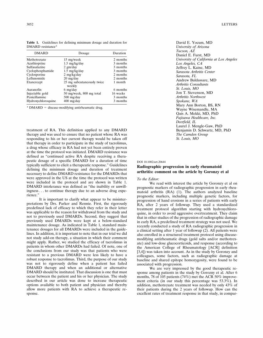
treatment of RA. This definition applied to any DMARDtherapy and was used to ensure that no patient whose RA wasresponding to his or her current therapy would be taken offthat therapy in order to participate in the study of tacrolimus,a drug whose efficacy in RA had not yet been entirely provenat the time the protocol was initiated. DMARD resistance wasdefined as “continued active RA despite receiving a thera-peutic dosage of a specific DMARD for a duration of timetypically sufficient to elicit a therapeutic response.” Guidelinesdefining the minimum dosage and duration of treatmentnecessary to define DMARD resistance for the DMARDs thatwere approved in the US at the time the protocol was writtenwere included in the protocol and are shown in Table 1.DMARD intolerance was defined as “the inability or unwill-ingness . . . to continue therapy due to an adverse drug expe-rience.”
It is important to clarify what appear to be misinter-pretations by Drs. Parker and Rennie. First, the rigorouslypredefined lack of efficacy to which they refer in their letterwas applicable to the reason for withdrawal from the study andnot to previously used DMARDs. Second, they suggest thatpreviously used DMARDs were kept at a below-standardmaintenance dosage. As indicated in Table 1, standard main-tenance dosages for all DMARDs were included in the guide-lines. In addition, it is important to note that in our trial we didnot study add-on therapy, a situation in which their commentmight apply. Rather, we studied the efficacy of tacrolimus inpatients in whom other DMARDs had failed. Of note, one ofthe conclusions from our study was that patients who wereresistant to a previous DMARD were less likely to have arobust response to tacrolimus. Third, the purpose of our studywas not to rigorously define when a patient has failedDMARD therapy and when an additional or alternativeDMARD should be instituted. That discussion is one that mustoccur between the patient and his or her physician. The studydescribed in our article was done to increase therapeuticoptions available to both patient and physician and therebyallow more patients with RA to achieve a therapeutic re-sponse.
David E. Yocum, MDUniversity of ArizonaTucson, AZDaniel E. Furst, MDUniversity of California at Los AngelesLos Angeles, CAJeffrey L. Kaine, MDSarasota Arthritis CenterSarasota, FLAndrew Baldassare, MDArthritis ConsultantsSt. Louis, MOJon T. Stevenson, MDArthritis NorthwestSpokane, WAMary Ann Borton, BS, RNWayne Wisemandle, MAQais A. Mekki, MD, PhDFujisawa Healthcare, Inc.Deerfield, ILLaurel J. Mengle-Gaw, PhDBenjamin D. Schwartz, MD, PhDThe Camden GroupSt. Louis, MO
DOI 10.1002/art.20644
Radiographic progression in early rheumatoidarthritis: comment on the article by Goronzy et al
To the Editor:We read with interest the article by Goronzy et al on
prognostic markers of radiographic progression in early rheu-matoid arthritis (RA) (1). The authors analyzed baselineprognostic markers, including multiple genetic factors, forprogression of hand erosions in a series of patients with earlyRA, after 2 years of followup. They used a standardizedtreatment protocol algorithm starting with hydroxychloro-quine, in order to avoid aggressive overtreatment. They claimthat in other studies of the progression of radiographic damagein early RA, a predefined treatment strategy was not used. Werecently conducted a study of RA radiographic progression ina clinical setting after 1 year of followup (2). All patients werealso enrolled in a structured treatment protocol using disease-modifying antirheumatic drugs (gold salts and/or methotrex-ate) and low-dose glucocorticoids, and response (according tothe American College of Rheumatology [ACR] definition[3,4]) was taken into account. As in the study by Goronzy andcolleagues, some factors, such as radiographic damage atbaseline and shared epitope homozygosity, were found to beassociated with progression.
We are very impressed by the good therapeutic re-sponse among patients in the study by Gorozny et al. After 6months, 78 of 105 patients (74%) met the ACR 50% improve-ment criteria (in our study this percentage was 53.3%). Inaddition, methotrexate treatment was needed by only 43% oftheir patients during the 2 years of followup. How can theexcellent rates of treatment response in that study, in compar-
Table 1. Guidelines for defining minimum dosage and duration forDMARD resistance*
DMARD Dosage Duration
Methotrexate 15 mg/week 2 monthsAzathioprine 1.5 mg/kg/day 3 monthsSulfasalazine 2 gm/day 3 monthsCyclophosphamide 1.5 mg/kg/day 3 monthsCyclosporine 2 mg/kg/day 2 monthsLeflunomide 20 mg/day 2 monthsEtanercept 25 mg subcutaneously twice
weekly1 month
Auranofin 6 mg/day 6 monthsInjectable gold 50 mg/week, 800 mg total 16 weeksPenicillamine 500 mg/day 3 monthsHydroxychloroquine 400 mg/day 3 months
* DMARD � disease-modifying antirheumatic drug.
3052 LETTERS
ison with ours and other studies using hydroxychloroquine, adrug considered to have a modest therapeutic effect in RA (5),be explained? Although the short disease duration of Goronzyand colleagues’ patients at study enrollment (mean 6 months),the high percentage of patients who were negative for rheu-matoid factor (41.8%), and the type of population analyzed(recruited from the local community) may partly explain thesehigh response rates, we believe other, as-yet-unknown, factorsmay also contribute to the strikingly benign disease evolution.Moreover, recent studies do not support the notion that RA inthe community population has a benign course (6). Thus, itwould be interesting to know the number of patients in whomcomplete remission was achieved and the percentage andclinical course of patients with seronegative disease or withdisease duration of �3 months at enrollment. It has beenreported that, in these patients, the rate of remission was veryhigh and probably unrelated to antirheumatic therapy (7).Moreover, the measurement of specific serologic markers ofRA, such as anti–cyclic citrullinated peptide antibodies (8),would be of interest in order to better classify this type ofarthritis in the early stages.
In conclusion, the study by Goronzy et al makesimportant contributions to the knowledge of factors related toradiographic progression in early RA, but in our opinion, theunexpected benign course in the patients reported is difficultto explain, and extrapolation of the results to other populationswith early RA is questionable.
Raimon Sanmarti, MDHospital ClinicBarcelona, SpainAntoni Gomez-Centeno, MDJordi Gratacos, MDHospital Parc TauliSabadell, SpainJuan D. Canete, MD, PhDHospital ClinicBarcelona, Spain
1. Goronzy JJ, Matteson EL, Fulbright JW, Warrington KJ, Chang-Miller A, Hunder GG, et al. Prognostic markers of radiographicprogression in early rheumatoid arthritis. Arthritis Rheum 2004;50:43–54.
2. Sanmarti R, Gomez A, Ercilla G, Gratacos J, Larrosa M, Suris X,et al. Radiological progression in early rheumatoid arthritis afterDMARDS: a one-year follow-up study in a clinical setting. Rheu-matology (Oxford) 2003;42:1044–9.
3. Felson DT, Anderson JJ, Boers M, Bombardier C, Furst D,Goldsmith C, et al. American College of Rheumatology prelimi-nary definition of improvement in rheumatoid arthritis. ArthritisRheum 1995;38:727–35.
4. Felson DT, Anderson JJ, Lange MLM, Wells G, LaValley MP.Should improvement in rheumatoid arthritis clinical trials bedefined as fifty percent or seventy percent improvement in core setmeasures rather than twenty percent? Arthritis Rheum 1998;41:1564–70.
5. Felson DT, Anderson JJ, Meenan RF. The comparative efficacyand toxicity of second-line drugs in rheumatoid arthritis: results oftwo metaanalyses. Arthritis Rheum 1990;33:1449–61.
6. Harrison BJ, Symmons DP, Brennan P, Bankhead CR, BarrettEM, Scott DG, et al. Inflammatory polyarthritis in the communityis not a benign disease: predicting functional disability one yearafter presentation. J Rheumatol 1996;23:1326–31.
7. Green M, Marzo-Ortega H, McGonagle D, Wakefield R, Proud-man S, Conaghan P, et al. Persistence of mild, early inflammatoryarthritis: the importance of disease duration, rheumatoid factor,and the shared epitope. Arthritis Rheum 1999;42:2184–8.
8. Schellekens GA, Visser H, de Jong BA, van den Hoogen FH,Hazes JM, Breedveld FC, et al. The diagnostic properties ofrheumatoid arthritis antibodies recognizing a cyclic citrullinatedpeptide. Arthritis Rheum 2000;43:155–63.
DOI 10.1002/art.20645
Reply
To the Editor:Sanmarti et al raise the question as to whether patients
with RA enrolled in our prospective study had unusuallybenign disease compared with other prospective cohorts, inparticular, the one they have followed up in Spain at theHospital Clinic of Barcelona and Hospital Parc Tauli ofSabadell (Sanmarti R, Gomez A, Ercilla G, Gratacos J,Larrosa M, Suris X, et al. Radiological progression in earlyrheumatoid arthritis after DMARDS: a one-year follow-upstudy in a clinical setting. Rheumatology [Oxford] 2003;42:1044–9). There have been few prospective studies that haveexplored prognostic markers and used predetermined treat-ment algorithms, among them the cohort study reported bySanmarti and colleagues in 2003 (we did not claim that ourstudy was the only one, as they imply). If one compares thesestudies carefully, the outcome results are actually quite similarand within the margin of error given the sample sizes: theSpanish cohort had 60 patients and our cohort had 111patients. Specifically, after 1 year (a comparison of the dataafter 2 years is not possible because the followup in the Spanishstudy was only 1 year), 38% of the Spanish patients had notachieved an ACR50 response and were taking methotrexate.The ACR50 response rate in the US cohort was almostidentical at 57%. Seventy-four percent of the Spanish patientshad no progression in the number of erosions within 1 year,again very similar to the US patients, who on average gained 1erosion per 2 years and of whom 48% did not have any erosiveprogression over 2 years.
It is correct that the 2 studies had slightly differentdemographics; the Spanish study allowed for the enrollment ofpatients with disease duration up to 2 years and showed thatlonger disease duration was associated with more radiographicprogression, whereas the US study only enrolled patients whohad had symptoms for �1 year. However, there is no indicationthat patients enrolled in our study did not have RA but ratherhad a self-limited arthritic disease. Complete remission after 2years and change in diagnosis were the rare exceptions, evenamong patients whose disease duration was �3 months at thetime of enrollment. Also, �10% of our patients who werenegative for rheumatoid factor at enrollment subsequentlybecame positive, and the percentage of rheumatoid factor–positive patients was comparable in the 2 studies.
Thus, the lesson from both studies is that there is asubset of patients with RA who tend to do well with nonag-gressive treatment. Prognostic markers are needed to identify
LETTERS 3053
such patients in order to avoid overtreatment, particularly if wemove from a step-up treatment strategy to an early aggressiveapproach.
Jorg J. Goronzy, MDCornelia M. Weyand, MDEmory University School of MedicineAtlanta, GA
DOI 10.1002/art.20646
Ultrasonography of the shoulder in patients withrheumatoid arthritis: comment on the article byHermann et al
To the Editor:We read with interest the article by Hermann et al (1),
reporting on a study in which they compared 3 imagingmethods in rheumatoid arthritis (RA) patients with shoulderpain. They detected erosions of the glenohumeral joint byradiography in 60% of patients, by ultrasonography (US) in70%, and by magnetic resonance imaging (MRI) in 91%.Although US was superior to conventional radiography indetecting erosions, significantly more erosions of the humeralhead were detected by MRI than by the other 2 imagingmethods. MRI was also superior to US in identifying synovitisof the glenohumeral joint (28% versus 63%), tenosynovitis ofthe biceps tendon (35% versus 65%); and bursitis (30% versus42%). We would like to raise some concerns about theseresults.
First, the finding of the low rate of erosions by US inthe RA group is difficult to understand. The quality of an USexamination depends not only on the experience of the exam-iner, but also on the equipment. Even in healthy adults,high-resolution US detects erosions (defined as a pit in thebone surface of �1 mm diameter in all 3 diameters) in 23% ofshoulders (2). The low resolution of the rather old US equip-ment used by Hermann and colleagues could explain the lowfrequency of erosions detected in the RA group. Furthermore,comparison of the rates of erosions identified by MRI and USis a perilous undertaking, since there is no internationalconsensus regarding the definition of erosions of the shoulderas detected by US. Because there is no gold standard, itremains uncertain whether the authors’ classification of ero-sions for US and MRI pertains to the same pathology. Forexample, the observed cortical defects with hypointense signalon T1-weighted spin-echo images could represent osteoar-thritic changes rather than RA-related pathology.
Second, gray-scale US is an excellent method fordetecting (para)articular abnormalities such as synovial mem-brane thickening and proliferation, joint effusion, bursal effu-sion, peritendinous effusion, and rotator cuff tears (3). Theauthors stated that their US findings of glenohumeral jointeffusion (28%), biceps sheath effusion (35%), and fluid withinthe bursa (30%) were consistent with those in a study byAlasaarela et al (4), but in fact they were not. The latter group
reported these abnormalities in 92%, 83%, and 89%, respec-tively, of cases. This large discrepancy between the results ofHermann et al and those of Alasaarela et al again raisesconcerns about the quality of the US equipment. It is also apity that the authors did not compare clinical examination forsoft tissue changes with US and MRI findings. Although USmay be less sensitive than MRI, it is likely to be more sensitiveand specific than clinical examination.
Third, Hermann et al limited their examination to theglenohumeral joint. As control subjects, they examined indi-viduals with shoulder pain. However, it is well known thatshoulder pain may originate from the acromioclavicular joint,which appears to be involved more often than the hum-eroscapular joint in patients with RA (5). This raises questionsas to whether the authors examined the correct joint. Exam-ining both the glenohumeral and the acromioclavicular jointcould have settled this issue.
We would like to make a final point. The report byHermann and colleagues focuses on erosions in the shoulderjoint. However, considering the fact that erosions in theglenoid fossa cannot be visualized by US it is questionablewhether US should be used in the first place for this purpose.We believe the future of US lies in the direction of visualiza-tion of synovitis and bursitis, and making the distinctionbetween inflammatory and noninflammatory disease.
George A. W. Bruyn, MD, PhDMedisch Centrum LeeuwardenLeeuwarden, The NetherlandsAnnamaria Iagnocco, MDUniversity of Rome La SapienzaRome, ItalyEsperanza Naredo, MDHospital Severo OchoaMadrid, SpainRichard J. Wakefield, BM, MRCPUniversity of LeedsLeeds, UKWolfgang A. Schmidt, MDHospital Berlin-BuchBerlin, Germany
1. Hermann KG, Backhaus M, Schneider U, Labs K, Loreck D,Zuhlsdorf S, et al. Rheumatoid arthritis of the shoulder joint:comparison of conventional radiography, ultrasound, and dynamiccontrast-enhanced magnetic resonance imaging. Arthritis Rheum2003;48:3338–49.
2. Schmidt WA, Schmidt H, Schicke B, Gromnica-Ihle E. Standardreference values for musculoskeletal ultrasonography. Ann RheumDis. In press.
3. Alasaarela E, Alasaarela EL. Ultrasound evaluation of painfulrheumatoid shoulders. J Rheumatol 1994;21:1642–8.
4. Alasaarela E, Takalo R, Tervonen O, Hakala M, Suramo I.Sonography and MRI in the evaluation of painful arthritic shoulder.Br J Rheumatol 1997;36:996–1000.
5. Lehtinen JT, Kaarela K, Belt EA, Kautiainen HJ, Kauppi MJ,Lehto MU. Relation of glenohumeral and acromioclavicular jointdestruction in rheumatoid shoulder; a 15 year follow up study. AnnRheum Dis 2000;59:158–60.
3054 LETTERS
DOI 10.1002/art.20647
Reply
To the Editor:We appreciate the detailed observations by Bruyn et al
concerning our study comparing conventional radiography,US, and MRI of the shoulder joint in RA. Their criticalcomments highlight the fact that investigators are far fromagreement on the most suitable imaging modalities for thediagnostic assessment of RA, and provide a good starting pointfor fruitful discussion.
Bruyn and colleagues raise 3 major issues. First, theyexpress doubt about the low rate of detection of erosions byUS, which they partly attribute to the low resolution of the USequipment used in our study. We did indeed use a US machinewith average performance and a maximum frequency of 7.5MHz. However, we deliberately chose this machine because itis the standard equipment typically used by rheumatologists.State-of-the-art US equipment with high-resolution lineartransducers (�10 MHz) is available at our institution forfuture studies but is currently not in wide use in the routinediagnostic setting. The rate of detection of erosions in ourstudy was 70% with US and 91% with MRI. These are belowthe rates reported by Alasaarela et al (1,2). All assessable areasof the humeral head were very carefully evaluated ultrasono-graphically in our study, by a rheumatologist experienced inthe field of musculoskeletal US. Superficial bone lesions areclearly identified by US, while deeper erosions are not acces-sible and are better identified by MRI. Another reason for thedifference in the detection of erosions by US and MRIbetween Alasaarela and colleagues’ study and ours is the factthat we used high-resolution 1.5T MRI and a high-resolutionmatrix of up to 512 pixels, while they performed MRI at 1.0T.The thin slice thickness of 3 mm used with some sequenceslikewise improves the detection of very small erosions by MRI.
Bruyn et al quote the most recent study on the USmorphology of clinically healthy joints (3), which was per-formed with state-of-the-art US equipment and yields interest-ing new insights. That study identified changes of the shoulderjoint resembling RA erosions in up to 23% of cases. The 10patients in our control group were examined only by MRI anddid not undergo US. MRI depicted erosive lesions in 20% ofthe controls, which is comparable with the above-quotedresults found by Schmidt et al (3). However, this finding needsto be verified in larger study groups. It would be useful to knowwhether Schmidt et al confirmed the bone lesions they identi-fied in healthy subjects with other imaging modalities (conven-tional radiography, MRI). It is well known that shoulderlesions become symptomatic later because the shoulder jointsare exposed to less strain than the legs. Moreover, the term“erosions” should not be used to refer to changes of the headof humerus in healthy subjects. The term “bone lesions” ismore appropriate, and these are most likely due to degenera-tive changes. Furthermore, it is important to confirm thesechanges using other imaging modalities, in particular, todetermine whether the changes are cystic bone lesions.
Second, Bruyn et al comment on our results concern-ing the detection of soft tissue lesions such as synovitis,tenosynovitis, and bursitis by US and compare these with thefindings published by Alasaarela et al (1). Synovitis wasdemonstrated by US in 28% of our study patients, as opposed
to 81% of the Finnish patients studied by Alasaarela andcolleagues (1). Agreement with MRI results is poor, as re-flected by a kappa value of 0.29 in both studies. However,kappa values in different studies cannot be compared when themarginal distribution is not the same (4). The difference in thepercentage of patients with US-detected synovitis in the 2study populations may be attributable to several factors, suchas differences in the duration of disease, rheumatoid factorstatus, or general disease activity. Moreover, the group of 30patients studied by Alasaarela et al was heterogeneous: mosthad RA, but some had other rheumatic diseases. Alasaarelaand colleagues likewise used a 7.5-MHz transducer and foundthe agreement between MRI and US in the detection ofbursitis and tenosynovitis to be good (� �0.40), whereas it waspoor in our study population (� �0.40).
Bruyn et al calculate a high incidence of soft tissueinvolvement in RA of the shoulder joint in the study byAlasaarela et al (1) by relating the number of joints positive byUS to the number positive by MRI, but not to the total numberof patients. This calculation is incorrect since it does not takeinto account the fact that MRI is not the gold standard, norwas it treated as a gold standard in our study or the study byAlasaarela and colleagues. For this reason, the possibility offalse-positive findings cannot be excluded. The internationalOMERACT (Outcome Measures in Rheumatology ClinicalTrials) group is currently discussing the recognition of MRI asthe gold standard for imaging of RA changes in the small jointsof the hands (5,6), but this is not presently planned for theshoulder joint.
A third issue raised by Bruyn et al concerns theacromioclavicular joint, which they suggest should have beenincluded in our analysis. Our protocol included examination ofthe acromioclavicular joint, but the analysis was not included inour recent report. An additional statistical analysis of acromi-oclavicular joint involvement in RA would have been beyondthe scope of that article, which was already quite detailed andlong. This question will be addressed in a followup report.
In conclusion, we believe erosions of the shoulder jointcan be identified by US as well as MRI. The latter appears tobe superior in that it depicts all portions of the joint includingthe glenoid fossa. US has an important role in assessing softtissue involvement in RA since it is widely available and clearlydifferentiates inflammatory and noninflammatory changes,and also has a higher sensitivity and specificity compared withclinical examination (7). MRI has a role as a problem-solvingtool. Definitive conclusions regarding the discrepancies in theincidence of individual US findings in comparison with MRIfindings among various studies can be drawn only on the basisof multicenter studies conducted using different equipmentand involving different examiners.
Kay-Geert A. Hermann, MDMarina Backhaus, MDTania SchinkBernd Hamm, MDCharite HospitalBerlin, GermanyMatthias Bollow, MDAugust-Kranken-AnstaltBochum, Germany
LETTERS 3055
1. Alasaarela E, Takalo R, Tervonen O, Hakala M, Suramo I.Sonography and MRI in the evaluation of painful arthritic shoulder.Br J Rheumatol 1997;36:996–1000.
2. Alasaarela E, Suramo I, Tervonen O, Lahde S, Takalo R, HakalaM. Evaluation of humeral head erosions in rheumatoid arthritis: acomparison of ultrasonography, magnetic resonance imaging, com-puted tomography and plain radiography. Br J Rheumatol 1998;37:1152–6.
3. Schmidt WA, Schmidt H, Schicke B, Gromnica-Ihle E. Standardreference values for musculoskeletal ultrasonography. Ann RheumDis 2004;63:988–94
4. Feinstein AR, Cicchetti DV. High agreement but low kappa. I. Theproblems of two paradoxes. J Clin Epidemiol 1990;43:543–9.
5. McQueen F, Lassere M, Edmonds J, Conaghan P, Peterfy C, BirdP, et al. OMERACT rheumatoid arthritis magnetic resonanceimaging studies. summary of OMERACT 6 MR imaging module.J Rheumatol 2003;30:1387–92.
6. Ostergaard M, Szkudlarek M. Imaging in rheumatoid arthritis: whyMRI and ultrasonography can no longer be ignored. Scand J Rheu-matol 2003;32:63–73.
7. Iagnocco A, Coari G, Leone A, Valesini G. Sonographic study ofpainful shoulder. Clin Exp Rheumatol 2003;21:355–8.
DOI 10.1002/art.20648
Electroretinograms of children born to motherstreated with hydroxychloroquine during pregnancyand breast-feeding: comment on the article byCostedoat-Chalumeau et al
To the Editor:We read with interest the article by Costedoat-
Chalumeau et al (1), who reported evidence that hydroxychlo-roquine (HCQ) therapy during pregnancy is safe for the fetus.In particular, no vision, hearing, growth, or developmentalabnormalities were found in any of the 119 children at the lastfollowup (mean age 26 months). We would like to addinformation on our experience in performing flash electroret-inography (ERG) in a small group of babies who had beenexposed to HCQ in utero.
In our pediatric department we routinely follow upchildren born to mothers with autoimmune diseases, irrespec-tive of maternal treatment and autoantibody status. We usuallyperform ophthalmologic examination with funduscopy in ba-bies whose mothers had taken HCQ during pregnancy. In 2003we also performed ERG in 6 of these babies, all of whom hadbeen exposed to HCQ in utero and were being seen by us forthe first time. For infants and young children, in whom visualfield examination cannot be performed, ERG is an additionaltool with which to seek evidence of possible adverse visioneffects. The characteristics of the study children are shown inTable 1. All babies were born without malformations or majorcomplications, and at the time of examination they were all ingood general health, with growth and development appropri-ate for age. ERG was performed successfully in all cases, andall results were normal.
ERG measures the retinal response to a stimulus oflight, using surface electrodes placed on the lower lid. Itprovides objective information on global retinal function andenables the examiner to distinguish between different retinaldisorders. In the 6 babies we studied, binocular repeatedstimulation with a standard flash white light from a distance of15 cm was performed during spontaneous sleep, withoutpharmacologic mydriasis. Electrodes recorded the retinal po-tentials that developed as a response. Two repeated measuresafter at least 15 flashes were analyzed and averaged. Bothoscillatory potentials and combined maximal response weresimultaneously analyzed. As noted above, results were normalin all cases.
Although HCQ treatment is thought to be safe duringpregnancy (2–4), there has been no definite consensus on this(5). The recent demonstration that HCQ crosses the placenta(6), with cord blood concentrations nearly identical to thosefound in maternal blood, emphasizes the need for carefulevaluation of the fetuses and newborn babies of women whoreceived HCQ during pregnancy. Among the potential adverseeffects of HCQ, retinal toxicity is of concern, and abnormali-ties on ERG have been associated with HCQ treatment (7). Inthe study by Costedoat-Chalumeau and coworkers (1), data onthe children were collected from mothers, general practi-tioners, and/or pediatricians, but ophthalmologic examinations
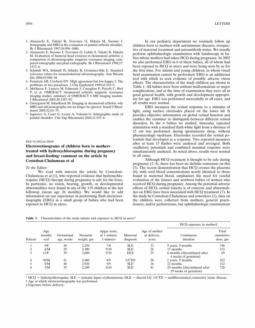
Table 1. Characteristics of the study infants and exposure to HCQ in utero*
Patient
Age,months/
sex†Gestationalage, weeks
Neonatalweight, gm
Apgar score,at 1 minute/
5 minutesMaternaldiagnosis
Age of motherat delivery,
years
HCQ exposure in mother‡
Continuousduration
Totalcumulativedose, gm
1 9/F 38 2,250 7/8 SLE 32 9 years, 9 months 7462 4/M 39 3,300 9/10 SLE 26 17 months 1533 13/F 39 2,680 9/10 DLE 25 6 months (discontinued after
4 weeks of gestation)69
4 30/M 41 2,480 8/9 UCTD 30 5 years, 9 months 4225 9/M 40 2,920 9/9 SLE 26 22 months 2126 3/M 35 2,100 8/10 SLE 41 29 months (discontinued after
19 weeks of gestation)728
* HCQ � hydroxychloroquine; SLE � systemic lupus erythematosus; DLE � discoid LE; UCTD � undifferentiated connective tissue disease.† Age at which electroretinography was performed.‡ Exposure before delivery.
3056 LETTERS

were not systematically performed. However, no clinical visionabnormality was observed. Our findings provide further evi-dence of the retinal safety of HCQ, in accordance withprevious reports (7–9). To our knowledge the use of ERGtesting of children exposed in utero to HCQ has not beenpreviously reported. All of the mothers in our study took HCQduring their pregnancies, and 5 of them took it also duringbreast-feeding. It is known that HCQ is excreted in breast milk,even if in small amounts (10). It is noteworthy that one motherbreast-fed her son for 30 months while taking HCQ (200 mgdaily); ERG was performed after this long period, and even inthis case the results were completely normal. Although ourstudy sample was small, our data reinforce the observation thatHCQ treatment is safe during pregnancy and lactation, even ifbreast-feeding is continued for many months.
Rolando Cimaz, MDInstituti Clinici di PerfezionamentoAntonio Brucato, MDOspedale NiguardaElisa Meregalli, MDIstituti Clinici di PerfezionamentoMarina Muscara, MDOspedale NiguardaPaola Sergi, MDIstituti Clinici di PerfezionamentoMilan, Italy
1. Costedoat-Chalumeau N, Amoura Z, Duhaut P, Huong DL,Sebbough D, Wechsler B, et al. Safety of hydroxychloroquine inpregnant patients with connective tissue diseases: a study of onehundred thirty-three cases compared with a control group. Arthri-tis Rheum 2003;48:3207–11.
2. Parke A, West B. Hydroxychloroquine in pregnant patients withsystemic lupus erythematosus. J Rheumatol 1996;23:1715–8.
3. Buchanan NM, Toubi E, Khamashta MA, Lima F, Kerslake S,Hughes GR. Hydroxychloroquine and lupus pregnancy: review ofa series of 36 cases. Ann Rheum Dis 1996;55:486–8.
4. Khamashta MA, Buchanan NM, Hughes GR. The use of hydroxy-chloroquine in lupus pregnancy: the British experience. Lupus1996;5 Suppl:S65–6.
5. Wallace DJ. Antimalarials: the “real” advance in lupus. Lupus2001;10:385–7.
6. Costedoat-Chalumeau N, Amoura Z, Aymard G, Huong DL,Wechsler B, Vauthier D, et al. Evidence of transplacental passageof hydroxychloroquine in humans. Arthritis Rheum 2002;46:1123–4.
7. Klinger G, Morad Y, Westall CA, Laskin C, Spitzer KA, Koren G,et al. Ocular toxicity and antenatal exposure to chloroquine orhydroxychloroquine for rheumatic diseases. Lancet 2001;358:813–4.
8. Motta M, Tincani A, Faden D, Zinzini E, Chirico G. Antimalarialagents in pregnancy. Lancet 2002;359:524–5.
9. Levy RA, Vilela VS, Cataldo MJ, Ramos RC, Duarte JL, TuraBR, et al. Hydroxychloroquine (HCQ) in lupus pregnancy: double-blind and placebo-controlled study. Lupus 2001;10:401–4.
10. Al-Herz A, Schulzer M, Esdaile JM. Survey of antimalarial use inlupus pregnancy and lactation. J Rheumatol 2002;29:700–6.
DOI 10.1002/art.20649
Reply
To the Editor:We thank Dr Cimaz and colleagues for their interest in
our report. Both retinal toxicity and ototoxicity in childrenborn to women treated with chloroquine have been reportedrarely (1,2). To our knowledge, no abnormalities have beennoted in relation to HCQ treatment, and we found no clinicalvisual abnormalities in 119 children at the last followup (meanage 26 months), as noted by Cimaz et al. Our clinical data arein accordance with those of Klinger et al, who found thatresults of ophthalmologic examinations and tests were normalin 14 children exposed to HCQ in utero and studied at a meanage of 1.9 years (3). In that study, ophthalmologic examina-tions and tests included slitlamp biomicroscopy of the anteriorsegment, dilated retinal examination using indirect ophthal-moscopy, cyclopegic refraction, visual acuity testing, visualfield assessment, and color vision assessment. These resultshave been confirmed in 26 additional children, by dilatedretinal examination with indirect ophthalmoscopy (4,5). Thenormal ERG results reported by Cimaz and colleagues thusreinforce the findings of previous studies.
We have also performed ERG in infants born tomothers who had been treated with HCQ during pregnancy(Table 1). All 4 mothers had systemic lupus erythematosus.HCQ treatment was continued throughout gestation (n � 4)and breast-feeding (n � 2). The mean gestational age of thesechildren was 38.6 weeks (range 38–39), and the mean weight at
Table 1. Characteristics of the study infants and exposure to HCQ in utero*
Patient
Age,months/
sex†Gestationalage, weeks
Neonatalweight, gm
Apgar score, at1 minute/5 minutes
Maternaldiagnosis
Age of motherat delivery,
years
HCQ exposure in mother‡
Continuousduration,months
Totalcumulativedose, gm
1 4/F 39 3,320 10/10 SLE 23 22 2642 13/F 39 3,000 10/10 SLE 30 84 1,0083 6/F 38 2,330 7/10 SLE 30 98 1,1764 7/F 38.5 2,630 10/10 SLE 32 74 888
* HCQ � hydroxychloroquine; SLE � systemic lupus erythematosus.† Age at which electroretinography was performed.‡ Exposure before delivery.
LETTERS 3057
birth was 2,820 gm (range 2,330–3,320). Ophthalmologic eval-uation included dilated retinal examination using indirectophthalmoscopy and ERG. The median age of the children atthe time of ERG was 6.5 months (range 4–13). No abnormal-ities were found.
Interestingly, Cimaz and colleagues emphasized that 5of the 6 children they studied were breast-fed. We havepreviously analyzed HCQ in the breast milk of 2 mothers (6);the HCQ concentrations were 344 ng/ml and 1,424 ng/ml,respectively. We later confirmed these results in 2 moremothers, whose breast milk HCQ concentrations were 1,131ng/ml and 1,392 ng/ml. It can thus be calculated that the HCQingestion by the infants was no more than 0.2 mg/kg/day. Theselevels are concordant with the daily ingestion of 0.11 mg/kg in1 infant reported by Nation et al (7). The amount of HCQreceived by children throughout lactation is very small com-pared with the daily therapeutic dosage (6.5 mg/kg in adults)(8). Additionally, given that HCQ concentrations in breastmilk are low compared with those found in cord blood(transplacental passage), it does not seem logical to adviseagainst breast-feeding if HCQ therapy has been maintainedthroughout pregnancy.
Taken altogether, these data provide support for pre-vious evidence of the safety of HCQ during pregnancy andlactation and are concordant with the experience of selectedexperts, as reported in a national survey concerning the use ofantimalarial drugs in lupus pregnancy (9). None of the respon-dents reported having seen any fetal toxicity related to the useof antimalarial agents.
However, since retinal toxicity of HCQ in adults is rare(3), the number of children studied may be insufficient todetect infrequent fetal retinal toxicity. A multicenter studyincluding complete ophthalmologic examinations and long-term followup of the children would be recommended in orderto obtain more definitive answers.
Nathalie Costedoat-Chalumeau, MDZahir Amoura, MDDjamel Sebbough, MDJean-Charles Piette, MDCentre Hospitalier Universitaire Pitie-SalpetriereParis, France
1. Phillips-Howard PA, Wood D. The safety of antimalarial drugs inpregnancy. Drug Saf 1996;14:131–45.
2. Paufique L, Magnard P. Retinal degeneration in 2 children follow-ing preventive antimalarial treatment of the mother during preg-nancy. Bull Soc Ophtalmol Fr 1969;69:466–7.
3. Klinger G, Morad Y, Westall CA, Laskin C, Spitzer KA, Koren G,et al. Ocular toxicity and antenatal exposure to chloroquine orhydroxychloroquine for rheumatic diseases. Lancet 2001;358:813–4.
4. Motta M, Tincani A, Faden D, Zinzini E, Chirico G. Antimalarialagents in pregnancy. Lancet 2002;359:524–5.
5. Levy RA, Vilela VS, Cataldo MJ, Ramos RC, Duarte JL, Tura BR,et al. Hydroxychloroquine (HCQ) in lupus pregnancy: double-blindand placebo-controlled study. Lupus 2001;10:401–4.
6. Costedoat-Chalumeau N, Amoura Z, Aymard G, Huong DL,Wechsler B, Vauthier D, et al. Evidence of transplacental passageof hydroxychloroquine in humans. Arthritis Rheum 2002;46:1123–4.
7. Nation RL, Hackett LP, Dusci LJ, Ilett KF. Excretion of hydroxy-chloroquine in human milk. Br J Clin Pharmacol 1984;17:368–9.
8. Physician’s Desk Reference. 57th ed. Montvale (NJ): Thomson;2003.
9. Al-Herz A, Schulzer M, Esdaile JM. Survey of antimalarial use inlupus pregnancy and lactation. J Rheumatol 2002;29:700–6.
DOI 10.1002/art.20650
Elective pregnancy termination and microchimerism:comment on the article by Khosrotehrani et al
To the Editor:In continuation of the intriguing microchimerism story,
Khosrotehrani et al (1) make the point that it is during earlypregnancy when fetal loss is most likely to result in the releaseof progenitor cells, those cells that have the greatest potentialfor engraftment, expansion, and differentiation within mater-nal tissues. I would like to emphasize that elective terminationof pregnancy is unique in terms of the release of theseprogenitor cells as well as facilitating their access to thematernal circulation.
There is a major difference between a spontaneousmiscarriage and elective termination of pregnancy. In the veryearly months of pregnancy, the expulsion accompanying spon-taneous miscarriage is nearly always preceded by the death ofthe embryo or fetus (2). Any fetomaternal cell trafficking thatoccurs is, for the most part, between the mother and dead cells.In contrast, during elective termination, a live healthy fetus istorn from the uterine lining, producing breaches and bleeding.The maternal blood is exposed to a profusion of live undiffer-entiated cells deriving from the torn and macerated tissues ofthe live fetus. Analysis by quantitative polymerase chain reac-tion amplification demonstrates a large fetal–maternal trans-fusion (3). Because engraftment is directly related to the size,viability, and lack of differentiation of the fetal cellular innocu-lum, engraftment is far more likely to follow elective termina-tion of pregnancy than spontaneous miscarriage. With termdelivery, as pointed out by the authors, there is blood ex-change, but the cells are well differentiated, posing little threatof engraftment.
The authors’ plea that physicians obtain detailed preg-nancy histories in women with scleroderma or other diseasespossibly related to microchimerism (4,5) is judicious. In fact, athorough pregnancy history, looking in particular for electivetermination of pregnancies, may be advisable in all women,because the effects of a large fetal–maternal transfusion ofengraftment-prone cells on the development or expression ofvirtually any disorder are at this time unknown.
Hugh McGrath, Jr., MDLouisiana State University Health Sciences CenterNew Orleans, LA
1. Khosrotehrani K, Johnson KL, Lau J, Dupuy A, Cha DH, BianchiDW. The influence of fetal loss on the presence of fetal cellmicrochimerism: a systematic review. Arthritis Rheum 2003;48:3237–41.
2. Cunningham FG, Gant NF, Leveno KJ, Gilstrap LC, Hauth JC,Wenstrom KD, editors. Williams obstetrics. 21st ed. New York:McGraw-Hill; 2001. p. 856.
3058 LETTERS
3. Bianchi DW, Farina A, Weber W, Delli-Bovi LC, Deriso M,Williams JM, et al. Significant fetal-maternal hemorrhage aftertermination of pregnancy: implications for development of fetal cellmicrochimerism. Am J Obstet Gynecol 2001;184:703–6.
4. Johnson KL, Nelson JL, Furst DE, McSweeney PA, Roberts DJ,Zhen D-K, et al. Fetal cell microchimerism in tissue from multiplesites in women with systemic sclerosis. Arthritis Rheum 2001;44:1848–54.
5. Klintschar M, Schwaiger P, Mannweiler S, Regauer S, Kleiber M.Evidence of fetal microchimerism in Hashimoto’s thyroiditis. J ClinEndocrinol Metab 2001;86:2494–8.
DOI 10.1002/art.20651
Reply
To the Editor:We thank Dr. McGrath for his insightful comments. In
our study, we analyzed the association between fetal cellmicrochimerism, defined by the persistence of male fetal cellsin women after pregnancy, and several pregnancy-relatedvariables. We observed a significant association between fetalloss, defined as both spontaneous abortion or elective termi-nation, and microchimerism. Most of the studies included inour meta-analysis did not distinguish between spontaneousabortions or elective termination. Therefore, we could notanalyze these 2 entities separately and do not have any data atpresent to strengthen Dr. McGrath’s hypothesis.
In his comments, Dr. McGrath made the interestingsuggestion that spontaneous abortion with preceding fetaldeath may not be a source of fetal cell microchimerism.Conversely, elective termination, as we have previously shownby quantitative polymerase chain reaction analysis, results in alarge transfer of fetal nucleated cells into the maternal circu-lation (1). We agree with Dr. McGrath that the frequency ofpregnancy-associated progenitor cells is likely to be higher inearly gestation, and that the transfer of fetal progenitor cells isimportant for the long-term development of microchimerism.
However, recent reports have described the detectionof fetal cell microchimerism in 30–50% of healthy women (2).Spontaneous abortion (including early pregnancy loss, definedas the loss of an embryo before the clinical diagnosis ofpregnancy) occurs in 32% of all conceptions in healthy youngnulliparous women (3). Given the high and somewhat similarfrequencies of microchimerism and spontaneous miscarriagein women, we cannot exclude the possibility that fetal cellsfrom a spontaneous miscarriage contribute to the developmentof microchimerism. Further research should address the ques-tion as to whether there is a difference in the development offetal cell microchimerism following spontaneous abortion ver-sus elective termination.
Kiarash Khosrotehrani, MDKirby L. Johnson, PhDDiana W. Bianchi, MDTufts–New England Medical CenterBoston, MA
1. Bianchi DW, Farina A, Weber W, Delli-Bovi LC, Deriso M,Williams JM, et al. Significant fetal-maternal hemorrhage after
termination of pregnancy: implications for development of fetal cellmicrochimerism. Am J Obstet Gynecol 2001;184:703–6.
2. Lambert NC, Lo YM, Erickson TD, Tylee TS, Guthrie KA, FurstDE, et al. Male microchimerism in healthy women and women withscleroderma: cells or circulating DNA? A quantitative answer.Blood 2002;100:2845–51.
3. Wang X, Chen C, Wang L, Chen D, Guang W, French J. Concep-tion, early pregnancy loss, and time to clinical pregnancy: a popu-lation-based prospective study. Fertil Steril 2003;79:577–84.
DOI 10.1002/art.20652
Causes of familial aggregation of fibromyalgia:comment on the article by Arnold et al
To the Editor:We read with interest the report by Arnold et al on
familial aggregation of fibromyalgia (FM) (1). Although wetend to agree with the authors’ statement that their finding offamilial aggregation of FM supports the validity of the diag-nosis, there should be at least 2 caveats with regard to theirfurther contention that the finding of familial aggregation alsosupports the notion of a genetic etiology.
First, the increased odds ratio for FM among first-degree relatives of index patients could reflect similar intra-uterine conditions among siblings. It is established that com-ponents of the hypothalamic–pituitary–adrenal axis, such ascorticotropin-releasing hormone, which is responsible for thehuman stress reaction, can cross the placenta and may havedeleterious effects that manifest many years after birth (2,3).Since this axis is considered to be dysfunctional in FM, it is notinconceivable that intrauterine development of siblings born toa mother with a high level of stress may predispose suchsiblings to the eventual development of FM, quite independentof any genetic determinant.
Second, it is important to point out that to the extentthat FM is describable in terms of a somatization-like disorder(4), environmental and behavioral influences may be of primeimportance. Specifically, the “language of the body” (5), or theuse of somatic symptoms as a means by which to expressanguish, to acquire attention, or to deal with anxiety and guilt,are all pathologic forms of expression that may be learnedwithin a family context, thus increasing the likelihood of FMoccurring in more than one member of a family.
Viewed in this context, Arnold and colleagues’ findingsare indeed intriguing, but multifactorial causation, or conceiv-ably a combination of genetic predisposition and a summationof life events (similar to the elegant model recently suggestedfor depression, linking life events with a polymorphism in theserotonin transporter gene [6]), better serve to explain thesefindings than do genetics alone.
Jacob N. Ablin, MDValerie Aloush, MDTel Aviv Souraski Medical CenterTel Aviv, Israel
1. Arnold LM, Hudson JI, Hess EV, Ware AE, Fritz DA, AuchenbachMB, et al. Family study of fibromyalgia. Arthritis Rheum 2004;50:944–52.
LETTERS 3059
2. Wadhwa PD, Sandman CA, Garite TJ. The neurobiology of stressin human pregnancy implications for prematurity and developmentof the fetal central nervous system. Prog Brain Res 2001;133:131–42.
3. Couzin J. Quirks of fetal environment felt decades later. Science2002;296:2167–9.
4. McBeth J, Macfarlane GJ, Benjamin S, Silman AJ. Features ofsomatization predict the onset of chronic widespread pain: resultsof a large population-based study. Arthritis Rheum 2001;44:940–6.
5. Shapiro B. Building bridges between body and mind: the analysis ofan adolescent with paralyzing chronic pain. Int J Psychoanal2003;84:547–61.
6. Caspi A, Sugden K, Moffitt TE, Taylor A, Craig IW, Harrington H,et al. Influence of life stress on depression: moderation by apolymorphism in the 5-HTT gene. Science 2003;301:386–9.
DOI 10.1002/art.20653
Reply
To the Editor:We welcome the comments of Drs. Ablin and Aloush,
which provide us with an opportunity to clarify 2 issues. First,we did not claim in our article that genetics alone causesfibromyalgia. Indeed, we agree that environmental factors verylikely play a role. Second, we agree that familial aggregationcan be due to either shared familial environmental factors,genetic factors, or a combination of the 2.
However, the high degree of familial aggregationfound in our study (an odds ratio or hazard ratio of 5.8–8.5,depending on the type of analysis) cannot be plausibly ex-plained in the absence of genetic factors. For example, con-sider an analysis by Khoury and colleagues (Khoury MJ, BeatyTH, Liang KY. Can familial aggregation of disease be ex-plained by familial aggregation of environmental risk factors?Am J Epidemiol 1988;127:674–83): if one makes the unrealis-tic assumption of complete correlation of exposure to anenvironmental risk factor among relatives, and then further
assumes that the environmental risk factor has a relative riskfor disease of 10—an extraordinarily high effect—one wouldstill obtain only modest levels of familial aggregation (relativerisk �2.0) in the absence of genetic effects. Similarly, Guo hasshown that even under the slightly more realistic, but stillunlikely, scenario of 2 environmental risk factors, each with acorrelation of 0.5 among relatives and with a relative risk ofdisease of 5, acting multiplicatively, the relative risk for familialaggregation in the absence of genetic effects is still only 2 (GuoM. Familial aggregation of environmental risk factors andfamilial aggregation of disease. Am J Epidemiol 2000;151:1121–31). If we return to our study and convert our estimatesfrom odds ratios and hazard ratios to relative risk measures,these are all 5 or greater—effect sizes that are unachievableunder any realistic scenario in the absence of any geneticinfluence. Therefore, it is not plausible that shared intrauterineconditions (see Khoury and colleagues’ discussion of terato-gens) or patterns of pathologic expression learned withinfamilies could account entirely for the high level of familialaggregation observed—unless these factors themselves werestrongly determined by genetics.
In conclusion, while not precluding a potentially im-portant role of environmental factors, our findings stronglysuggest that genetic factors contribute to the causation offibromyalgia, and it will be important to search for such factorsin subsequent studies.
Lesley M. Arnold, MDUniversity of Cincinnati
College of MedicineCincinnati, OHJames I. Hudson, MD, ScDMcLean HospitalBelmont, MA
and Harvard Medical SchoolBoston, MAPaul E. Keck, Jr., MDUniversity of Cincinnati
College of MedicineCincinnati, OH
3060 LETTERS