Embed Size (px)
Citation preview
371
a few cases11). To our knowledge, only one case of simultaneous cranial subarachnoid hemorrhage (SAH) and spinal SDH exists in the literature17). In this report, we discuss a female patient on anticoagulant after aortic valve replacement surgery who showed simultaneous spinal SDH and cranial SAH.
CASE REPORT
The 53-year-old female patient was transferred to the emer-gency room of our institute due to the sudden onset of headache, nausea, and vomiting, all of which developed while she was tak-ing a shower. A neurological examination revealed a slightly drowsy conscious mentality, nuchal rigidity with bilateral weak-ness of the lower limbs (Grade I) and sensory loss below T4 der-matome. Anal tone and perineal sensory capability were also severely impaired. The patient complained of the initial sudden onset of a headache and giddiness followed by the acute onset of weakness in the lower limbs with backache.
The patient had undertaken aortic valve replacement surgery about 10 years ago, and had been taking oral anticoagulant (Cou-madine 5 mg, daily) since the operation. Her blood chemistry showed a platelet count of 199×103/μL, a prothrombin time of 46.5 s (normal range, 9.4–12.5 s), a partial thromboplastin time of 47.3 s (normal range, 28.0–44.0 s), and an International Normal-ized Ratio (INR) of 4.39 (range 0.9–1.27).
INTRODUCTION
A spinal subdural hemorrhage (SDH) is a rare clinical entity that accounts for only 4.1% of all spinal hematomas13). The most com-mon form of intra-spinal hemorrhage occurs in the epidural space26), and the lower incidence of SDH in the spine has been at-tributed to the protection of the spinal subdural space by the ver-tebrae, broad paravertebral muscles, and the rare passage of blood vessels such as bridging veins through the subdural space11). Spinal SDH is most frequently observed around the thoracic and thora-columbar regions12).
The reasons for development of spinal SDH include hemorrhag-ic disorders, traumas, spinal surgery, lumbar puncture, or spinal anesthesia1,5). In addition, spinal vascular malformations or spinal tumors are sometimes reported as the causes of spinal SDH7). Dia-betes, chronic kidney failure, and alcoholism are among the oth-er etiological reasons.
Although the incidence of spinal SDH is quite low, it can also occur without any risk factors. This kind of spinal SDH is called “spontaneous spinal subdural hematoma”5,7). Spontaneous spinal hematomas were first described more than 60 years ago, by Schil-ler et al.23). Most cases are frequently associated with underlying coagulopathy and those receiving anticoagulants or antiplatelet agents8).
Simultaneous cranial and spinal SDH has been reported in only
Spontaneous Spinal Subdural Hematoma with Simultaneous Cranial Subarachnoid Hemorrhage
Hwan-Su Jung, M.D., Ikchan Jeon, M.D., Sang Woo Kim, M.D.
Department of Neurosurgery, Yeungnam University College of Medicine, Daegu, Korea
Spontaneous spinal subdural hematoma is reported at a rare level of incidence, and is frequently associated with underlying coagulopathy or those receiving anticoagulant or antiplatelet agents; some cases accompany concomitant intracranial hemorrhage. The spontaneous development of spinal subdural hemorrhage (SDH) is a neurological emergency; therefore, early diagnosis, the discontinuation of anticoagulant, and urgent surgical decom-pression are required to enable neurological recovery. In this report, we present a simultaneous spinal subdural hematoma and cranial subarachnoid hemorrhage, which mimicked an aneurysmal origin in a female patient who had been taking warfarin due to aortic valve replacement surgery.
Key Words : Spontaneous · Spinal subdural hematoma · Subarachnoid hemorrhage.
Case Report
• Received : January 30, 2014 • Revised : May 4, 2014 • Accepted : May 5, 2014• Address for reprints : Ikchan Jeon, M.D. Department of Neurosurgery, Yeungnam University College of Medicine, 170 Hyeonchung-ro, Nam-gu, Daegu 705-703, Korea Tel : +82-53-620-3790, Fax : +82-53-620-3770, E-mail : [email protected]• This is an Open Access article distributed under the terms of the Creative Commons Attribution Non-Commercial License (http://creativecommons.org/licenses/by-nc/3.0) which permits unrestricted non-commercial use, distribution, and reproduction in any medium, provided the original work is properly cited.
J Korean Neurosurg Soc 57 (5) : 371-375, 2015
http://dx.doi.org/10.3340/jkns.2015.57.5.371
Copyright © 2015 The Korean Neurosurgical Society
Print ISSN 2005-3711 On-line ISSN 1598-7876www.jkns.or.kr
372
J Korean Neurosurg Soc 57 | May 2015
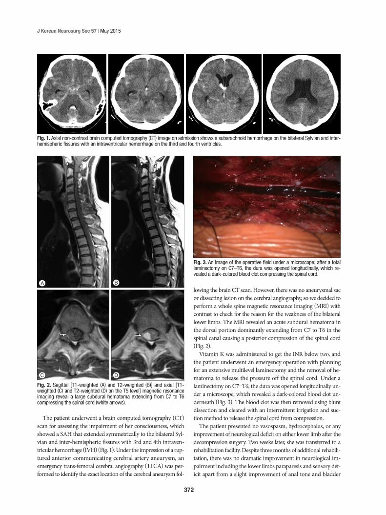
lowing the brain CT scan. However, there was no aneurysmal sac or dissecting lesion on the cerebral angiography, so we decided to perform a whole spine magnetic resonance imaging (MRI) with contrast to check for the reason for the weakness of the bilateral lower limbs. The MRI revealed an acute subdural hematoma in the dorsal portion dominantly extending from C7 to T6 in the spinal canal causing a posterior compression of the spinal cord (Fig. 2).
Vitamin K was administered to get the INR below two, and the patient underwent an emergency operation with planning for an extensive multilevel laminectomy and the removal of he-matoma to release the pressure off the spinal cord. Under a laminectomy on C7–T6, the dura was opened longitudinally un-der a microscope, which revealed a dark-colored blood clot un-derneath (Fig. 3). The blood clot was then removed using blunt dissection and cleared with an intermittent irrigation and suc-tion method to release the spinal cord from compression.
The patient presented no vasospasm, hydrocephalus, or any improvement of neurological deficit on either lower limb after the decompression surgery. Two weeks later, she was transferred to a rehabilitation facility. Despite three months of additional rehabili-tation, there was no dramatic improvement in neurological im-pairment including the lower limbs paraparesis and sensory def-icit apart from a slight improvement of anal tone and bladder
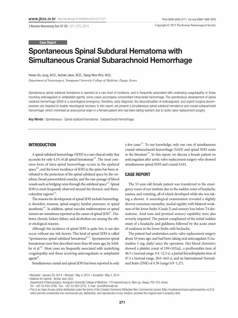
The patient underwent a brain computed tomography (CT) scan for assessing the impairment of her consciousness, which showed a SAH that extended symmetrically to the bilateral Syl-vian and inter-hemispheric fissures with 3rd and 4th intraven-tricular hemorrhage (IVH) (Fig. 1). Under the impression of a rup-tured anterior communicating cerebral artery aneurysm, an emergency trans-femoral cerebral angiography (TFCA) was per-formed to identify the exact location of the cerebral aneurysm fol-
Fig. 1. Axial non-contrast brain computed tomography (CT) image on admission shows a subarachnoid hemorrhage on the bilateral Sylvian and inter-hemispheric fissures with an intraventricular hemorrhage on the third and fourth ventricles.
Fig. 2. Sagittal [T1-weighted (A) and T2-weighted (B)] and axial [T1-weighted (C) and T2-weighted (D) on the T5 level] magnetic resonance imaging reveal a large subdural hematoma extending from C7 to T6 compressing the spinal cord (white arrows).
A B
C D
Fig. 3. An image of the operative field under a microscope; after a total laminectomy on C7–T6, the dura was opened longitudinally, which re-vealed a dark-colored blood clot compressing the spinal cord.
373
Spontaneous Spinal Subdural Hematoma with Intracranial Hemorrhage | HS Jung, et al.
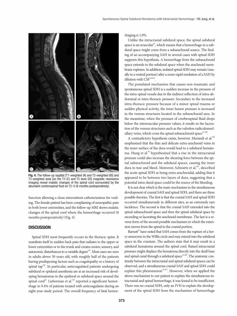
function allowing a clean intermittent catheterization for void-ing. The female patient has been complaining of neuropathic pain in both lower extremities, and the follow-up MRI shows malatic changes of the spinal cord where the hemorrhage occurred (8 months postoperatively) (Fig. 4).
DISCUSSION
Spinal SDH most frequently occurs in the thoracic spine. It manifests itself in sudden back pain that radiates to the upper or lower extremities or to the trunk and creates motor, sensory, and autonomic disturbances to a variable degree14). Most cases are seen in adults above 50 years old, with roughly half of the patients having predisposing factors such as coagulopathy or a history of spinal tap16). In particular, anticoagulated patients undergoing subdural or epidural anesthesia are at an increased risk of devel-oping hematomas in the epidural or subdural space around the spinal cord4). Lieberman et al.15) reported a significant hemor-rhage in 9.4% of patients treated with anticoagulants during an eight-year study period. The overall frequency of fatal hemor-
rhaging is 1.0%. Unlike the intracranial subdural space, the spinal subdural
space is an avascular6), which means that a hemorrhage in a sub-dural space might come from a subarachnoid source. The find-ing of an accompanying SAH in several cases with spinal SDH supports this hypothesis. A hemorrhage from the subarachnoid space extends to the subdural space when the arachnoid mem-brane ruptures. In addition, isolated spinal SDH may remain (usu-ally in a ventral portion) after a more rapid resolution of a SAH by dilution with CSF14,16).
The postulated mechanism that causes non-traumatic and spontaneous spinal SDH is a sudden increase in the pressure of the intra-spinal vessels due to the indirect reflection of intra-ab-dominal or intra-thoracic pressure. Secondary to the increased intra-thoracic pressure because of a minor spinal trauma or sudden physical activity, the inner lumen pressure is increased in the venous structures located in the subarachnoid area. In the meantime, when the pressure of cerebrospinal fluid drops below the intravascular pressure values, it results in the lacera-tion of the venous structures such as the valveless radiculomed-ullary veins, which cross the spinal subarachnoid space17,18).
A contradictory hypothesis exists, however. Morandi et al.18) emphasized that the thin and delicate extra-arachnoid veins in the inner surface of the dura would lead to a subdural hemato-ma. Hung et al.9) hypothesized that a rise in the intracranial pressure could also increase the shearing force between the spi-nal subarachnoid and the subdural spaces, causing the inner dura to tear and bleed. Moreover, Schwartz et al.24), described the acute spinal SDH as being extra-arachnoidal, adding that it appeared to be between two layers of dura, suggesting that a potential intra-dural space could be the source of the bleeding.
It is not clear which is the main mechanism in the simultaneous development of cranial SAH and spinal SDH, and there are three possible theories. The first is that the cranial SAH and spinal SDH occurred simultaneously in different sites, as an extremely rare incidence. The second is that the cranial SAH extended into the spinal subarachnoid space and then the spinal subdural space by exceeding or lacerating the arachnoid membrane. The last is a re-verse form of the second possible mechanism in which the exten-sion moves from the spinal to the cranial portion.
Barnett2) have noted that SAH comes from the rupture of a ber-ry aneurysm in the Willis circle and may extend into the subdural space in the cranium. The authors state that it may result in a subdural hematoma around the spinal cord. Raised intracranial pressure might displace the hematoma directly into the skull base and spinal canal through a subdural space11,28). The anatomic con-tinuity between the intracranial and spinal subdural spaces can be observed, and a simultaneous cranial SAH and spinal SDH could explain this phenomenon19,27). However, when we applied the above mechanism to our patient to explain the simultaneous in-tracranial and spinal hemorrhage, it was found to be insufficient. There was no cranial SDH, only an IVH to explain the develop-ment of the spinal SDH from the mechanism of hemorrhage
Fig. 4. The follow-up sagittal [T1-weighted (A) and T2-weighted (B)] and T2-weighted axial [on the T3 (C) and T5 level (D)] magnetic resonance imaging reveal malatic changes of the spinal cord surrounded by the abundant cerebrospinal fluid on T2–5 (8 months postoperatively).
A B
C D

374
J Korean Neurosurg Soc 57 | May 2015
migration from the cranial to the spinal portion. In addition, if the cranial SAH developed as a non-aneurysmal type, there would not be sufficient pressure to move the hematoma to the spinal canal and make an intracranial SDH. In addition, the extension of a large amount of a SAH from the cranium to the spinal portion could compress the spinal cord or lacerate the arachnoid mem-brane, resulting in the formation of a spinal SDH, which is also un-common, even in a case of an aneurysmal SAH.
The initial treatment for spinal SDH should consist of a wide laminectomy and the evacuation of the hematoma. However, there are some cases in the literature that have been treated via conservative treatment, without any surgical intervention10,11,24). The exact mechanism of the spontaneous resolution of the symptoms is unclear. The rapid redistribution of a hematoma with rostro-caudal expansion within a subdural space may avoid transverse expansions, and subsequently reduce the de-gree of cord compression20). Another possible mechanism was recently suggested, stating that the redistribution of the spinal SDH could occur due to the rapid dilution of the hematoma, in-termingled with cerebrospinal fluid, in the presence of arachnoid membrane tearing16).
Intra-spinal hematoma is the first diagnosis that should be considered in patients on anticoagulant therapies, presenting it-self as acute cord compression or cauda equina syndrome18,21). It is therefore advisable to assess the patients on anticoagulation therapy thoroughly for the possibility of an intra-spinal hema-toma in order to avoid any major neurological complications. The spontaneous onset of spinal SDH in patients on an antico-agulation therapy is a neurological emergency. With this in mind, early diagnosis, the discontinuation of the anticoagulant, and urgent surgical decompression are recommended to allow for neurological recovery18,22).
The timing of the surgery and the anatomic location of the hematoma determine a patient’s functional outcomes3,18). Addi-tionally, the degree of preoperative neurological deficit and level of the SDH correlate significantly with the early and long-term functional outcomes, despite prompt evacuation18,25).
CONCLUSION
When a patient is taking anticoagulation therapy and presents with a subarachnoid hemorrhage without hydrocephalus and se-vere lower extremity paraparesis or paraplegia with backache, the possibility of spinal hemorrhage should be considered. The spon-taneous development of a spinal SDH is a neurological emergen-cy. Therefore, early diagnosis, the discontinuation of anticoagu-lants, and urgent surgical decompression are recommended to facilitate neurological recovery.
References1. Barker GL : Spinal subdural haematoma following spinal anaesthesia.
Anaesthesia 43 : 664-665, 19882. Barnett HJ : Some clinical features of intracranial aneurysms. Clin Neu-
rosurg 16 : 43-72, 19693. Chau SY, Tiu SC : Spinal subdural haematoma : a rare complication of
low-molecular-weight heparin therapy. Hong Kong Med J 14 : 64-66, 20084. DeAngelis J : Hazards of subdural and epidural anesthesia during antico-
agulant therapy : a case report and review. Anesth Analg 51 : 676-679, 19725. Domenicucci M, Ramieri A, Ciappetta P, Delfini R : Nontraumatic acute
spinal subdural hematoma : report of five cases and review of the litera-ture. J Neurosurg 91 (1 Suppl) : 65-73, 1999
6. Gillilan LA : Veins of the spinal cord. Anatomic details; suggested clinical applications. Neurology 20 : 860-868, 1970
7. Han PP, Theodore N, Porter RW, Detwiler PW, Lawton MT, Spetzler RF : Subdural hematoma from a Type I spinal arteriovenous malformation. Case report. J Neurosurg 90 (2 Suppl) : 255-257, 1999
8. Harik SI, Raichle ME, Reis DJ : Spontaneously remitting spinal epidural hematoma in a patient on anticoagulants. N Engl J Med 284 : 1355-1357, 1971
9. Hung KS, Lui CC, Wang CH, Wang CJ, Howng SL : Traumatic spinal subdural hematoma with spontaneous resolution. Spine (Phila Pa 1976) 27 : E534-E538, 2002
10. Hurt RW, Shaw MD, Russell JA : Spinal subdural haematoma : an un-usual complication of lumbar puncture. Surg Neurol 8 : 296-297, 1977
11. Jain V, Singh J, Sharma R : Spontaneous concomitant cranial and spinal subdural haematomas with spontaneous resolution. Singapore Med J 49 : e53-e58, 2008
12. Khosla VK, Kak VK, Mathuriya SN : Chronic spinal subdural hematomas. Report of two cases. J Neurosurg 63 : 636-639, 1985
13. Kim HY, Ju CI, Kim SW : Acute cervical spinal subdural hematoma not related to head injury. J Korean Neurosurg Soc 47 : 467-469, 2010
14. Kyriakides AE, Lalam RK, El Masry WS : Acute spontaneous spinal sub-dural hematoma presenting as paraplegia : a rare case. Spine (Phila Pa 1976) 32 : E619-E622, 2007
15. Lieberman A, Hass WK, Pinto R, Isom WO, Kupersmith M, Bear G, et al. : Intracranial hemorrhage and infarction in anticoagulated patients with prosthetic heart valves. Stroke 9 : 18-24, 1978
16. Mavroudakis N, Levivier M, Rodesch G : Central cord syndrome due to a spontaneously regressive spinal subdural hematoma. Neurology 40 : 1306-1308, 1990
17. Mete A, Erkutlu I, Akcali A, Mete A : Simultaneous cranial subarachnoid hemorrhage and spinal subdural hematoma. Turk Neurosurg 22 : 349-352, 2012
18. Morandi X, Riffaud L, Chabert E, Brassier G : Acute nontraumatic spinal subdural hematomas in three patients. Spine (Phila Pa 1976) 26 : E547-E551, 2001
19. Nicholas DS, Weller RO : The fine anatomy of the human spinal meninges. A light and scanning electron microscopy study. J Neurosurg 69 : 276-282, 1988
20. Oh SH, Han IB, Koo YH, Kim OJ : Acute spinal subdural hematoma pre-senting with spontaneously resolving hemiplegia. J Korean Neurosurg Soc 45 : 390-393, 2009
21. Prasad SS, O’Malley M, Machani B, Shackleford IM : A case report of a spinal epidural haematoma associated with warfarin therapy. Ann R Coll Surg Engl 85 : 277-278, 2003
22. Roscoe MW, Barrington TW : Acute spinal subdural hematoma. A case report and review of literature. Spine (Phila Pa 1976) 9 : 672-675, 1984
23. Schiller F, Neligan G, Budtz-Olsen O : Surgery in haemophilia; a case of spi-nal subdural haematoma producing paraplegia. Lancet 2 : 842-845, 1948
24. Schwartz FT, Sartawi MA, Fox JL : Unusual hematomas outside the spinal cord. Report of two cases. J Neurosurg 39 : 249-251, 1973
25. Thiex R, Thron A, Gilsbach JM, Rohde V : Functional outcome after sur-gical treatment of spontaneous and nonspontaneous spinal subdural he-matomas. J Neurosurg Spine 3 : 12-16, 2005
26. Toledo E, Shalit MN, Segal R : Spinal subdural hematoma associated with

375
Spontaneous Spinal Subdural Hematoma with Intracranial Hemorrhage | HS Jung, et al.
anticoagulant therapy in a patient with spinal meningioma. Neurosurgery 8 : 600-603, 1981
27. Wang US, Ju CI, Kim SW, Kim SH : Spontaneous concomitant intracranial and spinal subdural hematomas in association with anticoagulation
therapy. J Korean Neurosurg Soc 51 : 237-239, 201228. Yamaguchi S, Kurisu K, Arita K, Takeda M, Tani I, Araki O : Simultaneous
cranial and spinal subdural hematoma. Neurol Med Chir (Tokyo) 45 : 645-649, 2005