Embed Size (px)
Citation preview
CASE REPORT Open Access
Benign multicystic peritoneal mesothelioma:a case reportXanthi Pitta1*, Efstathios Andreadis2, Athanasios Ekonomou2, Athanasia Papachristodoulou3,Chrisostomos Tziouvaras2, Leonidas Papapaulou4, Nikolaos Sapidis2, Thomas Chrisidis2
Abstract
Introduction: We report the case of a patient with a benign multicystic peritoneal mesothelioma and describe itsappearance on computed tomography scans and ultrasonography, in correlation with gross clinical andpathological findings.
Case presentation: A 72-year-old Caucasian woman presented to our emergency department with acuteabdomen signs and symptoms. A clinical examination revealed a painful palpable mass in her left abdomen.Abdominal ultrasonography and computed tomography demonstrated the presence of a large cystic mass in herleft upper abdomen, adjacent to her left hemidiaphragm. The lower border of the mass extended to the uppermargin of her pelvis. A complete resection of the lesion was performed. Pathological analysis showed a benignmulticystic peritoneal mesothelioma.
Conclusions: Benign multicystic peritoneal mesothelioma is a rare lesion with a non-specific appearance onimaging. Its diagnosis always requires pathological analysis.
IntroductionBenign multicystic peritoneal mesothelioma is anuncommon lesion arising from the peritoneal mesothe-lium. It is often diffuse and shows a marked predilectionfor the surfaces of the pelvic viscera [1-8]. In our casereport, the lesion was solitary and situated in the leftabdomen. This disease is a rare medical entity and thereare challenges in determining its origin, pathogenesis,diagnosis and therapy.
Case presentationA 72-year-old Caucasian woman was admitted to oursurgical department having experienced diffuse abdom-inal pain and discomfort, nausea and vomiting for theprevious two days. Her medical history included dia-betes mellitus and arterial hypertension, for which shewas on medication. She had no relevant family historyand did not smoke or drink alcohol.On physical examination, she showed signs of acute
abdomen and a palpable painful mass in her left
abdomen was noted. She was tachycardic and laboratorytests showed a white blood cell count of 13,000 cells percubic millimeter. Her chest and abdominal radiographsdid not reveal any abnormalities.An ultrasonography (US) examination demonstrated a
complex cystic mass with internal septa, withoutincreased vascularity. The source organ could not beidentified (Figure 1).Computed tomography (CT) examination demon-
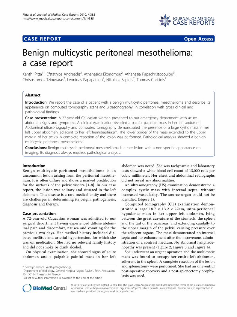
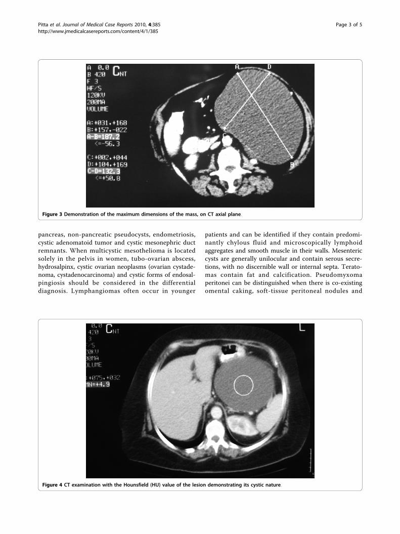
strated a large 18.7 × 13.2 × 22cm, intra-peritonealhypodense mass in her upper left abdomen, lyingbetween the great curvature of the stomach, the spleenand the tail of the pancreas, and extending caudally tothe upper margin of the pelvis, causing pressure overthe adjacent organs. The mass demonstrated no internalsepta and no enhancement after the intravenous admin-istration of a contrast medium. No abnormal lymphade-nopathy was present (Figure 2, Figure 3 and Figure 4).She underwent an urgent operation and the multicystic
mass was found to occupy her entire left abdomen,adherent to the spleen. A complete resection of the lesionand splenectomy were performed. She had an uneventfulpost-operative recovery and a post-splenectomy prophy-laxis was used.
* Correspondence: [email protected] of Radiology, General Hospital “Agios Pavlos”, Ethn. Antistaseos161, 55134 Thessaloniki, GreeceFull list of author information is available at the end of the article
Pitta et al. Journal of Medical Case Reports 2010, 4:385http://www.jmedicalcasereports.com/content/4/1/385 JOURNAL OF MEDICAL
CASE REPORTS
© 2010 Pitta et al; licensee BioMed Central Ltd. This is an Open Access article distributed under the terms of the Creative CommonsAttribution License (http://creativecommons.org/licenses/by/2.0), which permits unrestricted use, distribution, and reproduction inany medium, provided the original work is properly cited.

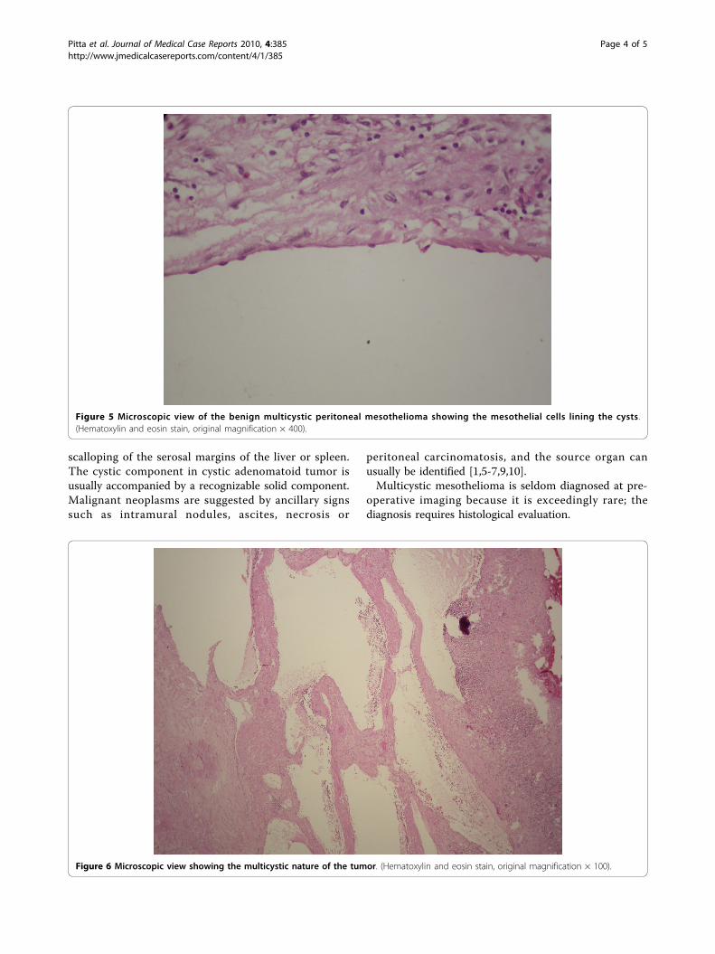
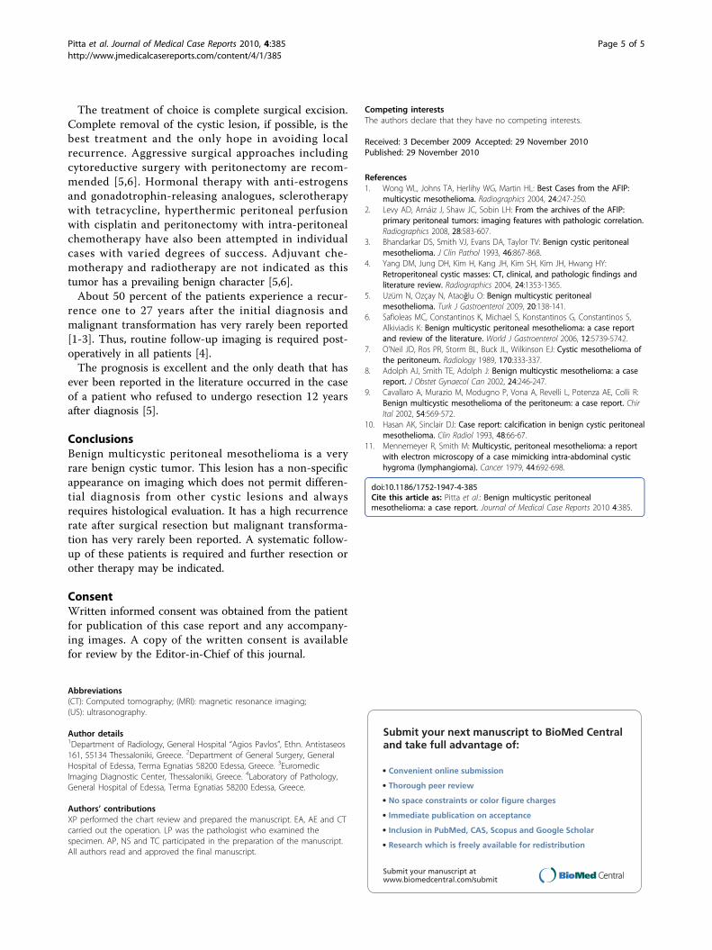
Gross examination of the specimen showed a largegelatinous cystic mass containing multiple smaller cysticspaces. Her immunohistochemical stains were positivefor calretinin and cytokeratins, confirming the mesothe-lial origin of the mass. The final diagnosis was benignmulticystic peritoneal mesothelioma. (Figures 5 and 6).Six months post-operatively, she had experienced no
recurrence and was free of symptoms.
DiscussionMesotheliomas are mesenchymal neoplasms originatingfrom the serous lining of the pleural, pericardial or peri-toneal space. Multicystic peritoneal mesotheliomainvolves the peritoneum or extra-peritoneal space,
omentum, pelvic or abdominal viscera. It most com-monly arises from the pelvic surfaces of the peritoneumand has benign or indolent biologic behavior. Multicys-tic mesothelioma of the peritoneum was first describedin 1979 by Mennemeyer and Smith and since thenapproximately 130 cases have been described in the lit-erature. It is an intermediate-grade tumor, among thebenign adenomatoid tumors of the peritoneum and themore common malignant asbestos-related peritonealmesothelioma. It is not related to prior asbestos expo-sure and may recur locally [1-11].On histological examination, the mesothelial cells lin-
ing the cysts may vary from flattened and endothelial-like to cuboidal. The thin-walled cysts may be filledwith eosinophilic, serous fluid. Inflammatory cells andfibrous elements may be found within the stromabetween the cysts. Foci of mesothelial hyperplasia mayalso be present [2].It is usually large at the time of diagnosis (mean dia-
meter, 13 cm). Multifocality, free floating cysts and uni-locular cysts have been reported [2].It commonly occurs in young to middle-aged women
(mean age, 37 years). The presenting symptoms arechronic or intermittent lower abdominal or pelvic pain,tenderness, or distension with an abdominal or pelvicmass and, rarely, dyspareunia, constipation and urinaryhesitancy and/or frequency. Women with this lesionoften have a history of prior pelvic surgery, endometrio-sis or pelvic inflammatory disease [1-3,5,7-10].The pathogenesis of benign multicystic peritoneal
mesothelioma is unclear and there is some controversyregarding its neoplastic and reactive nature [2,6]. Thefact that the great majority of patients are women ofreproductive age suggests that a key role is played byfemale sex hormones in its pathogenesis [5].US demonstrates multiseptated anechoic cysts. The fluid
within the cysts is generally anechoic, but the cysts maycontain echoes from debris or hemorrhage. The numberand complexity of septations, as well as the size of thecysts, are quite variable. Calcification has not beendescribed in multicystic mesothelioma. CT provides moreinformation about the location and extent of the mass,and demonstrates a well-defined, low-attenuation masswith non-calcified septa. The septa become enhanced fol-lowing intravenous administration of a contrast material.Magnetic resonance imaging (MRI) provides additionalcoronal and sagittal planes. The watery serous content haslow signal intensity on T1-weighted images and intermedi-ate-to-high signal intensity on T2-weighted images. Septalenhancement has been reported [1,2].The differential diagnosis includes lymphangioma,
other mesenteric and/or omental cysts, cystic teratoma,pseudomyxoma peritonei, cystic smooth muscle tumors,visceral cysts, cystic mucinous neoplasms of the
Figure 1 US image showing a cystic mass with internal septa.
Figure 2 CT axial image after the intravenous administration ofa contrast medium demonstrating an intra-peritonealhypodense non-enhancing mass adjacent to the stomach andspleen.
Pitta et al. Journal of Medical Case Reports 2010, 4:385http://www.jmedicalcasereports.com/content/4/1/385
Page 2 of 5
pancreas, non-pancreatic pseudocysts, endometriosis,cystic adenomatoid tumor and cystic mesonephric ductremnants. When multicystic mesothelioma is locatedsolely in the pelvis in women, tubo-ovarian abscess,hydrosalpinx, cystic ovarian neoplasms (ovarian cystade-noma, cystadenocarcinoma) and cystic forms of endosal-pingiosis should be considered in the differentialdiagnosis. Lymphangiomas often occur in younger
patients and can be identified if they contain predomi-nantly chylous fluid and microscopically lymphoidaggregates and smooth muscle in their walls. Mesentericcysts are generally unilocular and contain serous secre-tions, with no discernible wall or internal septa. Terato-mas contain fat and calcification. Pseudomyxomaperitonei can be distinguished when there is co-existingomental caking, soft-tissue peritoneal nodules and
Figure 3 Demonstration of the maximum dimensions of the mass, on CT axial plane.
Figure 4 CT examination with the Hounsfield (HU) value of the lesion demonstrating its cystic nature.
Pitta et al. Journal of Medical Case Reports 2010, 4:385http://www.jmedicalcasereports.com/content/4/1/385
Page 3 of 5
scalloping of the serosal margins of the liver or spleen.The cystic component in cystic adenomatoid tumor isusually accompanied by a recognizable solid component.Malignant neoplasms are suggested by ancillary signssuch as intramural nodules, ascites, necrosis or
peritoneal carcinomatosis, and the source organ canusually be identified [1,5-7,9,10].Multicystic mesothelioma is seldom diagnosed at pre-
operative imaging because it is exceedingly rare; thediagnosis requires histological evaluation.
Figure 5 Microscopic view of the benign multicystic peritoneal mesothelioma showing the mesothelial cells lining the cysts.(Hematoxylin and eosin stain, original magnification × 400).
Figure 6 Microscopic view showing the multicystic nature of the tumor. (Hematoxylin and eosin stain, original magnification × 100).
Pitta et al. Journal of Medical Case Reports 2010, 4:385http://www.jmedicalcasereports.com/content/4/1/385
Page 4 of 5
The treatment of choice is complete surgical excision.Complete removal of the cystic lesion, if possible, is thebest treatment and the only hope in avoiding localrecurrence. Aggressive surgical approaches includingcytoreductive surgery with peritonectomy are recom-mended [5,6]. Hormonal therapy with anti-estrogensand gonadotrophin-releasing analogues, sclerotherapywith tetracycline, hyperthermic peritoneal perfusionwith cisplatin and peritonectomy with intra-peritonealchemotherapy have also been attempted in individualcases with varied degrees of success. Adjuvant che-motherapy and radiotherapy are not indicated as thistumor has a prevailing benign character [5,6].About 50 percent of the patients experience a recur-
rence one to 27 years after the initial diagnosis andmalignant transformation has very rarely been reported[1-3]. Thus, routine follow-up imaging is required post-operatively in all patients [4].The prognosis is excellent and the only death that has
ever been reported in the literature occurred in the caseof a patient who refused to undergo resection 12 yearsafter diagnosis [5].
ConclusionsBenign multicystic peritoneal mesothelioma is a veryrare benign cystic tumor. This lesion has a non-specificappearance on imaging which does not permit differen-tial diagnosis from other cystic lesions and alwaysrequires histological evaluation. It has a high recurrencerate after surgical resection but malignant transforma-tion has very rarely been reported. A systematic follow-up of these patients is required and further resection orother therapy may be indicated.
ConsentWritten informed consent was obtained from the patientfor publication of this case report and any accompany-ing images. A copy of the written consent is availablefor review by the Editor-in-Chief of this journal.
Abbreviations(CT): Computed tomography; (MRI): magnetic resonance imaging;(US): ultrasonography.
Author details1Department of Radiology, General Hospital “Agios Pavlos”, Ethn. Antistaseos161, 55134 Thessaloniki, Greece. 2Department of General Surgery, GeneralHospital of Edessa, Terma Egnatias 58200 Edessa, Greece. 3EuromedicImaging Diagnostic Center, Thessaloniki, Greece. 4Laboratory of Pathology,General Hospital of Edessa, Terma Egnatias 58200 Edessa, Greece.
Authors’ contributionsXP performed the chart review and prepared the manuscript. EA, AE and CTcarried out the operation. LP was the pathologist who examined thespecimen. AP, NS and TC participated in the preparation of the manuscript.All authors read and approved the final manuscript.
Competing interestsThe authors declare that they have no competing interests.
Received: 3 December 2009 Accepted: 29 November 2010Published: 29 November 2010
References1. Wong WL, Johns TA, Herlihy WG, Martin HL: Best Cases from the AFIP:
multicystic mesothelioma. Radiographics 2004, 24:247-250.2. Levy AD, Arnáiz J, Shaw JC, Sobin LH: From the archives of the AFIP:
primary peritoneal tumors: imaging features with pathologic correlation.Radiographics 2008, 28:583-607.
3. Bhandarkar DS, Smith VJ, Evans DA, Taylor TV: Benign cystic peritonealmesothelioma. J Clin Pathol 1993, 46:867-868.
4. Yang DM, Jung DH, Kim H, Kang JH, Kim SH, Kim JH, Hwang HY:Retroperitoneal cystic masses: CT, clinical, and pathologic findings andliterature review. Radiographics 2004, 24:1353-1365.
5. Uzüm N, Ozçay N, Ataoğlu O: Benign multicystic peritonealmesothelioma. Turk J Gastroenterol 2009, 20:138-141.
6. Safioleas MC, Constantinos K, Michael S, Konstantinos G, Constantinos S,Alkiviadis K: Benign multicystic peritoneal mesothelioma: a case reportand review of the literature. World J Gastroenterol 2006, 12:5739-5742.
7. O’Neil JD, Ros PR, Storm BL, Buck JL, Wilkinson EJ: Cystic mesothelioma ofthe peritoneum. Radiology 1989, 170:333-337.
8. Adolph AJ, Smith TE, Adolph J: Benign multicystic mesothelioma: a casereport. J Obstet Gynaecol Can 2002, 24:246-247.
9. Cavallaro A, Murazio M, Modugno P, Vona A, Revelli L, Potenza AE, Colli R:Benign multicystic mesothelioma of the peritoneum: a case report. ChirItal 2002, 54:569-572.
10. Hasan AK, Sinclair DJ: Case report: calcification in benign cystic peritonealmesothelioma. Clin Radiol 1993, 48:66-67.
11. Mennemeyer R, Smith M: Multicystic, peritoneal mesothelioma: a reportwith electron microscopy of a case mimicking intra-abdominal cystichygroma (lymphangioma). Cancer 1979, 44:692-698.
doi:10.1186/1752-1947-4-385Cite this article as: Pitta et al.: Benign multicystic peritonealmesothelioma: a case report. Journal of Medical Case Reports 2010 4:385.
Submit your next manuscript to BioMed Centraland take full advantage of:
• Convenient online submission
• Thorough peer review
• No space constraints or color figure charges
• Immediate publication on acceptance
• Inclusion in PubMed, CAS, Scopus and Google Scholar
• Research which is freely available for redistribution
Submit your manuscript at www.biomedcentral.com/submit
Pitta et al. Journal of Medical Case Reports 2010, 4:385http://www.jmedicalcasereports.com/content/4/1/385
Page 5 of 5


![Isolated Giant benign Multicysticperitoneal Mesothelioma ......Benign Multicystic Peritoneal Mesothelioma (BMPM) is an uncommon lesion of the serosal membranes [1-3]. In the works](https://img.dokumen.tips/doc/110x75/60f80e44e27060088c5b84aa/isolated-giant-benign-multicysticperitoneal-mesothelioma-benign-multicystic.jpg)