Embed Size (px)
Citation preview
Hindawi Publishing CorporationCase Reports in OtolaryngologyVolume 2013, Article ID 796713, 7 pageshttp://dx.doi.org/10.1155/2013/796713
Case ReportEndoscopic Resection of Sinonasal Hemangiopericytomafollowing Preoperative Embolisation: A Case Report andLiterature Review
Georg J. Ledderose,1 Donata Gellrich,1 Markus Holtmannspötter,2,3 and Andreas Leunig1,4
1 Department of Otorhinolaryngology, Head and Neck Surgery, Ludwig Maximilian University Munich, Marchioninistraße 15,81377 Munich, Germany
2 Institute for Neuroradiology, Ludwig Maximilian University Munich, Marchioninistraße 15, 81377 Munich, Germany3 Center for Diagnostic Radiology, Department of Neuroradiology, Rigshospitalet, University of Copenhagen, Denmark4Center for Rhinology, Starnberg/Munich, Germany
Correspondence should be addressed to Georg J. Ledderose; johannes [email protected]
Received 18 March 2013; Accepted 12 April 2013
Academic Editors: A. Harimaya, H. Sudhoff, A. Tas, and L.-F. Wang
Copyright © 2013 Georg J. Ledderose et al. This is an open access article distributed under the Creative Commons AttributionLicense, which permits unrestricted use, distribution, and reproduction in any medium, provided the original work is properlycited.
Objectives. Hemangiopericytoma is a rare tumor entity deriving from pericytes. Less than 5% of hemangiopericytoma occur in thenasal cavity and are characterised by a rather benign nature with low tendency of metastasis. However, as the recurrence rate in theliterature ranges from 9.5% to 50%—depending on the length of followup—a radical surgical resection is considered as the gold-standard treatment. Only a few years ago, a wide external approach, usually via lateral rhinotomy or Caldwell-Luc, was performed.Endoscopic techniques were regarded as appropriate for small low-vascularised tumors only. Methods. We present the case of a64-year-old patient with an extended sinonasal hemangiopericytoma, who was successfully treated by an endoscopic controlledendonasal tumor resection after embolisation with Onyx. Further, to support the new treatment option, we review the literatureconcerning all features of sinonasal hemangiopericytomas and their therapeutical management. Results/Conclusion. Onyx, whichhas not been described in the context of hemangiopericytoma yet, is a very effective embolic agent for a preoperative embolisationof sinonasal hemangiopericytoma allowing a safe endoscopic surgery.
1. Introduction
Sinonasal hemangiopericytomas are a rare upper aerodiges-tive tract tumor deriving from perivascular modified smoothmuscle cells. This vascular neoplasm firstly described in 1943by Stout andMurray [1] may arise in any part of the body [2];only 15%–30% are located in the head and neck region [3].Of these, only 5% are found in the nasal cavity and paranasalsinus [4]. Hemangiopericytoma located in the nasal region isfrequently characterized by a more benign nature with lowtendency of metastasis [5]; however, sinonasal hemangioper-icytoma exhibits a recurrence rate of approximately 25% [3].
In the last two decades, less than 250 cases have beenpublished so that sinonasal hemangiopericytoma representsa rather rare tumor entity. A total of 57 patients out of the 250analysed cases underwent an endoscopic tumor resection, of
which 23 patients received a preoperative embolisation of thehigh vascularised hemangiopericytoma.
Here, we review the relevant literature and report about a64-year-old patient presenting with sinonasal hemangioperi-cytoma.Weused a liquid embolic agent, consisting of an ethy-lene vinyl alcohol dissolved in the organic solvent dimethylsulfoxide (DMSO) and containing tantalum powder forradiopacity (Onyx) and performed an endoscopically con-trolled resection. To our knowledge, this is the first reportabout preoperative embolisation of sinonasal hemangioper-icytoma using this material.
2. Case Report
A 64-year-old Caucasian man in good general health pre-sented to our institution complaining of nasal obstruction
2 Case Reports in Otolaryngology
CI
S
∗
(a)
CI
S
∗
(b)
Figure 1: Endoscopic view of the operational field before surgery(a). After embolization, shrinking is clearly seen (b). Nasal septum(S), concha inferior (CI), concha media (CM), sphenoid sinus (SS),and tumor (∗).
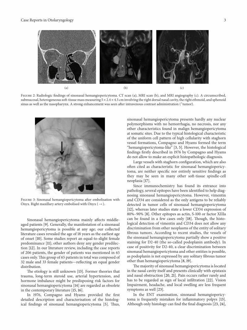
on the right side for 6 weeks. Further, the patient reporteda singular episode of epistaxis. Other nasal symptoms suchas recurrent epistaxis or sinusitis were absent. Head and neckexamination revealed a polypoid mass obstructing the rightnasal cavity subtotally (Figure 1(a)). The posterior rhinos-copia showed a partial obstruction of the nasopharynx aswell. There was no cervical adenopathy or other disorder inthe head and neck. A CT scan of the paranasal sinusesrevealed a soft-tissue mass involving the right dorsal nasalcavity, the right ethmoid, and the sphenoid sinus as well thenasopharynx (Figure 2(a)). A strong enhancement was seenafter intravenous contrast administration. Further, bonedestruction of the right ethmoid structures was demon-strated. In theCT staging, neither local nor distantmetastaseswere visible.TheMRI confirmed a circumscribed, submucos-al, heterogeneousmassmeasuring 5× 2.4× 4.5 cm, extendingfrom the cribriform plate to the platinum durum, infiltratingthe ethmoidal cells and the right maxillary and sphenoidsinus (Figure 2(b)). The cribriform plate also seemed to beinfiltrated. The tumor was brightly vascularised with flowsignal voids. Conventional digital angiography through theright external iliacal artery revealed a vascular network ofthe mass, mainly supplied by the right maxillary artery, as
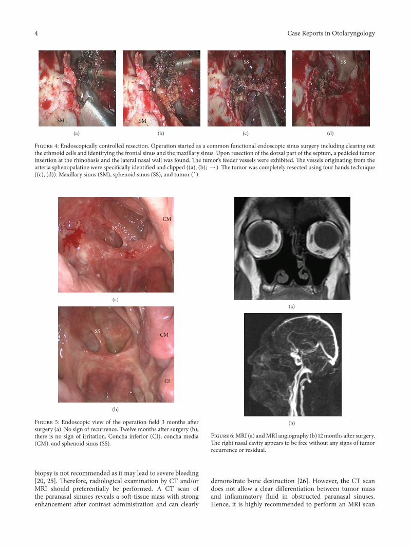
also seen in MRI angiography (Figure 2(c)). Additionally,the angiography showed a half-moon shaped blush, in par-ticular of the dorsocranial portion of the tumor, supplied byethmoidal branches of the right ophthalmic artery. A tumorresection by an endonasal approach was planned upon abiopsy revealing the suspicion of hemangiopericytoma. Asthe biopsy was followed by a prolonged bleeding and as theradiologic findings confirmed the high vascularisation of themass, the right maxillary artery as the main vascular feederwas selectively embolised with an ethylene vinyl alcoholdissolved in the organic solvent dimethyl sulfoxide (Onyx)24 hours before surgery (Figures 1(b) and 3). Following thepreoperative embolisation, the endoscopic tumor resectionwas performed via endonasal approach with a navigationsystem. Upon resection of the dorsal part of the septum, apedicled tumor insertion at the rhinobasis was exhibited infour hands technique and resected completely without anycomplication (Figure 4).
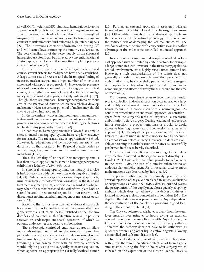
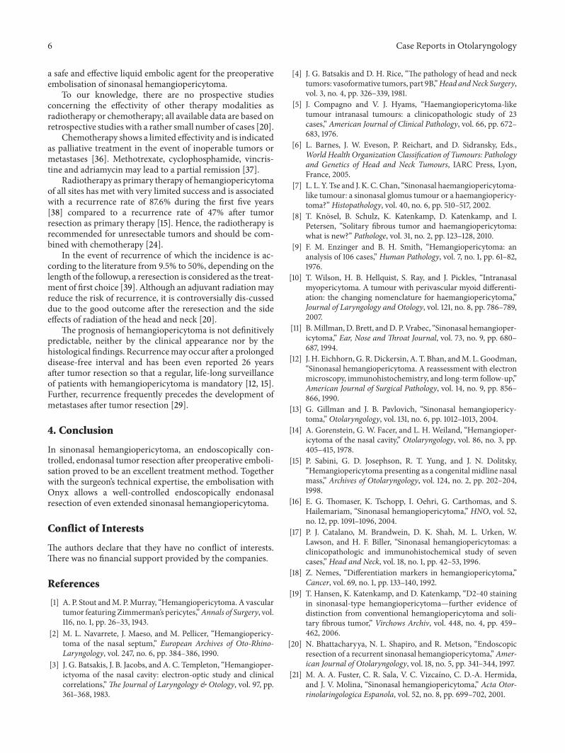
Histologically, the tumor was submucosal, unencapsu-lated, and showed fascicular pattern. The tumor cells wereuniform, spindle-shapedwith oval nuclei, accompanied by aninflammatory cell infiltrate including eosinophils. “Stag horn”vessels, as typical for hemangiopericytoma, were focally iden-tified as well as few nuclear atypia. Immunohistochemistryrevealed a strong staining pattern to vimentin but no reactionwith actin.The cells were immunoreactive for CD 34, slightlyfor Bcl2 and CD99 but negative for S-100 and epithelialmembrane antigen (EMA). The Ki67-proliferation index wasslightly increased at <2%. Due to a high recurrence rate of upto 25%, the patient has been followed up regularly by endo-scopic surveillance andMRI controls. One year after surgery,there was no sign of locoregional disease recurrence, neitherby endoscopy (Figures 5(a) and 5(b)) nor by MRI (Figures6(a) and 6(b)).
3. Discussion and Literature Review
The term “sinonasal hemangiopericytoma,” formed 60 yearsago, covers a wide range of neoplasia, all characterized bytheir histological appearance consisting of a uniform cellpattern of high cellularity and staghorn vessel formations.As a diversity of tumors meet this criterion, the term“sinonasal hemangiopericytoma” has become disputed. Someauthors prefer the term “intranasal hemangiopericytoma-like tumors” [5]; other studies postulate a close relationshipwith soft tissue hemangiopericytoma justifying the term“sinonasal hemangiopericytoma” [1].TheWHOclassificationof head and neck tumors proposed in 2005 that sinonasalhemangiopericytoma should be named glomangiopericy-toma due to their similarity with glomus tumors [6] espe-cially as there are studies revealing a closer relationship ofsinonasal hemangiopericytoma to glomus tumors than tohemangiopericytoma [7].
In the clinical-pathological routine, the practice has be-come accepted to use the term “hemangiopericytoma” for alltumors with hemangiopericytoma-like histology after ex-clusion of other tumor entities [8]—as done in this case re-port.
Case Reports in Otolaryngology 3
∗
(a)
∗
(b)
∗
(c)
Figure 2: Radiologic findings of sinonasal hemangiopericytoma. CT scan (a), MRI scan (b), and MRI angiography (c). A circumscribed,submucosal, heterogeneous soft-tissuemassmeasuring 5× 2.4× 4.5 cm involving the right dorsal nasal cavity, the right ethmoid, and sphenoidsinus as well as the nasopharynx. A strong enhancement was seen after intravenous contrast administration (∗tumor).
Figure 3: Sinonasal hemangiopericytoma after embolisation withOnyx. Right maxillary artery embolised with Onyx (→ ).
Sinonasal hemangiopericytoma mainly affects middle-aged patients [9]. Generally, the manifestation of a sinonasalhemangiopericytoma is possible at any age; our collectedliterature cases revealed the age of 18 years as the earliest ageof onset [10]. Some studies report an equal-to-slight femalepredominance [11]; other authors deny any gender predilec-tion [12]. In our literature review, including the case reportsof 206 patients, the gender of patients was mentioned in 65cases only.This group of 65 patients in total was composed of32 male and 33 female patients—reflecting an equal genderdistribution.
The etiology is still unknown [13]. Former theories thattrauma, long-term steroid use, arterial hypertension, andhormone imbalance might be predisposing risk factors forsinonasal hemangiopericytoma [14] are regarded as obsoletein the contemporary literature [15, 16].
In 1976, Compagno and Hyams provided the firstdetailed description and characterisation of the histolog-ical findings of sinonasal hemangiopericytoma [5]. Thus,
sinonasal hemangiopericytoma presents hardly any nuclearpolymorphisms with no hemorrhagia, no necrosis, nor anyother characteristics found in malign hemangiopericytomaat somatic sites. Due to the typical histological characteristicof the uniform cell pattern of high cellularity with staghornvessel formations, Compagno and Hyams formed the term“hemangiopericytoma-like” [3, 5]. However, the histologicalfindings firstly described in 1976 by Compagno and Hyamsdo not allow to make an explicit histopathologic diagnosis.
Large vessels with staghorn configuration, which are alsooften cited as characteristic for sinonasal hemangiopericy-toma, are neither specific nor entirely sensitive findings asthey may be seen in many other soft-tissue spindle-cellneoplasia [17].
Since immunochemistry has found its entrance intopathology, several epitopes have been identified to help diag-nosing sinonasal hemangiopericytoma. However, vimentinand CD34 are considered as the only antigens to be reliablydetected in tumor cells of sinonasal hemangiopericytoma[12], whereas later studies state a lower CD34 expression of80%–90% [8]. Other epitopes as actin, S-100 or factor XIIIacan be found in a few cases only [18]. Though, the histo-logical detection of vimentin and CD34 does not allow anydiscrimination from other neoplasms of the entity of solitaryfibrous tumors. According to recent studies, the vessels ofthe sinonasal hemangiopericytoma partially show a positivestaining for D2-40 (the so-called podoplanin antibody). Incase of positivity for D2-40, a clear discrimination betweensinonasal hemangiopericytoma and other entities is possible,as podoplanin is not expressed by any solitary fibrous tumorother than hemangiopericytoma [8, 19].
Themajority of sinonasal hemangiopericytoma is locatedin the nasal cavity itself and presents clinically with epistaxisand nasal obstruction [20, 21]. Pain occurs rather rarely andhas to be regarded as sign of local infiltration [22]. VisionImpairment, headache, and local swelling are less frequentsymptoms as well [23].
In the ENT examination, sinonasal hemangiopericy-toma is frequently mistaken for inflammatory polpys [13].Although only histology can find the final diagnosis [23, 24],
4 Case Reports in Otolaryngology
SM
(a)
SM
∗
(b)
SS
∗
∗
(c)
SS
(d)
Figure 4: Endoscopically controlled resection. Operation started as a common functional endoscopic sinus surgery including clearing outthe ethmoid cells and identifying the frontal sinus and the maxillary sinus. Upon resection of the dorsal part of the septum, a pedicled tumorinsertion at the rhinobasis and the lateral nasal wall was found. The tumor’s feeder vessels were exhibited. The vessels originating from thearteria sphenopalatine were specifically identified and clipped ((a), (b); → ). The tumor was completely resected using four hands technique((c), (d)). Maxillary sinus (SM), sphenoid sinus (SS), and tumor (∗).
CMSS
(a)
CMSS
CI
(b)
Figure 5: Endoscopic view of the operation field 3 months aftersurgery (a). No sign of recurrence. Twelve months after surgery (b),there is no sign of irritation. Concha inferior (CI), concha media(CM), and sphenoid sinus (SS).
biopsy is not recommended as it may lead to severe bleeding[20, 25]. Therefore, radiological examination by CT and/orMRI should preferentially be performed. A CT scan ofthe paranasal sinuses reveals a soft-tissue mass with strongenhancement after contrast administration and can clearly
(a)
(b)
Figure 6:MRI (a) andMRI angiography (b) 12months after surgery.The right nasal cavity appears to be free without any signs of tumorrecurrence or residual.
demonstrate bone destruction [26]. However, the CT scandoes not allow a clear differentiation between tumor massand inflammatory fluid in obstructed paranasal sinuses.Hence, it is highly recommended to perform an MRI scan
Case Reports in Otolaryngology 5
aswell. OnT1-weightedMRI, sinonasal hemangiopericytomaappears as solid isointense masses with strong enhancementafter intravenous contrast administration; on T2-weightedimaging, the tumor mass is isointense to low intense incontrast to inflammatory fluid reflecting high-intense signals[27]. The intravenous contrast administration during CTand MRI scan allows estimating the tumor vascularisation.The best visualisation of the vessel supply of the sinonasalhemangiopericytoma can be achieved by conventional digitalangiography, which helps at the same time to plan a preoper-ative embolisation [23].
In order to estimate the risk of an aggressive clinicalcourse, several criteria for malignance have been established.A large tumor size of >6.5 cm and the histological finding ofnecrosis, nuclear atypia, and a high number of mitosis areassociatedwith a poorer prognosis [9].However, the presenceof one of these features does not predict an aggressive clinicalcourse; it is rather the sum of several criteria for malig-nancy to be considered as prognostically unfavorable [1, 17].Though, there are sinonasal hemangiopericytoma withoutany of the mentioned criteria which nevertheless developmalignancy. Hence, a certain potential of malignancy shouldalways be taken into account [8].
In the meantime—concerning meningeal hemangioper-icytoma—it has become apparent thatmetastases are the onlyproven sign of a poor outcome, whereas histological featuresfail to show any prognostic value [16, 28].
In contrast to hemangiopericytoma located at somaticsites, sinonasal hemangiopericytoma has a very low tendencyfor metastasis. The metastasis rate is approximately 5% [3].However, lymphogenous and hematogenous metastases aredescribed in the literature [14]. Regional lymph nodes aswell as lungs, liver, and bone are the most common site ofmetastasis [11, 20].
Thus, the lethality of sinonasal hemangiopericytoma isless than 5%, in opposition to somatic hemangiopericytomaexhibiting a lethality of 25%–60% [3, 16].
In sinonasal hemangiopericytoma, the therapy of choiceis indisputably the wide-field excision with negative margins[14, 29]. Only a few years ago, an external surgical approach,usually via lateral rhinotomy, was considered as the standardtreatment regimen [22, 24] and was even regarded as obliga-tory when the tumor breached the cribriform plate [30] orspread beyond the sinonasal region [24]. An elective neckdissection is not indicated as lymphogenousmetastases occurrarely [29].
Recently, the tumor resection via endonasal approachbecamemore important in the treatment of sinonasal heman-giopericytoma. Out of 206 cases published in the last twodecades and collected in this literature review, 57 patientsreceived an endoscopic endonasal resection, of which 23patients underwent a preoperative embolisation.
The endoscopic controlled endonasal approach offersmany advantages compared to the external approach—particularly, a better overview for accurate assessment of thetumor insertion, the margins and the surrounding tissue.Obtaining a comparable view with an external approachwould only be possible by a surgically extensive exposition,which appears less appropriate for a usually localized tumor
[20]. Further, an external approach is associated with anincreased amount of blood loss during the surgical exposure[31]. Other added benefits of an endonasal approach arethe preservation of the natural physiology of the nose andthe reduced risk of damaging the lacrimal structures. Theavoidance of outer incision with consecutive scars is anotheradvantage of the endoscopic controlled endonasal approach[20].
In very few cases only, an endoscopic controlled endon-asal approach may be limited by certain factors, for example,a large tumor size with invasion in the fossa pterygopalatina,orbital involvement, or a highly vascularised tumor [20].However, a high vascularisation of the tumor does notgenerally exclude an endoscopic resection provided thatembolisation may be successfully performed before surgery.A preoperative embolisation helps to avoid intraoperativehemorrhagia and affects positively the tumor size and the areaof resection [9].
Our personal experience let us to recommend an endo-scopic controlled endonasal resection even in case of a largeand highly vascularised tumor, preferably by using fourhands technique in cooperation with a neurosurgeon. Ascondition precedent to an endonasal approach, we consider—apart from the surgeon’s technical expertise—a successfulembolisation before surgery. During endonasal endoscopictumor resection, a proper hemostasis is crucial to avoidexcessive bleeding necessitating a conversion to an externalapproach [24]. Twenty-three patients out of 206 collectedliterature cases of sinonasal hemangiopericytoma underwentan embolisation before surgery. However, no data are avail-able concerning the embolisation with Onyx as successfullyperformed in the case hereby described.
Onyx is a liquid embolic agent, consisting of an ethylenevinyl alcohol dissolved in the organic solvent dimethyl sul-foxide (DMSO) with added tantalum powder for radiopacity.In the early 1990s, the use of a similar substance as anendovascular embolic agent for intracranial arteriovenousmalformations was described by Taki et al. [32].
The polymerisation commences quickly upon the intra-arterial injection of Onyx. When placed in aqueous solutionsor suspensions as blood, the DMSO diffuses out and causesthe precipitation of the copolymer. Consequently, a spongyembolus which does not adhere at the delivery catheter isformed allowing a slow, controlled embolisation [33]. Thedepth of the distal vascular penetration by Onyx depends onthe concentration of the copolymer providing a good han-dling of the embolic material [34].
The Onyx copolymer precipitates radially from the outerlayer inwards over minutes to hours giving an excellentcontrol throughout the embolisation with Onyx. Further, theOnyx embolus does not adhere to the delivery catheter.Therefore, the catheter does not have to be withdrawn asquickly as when using other liquid embolic agents, allowinga controlled and safe embolisation [35].
In the hereby described case of preoperative embolisationwith Onyx, there were no adverse effects apart from a garlicsimilar smell during the first 36 hours after surgery, whichis based on the expiration of the DMSO. Hence, Onyx is
6 Case Reports in Otolaryngology
a safe and effective liquid embolic agent for the preoperativeembolisation of sinonasal hemangiopericytoma.
To our knowledge, there are no prospective studiesconcerning the effectivity of other therapy modalities asradiotherapy or chemotherapy; all available data are based onretrospective studies with a rather small number of cases [20].
Chemotherapy shows a limited effectivity and is indicatedas palliative treatment in the event of inoperable tumors ormetastases [36]. Methotrexate, cyclophosphamide, vincris-tine and adriamycin may lead to a partial remission [37].
Radiotherapy as primary therapy of hemangiopericytomaof all sites has met with very limited success and is associatedwith a recurrence rate of 87.6% during the first five years[38] compared to a recurrence rate of 47% after tumorresection as primary therapy [15]. Hence, the radiotherapy isrecommended for unresectable tumors and should be com-bined with chemotherapy [24].
In the event of recurrence of which the incidence is ac-cording to the literature from 9.5% to 50%, depending on thelength of the followup, a reresection is considered as the treat-ment of first choice [39]. Although an adjuvant radiationmayreduce the risk of recurrence, it is controversially dis-cusseddue to the good outcome after the reresection and the sideeffects of radiation of the head and neck [20].
The prognosis of hemangiopericytoma is not definitivelypredictable, neither by the clinical appearance nor by thehistological findings. Recurrencemay occur after a prolongeddisease-free interval and has been even reported 26 yearsafter tumor resection so that a regular, life-long surveillanceof patients with hemangiopericytoma is mandatory [12, 15].Further, recurrence frequently precedes the development ofmetastases after tumor resection [29].
4. Conclusion
In sinonasal hemangiopericytoma, an endoscopically con-trolled, endonasal tumor resection after preoperative emboli-sation proved to be an excellent treatment method. Togetherwith the surgeon’s technical expertise, the embolisation withOnyx allows a well-controlled endoscopically endonasalresection of even extended sinonasal hemangiopericytoma.
Conflict of Interests
The authors declare that they have no conflict of interests.There was no financial support provided by the companies.
References
[1] A. P. Stout andM. P.Murray, “Hemangiopericytoma. A vasculartumor featuring Zimmerman’s pericytes,”Annals of Surgery, vol.116, no. 1, pp. 26–33, 1943.
[2] M. L. Navarrete, J. Maeso, and M. Pellicer, “Hemangiopericy-toma of the nasal septum,” European Archives of Oto-Rhino-Laryngology, vol. 247, no. 6, pp. 384–386, 1990.
[3] J. G. Batsakis, J. B. Jacobs, and A. C. Templeton, “Hemangioper-ictyoma of the nasal cavity: electron-optic study and clinicalcorrelations,”The Journal of Laryngology & Otology, vol. 97, pp.361–368, 1983.
[4] J. G. Batsakis and D. H. Rice, “The pathology of head and necktumors: vasoformative tumors, part 9B,”Head andNeck Surgery,vol. 3, no. 4, pp. 326–339, 1981.
[5] J. Compagno and V. J. Hyams, “Haemangiopericytoma-liketumour intranasal tumours: a clinicopathologic study of 23cases,” American Journal of Clinical Pathology, vol. 66, pp. 672–683, 1976.
[6] L. Barnes, J. W. Eveson, P. Reichart, and D. Sidransky, Eds.,World Health Organization Classification of Tumours: Pathologyand Genetics of Head and Neck Tumours, IARC Press, Lyon,France, 2005.
[7] L. L. Y. Tse and J. K. C. Chan, “Sinonasal haemangiopericytoma-like tumour: a sinonasal glomus tumour or a haemangiopericy-toma?” Histopathology, vol. 40, no. 6, pp. 510–517, 2002.
[8] T. Knosel, B. Schulz, K. Katenkamp, D. Katenkamp, and I.Petersen, “Solitary fibrous tumor and haemangiopericytoma:what is new?” Pathologe, vol. 31, no. 2, pp. 123–128, 2010.
[9] F. M. Enzinger and B. H. Smith, “Hemangiopericytoma: ananalysis of 106 cases,” Human Pathology, vol. 7, no. 1, pp. 61–82,1976.
[10] T. Wilson, H. B. Hellquist, S. Ray, and J. Pickles, “Intranasalmyopericytoma. A tumour with perivascular myoid differenti-ation: the changing nomenclature for haemangiopericytoma,”Journal of Laryngology and Otology, vol. 121, no. 8, pp. 786–789,2007.
[11] B.Millman,D. Brett, andD. P. Vrabec, “Sinonasal hemangioper-icytoma,” Ear, Nose and Throat Journal, vol. 73, no. 9, pp. 680–687, 1994.
[12] J. H. Eichhorn,G. R.Dickersin, A. T. Bhan, andM. L.Goodman,“Sinonasal hemangiopericytoma. A reassessment with electronmicroscopy, immunohistochemistry, and long-term follow-up,”American Journal of Surgical Pathology, vol. 14, no. 9, pp. 856–866, 1990.
[13] G. Gillman and J. B. Pavlovich, “Sinonasal hemangiopericy-toma,” Otolaryngology, vol. 131, no. 6, pp. 1012–1013, 2004.
[14] A. Gorenstein, G. W. Facer, and L. H. Weiland, “Hemangioper-icytoma of the nasal cavity,” Otolaryngology, vol. 86, no. 3, pp.405–415, 1978.
[15] P. Sabini, G. D. Josephson, R. T. Yung, and J. N. Dolitsky,“Hemangiopericytoma presenting as a congenital midline nasalmass,” Archives of Otolaryngology, vol. 124, no. 2, pp. 202–204,1998.
[16] E. G. Thomaser, K. Tschopp, I. Oehri, G. Carthomas, and S.Hailemariam, “Sinonasal hemangiopericytoma,” HNO, vol. 52,no. 12, pp. 1091–1096, 2004.
[17] P. J. Catalano, M. Brandwein, D. K. Shah, M. L. Urken, W.Lawson, and H. F. Biller, “Sinonasal hemangiopericytomas: aclinicopathologic and immunohistochemical study of sevencases,” Head and Neck, vol. 18, no. 1, pp. 42–53, 1996.
[18] Z. Nemes, “Differentiation markers in hemangiopericytoma,”Cancer, vol. 69, no. 1, pp. 133–140, 1992.
[19] T. Hansen, K. Katenkamp, and D. Katenkamp, “D2-40 stainingin sinonasal-type hemangiopericytoma—further evidence ofdistinction from conventional hemangiopericytoma and soli-tary fibrous tumor,” Virchows Archiv, vol. 448, no. 4, pp. 459–462, 2006.
[20] N. Bhattacharyya, N. L. Shapiro, and R. Metson, “Endoscopicresection of a recurrent sinonasal hemangiopericytoma,”Amer-ican Journal of Otolaryngology, vol. 18, no. 5, pp. 341–344, 1997.
[21] M. A. A. Fuster, C. R. Sala, V. C. Vizcaıno, C. D.-A. Hermida,and J. V. Molina, “Sinonasal hemangiopericytoma,” Acta Otor-rinolaringologica Espanola, vol. 52, no. 8, pp. 699–702, 2001.
Case Reports in Otolaryngology 7
[22] E. Serrano, A. Coste, J. Percodani, S. Herve, and L. Brugel,“Endoscopic sinus surgery for sinonasal haemangiopericy-tomas,” Journal of Laryngology and Otology, vol. 116, no. 11, pp.951–954, 2002.
[23] W. Weber, H. Henkes, K. A. Metz, E. Berg-Dammer, and D.Kuhne, “Haemangiopericytoma of the nasal cavity,” Neuroradi-ology, vol. 43, no. 2, pp. 183–186, 2001.
[24] P. Castelnuovo, F. Pagella, G. Delu, M. Benazzo, and M.Cerniglia, “Endoscopic resection of nasal haemangiopericy-toma,” European Archives of Oto-Rhino-Laryngology, vol. 260,no. 5, pp. 244–247, 2003.
[25] A. P. Stout, “Hemangiopericytoma; a study of 25 cases,” Cancer,vol. 2, no. 6, pp. 1027–1054, 1949.
[26] R. E. Mosesson and P. M. Som, “The radiographic evaluation ofsinonasal tumors: an overview,”Otolaryngologic Clinics of NorthAmerica, vol. 28, no. 6, pp. 1097–1115, 1995.
[27] E. Palacios, S. Restrepo, L. Mastrogiovanni, G. D. Lorusso, andR. Rojas, “Sinonasal hemangiopericytomas: clinicopathologicand imaging findings,” Ear, Nose andThroat Journal, vol. 84, no.2, pp. 99–102, 2005.
[28] C. Davies, B. Schick, H. Kronsbein, and J. Hendus, “Heman-giopericytoma and hepatic metastasis,” HNO, vol. 47, no. 3, pp.183–187, 1999.
[29] S. A. Reiner, G. J. Siegel, K. F. Clark, and K. W. Min, “Heman-giopericytoma of the nasal cavity,” Rhinology, vol. 28, no. 2, pp.129–136, 1990.
[30] H. P. Boey, S. Mitra, and E. Yanagisawa, “Intranasal heman-giopericytoma,” Ear, Nose andThroat Journal, vol. 77, no. 12, pp.944–945, 1998.
[31] H. M. Abdel-Fattah, G. L. Adams, and M. R. Wick, “Heman-giopericytoma of the maxillary sinus and skull base,” Head andNeck, vol. 12, no. 1, pp. 77–83, 1990.
[32] W. Taki, Y. Yonekawa, H. Iwata, A. Uno, K. Yamashita, and H.Amemiya, “A new liquidmaterial for embolization of arteriove-nous malformations,” American Journal of Neuroradiology, vol.11, no. 1, pp. 163–168, 1990.
[33] A. J. Molyneux, S. Cekirge, I. Saatci, and G. Gal, “CerebralAneurysmMulticenter EuropeanOnyx (CAMEO)Trial: resultsof a prospective observational study in 20 European centers,”American Journal of Neuroradiology, vol. 25, no. 1, pp. 39–51,2004.
[34] Y. P. Gobin, Y. Murayama, K. Milanese et al., “Head andneck hypervascular lesions: embolization with ethylene vinylalcohol copolymer—laboratory evaluation in swine and clinicalevaluation in humans,” Radiology, vol. 221, no. 2, pp. 309–317,2001.
[35] P. Gore, N. Theodore, L. Brasiliense et al., “The utility of onyxfor preoperative embolization of cranial and spinal tumors,”Neurosurgery, vol. 62, no. 6, pp. 1204–1211, 2008.
[36] Y. Cohen, C. Lichtig, and E. Robinson, “Combination chemo-therapy in the treatment of metastatic hemangiopericytoma,”Oncology, vol. 26, no. 2, pp. 180–187, 1972.
[37] P. P. Wong and A. Yagda, “Chemotherapy of malignant heman-giopericytoma,” Cancer, vol. 41, no. 4, pp. 1256–1260, 1978.
[38] K. D. Backwinkel and J. A. Diddams, “Hemangioperizytoma,”Cancer, vol. 25, pp. 896–901, 1970.
[39] A. K. El-Naggar, J. G. Batsakis, G. M. Garcia, M. L. Luna, andH. Goepfert, “Sinonasal hemangiopericytomas: a clinicopatho-logic and DNA content study,” Archives of Otolaryngology, vol.118, no. 2, pp. 134–137, 1992.

Submit your manuscripts athttp://www.hindawi.com
Stem CellsInternational
Hindawi Publishing Corporationhttp://www.hindawi.com Volume 2014
Hindawi Publishing Corporationhttp://www.hindawi.com Volume 2014
MEDIATORSINFLAMMATION
of
Hindawi Publishing Corporationhttp://www.hindawi.com Volume 2014
Behavioural Neurology
EndocrinologyInternational Journal of
Hindawi Publishing Corporationhttp://www.hindawi.com Volume 2014
Hindawi Publishing Corporationhttp://www.hindawi.com Volume 2014
Disease Markers
Hindawi Publishing Corporationhttp://www.hindawi.com Volume 2014
BioMed Research International
OncologyJournal of
Hindawi Publishing Corporationhttp://www.hindawi.com Volume 2014
Hindawi Publishing Corporationhttp://www.hindawi.com Volume 2014
Oxidative Medicine and Cellular Longevity
Hindawi Publishing Corporationhttp://www.hindawi.com Volume 2014
PPAR Research
The Scientific World JournalHindawi Publishing Corporation http://www.hindawi.com Volume 2014
Immunology ResearchHindawi Publishing Corporationhttp://www.hindawi.com Volume 2014
Journal of
ObesityJournal of
Hindawi Publishing Corporationhttp://www.hindawi.com Volume 2014
Hindawi Publishing Corporationhttp://www.hindawi.com Volume 2014
Computational and Mathematical Methods in Medicine
OphthalmologyJournal of
Hindawi Publishing Corporationhttp://www.hindawi.com Volume 2014
Diabetes ResearchJournal of
Hindawi Publishing Corporationhttp://www.hindawi.com Volume 2014
Hindawi Publishing Corporationhttp://www.hindawi.com Volume 2014
Research and TreatmentAIDS
Hindawi Publishing Corporationhttp://www.hindawi.com Volume 2014
Gastroenterology Research and Practice
Hindawi Publishing Corporationhttp://www.hindawi.com Volume 2014
Parkinson’s Disease
Evidence-Based Complementary and Alternative Medicine
Volume 2014Hindawi Publishing Corporationhttp://www.hindawi.com