Embed Size (px)
Citation preview
Int J Clin Exp Pathol 2015;8(9):11776-11784www.ijcep.com /ISSN:1936-2625/IJCEP0013008
Case ReportEctopic hamartomatous thymoma: report of a case and review of literature
Hongbiao Jing1, Jing Wang2, Hong Wei3, Mei Liu3, Fangshu Chen3, Qingda Meng3, Yanhong Tai1,4
1Department of Pathology, The General Hospital, Jinan Military Command, Jinan 250031, Shandong Province, China; 2Department of Pathology, The Fourth People Hospital of Jinan City, Jinan 250031, Shandong Province, China; 3The Sixth People Hospital of Jinan City, Zhangqiu 250200, Shandong Province, China; 4Department of Pathology, 307 Hospital of PLA, Beijing, China
Received July 17, 2015; Accepted August 25, 2015; Epub September 1, 2015; Published September 15, 2015
Abstract: Ectopic hamartomatous thymoma (EHT) is an exceedingly rare lesion that usually arises in the lower neck and mainly affects adult men. We present the clinicopathological features of a case of EHT in a 28-year-old Chinese male, together with a literature review. Ultrasound imaging and a computed tomography (CT) scan of the neck demonstrated a 3.0-cm well-defined nodule of heterogeneous density located within the left sternocleidomastoid muscle. The patient underwent a gross total resection of the tumor. Grossly, the well-demarcated, encapsulated mass had a predominantly solid and gray-white appearance admixed with microcystic foci filled with serous content and yellowish regions. The lesion consisted of an irregular admixture of spindle cells, epithelium, and mature adi-pose tissue. Immunohistochemistry showed that both the spindle cell and epithelial components were diffuse and had intense nuclear positivity for p63 and cytoplasmic reactivity for pan-cytokeratin, CK7, and CK19. The patient was followed for 18 months without any evidence of metastasis or recurrence.
Keywords: Ectopic hamartomatous thymoma, neck, thymoma, tumor
Introduction
Ectopic hamartomatous thymoma (EHT) is an exceedingly rare neoplasm that usually arises in the lower neck, including the supraclavicular, suprasternal, or presternal areas; on histologi-cal examination, EHT characteristically consists of an admixture of spindle cell, epithelial cell, and mature adipose components. Since it was first provisionally described as “a mixed tumor featuring mesenchymal and epithelial compo-nents” by Smith and McClure in 1982, 59 cases of EHT have been documented in the English-language literature (Table 1) [1-28]. In this study, we describe the clinicopathological and immunohistochemical features of an EHT locat-ed in the left supraclavicular region. In addition, we review the available literature on this subject.
Case report
A 28-year-old Chinese male, in otherwise good health, presented with a 1-year history of a
painless mass located in the left supraclavicu-lar region, with slight tenderness on palpation. The tumor had enlarged slowly for approximate-ly 3 months. His past medical history and family history were unremarkable. He did not use alcohol or tobacco. His routine laboratory data were all within normal ranges. Physical exami-nation showed a 3.0-cm, oval, movable lump in the left supraclavicular area. The firm mass was slightly tender and had distinct margins on palpation. Using ultrasound imaging, a 3.0-cm well-defined nodule of heterogeneous density was observed within the left sternocleidomas-toid muscle. A computed tomography (CT) scan of neck also revealed an oval heterogeneous mass with mixed components of soft tissue and fat density. The image was not enhanced with contrast administration. There were no abnor-malities on the chest X-ray. From the ultraso-nography and CT findings, the clinical impres-sion at this time was a schwannoma, lymph node or unusual lipoma. Fine-needle aspiration was performed and yielded the diagnosis of a
Ectopic hamartomatous thymoma
11777 Int J Clin Exp Pathol 2015;8(9):11776-11784
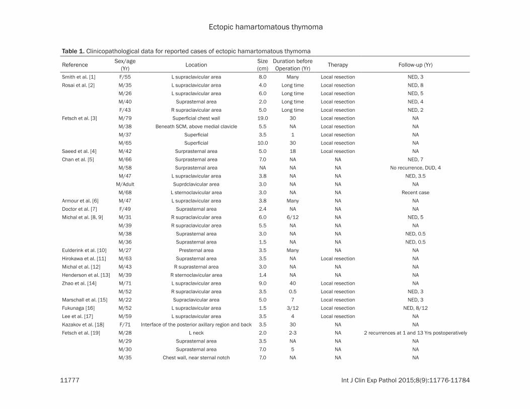
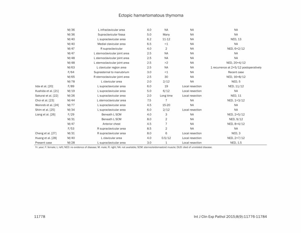
Table 1. Clinicopathological data for reported cases of ectopic hamartomatous thymoma
Reference Sex/age (Yr) Location Size
(cm)Duration beforeOperation (Yr) Therapy Follow-up (Yr)
Smith et al. [1] F/55 L supraclavicular area 8.0 Many Local resection NED, 3Rosai et al. [2] M/35 L supraclavicular area 4.0 Long time Local resection NED, 8
M/26 L supraclavicular area 6.0 Long time Local resection NED, 5M/40 Suprasternal area 2.0 Long time Local resection NED, 4F/43 R supraclavicular area 5.0 Long time Local resection NED, 2
Fetsch et al. [3] M/79 Superficial chest wall 19.0 30 Local resection NAM/38 Beneath SCM, above medial clavicle 5.5 NA Local resection NAM/37 Superficial 3.5 1 Local resection NAM/65 Superficial 10.0 30 Local resection NA
Saeed et al. [4] M/42 Surprasternal area 5.0 18 Local resection NAChan et al. [5] M/66 Surprasternal area 7.0 NA NA NED, 7
M/58 Surprasternal area NA NA NA No recurrence, DUD, 4M/47 L supraclavicular area 3.8 NA NA NED, 3.5
M/Adult Suprdclavicular area 3.0 NA NA NAM/68 L sternoclavicular area 3.0 NA NA Recent case
Armour et al. [6] M/47 L supraclavicular area 3.8 Many NA NADoctor et al. [7] F/49 Suprasternal area 2.4 NA NA NAMichal et al. [8, 9] M/31 R supraclavicular area 6.0 6/12 NA NED, 5
M/39 R supraclavicular area 5.5 NA NA NAM/38 Suprasternal area 3.0 NA NA NED, 0.5M/36 Suprasternal area 1.5 NA NA NED, 0.5
Eulderink et al. [10] M/27 Presternal area 3.5 Many NA NAHirokawa et al. [11] M/63 Suprasternal area 3.5 NA Local resection NAMichal et al. [12] M/43 R suprasternal area 3.0 NA NA NAHenderson et al. [13] M/39 R sternoclavicular area 1.4 NA NA NAZhao et al. [14] M/71 L supraclavicular area 9.0 40 Local resection NA
M/52 R supraclavicular area 3.5 0.5 Local resection NED, 3Marschall et al. [15] M/22 Supraclavicular area 5.0 7 Local resection NED, 3Fukunaga [16] M/52 L supraclavicular area 1.5 3/12 Local resection NED, 8/12Lee et al. [17] M/59 L supraclavicular area 3.5 4 Local resection NAKazakov et al. [18] F/71 Interface of the posterior axillary region and back 3.5 30 NA NAFetsch et al. [19] M/28 L neck 2.0 2-3 NA 2 recurrences at 1 and 13 Yrs postoperatively
M/29 Suprasternal area 3.5 NA NA NAM/30 Suprasternal area 7.0 5 NA NAM/35 Chest wall, near sternal notch 7.0 NA NA NA
Ectopic hamartomatous thymoma
11778 Int J Clin Exp Pathol 2015;8(9):11776-11784
M/36 L infraclavicular area 4.0 NA NA NAM/36 Supraclavicular fossa 5.0 Many NA NAM/40 L supraclavicular area 6.2 11-12 NA NED, 13M/40 Medial clavicular area 6.5 <1 NA NAM/47 R supraclavicular 4.0 2 NA NED, 6+2/12M/47 L sternoclavicular joint area 2.5 NA NA NAM/48 L sternoclavicular joint area 2.5 NA NA NAM/48 L sternoclavicular joint area 2.5 >2 NA NED, 20+4/12M/63 L clavicular region area 2.5 NA NA 1 recurrence at 2+5/12 postoperativelyF/64 Suprasternal to manubrium 3.0 <1 NA Recent caseM/65 R sternoclavicular joint area 2.5 30 NA NED, 16+8/12M/78 L clavicular area 2.0 2/12 NA NED, 5
Iida et al. [20] F/89 L supraclavicular area 6.0 19 Local resection NED, 11/12Kushida et al. [21] M/19 L supraclavicular area 5.0 6/12 Local resection NASakurai et al. [22] M/26 L supraclavicular area 2.0 Long time Local resection NED, 11Choi et al. [23] M/44 L sternoclavicular area 7.5 7 NA NED, 1+3/12Weinreb et al. [24] M/77 L supraclavicular area 4.5 15-20 NA NAShim et al. [25] M/34 L supraclavicular area 6.0 2/12 Local resection NALiang et al. [26] F/29 Beneath L SCM 4.0 3 NA NED, 2+5/12
M/31 Beneath L SCM 8.0 2 NA NED, 9/12M/47 Anterior chest 4.5 7 NA NED, 8+4/12F/53 R supraclavicular area 8.5 2 NA NA
Cheng et al. [27] M/31 R supraclavicular area 8.0 6 Local resection NED, 3Huang et al. [28] M/40 L clavicular area 4.0 0.5/12 Local resection NED, 2+7/12Present case M/28 L supraclavicular area 3.0 1 Local resection NED, 1.5Yr: year; F: female; L: left; NED: no evidence of disease; M: male; R: right; NA: not available; SCM: sternocleidomastoid muscle; DUD: died of unrelated disease.
Ectopic hamartomatous thymoma
11779 Int J Clin Exp Pathol 2015;8(9):11776-11784
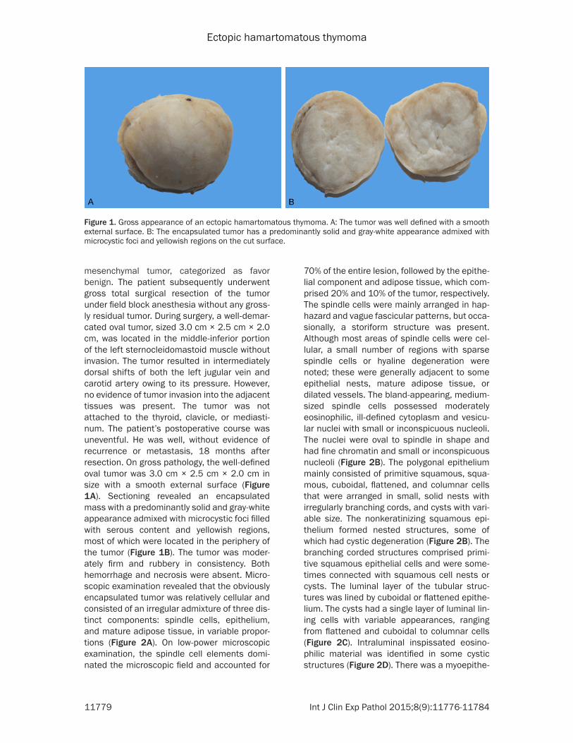
mesenchymal tumor, categorized as favor benign. The patient subsequently underwent gross total surgical resection of the tumor under field block anesthesia without any gross-ly residual tumor. During surgery, a well-demar-cated oval tumor, sized 3.0 cm × 2.5 cm × 2.0 cm, was located in the middle-inferior portion of the left sternocleidomastoid muscle without invasion. The tumor resulted in intermediately dorsal shifts of both the left jugular vein and carotid artery owing to its pressure. However, no evidence of tumor invasion into the adjacent tissues was present. The tumor was not attached to the thyroid, clavicle, or mediasti-num. The patient’s postoperative course was uneventful. He was well, without evidence of recurrence or metastasis, 18 months after resection. On gross pathology, the well-defined oval tumor was 3.0 cm × 2.5 cm × 2.0 cm in size with a smooth external surface (Figure 1A). Sectioning revealed an encapsulated mass with a predominantly solid and gray-white appearance admixed with microcystic foci filled with serous content and yellowish regions, most of which were located in the periphery of the tumor (Figure 1B). The tumor was moder-ately firm and rubbery in consistency. Both hemorrhage and necrosis were absent. Micro- scopic examination revealed that the obviously encapsulated tumor was relatively cellular and consisted of an irregular admixture of three dis-tinct components: spindle cells, epithelium, and mature adipose tissue, in variable propor-tions (Figure 2A). On low-power microscopic examination, the spindle cell elements domi-nated the microscopic field and accounted for
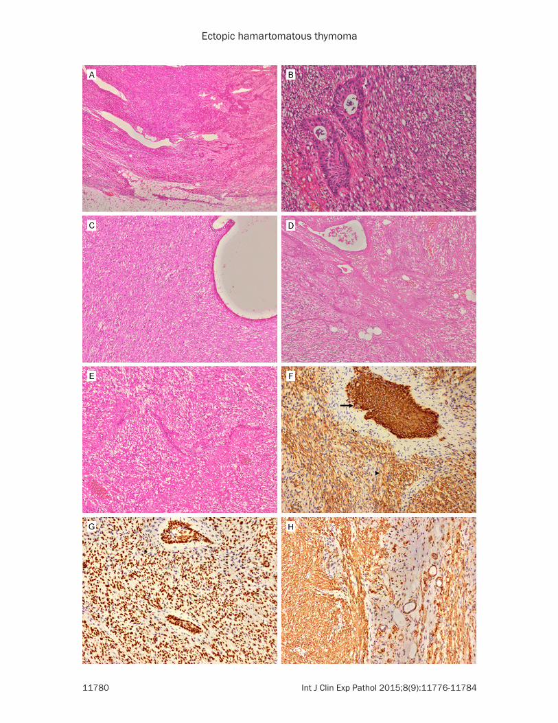
70% of the entire lesion, followed by the epithe-lial component and adipose tissue, which com-prised 20% and 10% of the tumor, respectively. The spindle cells were mainly arranged in hap-hazard and vague fascicular patterns, but occa-sionally, a storiform structure was present. Although most areas of spindle cells were cel-lular, a small number of regions with sparse spindle cells or hyaline degeneration were noted; these were generally adjacent to some epithelial nests, mature adipose tissue, or dilated vessels. The bland-appearing, medium-sized spindle cells possessed moderately eosinophilic, ill-defined cytoplasm and vesicu-lar nuclei with small or inconspicuous nucleoli. The nuclei were oval to spindle in shape and had fine chromatin and small or inconspicuous nucleoli (Figure 2B). The polygonal epithelium mainly consisted of primitive squamous, squa-mous, cuboidal, flattened, and columnar cells that were arranged in small, solid nests with irregularly branching cords, and cysts with vari-able size. The nonkeratinizing squamous epi-thelium formed nested structures, some of which had cystic degeneration (Figure 2B). The branching corded structures comprised primi-tive squamous epithelial cells and were some-times connected with squamous cell nests or cysts. The luminal layer of the tubular struc-tures was lined by cuboidal or flattened epithe-lium. The cysts had a single layer of luminal lin-ing cells with variable appearances, ranging from flattened and cuboidal to columnar cells (Figure 2C). Intraluminal inspissated eosino-philic material was identified in some cystic structures (Figure 2D). There was a myoepithe-
Figure 1. Gross appearance of an ectopic hamartomatous thymoma. A: The tumor was well defined with a smooth external surface. B: The encapsulated tumor has a predominantly solid and gray-white appearance admixed with microcystic foci and yellowish regions on the cut surface.
Ectopic hamartomatous thymoma
11780 Int J Clin Exp Pathol 2015;8(9):11776-11784
Ectopic hamartomatous thymoma
11781 Int J Clin Exp Pathol 2015;8(9):11776-11784
lial layer around the circumference of the tubu-lar and cystic structures. Although the epitheli-um, especially in the cystic and squamous cell nested areas, was well separated from the spindle cell component, a gradual transition from branching cords and strands of epithelial cells to spindle cells was frequently observed (Figure 2E). Mature fat cells and lymphocytes were scattered throughout the lesion. The for-mer were in various proportions in different areas of the tumor, such as in clusters and foci of adipocytes, but had a tendency to be pre-dominantly distributed at the periphery of the tumor. The diffuse but sparse infiltration of small lymphocytes was present mostly in the background of the spindle cell component. Atypical or malignant features such as nuclear atypia, cellular pleomorphism, vascular and capsular invasion, and necrosis were absent from the entire lesion. Mitoses were scarce, with 1.4 per 50 high-power fields (HPFs), but no abnormal mitoses were observed. Immuno- histochemical staining demonstrated that both the spindle cell and epithelial components were diffuse and intense cytoplasmic reactivity for pan-cytokeratin (Figure 2F), CK7, and CK19, and nuclear positivity for p63 (Figure 2G). Moreover, the extent of reactivity for pan-cyto-keratin, CK7, and CK19 in epithelial compo-nents was stronger than in the spindle cell por-tion. The spindle cells were also strongly and uniformly positive for smooth muscle actin (SMA) (Figure 2H) but focally and weakly reac-tive for vimentin and CD99. The epithelial cells were negative for CD99, vimentin, and SMA; however, the outer layer cells of the cystic and tubular structures were positive for SMA and vimentin, which corresponded to the myoepi-thelial cell position (Figure 2H). Staining for S-100 protein, epithelial membrane antigen (EMA), desmin, Myo-D1, and HMB45 was nega-tive in the spindle cell and epithelial compo-
nents. The scattered intratumoral lymphoid component was positive for CD45RO and CD3 but negative for CD20, CD79a, CD1a, and CD99. MIB-1 stained only approximately 1.5% of the spindle and epithelial cells, with 1000 cells counted. These overall clinicopathological features found in the patient met the diagnos-tic criteria for EHT.
Discussion
An EHT, which was first formally described as a new entity in the 2002 edition of the WHO clas-sification of soft-tissue tumors, is an extremely rare benign tumor characteristically comprising an admixture of spindle cells, epithelial islands, and mature adipose tissue [29]. It was first described by Smith and McClure in 1982 and subsequently was designated as an “ectopic hamartomatous thymoma” by Rosai et al. in 1984 [1, 2].
So far, a total of 60 pathologically confirmed cases of EHT have been reported in the English literature, including our patient (Table 1) [1-28]. As documented in Table 1, EHT is usually a slowly growing mass with a long duration, rang-ing from 15 days to 40 years, prior to surgery. EHT often occurs in middle-aged adults (age range: 19 to 89 years; mean: 46.1 years). The male-to-female ratio is 52:8, demonstrating the remarkable male predominance. The most common locations of EHT are the supraclavicu-lar, suprasternal, and sternoclavicular areas. Other sites more rarely affected include the chest, the interface of the posterior axillary region and back, and the presternal, infracla-vicular, and clavicular areas. In the 58 cases of EHT that documented tumor size, the tumors ranged from 1.4 cm to 19.0 cm (mean, 4.8 cm), and the follow-up interval of the 30 cases of EHT with available follow-up information ranged
Figure 2. Microscopic and immunohistochemistry findings of the tumor. A: The neoplasm comprised an admixture of spindled cells, epithelial cells, and mature adipose tissue (hematoxylin and eosin stain, original magnification, × 40). B: Nonkeratinizing squamous epithelium with cystic degeneration and bland spindle cells in which no pleo-morphism, abnormal mitosis, or nuclear atypia were present (hematoxylin and eosin stain, original magnification, × 200). C: A cystic structure lined by flattened, cuboidal, or columnar epithelial cells (hematoxylin and eosin stain, original magnification × 100). D: Branching corded structures of epithelium and cystic structures with inspissated eosinophilic material (hematoxylin and eosin stain, original magnification, × 100). E: Gradual transition from branch-ing cords of the epithelium to spindle cells (hematoxylin and eosin stain, original magnification, × 100). F: Both the spindle cell and epithelial components were positive for pan-cytokeratin, and the level of immunostaining of the latter (arrow) was stronger than that in the former (arrowhead) (original magnification, × 200). G: Strong and diffuse P63 immunoreactivity was present in epithelial cells and spindle cells (original magnification, × 200). H: Immuno-histochemical staining for SMA was positive in the spindle cells and myoepithelial cells of the tubular and cystic structures but negative in the epithelial cells (original magnification, × 200).
Ectopic hamartomatous thymoma
11782 Int J Clin Exp Pathol 2015;8(9):11776-11784
from 0.5 years to 13 years (mean: 5.2 years). All patients with available follow-up information were alive except one who had died 4 years postoperatively of unrelated causes. One patient’s tumor recurred once at a post-opera-tive interval of 2 years and 5 months, and one patient relapsed twice at 1 year and 13 years after surgery. However, the recurrences of two patients reported by Fetsch et al. were due to incomplete local excision, and both patients had no evidence of disease for a long time after a complete re-excision [19].
It is well known that nearly all of EHTs show no evidence of atypical or malignant features and pursue a benign clinical course, and complete local excision is the first treatment of choice. The malignant epithelial portions, consisting of a dysplastic glandular component that is usu-ally arranged in a cribriform structure bridging several glandular configurations, were observed in 2 cases of EHT reported by Michal et al. [8, 9]. As a result, some authors have advocated that the probability of the malignant convert of tumor cells within EHT must be considered and that sufficient sampling and careful assess-ment of the malignant component in each instance of EHT are essential [19, 22]. However, no adverse outcome was documented in one of the two patients with an EHT with a malignant epithelial component; there was no document-ed follow up on the other patient with an EHT with a malignant epithelium [8, 9].
Myoid cells were observed among the spindle cell areas in variable proportions in three cases of EHT, which immunohistochemically were myoglobin positive and desmin negative [4, 6, 14]. Interestingly, immunostaining for cytokera-tin and EMA was positive in the myoid compo-nent in one case of EHT [14] and negative in the remaining two cases [4, 6]. Occasional poorly formed cross-striations were demonstrated in such cells in one case by phosphotungstic acid hematoxylin (PTAH) staining [4].
The exact origin of EHT remains controversial. The prevailing original view is that this tumor originates in the thymic anlage; possible sourc-es include the third and fourth pharyngeal pouches, the cervical sinus of His, and the ulti-mobranchial body [1-4, 6]. However, many char-acteristics of this lesion have challenged the hypothesis of thymic origin. Residual thymic structures have never been described in asso-
ciation with EHT and a mediastinal counterpart was absent in all cases of EHT reported to date [8, 9, 13, 15, 19]. Furthermore, the reported unusual locations of EHT, including the prester-nal and dorsal locations, could also exclude ori-gin in a remnant of thymic tissue because of the scarcity of thymic remnants in both the pre-sternal and dorsal areas [10, 18]. Considering the adenocarcinomatous differentiation, myo-epithelial cell component, and clusters of gland acini component in EHT, Michal et al. believed that there could be salivary gland differentia-tion in EHT [8]. In 2000, Henderson and Gupta proposed an EHT origin from the branchial pouch endoderm rather than from thymic rem-nants [13]. After analyzing the clinicopathologi-cal and immunohistochemical features of 21 cases of so-called “ectopic hamartomatous thymoma”, Fetsch et al. suggested that the nomenclature “branchial anlage mixed tumor” more accurately reflects the nature of this rare lesion [19]. Weinreb et al. reported a case of EHT with skin adnexal differentiation with immunoreactivity for EMA, androgen, and BRST-2 and suggested that “a mixed tumor of skin adnexal origin” is another possible desig-nation for EHT [24].
The pathologic differential diagnosis of EHT includes biphasic synovial sarcoma, malignant schwannoma with epithelial differentiation, a mixed tumor of skin or salivary gland tissue, and thymolipoma. Briefly, the presence of a well-defined lesion usually located in the lower neck with characteristic spindle cell and epithe-lial cell components and mature adipose tis-sue, together with the immunohistochemical features of both the spindle cell and epithelial cell components having cytokeratin positivity should facilitate an accurate diagnosis of EHT. Biphasic synovial sarcoma can easily be exclud-ed because it rarely shows squamous differen-tiation and the spindle tumor cells have higher cellularity, greater nuclear hyperchromasia, and mitotic figures. Furthermore, the spindle cell component in synovial sarcoma shows foci of patchy positivity for cytokeratin [30]. Tumor cells in malignant schwannoma with epithelial differentiation are S-100 protein positive and cytokeratin negative. In contrast, tumor cells in EHT are cytokeratin positive and S-100 protein negative [31]. Differentiating a mixed tumor of skin or salivary gland tissue from an EHT is based on the former’s chondromyxoid stroma
Ectopic hamartomatous thymoma
11783 Int J Clin Exp Pathol 2015;8(9):11776-11784
and immunoreactivity for S-100 protein, which are absent in the latter [32, 33]. Thymolipoma could be excluded, as it nearly always arises in the mediastinum rather than in the neck, and lacks the prominent spindle cell component of EHT [34].
Conclusion
In conclusion, we have reported a case of EHT in a 28-year-old male and presented a review of the literature to clarify the clinical and patho-logical characteristics of this rare tumor. EHT is a rare lesion with characteristic histological and immunohistochemical features and is fre-quently located in the lower neck area. Correct identification of this unique and distinctive neo-plasm is important because the tumor shows a benign biologic behavior and the therapy, if not complicated by a malignant component, is local excision. More cases of EHT must be studied to draw definitive conclusions about the tumor’s exact histogenesis.
Acknowledgements
The authors thank American Journal Experts for editing the present paper. This work was supported by the Natural Science Foundation of Shandong Province (Grant No. ZR2012HM- 018).
Disclosure of conflict of interest
None.
Address correspondence to: Dr. Yanhong Tai, De- partment of Pathology, The General Hospital, Jinan Military Command, 25 Shifan Road , Jinan 250031, Shandong Province, China. Tel: +86 531 51666857; Fax: +86 531 51666284; E-mail: taiyanhongcheng@ 163.com
References
[1] Smith PS, McClure J. Unusual subcutaneous mixed tumour exhibiting adipose, fibroblastic, andepithelial components. J Clin Pathol 1982; 35: 1074-7.
[2] Rosai J, Limas C, Husband EM. Ectopic hamar-tomatous thymoma. A distinctive benign lesion of lower neck. Am J Surg Pathol 1984; 8: 501-13.
[3] Fetsch JF, Weiss SW. Ectopic hamartomatous thymoma: clinicopathologic, and histogenetic considerations in four new cases. Hum Pathol 1990; 21: 662-8.
[4] Saeed IT, Fletcher CD. Ectopic hamartomatous thymoma containing myoid cells. Histopath- ology 1990; 17: 572-4.
[5] Chan JK, Rosai J. Tumors of the neck showing thymic or related branchial pouch differentia-tion: a unifying concept. Hum Pathol 1991; 22: 349-67.
[6] Armour A, Williamson JM. Ectopic cervical hamartomatous thymoma showing extensive myoid differentiation. J Laryngol Otol 1993; 107: 155-8.
[7] Doctor VM, Simha MR. Ectopic hamartoma-tous thymoma-a case report and review of the literature. Indian J Cancer 1993; 30: 192-5.
[8] Michal M, Zámecník M, Gogora M, Mukensnabl P, Neubauer L. Pitfalls in the diagnosis of ecto-pic hamartomatous thymoma. Histopathology 1996; 29: 549-55.
[9] Michal M, Neubauer L, Fakan F. Carcinoma arising in ectopic hamartomatous thymoma. An ultrastructural study. Pathol Res Pract 1996; 192: 610-8.
[10] Eulderink F, de Graaf PW. Ectopic hamartoma-tous thymoma located presternally. Eur J Surg 1998; 164: 629-30.
[11] Hirokawa M, Tadaoka Y, Shimizu M, Monobe Y, Kanahara T, Manabe T. Ectopic hamartoma-tous thymoma. Report of a case with fine nee-dle aspiration biopsy findings. Acta Cytol 1999; 43: 232-4.
[12] Michal M, Mukensnabl R. Clear cell epithelial cords in an ectopic hamartomatous thymoma. Histopathology 1999; 35: 89-90.
[13] Henderson CJ, Gupta L. Ectopic hamartoma-tous thymoma: a case study and review of the literature. Pathology 2000; 32: 142-6.
[14] Zhao C, Yamada T, Kuramochi S, Yamazaki K, Mukai M, Kameyama K, Hata J. Two cases of ectopic hamartomatous thymoma. Virchows Arch 2000; 437: 643-7.
[15] Marschall J, Kanthan R. The sarcomatous guise of cervical ectopic hamartomatous thy-moma. Head Neck 2002; 24: 800-4.
[16] Fukunaga M. Ectopic hamartomatous thymo-ma: a case report with immunohistochemical and ultrastructural studies. APMIS 2002; 110: 565-70.
[17] Lee SN, Cho MS, Koo H, Han WS. Ectopic ham-artomatous thymoma: A case report showing CD99 + lymphocytes and a low proliferation index. Arch Pathol Lab Med 2003; 127: e378-81.
[18] Kazakov DV, Mukensnabl P, Hes O, Michal M. ‘Ectopic’ ectopic hamartomatous thymoma. Histopathology 2004; 45: 202-4.
[19] Fetsch JF, Laskin WB, Michal M, Remotti F, Heffner D, Ellis G, Furlong M, Miettinen M. Ectopic hamartomatous thymoma: A clinico-pathologic and immunohistochemical analysis
Ectopic hamartomatous thymoma
11784 Int J Clin Exp Pathol 2015;8(9):11776-11784
of 21 cases with data supporting reclassifica-tion as a branchial anlage mixed tumor. Am J Surg Pathol 2004; 28: 1360-70.
[20] Iida E, Okazaki M, Sarukawa S, Motoi T, Kikuchi Y. Ectopic hamartomatous thymoma in the sternocleidomastoid muscle masquerading as sarcoma. Scand J Plast Reconstr Surg Hand Surg 2006; 40: 249-52.
[21] Kushida Y, Haba R, Kobayashi S, Ishikawa M, Doi T, Kadota K. Ectopic hamartotoous thymo-ma: a case report with immunohistochemical study and review of the literature. J Cutan Pathol 2006; 33: 369-72.
[22] Sakurai H, Kaji M, Mukai K, Suemasu K. Ectopic hamartomatous thymoma--a truly rare neoplasm: report of a case. Surg Today 2010; 40: 146-9.
[23] Choi JH, Shim YR, Song SY. Fine needle aspira-tion cytology of ectopic hamartomatous ham-artomatous thymoma. Acta Cytol 2007; 51: 672-5.
[24] Weinreb I, O’Malley F, Ghazarian D. Ectopic hamartomatous thymoma: a case demonstrat-ing skin adnexal differentiation with positivity for epithelial membrane antigen, androgen re-ceptors, and BRST-2 by immunohistochemis-try. Hum Pathol 2007; 38: 1092-5.
[25] Shim DB, Song JS, Baek SJ. An ectopic hamar-tomatous thymoma. Auris Nasus Larynx 2012; 39: 110-3.
[26] Liang PI, Li CF, Sato Y, Lee VK, Bahrami A, Chuang SS. Ectopic hamartomatous thymoma is distinct from lipomatous pleomorphic ade-noma in lacking PLAG1 aberration. Histopath- ology 2013; 62: 518-22.
[27] Cheng YS, Zhou ZR, Wang J. Ectopic haamrto-matous thymoma: radiographic and clinico-pathological features. Chin Med J (Engl) 2013; 126: 798-9.
[28] Huang L, Zhao L, Chai Y. An ectopic hamarto-matous thymoma compressing left jugular vein. Niger J Clin Pract 2014; 17: 814-6.
[29] Chan JKC. Ectopic hamartomatous thymoma. In: Fletcher CDM, Unni KK, Mertens F, editors. Pathology and genetics of tumours of soft tis-sue and bone. Lyon: IARC Press; 2002. pp. 192-3.
[30] Fisher C, de Bruijn DRH, van Kessel AG. Synovial sarcoma. In: Fletcher CDM, Unni KK, Mertens F, editors. Pathology and genetics of tumours of soft tissue and bone. Lyon: IARC Press; 2002. pp. 200-4.
[31] Scheithauer BW, Louis DN, Hunter S, Woodruff JM, Antonescu CR. In: Louis DN, Ohgaki H, Wiestler OD, Cavenee WK, editors. Pathology and genetics of tumours of the central nervous system. Lyon: IARC Press; 2007. pp. 160-2.
[32] McNiff J, McCalmont TH, Requena L, Sangüeza OP, Vassallo C, Rosso R, Borroni G, Glusac EJ, Pichardo RO. Mixed tumour. In: LeBoit PE, Burg G, Weedon D, Sarasin A, editors. Pathology and genetics of skin tumours. Lyon: IARC Press; 2006. pp. 147-8.
[33] Eveson JW, Kusafuka K, Stenman G, Nagao T. Pleomorphic adenoma. In: Barnes L, Eveson JW, Reichart P, Sidransky D, editors. Pathology and genetics of head and neck tumours. Lyon: IARC Press; 2005. pp. 254-8.
[34] Romero-Guadarrama MB, Duran-Padilla MA, Cruz-Ortiz H, Castro-Gomez L, Lopez-Vancell D, Novelo-Retana V, Santia-go-Prieto AC, Fierro-Chavez E, Rodriguez-Martinez HA. Diagnosis of thymolipoma with fine needle aspiration biop-sy: Report of a case initially misdiagnosed as liposarcoma. Acta Cytol 2004; 48: 441-6.


![Parathyroid Adenoma/Thymoma Case Reportadenoma and thymoma without mention of sestamibi uptake by the thymoma (whether such imaging was performed or not). Byrne et al. [13] demonstrated](https://img.dokumen.tips/doc/110x75/5e2f040ac0577556e1278f0b/parathyroid-adenomathymoma-case-adenoma-and-thymoma-without-mention-of-sestamibi.jpg)