Embed Size (px)
Citation preview

Int J Clin Exp Pathol 2014;7(11):8198-8204www.ijcep.com /ISSN:1936-2625/IJCEP0002758
Case ReportCongenital neuroblastoma with placental involvement
Ayako Kume1,2, Teppei Morikawa1, Makiko Ogawa1, Aki Yamashita3, Shunichi Yamaguchi3, Masashi Fukayama1
1Department of Pathology, The University of Tokyo Hospital, Tokyo, Japan; 2School of Medicine, The University of Kyushu, Fukuoka, Japan; 3Department of Obstetrics and Gynecology, The University of Tokyo Hospital, Tokyo, Japan
Received September 24, 2014; Accepted November 8, 2014; Epub October 15, 2014; Published November 1, 2014
Abstract: We describe an extremely rare case of congenital neuroblastoma with placental involvement. A fetus with a left abdominal mass detected during ultrasonography at 23 weeks’ gestation developed hydrops fetalis by 26 weeks’ gestation. The mother developed hypertension at 26 5/7 weeks’ gestation. Based on a clinical diagnosis of pregnancy-induced hypertension, labor was induced at 26 6/7 weeks. However, intrauterine fetal death was diagnosed during delivery. Postmortern examination revealed a solid tumor at the site of the left adrenal gland. Histological examination of the tumor revealed dense proliferation of small round tumor cells with sparse cyto-plasm and hyperchromatic nuclei. Some tumor-cell complexes contained abundant neurofibrils and Hormer-Wright rosettes were observed. A diagnosis of neuroblastoma of the left adrenal gland was made. The liver was markedly enlarged and was extensively replaced by neuroblastoma cells. In addition, small nests of tumor cells were detected in the blood vessels of various organs including the heart, lung, spleen, kidneys, stomach, small and large intestine, thyroid gland, testis, spinal cord, and bone marrow. Histological examination of the enlarged placenta revealed numerous neuroblastoma cells in the villous fetal capillary spaces. The present case was unusual in that the tumor cells were found not only in the chorionic villi, but also in the intervillous space of the maternal vascular system. However, there was no clinical evidence of maternal metastasis.
Keywords: Autopsy, fetal malignancy, immunohistochemistry, tumor thrombus, metastasis
Introduction
Congenital malignant tumors are rare [1], and placental involvement of such tumors is exceed-ingly rare. Most tumors involving the placenta are neuroblastomas [2-7]; however, tumor cells have been confined to the chorionic villi in all reported cases of neuroblastomas involving the placenta. Here, we describe the clinical and pathological features of a stillborn infant with a congenital neuroblastoma with widespread metastases involving multiple organs, including the placenta. This case was unusual because the tumor cells were found not only in the chori-onic villi, but also in the intervillous space of the maternal vascular system. We also discuss the importance of histological examination of the placenta for establishing a diagnosis.
Case report
Clinical history
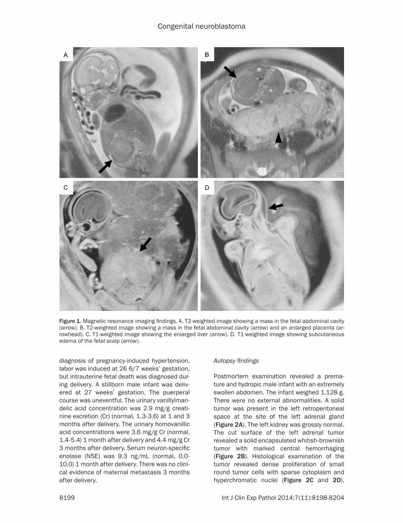
A 29-year-old, gravida 2, para 1 woman with a past history of hyperthyroidism was referred to our hospital because of a fetal abdominal mass detected during a routine prenatal ultrasono-gram at 23 weeks’ gestation. A left renal tumor was suspected on the basis of magnetic reso-nance imaging at 25 weeks’ gestation (Figure 1A and 1B). Enlargement of the liver and pla-centa, and subcutaneous edema were also detected (Figure 1B-D). Fetal pericardial fluid was pointed out by ultrasonography at 26 weeks’ gestation, and the mother’s blood pres-sure was elevated to 143/86 mmHg at 26 5/7 weeks’ gestation. Albuminuria and generalized edema were also observed. Based on a clinical
Congenital neuroblastoma
8199 Int J Clin Exp Pathol 2014;7(11):8198-8204
diagnosis of pregnancy-induced hypertension, labor was induced at 26 6/7 weeks’ gestation, but intrauterine fetal death was diagnosed dur-ing delivery. A stillborn male infant was deliv-ered at 27 weeks’ gestation. The puerperal course was uneventful. The urinary vanillylman-delic acid concentration was 2.9 mg/g creati-nine excretion (Cr) (normal, 1.3-3.6) at 1 and 3 months after delivery. The urinary homovanillic acid concentrations were 3.6 mg/g Cr (normal, 1.4-5.4) 1 month after delivery and 4.4 mg/g Cr 3 months after delivery. Serum neuron-specific enolase (NSE) was 9.3 ng/mL (normal, 0.0-10.0) 1 month after delivery. There was no clini-cal evidence of maternal metastasis 3 months after delivery.
Autopsy findings
Postmortern examination revealed a prema-ture and hydropic male infant with an extremely swollen abdomen. The infant weighed 1,128 g. There were no external abnormalities. A solid tumor was present in the left retroperitoneal space at the site of the left adrenal gland (Figure 2A). The left kidney was grossly normal. The cut surface of the left adrenal tumor revealed a solid encapsulated whitish-brownish tumor with marked central hemorrhaging (Figure 2B). Histological examination of the tumor revealed dense proliferation of small round tumor cells with sparse cytoplasm and hyperchromatic nuclei (Figure 2C and 2D).
Figure 1. Magnetic resonance imaging findings. A. T2-weighted image showing a mass in the fetal abdominal cavity (arrow). B. T2-weighted image showing a mass in the fetal abdominal cavity (arrow) and an enlarged placenta (ar-rowhead). C. T1-weighted image showing the enlarged liver (arrow). D. T1-weighted image showing subcutaneous edema of the fetal scalp (arrow).

Congenital neuroblastoma
8200 Int J Clin Exp Pathol 2014;7(11):8198-8204
Some tumor-cell complexes contained abun-dant neurofibrils, and Hormer-Wright rosettes were observed. The left adrenal gland was almost completely replaced by tumor cells, though a few normal adrenocortical cells were found at the periphery of the tumor (Figure 2C). The tumor was covered by a fibrous capsule, but the tumor cells invaded directly into the pancreas. Direct invasion of the left kidney was not observed.
Immunohistochemical analysis [8] revealed that the tumor cells were immunoreactive for synaptophysin, chromogranin A, CD56, and NSE, but negative for leukocyte common anti-gen (LCA) and cytokeratin. Based on the histo-logical and immunohistochemical findings, a diagnosis of neuroblastoma of the left adrenal gland was made.
The liver was markedly enlarged and weighed 225 g. The cut surface of the liver showed mul-tiple subcapsular, ill-defined, whitish lesions (Figure 3A). Microscopically, the liver was extensively replaced by neuroblastoma cells (Figure 3B). Involvement of neuroblastoma cells in the right adrenal grand was also observed (Figure 3C). In addition, small nests of tumor cells were detected in the blood ves-sels of various organs including the heart (Figure 3D), lung, spleen, kidneys, stomach, small and large intestine, thyroid gland, testis, spinal cord, and bone marrow.
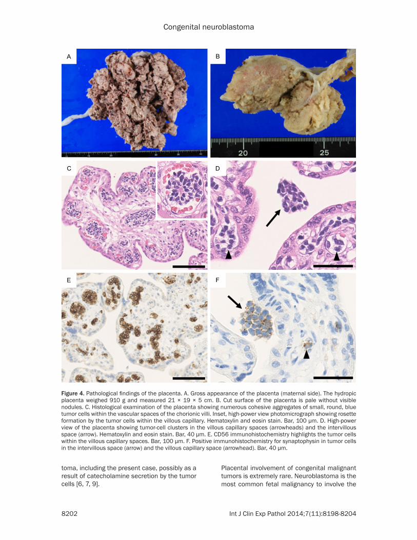
Histopathology of the placenta
The placenta weighed 910 g and measured 21 × 19 × 5 cm (Figure 4A). The cut surface of the
Figure 2. Pathological findings of the left adrenal gland tumor. A. Gross appearance of the bilateral kidneys and ad-renal glands. The left adrenal gland is markedly enlarged. RA, right adrenal gland; RK, right kidney; LA, left adrenal gland; LK, left kidney. B. Cut surface of the left adrenal gland tumor with marked central hemorrhage. Arrow indi-cates the left kidney. C. Histological findings of the left adrenal gland tumor. Diffuse infiltration of tumor cells with areas of necrosis (left bottom) and residual adrenal cortex and capsule (right). Hematoxylin and eosin stain. Bar, 500 µm. D. High-power view of the left adrenal gland tumor showing proliferation of small, round, blue tumor cells with sparse cytoplasm and hyperchromatic nuclei, and formation of Hormer-Wright rosettes (arrows). Hematoxylin and eosin stain. Bar, 50 µm.

Congenital neuroblastoma
8201 Int J Clin Exp Pathol 2014;7(11):8198-8204
chorionic villi was edematous and pale, with no visible nodules (Figure 4B). The umbilical cord was normal in length and diameter and showed the usual spiraling. Microscopically, the villous fetal capillary spaces contained numerous cohesive aggregates of small, round, blue tumor cells with significant nuclear atypia (Figure 4C). Rosette formation was sometimes observed. The tumor cells were widely dis-persed throughout the placenta, with > 90% of the examined villi containing tumor cells. Focal invasion of the villous stroma was observed, and necrotic debris was sometimes observed in the villous stroma. Tumor cell clusters were occasionally observed in the intervillous space (Figure 4D).
Immunohistochemical analysis revealed that the tumor cells in the placenta were immunore-
active for CD56 (Figure 4E), synaptophysin (Figure 4F), chromogranin A, and NSE, but neg-ative for LCA and cytokeratin. The morphology and immunophenotype of the tumor in the pla-centa confirmed the diagnosis of placental involvement of neuroblastoma.
Discussion
Neuroblastoma is the most frequently occur-ring malignant tumor in the newborn period and early infancy [1]. It is estimated that 25% of congenital malignant tumors are neuroblasto-mas [1]. In the present case, congenital neuro-blastoma of the left adrenal gland with placen-tal involvement was confirmed by histological and immunohistochemical examinations of the placenta, as well as by autopsy. Maternal hyper-tension and other symptoms have been report-ed in some cases of fetal congenital neuroblas-
Figure 3. Pathological findings of metastatic lesions. A. Cut surface of the liver showing multiple subcapsular, ill-defined whitish lesions. B. On microscopic examination, the liver was extensively replaced by tumor cells. Residual hepatocytes are seen in the right-bottom corner. Hematoxylin and eosin stain. Bar, 100 μm. C. Histological examina-tion of the right adrenal gland showing tumor-cell infiltration mainly in the medulla. Inset, tumor cell invasion in the cortex. Hematoxylin and eosin stain. Bar, 2 mm. D. Histological examination of the heart showing multiple tumor-cell emboli within the vessels in the myometrium. Inset, high-power view. Hematoxylin and eosin stain. Bar, 200 μm.
Congenital neuroblastoma
8202 Int J Clin Exp Pathol 2014;7(11):8198-8204
toma, including the present case, possibly as a result of catecholamine secretion by the tumor cells [6, 7, 9].
Placental involvement of congenital malignant tumors is extremely rare. Neuroblastoma is the most common fetal malignancy to involve the
Figure 4. Pathological findings of the placenta. A. Gross appearance of the placenta (maternal side). The hydropic placenta weighed 910 g and measured 21 × 19 × 5 cm. B. Cut surface of the placenta is pale without visible nodules. C. Histological examination of the placenta showing numerous cohesive aggregates of small, round, blue tumor cells within the vascular spaces of the chorionic villi. Inset, high-power view photomicrograph showing rosette formation by the tumor cells within the villous capillary. Hematoxylin and eosin stain. Bar, 100 μm. D. High-power view of the placenta showing tumor-cell clusters in the villous capillary spaces (arrowheads) and the intervillous space (arrow). Hematoxylin and eosin stain. Bar, 40 μm. E. CD56 immunohistochemistry highlights the tumor cells within the villous capillary spaces. Bar, 100 μm. F. Positive immunohistochemistry for synaptophysin in tumor cells in the intervillous space (arrow) and the villous capillary space (arrowhead). Bar, 40 μm.

Congenital neuroblastoma
8203 Int J Clin Exp Pathol 2014;7(11):8198-8204
placenta, followed by hepatoblastoma and leu-kemia [5]. Fewer than 20 cases of congenital neuroblastoma involving the placenta have been reported in the English literature [2-7]. Microscopically, the congenital malignant tumor cells are usually confined to the villous capillaries (fetal circulation), except in cases of placental involvement of leukemia, which often diffusely infiltrates the villous stroma [5, 10]. In contrast, the tumor cells typically occupy the intervillous space (a component of the mater-nal vascular system) in most cases of placental metastases from maternal malignant tumors (most commonly malignant melanoma) [5]. Massive liver metastasis is common in congen-ital neuroblastoma with placental involvement, as in the present case, and the prognosis is extremely poor [3, 6].
In the present case, tumor cells were found not only in the chorionic villi, but also in the intervil-lous space of the maternal vascular system, in contrast to all but one previous reports of pla-cental involvement by neuroblastoma, in which microscopic examination revealed that the tumor cells were confined to the villous capillar-ies [5]. Perkins et al. reported a case of con-genital neuroblastoma that infiltrated the vil-lous stroma without extension to the intervil-lous space [11]. To the best of our knowledge, the present case is the first reported incidence of congenital neuroblastoma cells in the inter-villous space of the placenta. To date, no cases of fetal malignancy resulting in maternal metas-tases have been reported in the literature [5]. It is possible that the maternal immune system suppresses the fetal tumor cells [11]. Likewise, there was no clinical evidence of maternal metastasis in the present case. However, it is possible that tumor cells may have entered the maternal circulation, and given that late recur-rences of neuroblastoma have been reported [12], caution should be exercised if the mother requires immunosuppressive therapy (e.g., for autoimmune disease) in the future.
The placenta can be sampled easily and non-invasively at birth. Although the fatal outcome could not have been prevented in the present case, the prognosis of neuroblastoma is not necessarily hopeless, even when metastases are present [13, 14]. Most previously reported patients with placental involvement of neuro-blastoma have died within 3 months, but rapid
treatment employing modern modes of chemo-therapy might improve the prognosis in some cases [4]. Ohyama et al. reported two neonates with congenital neuroblastoma initially diag-nosed by histological examination of the pla-centa [4]. Both neonates had multiple metasta-ses and were treated with chemotherapy. One patient was in remission at 8 months old and the other showed good partial remission at 14 months old [4]. It is therefore necessary to make the diagnosis as early as possible to allow optimal treatment. Placental examination may provide valuable information, but careful examination is needed given that only a few tumor cells may be found in the placenta [15, 16].
In summary, we report a case of congenital neuroblastoma with placental involvement. The diagnosis was made on the basis of histological and immunohistochemical examinations of the placenta, as well as an autopsy. Careful exami-nation of the placenta may help in documenting the presence of metastatic disease, as well as in determining the optimal treatment for live-born infants with congenital malignant tumors.
Acknowledgements
We are grateful to Kei Sakuma for excellent technical support.
Disclosure of conflict of interest
None.
Address correspondence to: Dr. Teppei Morikawa, Department of Pathology, Graduate School of Medicine, The University of Tokyo, 7-3-1 Hongo, Bunkyo-Ku, Tokyo 113-0033, Japan. Tel: +81-3-5841-3341; Fax: +81-3-3815-8379; E-mail: [email protected]
References
[1] Wells HG. Occurrence and significance of con-genital malignant neoplasms. Arch Pathol 1940; 30: 535-601.
[2] Strauss L, Driscoll SG. Congenital Neuro- blastoma Involving the Placenta. Reports of Two Cases. Pediatrics 1964; 34: 23-31.
[3] Lynn AA, Parry SI, Morgan MA, Mennuti MT. Disseminated congenital neuroblastoma in-volving the placenta. Arch Pathol Lab Med 1997; 121: 741-744.
[4] Ohyama M, Kobayashi S, Aida N, Toyoda Y, Ijiri R, Tanaka Y. Congenital neuroblastoma diag-
Congenital neuroblastoma
8204 Int J Clin Exp Pathol 2014;7(11):8198-8204
nosed by placental examination. Med Pediatr Oncol 1999; 33: 430-431.
[5] Roberts DJ, Oliva E. Clinical significance of pla-cental examination in perinatal medicine. J Matern Fetal Neonatal Med 2006; 19: 255-264.
[6] Allen AT, Dress AF, Moore WF. Mirror syndrome resulting from metastatic congenital neuro-blastoma. Int J Gynecol Pathol 2007; 26: 310-312.
[7] Inoue T, Ito Y, Nakamura T, Matsuoka K, Sago H. A catecholamine-secreting neuroblastoma leading to hydrops fetalis. J Perinatol 2014; 34: 405-407.
[8] Ichimura T, Morikawa T, Kawai T, Nakagawa T, Matsushita H, Kakimi K, Kume H, Ishikawa S, Homma Y, Fukayama M. Prognostic signifi-cance of CD204-positive macrophages in up-per urinary tract cancer. Ann Surg Oncol 2014; 21: 2105-2112.
[9] Newton ER, Louis F, Dalton ME, Feingold M. Fetal neuroblastoma and catecholamine-in-duced maternal hypertension. Obstet Gynecol 1985; 65: 49S-52S.
[10] Las Heras J, Leal G, Haust MD. Congenital leu-kemia with placental involvement. Report of a case with ultrastructural study. Cancer 1986; 58: 2278-2281.
[11] Perkins DG, Kopp CM, Haust MD. Placental in-filtration in congenital neuroblastoma: a case study with ultrastructure. Histopathology 1980; 4: 383-389.
[12] Hata Y, Sasaki F, Naito H, Takahashi H, Namieno T, Uchino J. Late recurrence in neuro-blastoma. J Pediatr Surg 1991; 26: 1417-1419.
[13] Maris JM, Hogarty MD, Bagatell R, Cohn SL. Neuroblastoma. Lancet 2007; 369: 2106-2120.
[14] Maris JM. Recent advances in neuroblastoma. N Engl J Med 2010; 362: 2202-2211.
[15] Anders D, Kindermann G, Pfeifer U. Meta- stasizing fetal neuroblastoma with involve-ment of the placenta simulating fetal erythro-blastosis. Report of two cases. J Pediatr 1973; 82: 50-53.
[16] Smith CR, Chan HS, deSa DJ. Placental involve-ment in congenital neuroblastoma. J Clin Pathol 1981; 34: 785-789.