Embed Size (px)
Citation preview
Int J Clin Exp Med 2017;10(7):11097-11100www.ijcem.com /ISSN:1940-5901/IJCEM0050895
Case ReportCavernous hemangioma with multiple phleboliths of the parotid gland in adult masquerading assialolithiasis
Bo-Nien Chen1,2
1Department of Otolaryngology-Head and Neck Surgery, Hsinchu MacKay Memorial Hospital, Hsinchu City, Hsinchu 30071, Taiwan; 2Mackay Junior College of Medicine, Nursing, and Management, New Taipei City, Taiwan
Received January 18, 2017; Accepted May 26, 2017; Epub July 15, 2017; Published July 30, 2017
Abstract: Objectives: Hemangiomas account for 0.4%-0.6% of all tumors of the parotid gland and are extremely rare in adults. Changes in blood flow dynamics within hemangiomas result in thrombus formation and phleboliths. We present a case of cavernous hemangioma with multiple phleboliths of the parotid gland masquerading assi-alolithiasis in an adult patient. Methods: A 43-year-old woman visited our hospital, presenting with a 3-year history of a slowly progressive painless mass at the left infraauricular area associated with intermittent swelling episodes related to meals. Computed tomography revealed multiple calcified nodules located within the enlarged, contrast-enhanced left parotid gland. The patient subsequently underwent total parotidectomy. Results: Cavernous heman-gioma with multiple phleboliths of the parotid gland was diagnosed. The patient recovered well from the surgery and was disease free at the most recent follow-up, 10 years later. Conclusions: No previous case reports have described a cavernous hemangioma with multiple phleboliths of the parotid gland. This condition requires further study be-cause its rarity, clinical presentation, and imaging features often lead physicians to a misdiagnosis of sialolithiasis. It emphasizes that the possibility of a cavernous hemangioma should be considered in the differential diagnosis of parotid tumors when multiple intraglandular calcification nodules are observed in imaging studies.
Keywords: Cavernous hemangioma, phlebolith, parotid gland, sialolithiasis, mealtime syndrome
Introduction
Hemangiomas account for 0.4%-0.6% of all tumors of the parotid gland and are extremely rare in adults [1]. Changes in blood flow dynam-ics within hemangiomas result in thrombus for-mation and phleboliths. The changes may origi-nate from injury to a vessel wall or result from stagnation of the flow of blood [2-4]. To date, a few reports of hemangioma with phlebolith of the parotid gland have been reported (Table 1) [4-6]. Herein, we present a case of cavernous hemangioma with multiple phleboliths of the parotid gland masquerading assialolithiasis in an adult patient. Based on our research, this is the first case report of cavernous hemangioma with multiple phleboliths of the parotid gland in the English-language literature.
Case report
A 43-year-old woman visited our hospital, pre-senting with a 3-year history of a slowly pro-gressive painless mass at the left infraauricular
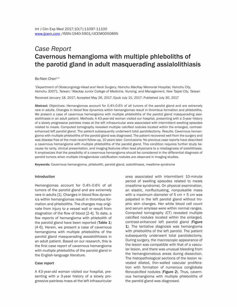
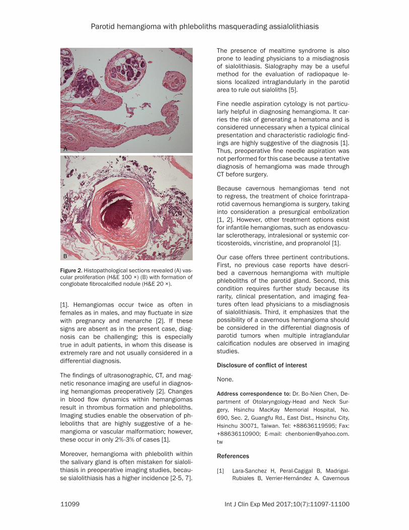
area associated with intermittent 10-minute period of swelling episodes related to meals (mealtime syndrome). On physical examination, an elastic, nonfluctuating, nonpulsatile mass with a maximum diameter of 5 cm × 5 cm was palpated in the left parotid gland without tro-phic skin changes. Her white blood cell count and serum amylase were within normal ranges. Computed tomography (CT) revealed multiple calcified nodules located within the enlarged, contrast-enhanced left parotid gland (Figure 1). The tentative diagnosis was hemangioma with phleboliths of the left parotid. The patient subsequently underwent total parotidectomy. During surgery, the macroscopic appearance of the lesion was compatible with that of a vascu-lar lesion, and there was unusual bleeding from the hemangiomatous areas during dissection. The histopathological sections of the lesion re- vealed dilated, thin-walled vascular prolifera-tion with formation of numerous conglobate fibrocalcified nodules (Figure 2). Thus, cavern-ous hemangioma with multiple phleboliths of the parotid gland was diagnosed.
Parotid hemangioma with phleboliths masquerading assialolithiasis
11098 Int J Clin Exp Med 2017;10(7):11097-11100
The patient recovered well from the surgery and was disease free at the most recent fo- llow-up, 10 years later.
Discussion
In the head and neck, hemangiomas principally affect the salivary glands, with the parotid as the most common site. Hemangiomas mostly occur in children [1-3]. Hemangiomas are clas-
sified as cavernous, capillary, and mixed hem-angiomas [1]. Adult salivary gland hemangio-mas are of the cavernous type, whereas infan-tile hemangiomas are usually capillary [2].
The classic clinical presentation of a parotid hemangioma is an intraglandular mass that may be associated with skin lesions charac- terized by reddish macules, and a vibration or pulsation when palpating the parotid region
Table 1. Previous reports of hemangioma with phlebolith of the parotid glandAuthors CasesMandel et al. (2010) 1 56 y/o, femaleChoi et al. (2013) 1 44 y/o, male, left side, tumor size: 6 × 4 × 3 cm, total parotidectomyGao et al. (2014) 5 4 total parotidectomy, 1 partial parotidectomy, no recurrence after 1-8 years of follow up
Figure 1. CT revealed multiple calcification nodules (arrows) located within the enlarged, contrast-enhanced left parotid gland.
Parotid hemangioma with phleboliths masquerading assialolithiasis
11099 Int J Clin Exp Med 2017;10(7):11097-11100
[1]. Hemangiomas occur twice as often in females as in males, and may fluctuate in size with pregnancy and menarche [2]. If these signs are absent as in the present case, diag-nosis can be challenging; this is especially true in adult patients, in whom this disease is extremely rare and not usually considered in a differential diagnosis.
The findings of ultrasonographic, CT, and mag-netic resonance imaging are useful in diagnos-ing hemangiomas preoperatively [2]. Changes in blood flow dynamics within hemangiomas result in thrombus formation and phleboliths. Imaging studies enable the observation of ph- leboliths that are highly suggestive of a he- mangioma or vascular malformation; however, these occur in only 2%-3% of cases [1].
Moreover, hemangioma with phlebolith within the salivary gland is often mistaken for sialoli-thiasis in preoperative imaging studies, becau- se sialolithiasis has a higher incidence [2-5, 7].
The presence of mealtime syndrome is also prone to leading physicians to a misdiagnosis of sialolithiasis. Sialography may be a useful method for the evaluation of radiopaque le- sions localized intraglandularly in the parotid area to rule out sialoliths [5].
Fine needle aspiration cytology is not particu-larly helpful in diagnosing hemangioma. It car-ries the risk of generating a hematoma and is considered unnecessary when a typical clinical presentation and characteristic radiologic find-ings are highly suggestive of the diagnosis [1]. Thus, preoperative fine needle aspiration was not performed for this case because a tentative diagnosis of hemangioma was made through CT before surgery.
Because cavernous hemangiomas tend not to regress, the treatment of choice forintrapa-rotid cavernous hemangioma is surgery, taking into consideration a presurgical embolization [1, 2]. However, other treatment options exist for infantile hemangiomas, such as endovascu-lar sclerotherapy, intralesional or systemic cor-ticosteroids, vincristine, and propranolol [1].
Our case offers three pertinent contributions. First, no previous case reports have descri- bed a cavernous hemangioma with multiple phleboliths of the parotid gland. Second, this condition requires further study because its rarity, clinical presentation, and imaging fea-tures often lead physicians to a misdiagnosis of sialolithiasis. Third, it emphasizes that the possibility of a cavernous hemangioma should be considered in the differential diagnosis of parotid tumors when multiple intraglandular calcification nodules are observed in imaging studies.
Disclosure of conflict of interest
None.
Address correspondence to: Dr. Bo-Nien Chen, De- partment of Otolaryngology-Head and Neck Sur- gery, Hsinchu MacKay Memorial Hospital, No. 690, Sec. 2, Guangfu Rd., East Dist., Hsinchu City, Hsinchu 30071, Taiwan. Tel: +88636119595; Fax: +88636110900; E-mail: [email protected]
References
[1] Lara-Sanchez H, Peral-Cagigal B, Madrigal-Rubiales B, Verrier-Hernández A. Cavernous
Figure 2. Histopathological sections revealed (A) vas-cular proliferation (H&E 100 ×) (B) with formation of conglobate fibrocalcified nodule (H&E 20 ×).
Parotid hemangioma with phleboliths masquerading assialolithiasis
11100 Int J Clin Exp Med 2017;10(7):11097-11100
hemangioma of the parotid gland in adults. J Clin Exp Dent 2014; 6: e592-4.
[2] Chuang CC, Lin HC, Huang CW. Submandibular cavernous hemangiomas with multiple phlebo-liths masquerading as sialolithiasis. J Chin Med Assoc 2005; 68: 441-443.
[3] Aynalı G, Unal F, Yarıktaş M, Yasan H, Ciriş M, Yılmaz O. Submandibular hrmangioma with multiple phleboliths mimicking sialolithiasis: the first pediatric case. Kulak Burun Bogaz Ihtis Derg 2014; 24: 168-171.
[4] Mandel L, Perrino MA. Phleboliths and the vascular maxillofacial lesion. J Oral Maxillofac Surg 2010; 68: 1973-1976.
[5] Choi HJ, Lee JC, Kim JH, Lee YM, Lee HJ. Cav-ernous hemangioma with large phlebolith of the parotid gland. J Craniofac Surg 2013; 24: 621-623.
[6] Gao Y, LW, Yi XL, Zhong T. Surgical treatment for venous malformations of the parotid gland with phlebolith formation. Chin Arch Otolaryn-gol Head Neck Surg 2014; 11: 585-587.
[7] Gooi Z, Mydlarz WK, Tunkel DE, Eisele DW. Submandibular venous malformation phlebo-liths mimicking sialolithiasis in children. Laryn-goscope 2014; 124: 2826-2828.


![Case Report Cavernous Hemangioma of the Skull and ...downloads.hindawi.com/journals/crinm/2015/716837.pdf · etiology for brain tumors like meningiomas and cavernous hemangiomas,gliomas,andsarcomas[].Radiation-induced](https://img.dokumen.tips/doc/110x75/608fef3819cb3a1b7677deab/case-report-cavernous-hemangioma-of-the-skull-and-etiology-for-brain-tumors.jpg)