Embed Size (px)
Citation preview

Case ReportA Case of Pyomyoma following Uterine FibroidEmbolization and a Review of the Literature
Chika C. Obele,1 Samantha Dunham,2 Genevieve Bennett,1 Johanna Pagan,3
Lok Yun Sung,4 and Hearns W. Charles3
1Department of Radiology, Tisch Hospital, NYU School of Medicine, 660 First Avenue, 3rd Floor, New York, NY 10016, USA2Department of Obstetrics and Gynecology, Tisch Hospital, NYU School of Medicine, 111 Broadway 2nd Floor, New York,NY 10006, USA3Division of Vascular & Interventional Radiology, Department of Radiology, Tisch Hospital, NYU School of Medicine,660 First Avenue, 7th Floor, New York, NY 10016, USA4Department of Radiology, North Shore University Hospital, Hofstra University School of Medicine, 300 Community Drive,Manhasset, NY 11030, USA
Correspondence should be addressed to Hearns W. Charles; [email protected]
Received 4 September 2015; Accepted 7 December 2015
Academic Editor: Yoshio Yoshida
Copyright © 2016 Chika C. Obele et al.This is an open access article distributed under the Creative Commons Attribution License,which permits unrestricted use, distribution, and reproduction in any medium, provided the original work is properly cited.
Background. Since its introduction in 1996, uterine fibroid embolization (UFE) has become standard medical practice in themanagement of symptomatic uterine fibroids. An extremely rare complication, pyomyoma, has been reported only 5 timespreviously in the literature following UFE. Case.A 37-year-old woman underwent UFE for symptomatic leiomyomas of the uterus.Signs and symptoms of uterine infection ensued, beginning at 6 days following the procedure. Recurrent fevers and increasingleukocytosis despite the intravenous administration of appropriate antibiotics eventually necessitated surgical intervention onpostprocedure day #18. Conclusion. An extremely rare complication of UFE is herein presented, pyomyoma, with a review of otherreported cases. Commonalities are sought among these few reported cases with the hope of increasing diagnostic acumen in thedetection of this disease.
1. Introduction
Uterine leiomyoma is the most common benign tumor of theuterus and a common indication for uterine artery emboliza-tion (UAE) for patients wishing to have an alternative tomed-ical and surgical management. Severe complications fromfibroid embolization are rare. One of themost rarely reportedcomplications may be pyomyoma. A suppurative leiomyoma,pyomyoma, has been reported to occur spontaneously, andalso in postpartum, postinstrumentation, and postsurgerystates. There have been 67 cases reported in the literaturesince 1871, with 13.6% mortality rate after the introductionof antibiotic in 1945 [1]. Most reported cases are related topregnancy or gynecological procedures. Between 1945 and2010, only 22 cases of pyomyoma have been reported in theliterature and confirmed on postoperative or postmortempathology. Postembolization infections in general are often
related to ischemia of the uterus or leiomyomas. Pyomy-omas are caused by infarction, necrosis, and infection of aleiomyoma [2]. In this report, we present a case of a 37-year-old woman who developed pyomyoma after uterine fibroidembolization (UFE).
2. Case Report
Institutional review board approval is not required for thistype of case reporting at our institution.
A 37-year-old G0P0 woman with chronic severe anemiahad persistent severe menometrorrhagia, requiring her tochange her tampon and pads every 15 minutes during themenstrual cycle. She also had symptoms of urinary frequency,presumably secondary to her enlarged myomatous uterus.The patient had a myomectomy 8 years prior to presentationand had received Lupron for approximately 1.5 years, without
Hindawi Publishing CorporationCase Reports in Obstetrics and GynecologyVolume 2016, Article ID 9835412, 5 pageshttp://dx.doi.org/10.1155/2016/9835412

2 Case Reports in Obstetrics and Gynecology
(a) (b)
Figure 1: Sagittal view of pre-UFE pelvic MRI (a) prior to and (b) after gadolinium contrast injection. An enlarged uterus, at the level of thecervical canal (asterisk), contains numerous fibroids, with heterogeneous enhancement. One such midanterior mass is annotated (circle).
(a) (b) (c)
Figure 2: Left (a) and right (b) uterine and (c) right ovarian arteriographic images. (a) Enlarged left uterine artery supplying more than halfof the enlarged uterus, with no discrete differentiation of the numerous hypervascular myomas. (b) Enlarged right uterine artery, with similarfindings, albeit in a smaller degree, of the left side. (c) Following UAE, tortuous and enlarged right ovarian artery shows continuity with asuperolateral branch of the right uterine artery, via a prominent uteroovarian anastomotic arterial branch (black arrow). Not embolized, thisovarian artery maintains some intrauterine flow following fibroid embolization.
clinical success. There was no history of pelvic inflammatorydisease and there was no concomitant diabetes mellitus. Thepatient was seen in Vascular and Interventional Radiology(VIR) clinic and, due to her young age and childless state,she was advised to undergo a second myomectomy, but sherefused. Due to a lapse in her insurance coverage, there wasno intervention for two years. She was subsequently seen twoyears after the initial clinic encounter when she returned,insisting on UFE.
Pelvic MRI performed two weeks prior to UFE (Figure 1)depicted approximately twenty fibroids, predominantly intra-mural in location. The largest mass measured approximately7.3 × 5.2 × 6.9 cm.There were no fibroids within the distortedendometrial cavity. No extrauterine arterial contribution tothe fibroids was reported. Patient underwent UFE (Figure 2)without procedural or immediate postprocedural complica-tions. The left uterine artery was embolized with six vialsof 500–700𝜇m and two vials of 700–900𝜇m microspheres

Case Reports in Obstetrics and Gynecology 3
(a) (b)
Figure 3: Pelvic CT performed on postprocedure day #26. Axial image (a), at the level of the ovaries (star), and (b) coronal image of theuterus show a large amount of intrauterine air (asterisk), including nonlinear, extravascular air, raising the suspicion of pyomyoma in theproper clinical setting. Patient also had unexpected small bilateral pleural effusions and moderately sized pericardial effusion (not shown).
(Embospheres, Merit Medical Systems, South Jordan, UT,USA).The right uterine artery was embolized with three vialsof 500–700𝜇m microspheres. Both arteries were embolizedto near stasis. Completion abdominopelvic aortographyshowed, with right being greater than left, ovarian arterialcontribution to the uterine masses. Stagnation of flow in theopacified intrauterine right ovarian arterial territory obviatedthe need to reembolize this already embolized territory.
On postprocedural day (PPD) #10, the patient presentedto the emergency department with a 4-day history of feversup to 103.7∘F.Thepatient also had associated lightheadedness,diaphoresis, and dizziness. She reported a 1-day history ofnonradiating right pelvic pain and vaginal spotting withnonmalodorous discharge. In the emergency room, hermaxi-mum temperature was 102.7∘F.The complete blood count waspositive for elevated white blood count of 14.6 K/𝜇L (normalrange: 3.7–11.4 K/𝜇L). An absolute neutrophil count was alsoelevated, 11.5. The hemoglobin was 9.4 g/dL [normal, 12.5–15.5 g/dL]. An abdominopelvic CT showed multiple globularfoci of air in multiple necrotic leiomyoma (Figure 3).
Blood and urine cultures were collected. The patient wastreatedwith antibioticmedications, observed for one day, andthen discharged. On PPD #13, the patient was recalled andadmitted for positive blood cultures, gram-positive cocci inanaerobic bottles. Subsequent blood culture analysis revealedno growth. During the hospital course, she continued tohave recurrent fevers on intravenous vancomycin (Hospira,Austin, TX) and zosyn (Pfizer,NewYork,NY).As seen duringthe UFE procedure, persistent right hydroureteronephrosisfrom the enlarged uterus led to a clinical suspicion ofassociated infection. On HAD #2, she was transfused withtwo units of packed red blood cells, with Hgb responsefrom 8.8 to 12.4 g/dL. On hospital admission day (HAD)#3, a ureteral stent was placed via cystoscopy, with thepatient declining a hysterectomy, despite gynecologic and
interventional radiologist’s counsel about ongoing fevers,bacteremia, increasing leukocytosis, and the risk of sepsis.
On HAD #4 (PPD #18), the degree of leukocytosisand neutrophilia increased to 24.3 K/𝜇L and 21.4, respec-tively, despite the intravenous antibiotic regimen.The patientwas taken to the operating room for an uncomplicatedexploratory laparotomy, supracervical hysterectomy, andbilateral salpingectomy. A 20-week size myomatous uteruswith purulent discharge was found.The uterus was bulky andboggy, with normal appearing fallopian tubes and ovaries.Endometrial culture showed moderate white blood cells andrare gram-positive cocci in pairs. The patient was dischargedat postoperative day #4. The pathologic specimen revealedendometrium with ulceration, acute and chronic endometri-tis, and abscess in the myometrium. Embolization materialwas seen in leiomyomata, with extensive infarction. Fallopiantubes showed acute inflammation, congestion, and edema.Endocervical tissue appeared benign.
Patient had a benign immediate postsurgical convales-cence and she was discharged to home on postsurgical day#4. The leukocytosis and neutrophilia normalized over aperiod of three days, with predischarge values of 7.4 and 7.9,respectively.
3. Discussion
Pyomyoma, a suppurative myoma of the uterus, is a rarecomplication of leiomyoma that has been reported in thepostpartum setting and postgynecological procedures andfollowing uterine artery fibroid embolization [1, 6–10]. Mostcases of pyomyoma reported in the gynecological literaturearise spontaneously, postpartum, or postsurgical. The routeof postpartum gynecologic infection is often an ascendinginfection from the genital tract [8]. After UAE, the route ofinfection often includes direct spread from the endometrial
4 Case Reports in Obstetrics and Gynecology
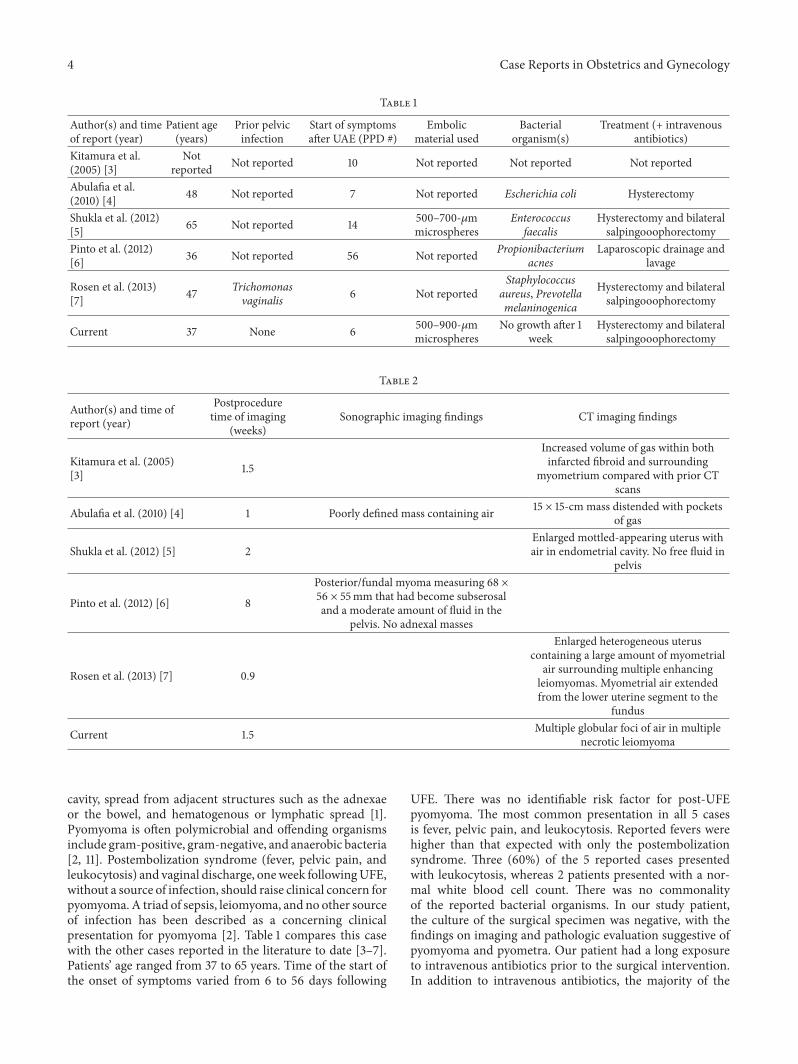
Table 1
Author(s) and timeof report (year)
Patient age(years)
Prior pelvicinfection
Start of symptomsafter UAE (PPD #)
Embolicmaterial used
Bacterialorganism(s)
Treatment (+ intravenousantibiotics)
Kitamura et al.(2005) [3]
Notreported Not reported 10 Not reported Not reported Not reported
Abulafia et al.(2010) [4] 48 Not reported 7 Not reported Escherichia coli Hysterectomy
Shukla et al. (2012)[5] 65 Not reported 14 500–700-𝜇m
microspheresEnterococcus
faecalisHysterectomy and bilateralsalpingooophorectomy
Pinto et al. (2012)[6] 36 Not reported 56 Not reported Propionibacterium
acnesLaparoscopic drainage and
lavage
Rosen et al. (2013)[7] 47 Trichomonas
vaginalis 6 Not reportedStaphylococcus
aureus, Prevotellamelaninogenica
Hysterectomy and bilateralsalpingooophorectomy
Current 37 None 6 500–900-𝜇mmicrospheres
No growth after 1week
Hysterectomy and bilateralsalpingooophorectomy
Table 2
Author(s) and time ofreport (year)
Postproceduretime of imaging
(weeks)Sonographic imaging findings CT imaging findings
Kitamura et al. (2005)[3] 1.5
Increased volume of gas within bothinfarcted fibroid and surrounding
myometrium compared with prior CTscans
Abulafia et al. (2010) [4] 1 Poorly defined mass containing air 15 × 15-cm mass distended with pocketsof gas
Shukla et al. (2012) [5] 2Enlarged mottled-appearing uterus withair in endometrial cavity. No free fluid in
pelvis
Pinto et al. (2012) [6] 8
Posterior/fundal myoma measuring 68 ×56 × 55mm that had become subserosaland a moderate amount of fluid in the
pelvis. No adnexal masses
Rosen et al. (2013) [7] 0.9
Enlarged heterogeneous uteruscontaining a large amount of myometrial
air surrounding multiple enhancingleiomyomas. Myometrial air extendedfrom the lower uterine segment to the
fundus
Current 1.5 Multiple globular foci of air in multiplenecrotic leiomyoma
cavity, spread from adjacent structures such as the adnexaeor the bowel, and hematogenous or lymphatic spread [1].Pyomyoma is often polymicrobial and offending organismsinclude gram-positive, gram-negative, and anaerobic bacteria[2, 11]. Postembolization syndrome (fever, pelvic pain, andleukocytosis) and vaginal discharge, oneweek followingUFE,without a source of infection, should raise clinical concern forpyomyoma. A triad of sepsis, leiomyoma, and no other sourceof infection has been described as a concerning clinicalpresentation for pyomyoma [2]. Table 1 compares this casewith the other cases reported in the literature to date [3–7].Patients’ age ranged from 37 to 65 years. Time of the start ofthe onset of symptoms varied from 6 to 56 days following
UFE. There was no identifiable risk factor for post-UFEpyomyoma. The most common presentation in all 5 casesis fever, pelvic pain, and leukocytosis. Reported fevers werehigher than that expected with only the postembolizationsyndrome. Three (60%) of the 5 reported cases presentedwith leukocytosis, whereas 2 patients presented with a nor-mal white blood cell count. There was no commonalityof the reported bacterial organisms. In our study patient,the culture of the surgical specimen was negative, with thefindings on imaging and pathologic evaluation suggestive ofpyomyoma and pyometra. Our patient had a long exposureto intravenous antibiotics prior to the surgical intervention.In addition to intravenous antibiotics, the majority of the

Case Reports in Obstetrics and Gynecology 5
reported patients were treated with hysterectomy, with 1patient successfully treated with uterine sparing surgicaldrainage and lavage.
There was no commonality among the type and amountof embolic materials used for the 5 other reported cases.Only Shukla et al. [5] reported using 500–700-micronmicro-spheres. Neither the type nor the amount of embolic materialhas been reported as a risk factor for post-UFE infection,a rare and sporadic event [12]. Uterine ischemia has beenproposed as a potential contributor to post-UAE infection [5],but, in our patient, this was unexpected as the patient hadbilateral ovarian artery contribution to the uterus, neither ofwhich was embolized.
On imaging, visualization of gas in the uterus and uterinevessels is an expected finding and does not necessarilyindicate superinfection. This complicates imaging diagnosisof pyomyoma. Airmay be seenwithin the fibroid(s) followingUAE. This finding is thought to be the result of gas fillingpotential spaces left by tissue infarction desiccation. This canbe seen as early as one month after UFE and it has beendescribed as a normal finding without signs of infection[11], in contrast to a globular or localized gas collection ofa necrotic infected leiomyoma [13]. The specificity of thisfinding has not however been proven and it remains difficultto diagnose pyomyoma on all current imaging modalities.The most common radiologic finding in the reported cases[3–5, 7] is the presence of an abundance of intrauterine air,with the exception of Pinto et al. who reported a moderateamount of free pelvic fluid [6] on ultrasound, without acorresponding CT examination. This is outlined in Table 2.Quantification and characterization of intrauterine air maybe more objective by CT scanning than ultrasound, whereasthe latter does not utilize ionizing radiation.
Thus, a strong clinical acumen and a high index ofsuspicion are imperative. Prompt diagnosis is essential aspyomyoma can lead to serious complications such asmyome-trial rupture, sepsis, and death. Hysterectomy or myomec-tomy is the treatment of choice for pyomyoma, althoughthere have been several reports of more conservative surgicaldrainage [6, 8], including a case of nonsurgical managementof clostridial pyomyoma [14].
In summary, this case depicts a rare potentially life-threatening complication, suppurative myoma, and postuter-ine artery embolization in a young woman. Imaging can besupplementary to diagnose this complication, but diagnosingwith imaging alone is difficult given the presence of postpro-cedural gas up to one month following embolization. Signsand symptoms of postembolization syndrome over one weekas well as the triad of fevers, leiomyoma, and no other sourcefor infection should raise clinical concern for pyomyoma.
Disclosure
None of the authors have received any grant support or NIHfunding for this research.
Competing Interests
None of the authors have identified competing interests.
References
[1] J. S. Greenspoon, M. Ault, B. A. James, and L. Kaplan, “Pyomy-oma associated with polymicrobial bacteremia and fatal septicshock: case report and review of the literature,” Obstetrical andGynecological Survey, vol. 45, no. 9, pp. 563–569, 1990.
[2] A. Patwardhan and P. Bulmer, “Pyomyoma as a complication ofuterine fibroids,” Journal of Obstetrics and Gynaecology, vol. 27,no. 4, pp. 444–445, 2007.
[3] Y. Kitamura, S. M. Ascher, C. Cooper et al., “Imaging man-ifestations of complications associated with uterine arteryembolization,” RadioGraphics, vol. 25, pp. S119–S132, 2005.
[4] O. Abulafia, T. Shah, G. Salame et al., “Sonographic featuresassociated with post-uterine artery embolization pyomyoma,”Journal of Ultrasound in Medicine, vol. 29, no. 5, pp. 839–842,2010.
[5] P. A. Shukla, A. Kumar, D. Klyde, and S. Contractor, “Pyomy-oma after uterine artery embolization,” Journal of Vascular andInterventional Radiology, vol. 23, no. 3, pp. 423–424, 2012.
[6] E. Pinto, A. Trovao, S. Leitao, C. Pina, F. K. Mak, and A.Lanhoso, “Conservative laparoscopic approach to a perforatedpyomyoma after uterine artery embolization,” Journal of Mini-mally Invasive Gynecology, vol. 19, no. 6, pp. 775–779, 2012.
[7] M. L. Rosen, M. L. Anderson, and S. M. Hawkins, “Pyomyomaafter uterine artery embolization,” Obstetrics and Gynecology,vol. 121, no. 2, pp. 431–433, 2013.
[8] M. Magro and I. Gafson, “Postpartum pyomyoma: a rare com-plication of leiomyoma,” Journal of Obstetrics and Gynaecology,vol. 34, no. 2, pp. 202–203, 2014.
[9] H. Ono, M. Kanematsu, H. Kato et al., “MR imaging findings ofuterine pyomyoma: radiologic-pathologic correlation,” Abdom-inal Imaging, vol. 39, no. 4, pp. 797–801, 2014.
[10] Z.H.-Y.Chen,H.-D. Tsai, andM.-J. Sun, “Pyomyoma: a rare andlife-threatening complication of uterine leiomyoma,” TaiwaneseJournal of Obstetrics and Gynecology, vol. 49, no. 3, pp. 351–356,2010.
[11] P. R. Genta,M. L.N.Dias, T. A. Janiszewski, J. P. Carvalho,M.H.Arai, and L. P. Meireles, “Streptococcus agalactiae endocarditisand giant pyomyoma simulating ovarian cancer,” SouthernMedical Journal, vol. 94, no. 5, pp. 508–511, 2001.
[12] D. K. Rajan, J. R. Beecroft, T.W. I. Clark et al., “Risk of intrauter-ine infectious complications after uterine artery embolization,”Journal of Vascular and Interventional Radiology, vol. 15, no. 12,pp. 1415–1421, 2004.
[13] S. K. Verma, C. F. Gonsalves, O. H. Baltarowich, D. G. Mitchell,A. S. Lev-Toaff, and D. Bergin, “Spectrum of imaging findingson MRI and CT after uterine artery embolization,” AbdominalImaging, vol. 35, no. 1, pp. 118–128, 2010.
[14] D. Stroumsa, E. Ben-David, N. Hiller, and D. Hochner-Celnikier, “Severe clostridial pyomyoma following an abortiondoes not always require surgical intervention,” Case Reports inObstetrics and Gynecology, vol. 2011, Article ID 364641, 3 pages,2011.

Submit your manuscripts athttp://www.hindawi.com
Stem CellsInternational
Hindawi Publishing Corporationhttp://www.hindawi.com Volume 2014
Hindawi Publishing Corporationhttp://www.hindawi.com Volume 2014
MEDIATORSINFLAMMATION
of
Hindawi Publishing Corporationhttp://www.hindawi.com Volume 2014
Behavioural Neurology
EndocrinologyInternational Journal of
Hindawi Publishing Corporationhttp://www.hindawi.com Volume 2014
Hindawi Publishing Corporationhttp://www.hindawi.com Volume 2014
Disease Markers
Hindawi Publishing Corporationhttp://www.hindawi.com Volume 2014
BioMed Research International
OncologyJournal of
Hindawi Publishing Corporationhttp://www.hindawi.com Volume 2014
Hindawi Publishing Corporationhttp://www.hindawi.com Volume 2014
Oxidative Medicine and Cellular Longevity
Hindawi Publishing Corporationhttp://www.hindawi.com Volume 2014
PPAR Research
The Scientific World JournalHindawi Publishing Corporation http://www.hindawi.com Volume 2014
Immunology ResearchHindawi Publishing Corporationhttp://www.hindawi.com Volume 2014
Journal of
ObesityJournal of
Hindawi Publishing Corporationhttp://www.hindawi.com Volume 2014
Hindawi Publishing Corporationhttp://www.hindawi.com Volume 2014
Computational and Mathematical Methods in Medicine
OphthalmologyJournal of
Hindawi Publishing Corporationhttp://www.hindawi.com Volume 2014
Diabetes ResearchJournal of
Hindawi Publishing Corporationhttp://www.hindawi.com Volume 2014
Hindawi Publishing Corporationhttp://www.hindawi.com Volume 2014
Research and TreatmentAIDS
Hindawi Publishing Corporationhttp://www.hindawi.com Volume 2014
Gastroenterology Research and Practice
Hindawi Publishing Corporationhttp://www.hindawi.com Volume 2014
Parkinson’s Disease
Evidence-Based Complementary and Alternative Medicine
Volume 2014Hindawi Publishing Corporationhttp://www.hindawi.com