Embed Size (px)
Citation preview
Cardiovascular PharmacologyCardiovascular Pharmacology
Review of Cardiovascular Form Review of Cardiovascular Form and Functionand Function
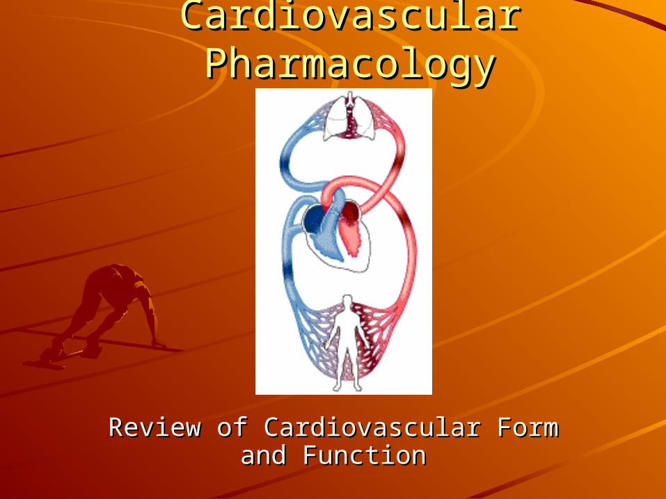
Introduction and BackgroundIntroduction and BackgroundCardiovascular disease is the major cause Cardiovascular disease is the major cause of death in the US (>50% of all deaths)of death in the US (>50% of all deaths)Cardiovascular function based onCardiovascular function based on– Cardiac pumping abilityCardiac pumping ability
Pace-making electrical signalsPace-making electrical signalsForce of contractionForce of contractionHeight of ventricle discharge pressureHeight of ventricle discharge pressure
– Integrity of vasculatureIntegrity of vasculaturePresence of blockagePresence of blockageMuscular tone/structural integrityMuscular tone/structural integrityPressure drop needed to move blood to and through Pressure drop needed to move blood to and through capillary bedscapillary beds
– Blood volume/compositionBlood volume/compositionWater, electrolyte, iron balancesWater, electrolyte, iron balancesLipid and protein compositionLipid and protein composition
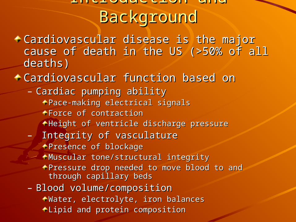
Major Cardiovascular Pathologies Major Cardiovascular Pathologies Requiring Pharmacological InterventionRequiring Pharmacological Intervention
HypertensionHypertensionArrhythmiaArrhythmiaHeart failureHeart failureReduced vascular blood flowReduced vascular blood flow
I. Background to Hypertension -I. Background to Hypertension -Regulation of Blood PressureRegulation of Blood Pressure
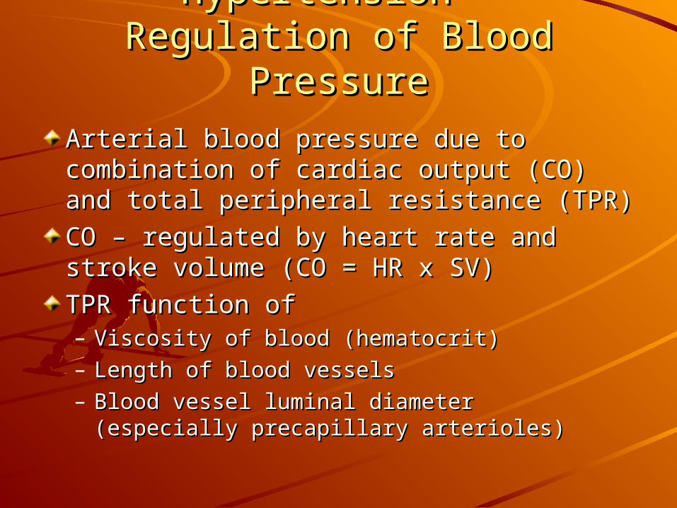
Arterial blood pressure due to combination Arterial blood pressure due to combination of cardiac output (CO) and total peripheral of cardiac output (CO) and total peripheral resistance (TPR)resistance (TPR)CO – regulated by heart rate and stroke CO – regulated by heart rate and stroke volume (CO = HR x SV)volume (CO = HR x SV)TPR function of TPR function of – Viscosity of blood (hematocrit)Viscosity of blood (hematocrit)– Length of blood vesselsLength of blood vessels– Blood vessel luminal diameter (especially Blood vessel luminal diameter (especially
precapillary arterioles) precapillary arterioles)
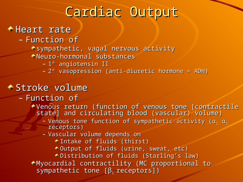
Cardiac OutputCardiac OutputHeart rateHeart rate– Function of Function of
sympathetic, vagal nervous activitysympathetic, vagal nervous activityNeuro-hormonal substancesNeuro-hormonal substances
– 1° angiotensin II1° angiotensin II– 2º vasopression (anti-diuretic hormone = ADH)2º vasopression (anti-diuretic hormone = ADH)
Stroke volumeStroke volume– Function of Function of
Venous return (function of venous tone [contractile state] Venous return (function of venous tone [contractile state] and circulating blood (vascular) volume)and circulating blood (vascular) volume)
– Venous tone function of sympathetic activity (Venous tone function of sympathetic activity (αα1, 1, αα2 2 receptors receptors))– Vascular volume depends onVascular volume depends on
Intake of fluids (thirst)Intake of fluids (thirst)Output of fluids (urine, sweat, etc)Output of fluids (urine, sweat, etc)Distribution of fluids (Starling’s law)Distribution of fluids (Starling’s law)
Myocardial contractility (MC proportional to sympathetic Myocardial contractility (MC proportional to sympathetic tone [tone [ββ1 1 receptors])receptors])
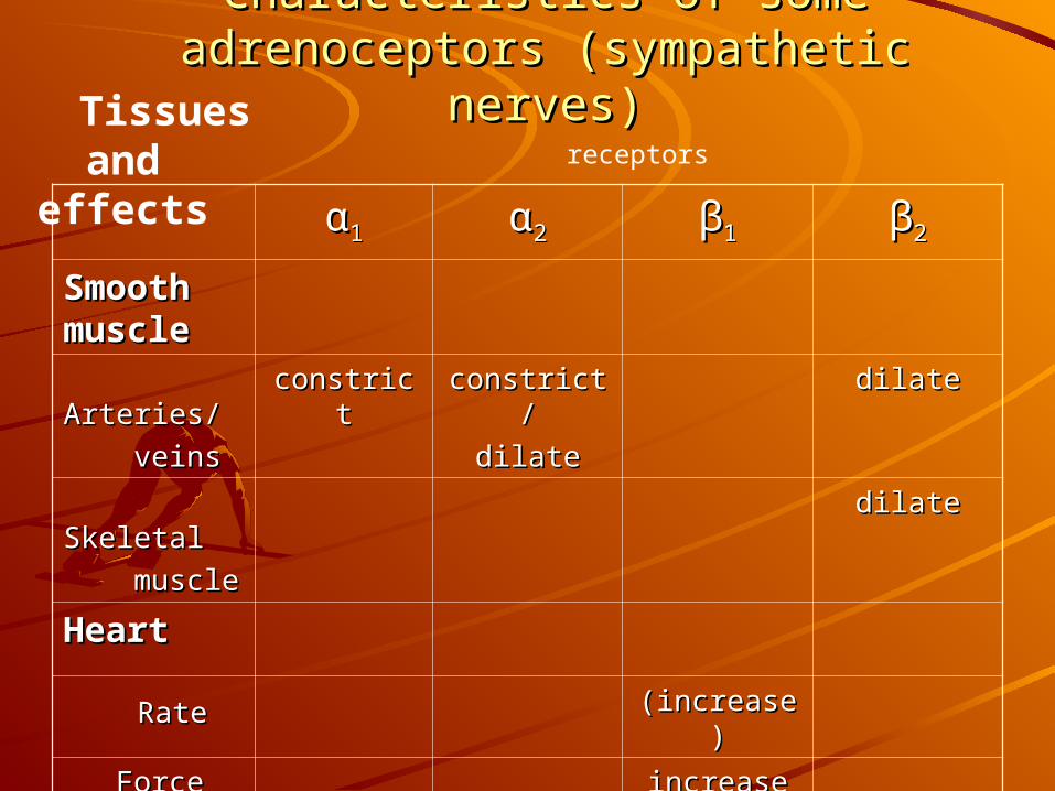
Characteristics of some adrenoceptors Characteristics of some adrenoceptors (sympathetic nerves)(sympathetic nerves)
αα11 αα22 ββ11 ββ22
Smooth Smooth musclemuscle Arteries/Arteries/ veinsveins
constrictconstrict constrict/constrict/dilatedilate
dilatedilate
SkeletalSkeletal musclemuscle
dilatedilate
HeartHeart
RateRate (increase)(increase)
Force of Force of contractioncontraction
increaseincrease
Tissues and effects receptors
Beat-to-Beat Modulation of Blood Beat-to-Beat Modulation of Blood PressurePressure
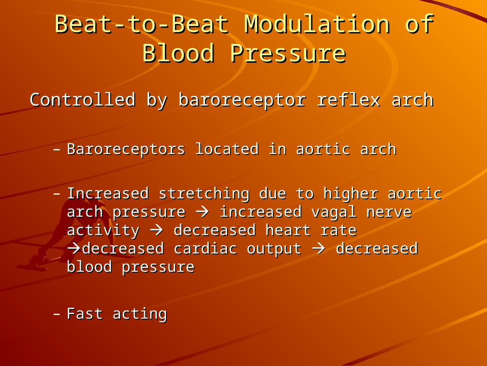
Controlled by baroreceptor reflex archControlled by baroreceptor reflex arch
– Baroreceptors located in aortic archBaroreceptors located in aortic arch
– Increased stretching due to higher aortic arch Increased stretching due to higher aortic arch pressure pressure increased vagal nerve activity increased vagal nerve activity decreased heart rate decreased heart rate decreased cardiac decreased cardiac output output decreased blood pressure decreased blood pressure
– Fast actingFast acting
Autonomic Regulation of Blood PressureAutonomic Regulation of Blood Pressure
Coordinates and integrates all Coordinates and integrates all regulators of cardiovascular functionregulators of cardiovascular function
Can regulate both cardiac output and Can regulate both cardiac output and blood vessel size via sympathetic blood vessel size via sympathetic and parasympathetic innervation of and parasympathetic innervation of cardiovascular end-organs (heart, cardiovascular end-organs (heart, vasculature, kidneys, adrenal glands, vasculature, kidneys, adrenal glands, etc)etc)
Autonomic Regulation of the HeartAutonomic Regulation of the Heart
Heart RateHeart Rate– Parasympathetic input via vagus nerve Parasympathetic input via vagus nerve
causes decrease in HR (dominates) causes decrease in HR (dominates) – Sympathetic input to sino-atrial node Sympathetic input to sino-atrial node
causes increase in HR (usually minor) causes increase in HR (usually minor)
Heart contractilityHeart contractility– Increased by sympathetic activity Increased by sympathetic activity
causing release of epinephrine, causing release of epinephrine, norepinephrine from adrenal glandnorepinephrine from adrenal gland
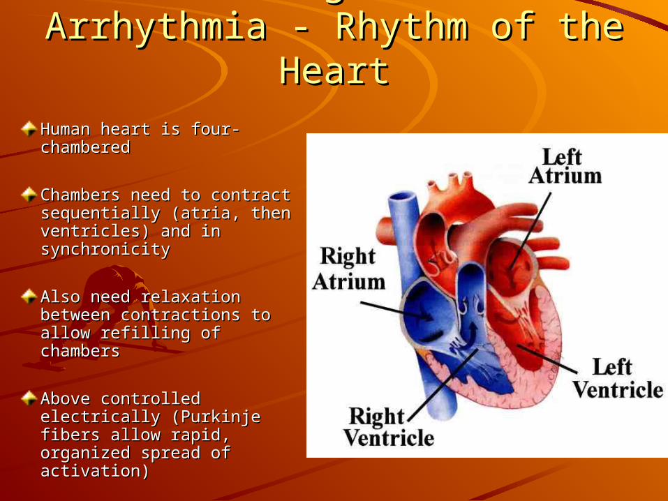
II. Background to Arrhythmia - II. Background to Arrhythmia - Rhythm of the HeartRhythm of the Heart
Human heart is four-Human heart is four-chamberedchambered
Chambers need to contract Chambers need to contract sequentially (atria, then sequentially (atria, then ventricles) and in ventricles) and in synchronicitysynchronicity
Also need relaxation Also need relaxation between contractions to between contractions to allow refilling of chambersallow refilling of chambers
Above controlled Above controlled electrically (Purkinje fibers electrically (Purkinje fibers allow rapid, organized allow rapid, organized spread of activation)spread of activation)
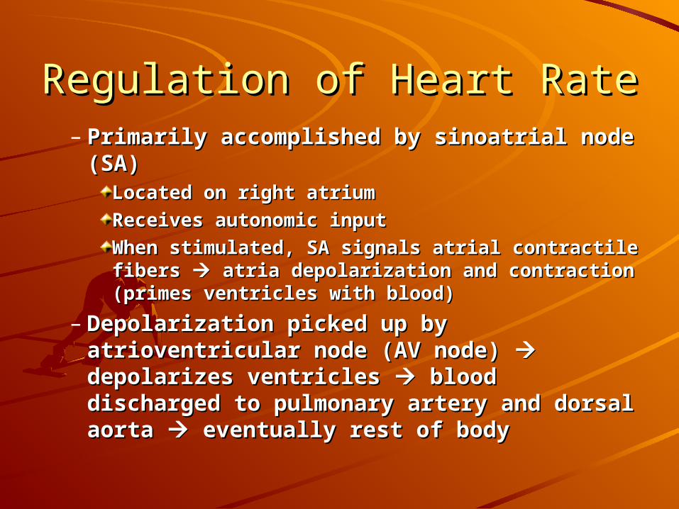
Regulation of Heart RateRegulation of Heart Rate– Primarily accomplished by sinoatrial node (SA)Primarily accomplished by sinoatrial node (SA)
Located on right atriumLocated on right atriumReceives autonomic inputReceives autonomic inputWhen stimulated, SA signals atrial contractile fibers When stimulated, SA signals atrial contractile fibers atria depolarization and contraction (primes atria depolarization and contraction (primes ventricles with blood)ventricles with blood)
– Depolarization picked up by atrioventricular Depolarization picked up by atrioventricular node (AV node) node (AV node) depolarizes ventricles depolarizes ventricles blood discharged to pulmonary artery and blood discharged to pulmonary artery and dorsal aorta dorsal aorta eventually rest of body eventually rest of body
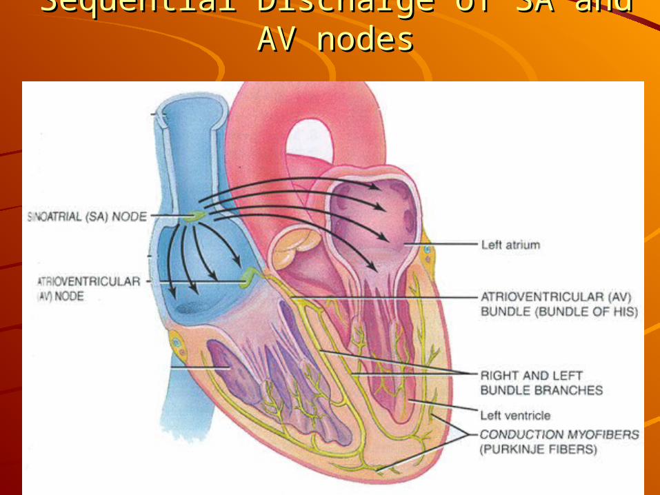
Sequential Discharge of SA and AV nodesSequential Discharge of SA and AV nodes
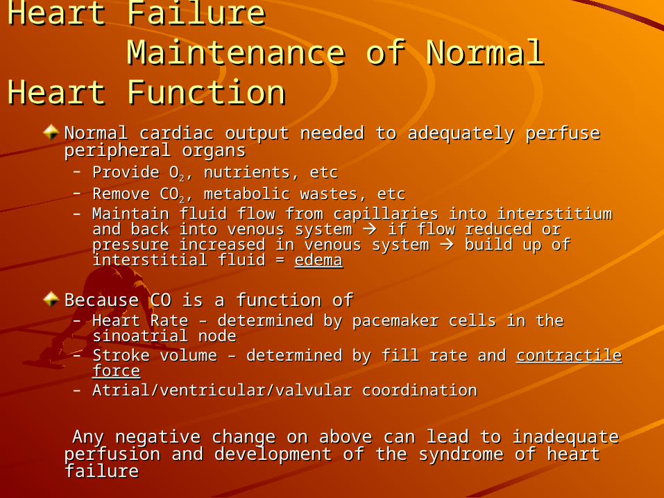
III. Background to Congestive Heart Failure III. Background to Congestive Heart Failure Maintenance of Normal Heart Function Maintenance of Normal Heart Function
Normal cardiac output needed to adequately perfuse Normal cardiac output needed to adequately perfuse peripheral organsperipheral organs– Provide OProvide O22, nutrients, etc, nutrients, etc– Remove CORemove CO22, metabolic wastes, etc, metabolic wastes, etc– Maintain fluid flow from capillaries into interstitium and back Maintain fluid flow from capillaries into interstitium and back
into venous system into venous system if flow reduced or pressure increased in if flow reduced or pressure increased in venous system venous system build up of interstitial fluid = build up of interstitial fluid = edemaedema
Because CO is a function ofBecause CO is a function of– Heart Rate – determined by pacemaker cells in the sinoatrial Heart Rate – determined by pacemaker cells in the sinoatrial
nodenode– Stroke volume – determined by fill rate and Stroke volume – determined by fill rate and contractile forcecontractile force– Atrial/ventricular/valvular coordinationAtrial/ventricular/valvular coordination
Any negative change on above can lead to inadequate Any negative change on above can lead to inadequate perfusion and development of the syndrome of heart failureperfusion and development of the syndrome of heart failure
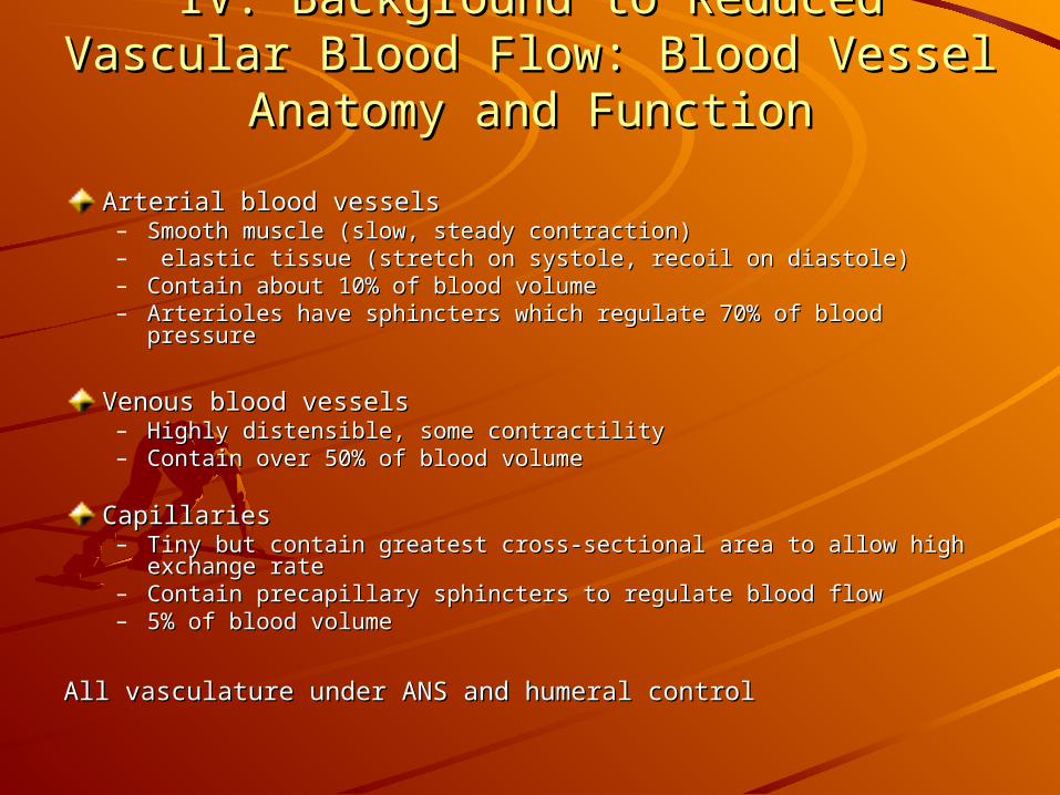
IV. Background to Reduced Vascular Blood IV. Background to Reduced Vascular Blood Flow: Blood Vessel Anatomy and FunctionFlow: Blood Vessel Anatomy and Function
Arterial blood vesselsArterial blood vessels– Smooth muscle (slow, steady contraction)Smooth muscle (slow, steady contraction)– elastic tissue (stretch on systole, recoil on diastole)elastic tissue (stretch on systole, recoil on diastole)– Contain about 10% of blood volumeContain about 10% of blood volume– Arterioles have sphincters which regulate 70% of blood pressureArterioles have sphincters which regulate 70% of blood pressure
Venous blood vesselsVenous blood vessels– Highly distensible, some contractilityHighly distensible, some contractility– Contain over 50% of blood volumeContain over 50% of blood volume
CapillariesCapillaries– Tiny but contain greatest cross-sectional area to allow high exchange Tiny but contain greatest cross-sectional area to allow high exchange
raterate– Contain precapillary sphincters to regulate blood flowContain precapillary sphincters to regulate blood flow– 5% of blood volume5% of blood volume
All vasculature under ANS and humeral controlAll vasculature under ANS and humeral control
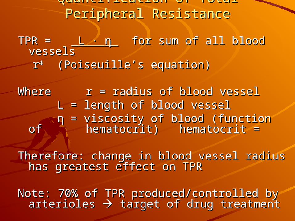
Quantification of Total Peripheral ResistanceQuantification of Total Peripheral Resistance
TPR = _TPR = _L · L · ηη_ for sum of all blood vessels_ for sum of all blood vessels rr44 (Poiseuille’s equation)(Poiseuille’s equation)
Where Where r = radius of blood vesselr = radius of blood vessel L = length of blood vesselL = length of blood vessel
ηη = viscosity of blood (function of = viscosity of blood (function of hematocrit) hematocrit = hematocrit) hematocrit =
Therefore: change in blood vessel radius has Therefore: change in blood vessel radius has greatest effect on TPRgreatest effect on TPR
Note: 70% of TPR produced/controlled by Note: 70% of TPR produced/controlled by arterioles arterioles target of drug treatment target of drug treatment
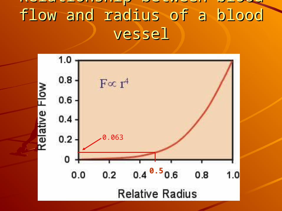
Relationship between blood flow Relationship between blood flow and radius of a blood vesseland radius of a blood vessel
0.5
0.063
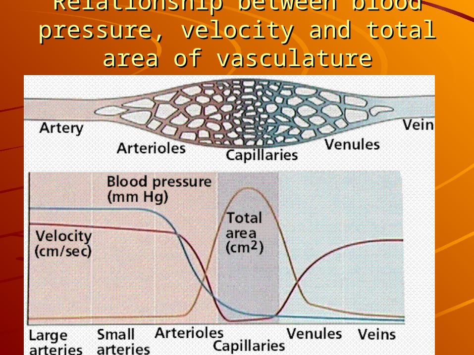
Relationship between blood pressure, Relationship between blood pressure, velocity and total area of vasculaturevelocity and total area of vasculature
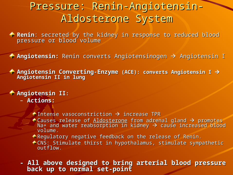
Humeral Regulation of Blood Pressure: Humeral Regulation of Blood Pressure: Renin-Angiotensin-Aldosterone SystemRenin-Angiotensin-Aldosterone System
ReninRenin:: secreted by the kidney in response to reduced blood pressure secreted by the kidney in response to reduced blood pressure or blood volumeor blood volume
Angiotensin:Angiotensin: Renin converts Angiotensinogen Renin converts Angiotensinogen Angiotensin I Angiotensin I
Angiotensin Converting-EnzymeAngiotensin Converting-Enzyme (ACE): converts Angiotensin I (ACE): converts Angiotensin I Angiotensin II in lungAngiotensin II in lung
Angiotensin IIAngiotensin II:: – Actions: Actions:
Intense vasoconstriction Intense vasoconstriction increase TPR increase TPR Causes release of Causes release of AldosteroneAldosterone from adrenal gland from adrenal gland promotes Na+ and promotes Na+ and water reabsorption in kidney water reabsorption in kidney cause increased blood volume. cause increased blood volume. Regulatory negative feedback on the release of Renin. Regulatory negative feedback on the release of Renin. CNS: Stimulate thirst in hypothalamus, stimulate sympathetic outflow. CNS: Stimulate thirst in hypothalamus, stimulate sympathetic outflow.
- All above designed to bring arterial blood pressure - All above designed to bring arterial blood pressure back up to normal set-pointback up to normal set-point
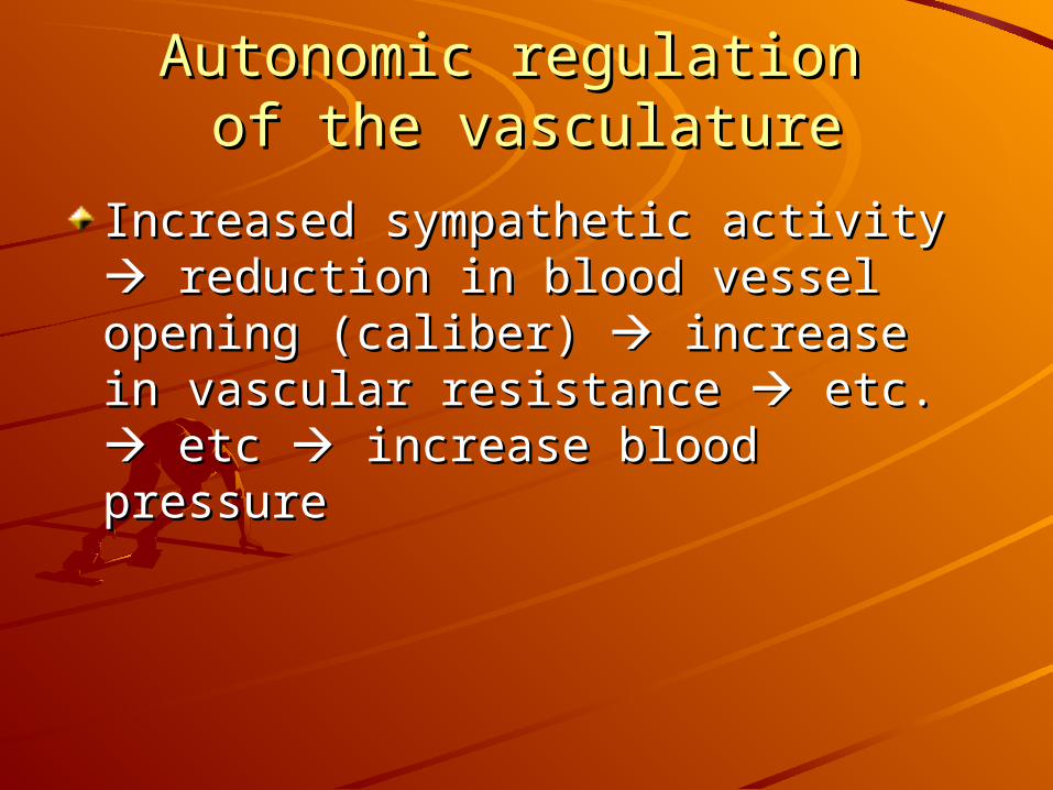
Autonomic regulation Autonomic regulation of the vasculatureof the vasculature
Increased sympathetic activity Increased sympathetic activity reduction in blood vessel opening reduction in blood vessel opening (caliber) (caliber) increase in vascular increase in vascular resistance resistance etc. etc. etc etc increase increase blood pressureblood pressure
Stop talking now and Stop talking now and let them go!let them go!
I’m outta’ here!