Embed Size (px)
Citation preview

PERIPARTUM CARDIOMYOPATHY
Fahad zakwan

NORMAL CLINICAL FINDINGS OF
CARDIOVASCULAR SYSTEM DURING PREGNANCY.

ANATOMICAL CHANGES
Occurs due to elevation of the diaphragm consequent to the
enlarged uterus, the heart is pushed upwards and outwards with slight
rotation to the left.
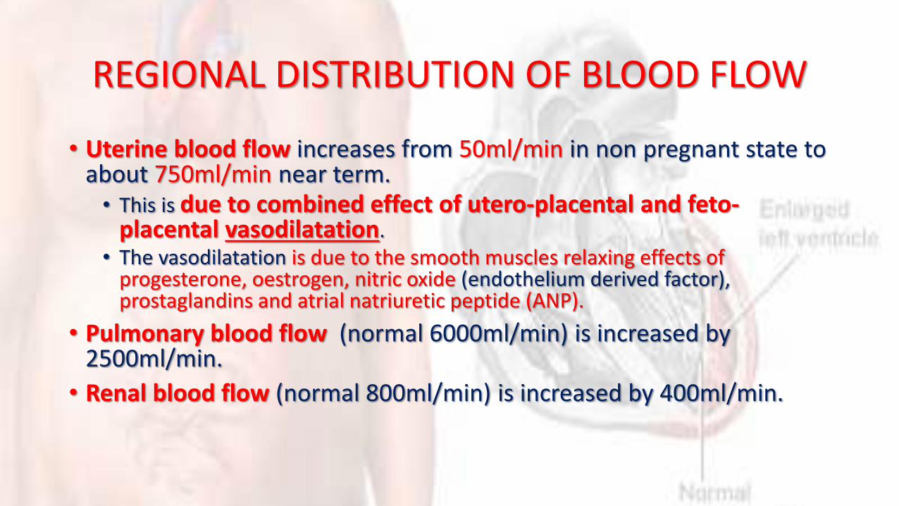
REGIONAL DISTRIBUTION OF BLOOD FLOW
• Uterine blood flow increases from 50ml/min in non pregnant state to about 750ml/min near term.• This is due to combined effect of utero-placental and feto-
placental vasodilatation.• The vasodilatation is due to the smooth muscles relaxing effects of
progesterone, oestrogen, nitric oxide (endothelium derived factor), prostaglandins and atrial natriuretic peptide (ANP).
• Pulmonary blood flow (normal 6000ml/min) is increased by 2500ml/min.
• Renal blood flow (normal 800ml/min) is increased by 400ml/min.
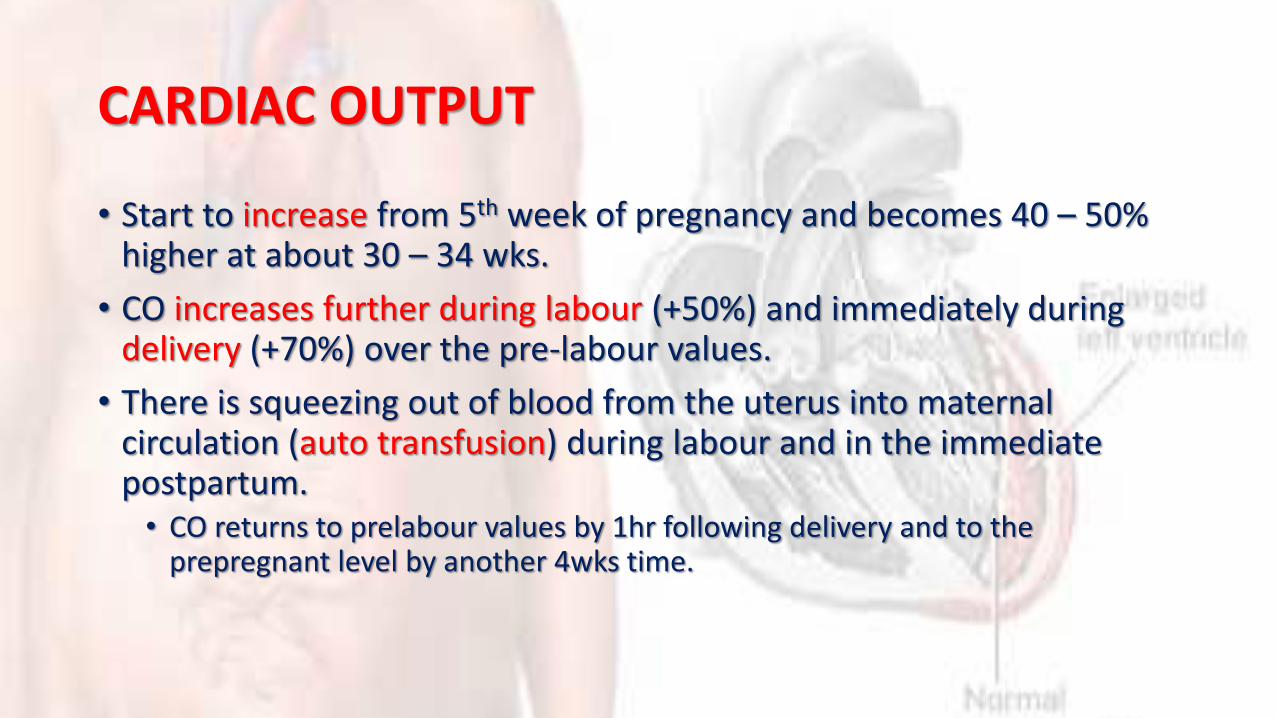
CARDIAC OUTPUT
• Start to increase from 5th week of pregnancy and becomes 40 – 50% higher at about 30 – 34 wks.
• CO increases further during labour (+50%) and immediately during delivery (+70%) over the pre-labour values.
• There is squeezing out of blood from the uterus into maternal circulation (auto transfusion) during labour and in the immediate postpartum.• CO returns to prelabour values by 1hr following delivery and to the
prepregnant level by another 4wks time.

BLOOD PRESSURE
• BP = CO × SVR
• Systemic vascular resistance (SVR) decreases to -21% due to smooth muscles relaxing effects of progesterone, prostaglandins etc.
• Inspite of the large increase in CO, the maternal BP is decreased due to decrease in SVR.
• Hence there is overall decrease in diastolic blood pressure(DBP) and mean arterial pressure (MAP) by 5 – 10mmHg.

VENOUS PRESSURE
• Antecubital venous pressure remains unaffected
• Femoral venous pressure is markedly raised specifically in the latter months of pregnancy.• It is due to the pressure exerted by the gravid uterus on the common iliac
veins,
• It is more on the right side due to dextrorotation of the uterus.
• The femoral venous pressure is raised from 8 – 10cm of water in non pregnant state to around 25cm of water during pregnancy in lying down position to about 80 – 100cm of water in standing position.• This explains the fact that the physiological oedema of pregnancy subsides by
rest alone.

ABNORMAL CLINICAL FINDINGS OF
CARDIOVASCULAR SYSTEM DURING PREGNANCY.

• due to the physiological changes in cardiovascular system during pregnancy….
• The patient may experience palpitations
• The apex beat is shifted to the 4th intercostal space 2.5cm outside the midclavicular line.
• Pulse rate is slightly raised often with extrasystoles.
• A systolic murmur may be audible in the apical or pulmonary area.• This is due to decreased blood viscosity and torsion of great vessels
• A continuous hissing murmur may be audible over the tricuspid area in the left 2nd and 3rd intercostal spaces called mammary murmur.• It is due to increased blood flow through the internal mammary vessels.

As a doctor you should be familiar with these physiological findings and should execute continuous approach in diagnosis of heart disease during
pregnancy.

HEART DISEASES IN PREGNANCY
•The incidence of cardiac lesions is less than 1% amongst hospital deliveries.
•The commonest cardiac lesson is of rheumatic origin followed by congenitalones.

1. RHEUMATIC HEART DISEASE• Mitral stenosis (80%)• Aortic stenosis
2. CONGENITAL HEART DISEASEA. ACYANOTIC
• Atrial septal defect (ASD)
• Patent ductus arteriosus (PDA)
• Ventricular septal defect (VSD)
• Mitral valve prolapse (MVP)
B. CYANOTIC• Tetralogy of Fallot
• Eisenmenger’s syndrome

C. OTHER CONGENITAL HEART LESIONS •Coarctation of aorta•Primary pulmonary hypertension•Marfan’s syndrome•Prosthetic valves
D.CARDIOMYOPATHIES•Peripartum cardiomyopathy•Myocardial infarction

PERIPARTUM CARDIOMYOPATHY
DEFINITION
Peripartum cardiomyopathy is defined as the onset of acute heart failure without demonstrable cause in the last trimester of pregnancy or within the first 5 months
after delivery.

•A form of Dilated Cardiomyopathy
•Left ventricular systolic dysfunction
•Results in signs and symptoms of heart failure
•Often unrecognized, as symptoms of normal pregnancy commonly mimic those of mild heart failure.

CRITERIA FOR PERIPARTUM CARDIOMYOPATHY
1.Development of Cardiac failure in the last month of pregnancy or within 5 month after delivery
2. Absence of an identifiable cause for the cardiac failure. 3.Absence of recognizable heart disease prior to the last
month of pregnancy. 4.Left ventricular systolic dysfunction demonstrated by
classic Echo Cardio Graphic criteria such as depressed shortening fraction or ejection fraction.
INCIDENCE The incidence in the west ranges from 1 in 4000 deliveries 60% present within the first 2 months postpartum Up to 7% may present in the last trimester of pregnancy. Geographic variations exist with a higher incidence
reported in areas of Africa because of malnutrition and local customs in the puerperium
ETIOLOGY
Still unknown. nutritional deficiencies small vessel coronary artery abnormalityhormonal effects toxemiamaternal immunologic response to fetal
antigen or myocarditis

RISK FACTORS
•Age >30 years old•Multiparity•African Descent•Maternal cocaine abuse•Long term tocolytic therapy (>4weeks)
•Pregnancy with multiple fetuses
•History of Preeclampsia, eclampsia, or postpartum HTN
•Nutritional deficiencies

SYMPTOMS
Dyspnoea on exertion fatigue ankle oedema embolic phenomena atypical chest pains
Haemoptysis. Palpitation Abdominal
discomfort Cough orthopnoea
Many of above symptoms may occur even in normal pregnancy and can be mistaken for a diseased state.
Symptoms of worsening cardiac failure like:
SIGNS•evidence of a raised CVP
• tachycardia
•cardiomegaly with a gallop rhythm (S3)
•mitral regurgitation
•pulmonary crackles and
•peripheral oedema.
TIMING OF DIAGNOSIS
•Diagnosis Requires being in the last month of pregnancy
•If earlier, consider underlying heart disease (ischemic, valvular, or myopathic)

DIAGNOSIS
Peripartum cardiomyopathy is a
diagnosis of exclusion.

INVESTIGATIONS
•EKG
•Auscultation of the heart
•Two-dimensional echocardiogram
•CXR
•Lab: CBC, CMP, BNP, TSH, Ferritin
On auscultation of the heart:
• loud first heart sound
• exaggerated splitting
•mid systolic murmur and
• continuous venous hum
These physical signs may confuse and there could be mistakes in the form of over diagnosis or disregarding of heart disease.

Chest radiograph:
cardiomegaly with pulmonary oedema
pulmonary venous congestion.
The Electrocardiogram:
•nonspecific ST and T wave changes•atrial or ventricular arrhythmias and •conduction defects.

Echocardiography / Doppler • may reveal enlargement of all four
chambers with marked reduction in left ventricular systolic function
• small to moderate pericardial effusion and
• mitral, tricuspid and pulmonary regurgitation
• Ventricular wall motion, ejection fraction and cardiac output are decreased and
• pulmonary wedge pressure is increased. • Spherical LV• Left Atrial enlargement
• The clinical presentation and hemodynamic features in PPCM are indistinguishable from those of other forms of dilated cardiomyopathy.
• In the absence of any cardiac symptoms, one of the early indications about this condition is revealed during evaluation of the fetus with a fetal monitor and ultrasound
• Fetal growth is dependent on good blood flow to the uterus and placenta
• An insufficient blood flow means decreased oxygenation resulting in slowed growth
• This should prompt further investigation to discover heart disease.
NOTE:
MANAGEMENT OF PPCM
• Bed rest•Delivery – preferably vaginal• Similar to other forms of CHF
• Diuretics• ß-blockers• Digoxin• Anticoagulants
• Epidural anaesthesia is idealMust consider pregnancy class/breast-feeding harm
potential!
Drugs
•Digoxin Class C•Symptomatic control•Requires level monitoring•Therapeutic levels 0.7-1.2

Diuretics
•Lasix Class C•Reserved for cardiac
conditions•Not recommended in
PIH•May decrease
placental perfusion
•Thiazide Diuretics•Reserved for cardiac
conditions•Not recommended in
PIH• Thrombocytopenia has
been reported in breast feeding infants

Vasodilators
•Hydralazine Class C•Compatible with breastfeeding
•ACE Inhibitors•Class D in 2nd/3rd
trimesters•Reserved for
postpartum use-compatible with BF•Renal toxicity in infants
exposed in utero
Beta-Blockers
•Class C•Compatible with breast feeding•Has been shown to cause IUGR in some infants in utero.

Anticoagulants
•Heparin Class C• Short half life-can be
discontinued prior to delivery to prevent maternal hemorrhage•Not excreted in breast
milk
•Warfarin Class D•Contraindicated in pregnancy•Safe in breast feeding. Not excreted in breast milk.
Other Therapy
• IV Immune Globulin•Cardiac Transplant
• Estimated that transplant is performed in up to 1/3 of PPCM patients
• Pts should be strongly advised against future pregnancies.• Increased risk of HTN, preeclampsia, and preterm labor• Also at risk for graft failure due to recurrent disease.

DIFFERENTIAL DIAGNOSIS
•PIH•However, HF associated with PIH represents a
diastolic failure, vs. systolic in PPCM
•Pulmonary Embolism•Again, usually ruled out by CXR• If still suspicious, can order spiral CT

THE PROGNOSIS
• 50-60% patients show complete or near complete recovery within the first 6 months postpartum
• In others, either continued clinical deterioration leading to early death or persistent left ventricular dysfunction and chronic heart failure results
• There is an initial high risk period with mortality of 25-50% in the first 3 months postpartum.
• Patients with persistent cardiomegaly at 6 months have a reported mortality of 85% at 5 years.
Future Pregnancies??• Opinions widely vary
• Most experts agree that patients should avoid future pregnancy if LV dysfunction is persistent greater than 6 months
• Highly Individual• Patient education of risks• Cardiologist involvement in decision
• If future pregnancy desired:• Maternal Echocardiogram per trimester• Serial sonograms for growth• Again, Subspecialty involvement