Embed Size (px)
Citation preview
Brit. Heart ., 1969, 31, 227.
Cardiac Abnormalities in the Ehlers-Danlos SyndromeP. BEIGHTON*
From St. Thomas' Hospital, London S.E.J
There have been several reports of cardiac abnor-malities in patients with the Ehlers-Danlos syn-drome, but it is not certain whether these anomaliesare part of the syndrome or whether they representchance concomitants (McKusick, 1966).
In an investigation in Southern England, 100patients with this syndrome have been examined.The results of this survey, from the cardiac pointof view, are presented in this paper, and the featuresthat were encountered are discussed with referenceto previous reports.
DESCRIPTION OF SYNDROMEAffected patients have hyperextensible skin,
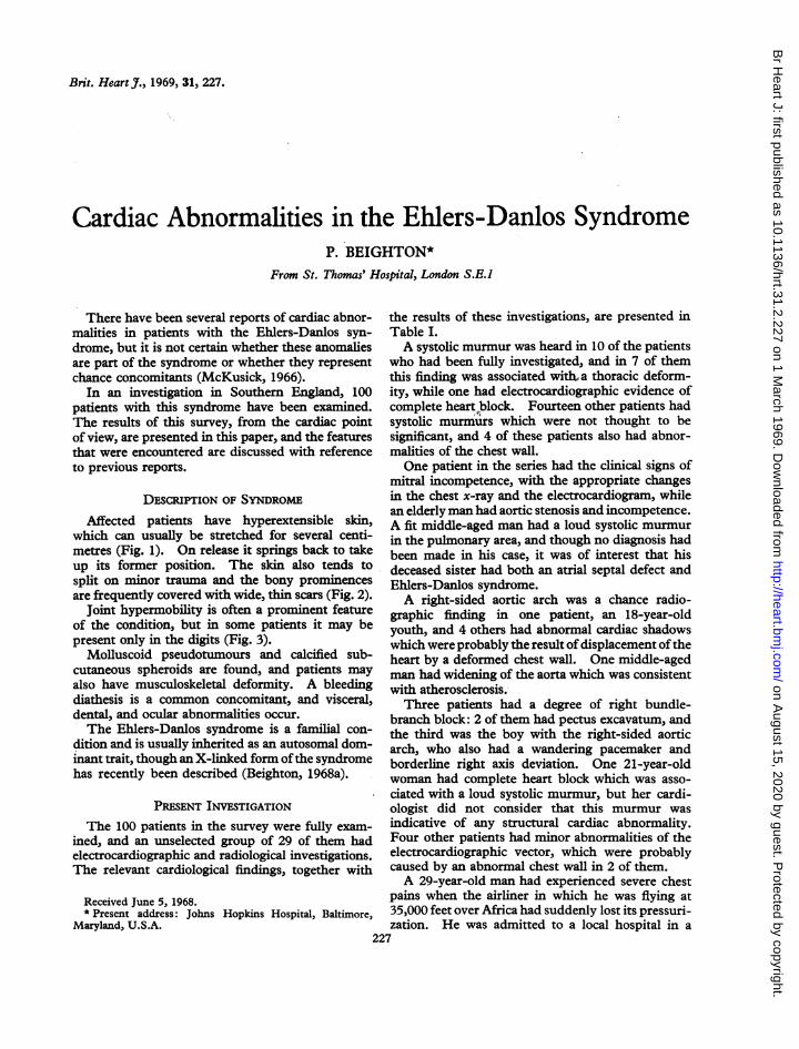
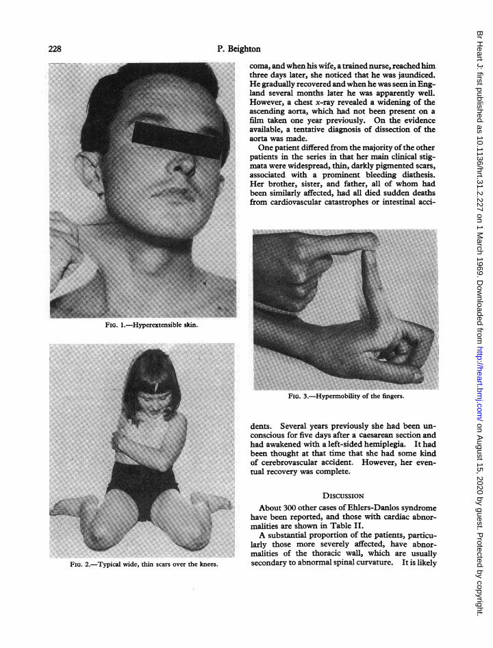
which can usually be stretched for several centi-metres (Fig. 1). On release it springs back to takeup its former position. The skin also tends tosplit on minor trauma and the bony prominencesare frequently covered with wide, thin scars (Fig. 2).
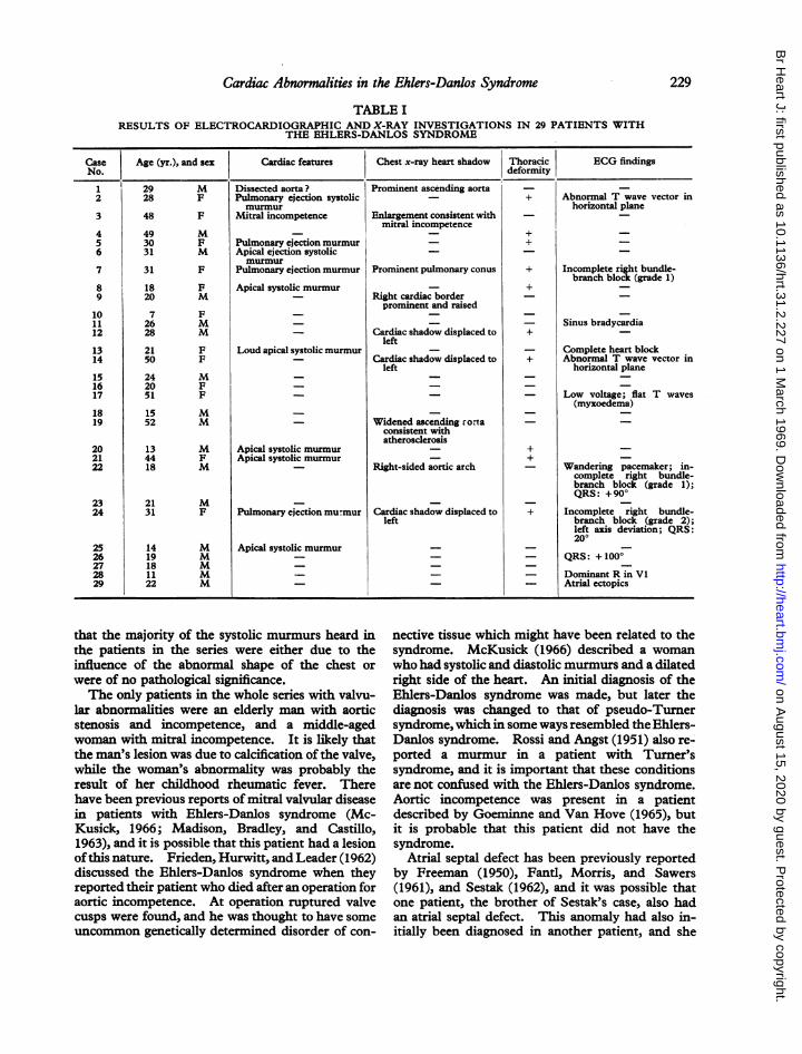
Joint hypermobility is often a prominent featureof the condition, but in some patients it may bepresent only in the digits (Fig. 3).
Molluscoid pseudotumours and calcified sub-cutaneous spheroids are found, and patients mayalso have musculoskeletal deformity. A bleedingdiathesis is a common concomitant, and visceral,dental, and ocular abnormalities occur.The Ehlers-Danlos syndrome is a familial con-
dition and is usually inherited as an autosomal dom-inant trait, though an X-linked form ofthe syndromehas recently been described (Beighton, 1968a).
PRESENT INVESTIGATIONThe 100 patients in the survey were fully exam-
ined, and an unselected group of 29 of them hadelectrocardiographic and radiological investigations.The relevant cardiological findings, together with
Received June 5, 1968.* Present address: Johns Hopkins Hospital, Baltimore,
Maryland, U.S.A.227
the results of these investigations, are presented inTable I.A systolic murmur was heard in 10 of the patients
who had been fully investigated, and in 7 of themthis finding was associated with a thoracic deform-ity, while one had electrocardiographic evidence ofcomplete heart block. Fourteen other patients hadsystolic murmurs which were not thought to besignificant, and 4 of these patients also had abnor-malities of the chest wall.One patient in the series had the clinical signs of
mitral incompetence, with the appropriate changesin the chest x-ray and the electrocardiogram, whilean elderly man had aortic stenosis and incompetence.A fit middle-aged man had a loud systolic murmurin the pulmonary area, and though no diagnosis hadbeen made in his case, it was of interest that hisdeceased sister had both an atrial septal defect andEhlers-Danlos syndrome.A right-sided aortic arch was a chance radio-
graphic finding in one patient, an 18-year-oldyouth, and 4 others had abnormal cardiac shadowswhich were probably the result ofdisplacement oftheheart by a deformed chest wall. One middle-agedman had widening of the aorta which was consistentwith atherosclerosis.-Three patients had a degree of right bundle-
branch block: 2 of them had pectus excavatum, andthe third was the boy with the right-sided aorticarch, who also had a wandering pacemaker andborderline right axis deviation. One 21-year-oldwoman had complete heart block which was asso-ciated with a loud systolic murmur, but her cardi-ologist did not consider that this murmur wasindicative of any structural cardiac abnormality.Four other patients had minor abnormalities of theelectrocardiographic vector, which were probablycaused by an abnormal chest wall in 2 of them.A 29-year-old man had experienced severe chest
pains when the airliner in which he was flying at35,000 feet over Africa had suddenly lost its pressuri-zation. He was admitted to a local hospital in a
on August 15, 2020 by guest. P
rotected by copyright.http://heart.bm
j.com/
Br H
eart J: first published as 10.1136/hrt.31.2.227 on 1 March 1969. D
ownloaded from
P. Beighton
FIG. 1.-Hyperextensible skin.
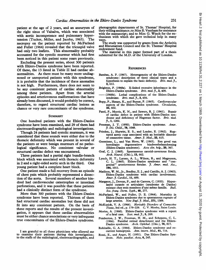
FIG. 2.-Typical wide, thin scars over the knees.
coma, and when his wife, a trained nurse, reached himthree days later, she noticed that he was jaundiced.He gradually recovered and when he was seen in Eng-land several months later he was apparently well.However, a chest x-ray revealed a widening of theascending aorta, which had not been present on afilm taken one year previously. On the evidenceavailable, a tentative diagnosis of dissection of theaorta was made.One patient differed from the majority ofthe other
patients in the series in that her main clinical stig-mata were widespread, thin, darkly pigmented scars,associated with a prominent bleeding diathesis.Her brother, sister, and father, all of whom hadbeen similarly affected, had all died sudden deathsfrom cardiovascular catastrophes or intestinal acci-
Fio. 3.-Hypermobility of the fingers.
dents. Several years previously she had been un-conscious for five days after a caesarean section andhad awakened with a left-sided hemiplegia. It hadbeen thought at that time that she had some kindof cerebrovascular accident. However, her even-tual recovery was complete.
DISCUSSIONAbout 300 other cases of Ehlers-Danlos syndrome
have been reported, and those with cardiac abnor-malities are shown in Table IIA substantial proportion of the patients, particu-
larly those more severely affected, have abnor-malities of the thoracic wall, which are usuallysecondary to abnormal spinal curvature. It is likely
228
on August 15, 2020 by guest. P
rotected by copyright.http://heart.bm
j.com/
Br H
eart J: first published as 10.1136/hrt.31.2.227 on 1 March 1969. D
ownloaded from
Cardiac Abnormalities in the Ehlers-Danlos Syndrome
TABLE IRESULTS OF ELECTROCARDIOGRAPHIC AND X-RAY INVESTIGATIONS IN 29 PATIENTS WITH
THE EHLERS-DANLOS SYNDROME
Case Age (yr.), and sex Cardiac features Chest x-ray heart shadow Thoracic ECG findingsNo. deformity
1 29 M Dissected aorta? Prominent ascending aorta _2 28 F Pulmonary ejection systolic - + Abnormal T wave vector in
murmur horizontal plane3 48 F Mitral incompetence Enlargement consistent with _
mitral incompetence4 49 M - + -5 30 F Pulmonary ejection murmur _ +6 31 M Apical ejection systolic _ _
murmur7 31 F Pulmonary ejection murmur Prominent pulmonary conus + Incomplete right bundle-
branch block (grade 1)8 18 F Apical systolic murmur - +9 20 M - Right cardiac border _
prominent and raised10 7 F _11 26 M _ - Sinus bradycardia12 28 M - Cardiac shadow displaced to +
left13 21 F Loud apical systolic murmur _ - Complete heart block14 50 F - Cardiac shadow displaced to + Abnormal T wave vector in
left horizontal plane15 24 M _ _ _16 20 F _ _ _17 51 F _- - Low voltage; flat T waves
(myxoedema)18 15 M _ _ _19 52 M - Widened ascending rorta -
consistent withatherosclerosis
20 13 M Apical systolic murmur - +21 44 F Apical systolic murmur - +22 18 M - Right-sided aortic arch - Wandering pacemaker; in-
complete right bundle-branch block (grade 1);QRS: +900
23 21 M _24 31 F Pulmonary ejection murmur Cardiac shadow displaced to + Incomplete right bundle-
left branch block (grade 2);left axis deviation; QRS:200
25 14 M Apical systolic murmur _ -
26 19 M _- - QRS: +100'27 18 M _ _ _ _28 11 M _- - Dominant R in Vl29 22 M - - Atrial ectopics
that the majority of the systolic murmurs heard inthe patients in the series were either due to theinfluence of the abnormal shape of the chest orwere of no pathological significance.The only patients in the whole series with valvu-
lar abnormalities were an elderly man with aorticstenosis and incompetence, and a middle-agedwoman with mitral incompetence. It is likely thatthe man's lesion was due to calcification of the valve,while the woman's abnormality was probably theresult of her childhood rheumatic fever. Therehave been previous reports of mitral valvular diseasein patients with Ehlers-Danlos syndrome (Mc-Kusick, 1966; Madison, Bradley, and Castillo,1963), and it is possible that this patient had a lesionofthis nature. Frieden, Hurwitt, and Leader (1962)discussed the Ehlers-Danlos syndrome when theyreported their patient who died after an operation foraortic incompetence. At operation ruptured valvecusps were found, and he was thought to have someuncommon genetically determined disorder of con-
nective tissue which might have been related to thesyndrome. McKusick (1966) described a womanwho had systolic and diastolic murmurs and a dilatedright side of the heart. An initial diagnosis of theEhlers-Danlos syndrome was made, but later thediagnosis was changed to that of pseudo-Turnersyndrome, which in some ways resembled the Ehlers-Danlos syndrome. Rossi and Angst (1951) also re-ported a murmur in a patient with Turner'ssyndrome, and it is important that these conditionsare not confused with the Ehlers-Danlos syndrome.Aortic incompetence was present in a patientdescribed by Goeminne and Van Hove (1965), butit is probable that this patient did not have thesyndrome.
Atrial septal defect has been previously reportedby Freeman (1950), Fantl, Morris, and Sawers(1961), and Sestak (1962), and it was possible thatone patient, the brother of Sestak's case, also hadan atrial septal defect. This anomaly had also in-itially been diagnosed in another patient, and she
229
on August 15, 2020 by guest. P
rotected by copyright.http://heart.bm
j.com/
Br H
eart J: first published as 10.1136/hrt.31.2.227 on 1 March 1969. D
ownloaded from
TABLE IICARDIAC ABNORMALITIES IN THE EHLERS-DANLOS SYNDROME
Case Age (yr.), and Family Condition Other features AuthorNo. sex history
1 13 F - Atrial septal defect _ Freeman (1950)2 43 F + Atrial septal defect with tricuspid Right bundle-branch block Sestak (1962)
incompetence3 11 F + Partial atrioventricular canal; left Deficient Hageman factor; success- Fantl et al. (1961)
leaflets of mitral and tricuspid ful repair of ASDvalves
4 26 F - Fallot's tetralogy Right bundle-branch block Wallach and Burkhart(1950)
5 2 F + "Congenital cardiac defect" Mother had Ehlers-Danlos and Rubinstein and Cohendied after operation on internal (1964)carotid aneurysm
6 10 M - Bifid pulmonary artery; abnormal _ Bopp et al. (1965)aortic arch
7 28 M - Abnormal aortic arch _ Robitaille (1964)8 40 M - Aortic stenosis and mitral incom- Murmurs first heard shortly after McKusick (1966)
petence birth; sudden death in 19579 17 M _ "Bicuspid" tricuspid valve Death from spontaneous rupture of McFarland and Fuller
subclavian artery; friable cardiac (1964)muscle at necropsy
10 17 M + Mitral and tricuspid incompetence Death in congestive cardiac failure Madison et al. (1963)11 42 F - Aneurysm of right sinus of Valsalva; - Tucker et al. (1963)
aortic incompetence; pulmonaryhypertension
12 9 M + Systolic murmurs Murmurs due to thoracic deformity Margarot, Deveze, and deCarrera (1933)
13 25 F _ Systolic and diastolic murmurs; Probably a pseudo-Turner syn- McKusick (1966)dilated right side of the heart drome
had a forceps delivery of her first child for thisreason. The diagnosis was later revised when itwas realized that her murmur was probably due tothe very narrow antero-posterior diameter of herchest.The right-sided aortic arch found in one patient
has not been previously described in the Ehlers-Danlos syndrome. Bopp, Hatam, and Bussat (1965)carried out angiographic studies on a boy with an"abnormal aortic arch" and showed a bifid pul-monary artery, while Robitaille (1964) investigateda man with an abnormal aortic arch in the same way.It is impossible to say whether these abnormalitieshave any direct relation with the Ehlers-Danlossyndrome.Three patients in the series had a right bundle-
branch block, and two of them also had abnormalchest walls, while the third was the patient with aright-sided aortic arch. A right bundle-branchblock was present in the patient with the atrial septaldefect described by Sestak (1962), and he also foundan incomplete right bundle-branch block in apatient with cutis laxa. McKusick (1966) alsoreported an incomplete right bundle-branch blockin a man who had no other demonstrable cardiaclesion.
This conduction defect is common in normalpeople, and there is nothing to suggest that it wasanything more than either a chance finding or anabnormality related to the other structural featuresin the patients described. The complete heartblock and the vector abnormalities were probablyalso chance findings.
It seems likely that Case 1 (Table I) survived agenuine dissection of the aorta. Similar eventshave been reported in three young men byMcKusick (1966), and Lynch et al. (1965) reporteda further case. Pemberton, Freeman, and Schepens(1966) mentioned the successful surgical repair of adissection of the aorta in a man of 39; however, hedied suddenly one month later, presumably fromanother dissection.The patient with the family history of arterial
rupture probably has a clinically distinct variety ofthe Ehlers-Danlos syndrome. Barabas (1967) sug-gested the existence of this entity, and describedtwo patients with similar stigmata who had spon-taneous arterial bleeding. There have been severalother reports of arterial rupture in Ehlers-Danlossyndrome, including those ofMories (1960), McFar-land and Fuller (1964), and McKusick (1966).This patient's cerebrovascular episode could havebeen due to an intracranial vascular malformationresembling those described by Schoolman andKepes (1967), Graf (1965), and Rubinstein andCohen (1964), which seem to occur in patientswith this type of the syndrome.However, arterial bleeding has occurred in
patients with the usual manifestations of the Ehlers-Danlos syndrome, and such patients are at some riskfrom this complication (Beighton, 1968b).Among the other cardiac abnormalities that have
been previously reported in patients with this syn-drome are Fallot's tetralogy (Wallach and Burkhart,1950), a "congenital cardiac defect" (Rubinsteinand Cohen, 1964) which caused the death of the
230 P. Beighton
on August 15, 2020 by guest. P
rotected by copyright.http://heart.bm
j.com/
Br H
eart J: first published as 10.1136/hrt.31.2.227 on 1 March 1969. D
ownloaded from
Cardiac Abnormalities in the Ehlers-Danlos Syndrome
patient at the age of 2 years, and an aneurysm ofthe right sinus of Valsalva, which was associatedwith aortic incompetence and pulmonary hyper-tension (Tucker, Miller, and Jacoby, 1963). Thenecropsy on the patient reported by McFarlandand Fuller (1964) revealed that the tricuspid valvehad only two leaflets. This abnormality probablyaccounted for the systolic murmur which had firstbeen noticed in this patient some years previously.
Excluding the present series, about 300 patientswith Ehlers-Danlos syndrome have been reported.Of these, the 13 listed in Table II had cardiac ab-normalities. As there must be many more undiag-nosed or unreported patients with this syndrome,it is probable that the incidence of the3e anomaliesis not high. Furthermore, there does not seem tobe any consistent pattern of cardiac abnormalityamong these patients. Apart from the arterialepisodes and arteriovenous malformations that havealready been discussed, it would probably be correct,therefore, to regard structural cardiac lesions aschance or very rare concomitants of the syndrome.
SUMMARYOne hundred patients with the Ehlers-Danlos
syndrome have been examined, and 29 of them hadelectrocardiographic and radiological investigations.Though 24 patients had systolic murmurs, it was
considered that these sounds were either due to thethoracic deformity which was present in several ofthe patients or were benign murmurs of no patho-logical significance. No consistent valvular orstructural cardiac defect was encountered.Three patients had a partial right bundle-branch
block which was associated with thoracic deformityin 2 and a right-sided aortic arch in the third. Oneyoung patient had a complete heart block.One patient made a full recovery from an episode
of chest pain which probably represented a dissec-tion of the aorta. Several members of another kin-dred had cardiovascular catastrophes or intestinalperforations, and it was possible that these patientshad a clinically distinct form of the syndrome.More than 300 patients with the Ehlers-Danlos
syndrome have been reported. Thirteen of themhad structural cardiac anomalies but these did notfit into any consistent pattern. On the basis ofthese reports and the results of the present investi-gation, it appears that these cardiac abnormalitiesmust be either chance associations or very infrequenttrue concomitants of the Ehlers-Danlos syndrome.
I am grateful to all those physicians who allowed meto examine their patients during this investigation;to the staffs of the radiological, electrocardiographic, and
photographic departments of St. Thomas' Hospital, fortheir willing assistance; to Miss E. Ventham for assistancewith the manuscript; and to Miss G. Winch for the en-thusiasm with which she gave technical help in manyways.The study was supported by grants from the Arthritis
and Rheumatism Council and the St. Thomas' Hospitalendowment fund.The material in this paper formed part of a thesis
submitted for the M.D. of the University of London.
REFERENCES
Barabas, A. P. (1967). Heterogeneity of the Ehlers-Danlossyndrome: description of three clinical types and ahypothesis to explain the basic defect(s). Brit. med. j.,2, 612.
Beighton, P. (1968a). X-linked recessive inheritance in theEhlers-Danlos syndrome. Brit. med. J7., 3, 409.(1968b). Lethal complications of the Ehlers-Danlossyndrome. Brit. med. J., 3, 656.
Bopp, P., Hatam, K., and Bussat, P. (1965). Cardiovascularaspects of the Ehlers-Danlos syndrome. Circulation,32, 602.
Fantl, P., Morris, K. N., and Sawers, R. J. (1961). Repairof cardiac defect in patient with Ehlers-Danlos syn-drome and deficiency of Hageman factor. Brit. med.J., 1, 1202.
Freeman, J. T. (1950). Ehlers-Danlos syndrome. Amer.J. Dis. Child., 79, 1049.
Frieden, J., Hurwitt, E. S., and Leader, E. (1962). Rup-tured aortic cusp associated with an heritable disorderof connective tissue. Amer. J. Med., 33, 615.
Goeminne, L., and Van Hove, W. (1965). Een geval vanhereditaire degeneratieve bindweefselaandoening(Ehlers-Danlos syndrome). Acta clin. belg., 20, 367.
Graf, C. J. (1965). Spontaneous carotid-cavemous fistula.Arch. Neurol. (Chic.), 13, 662.
Lynch, H. T., Larsen, A. L., Wilson, R., and Magnuson,C. L. (1965). Ehlers-Danlos syndrome and " con-genital" arteriovenous fistulae. J7. Amer. med. Ass.,194, 1011.
Madison, W. M., Jr., Bradley, E. J., and Castillo, A. J. (1963).Ehlers-Danlos syndrome with cardiac involvement.Amer. J. Cardiol., 11, 689.
Margarot, J., Deveze, P., and de Carrera, C. (1933). Hyper-laxit6 cutan6e et articulaire (syndrome de Danlos)existant chez trois membres d'une meme famille. Bull.Soc. franc. Derm. Syph., 40, 277.
McFarland, W., and Fuller, D. E. (1964). Mortality inEhlers-Danlos syndrome due to spontaneous rupture oflarge arteries. New Engl. J. Med., 271, 1309.
McKusick, V. A. (1966). Heritable Disorders of ConnectiveTissue, 3rd ed., p. 179-229. C. V. Mosby, Saint Louis.
Mories, A. (1960). Ehlers-Danlos syndrome with a reportof a fatal case. Scot. med. J., 5, 269.
Pemberton, J. W., Freeman, H. M., and Schepens, C. L.(1966). Familial retinal detachment and the Ehlers-Danlos syndrome. Arch. Ophthal. (Chic.), 76, 817.
Robitaille, G. A. (1964). Ehlers-Danlos syndrome and re-current hemoptysis. Ann. intern. Med., 61, 716.
Rossi, E., and Angst, H. (1951). Das Ehlers-Danlos Syn-drom. Helv. paediat. Acta, 6, 245.
231
on August 15, 2020 by guest. P
rotected by copyright.http://heart.bm
j.com/
Br H
eart J: first published as 10.1136/hrt.31.2.227 on 1 March 1969. D
ownloaded from

232 P. Beighton
Rubinstein, M. K., and Cohen, N. H. (1964). Ehlers-Danlos syndrome associated with multiple intracranialaneurysms. Neurology (Minneap.), 14, 125.
Schoolman, A., and Kepes, J. J. (1967). Bilateral spontane-ous carotid-cavernous fistulae in Ehlers-Danlos syn-drome. Case report. J. Neurosurg., 26, 82.
Sestak, Z. (1962). Ehlers-Danlos syndrome and cutis laxa:an account of families in the Oxford area. Ann. hum.
Genet., 25, 313.Tucker, D. H., Miller, D. E., and Jacoby, W. J., Jr. (1963),
Ehlers-Danlos syndrome with a sinus of Valsalvaaneurysm and aortic insufficiency simulating rheumaticheart disease. Amer. J. Med., 35, 715.
Wallach, E. A., and Burkhart, E. F. (1950). Ehlers-Danlossyndrome associated with the tetralogy of Fallot.Arch. Derm. Syph. (Chic.), 61, 750.
on August 15, 2020 by guest. P
rotected by copyright.http://heart.bm
j.com/
Br H
eart J: first published as 10.1136/hrt.31.2.227 on 1 March 1969. D
ownloaded from