Embed Size (px)
Citation preview
Protocol for the Examination of Specimens From Patients With Melanoma of the SkinVersion: Melanoma 4.0.1.0 Protocol Posting Date: June 2017Includes pTNM requirements from the 8th Edition, AJCC Staging Manual
For accreditation purposes, this protocol should be used for the following procedures AND tumor types:Procedure DescriptionExcisionTumor Type DescriptionInvasive melanoma Limited to melanoma of cutaneous surfaces only
This protocol is NOT required for accreditation purposes for the following:ProcedureBiopsyPrimary resection specimen with no residual cancer (eg, following neoadjuvant therapy)Cytologic specimens
AuthorsBruce Robert Smoller, MD*; Jeffrey E. Gershenwald, MD*; Richard A. Scolyer, BMedSci, MBBS, MD*; J. Ahmad Brown, MD; A. Neil Crowson, MD; Dimitrios Divaris, MD; David P. Frishberg, MD; Jennifer M. McNiff, MD; Thuy L. Phung, MD, PhD; Victor G. Prieto, MD, PhD; Maria Angelica Selim, MD; Sara C. Shalin, MD; Janis Taube, MD; Martin J. Trotter, MD, PhD
With guidance from the CAP Cancer and CAP Pathology Electronic Reporting Committees.* Denotes primary author. All other contributing authors are listed alphabetically.
© 2017 College of American Pathologists (CAP). All rights reserved. For Terms of Use please visit www.cap.org/cancerprotocols.
Skin • Melanoma 4.0.1.0
Accreditation RequirementsThis protocol can be utilized for a variety of procedures and tumor types for clinical care purposes. For accreditation purposes, only the definitive primary cancer resection specimen is required to have the core and conditional data elements reported in a synoptic format.
Core data elements are required in reports to adequately describe appropriate malignancies. For accreditation purposes, essential data elements must be reported in all instances, even if the response is “not applicable” or “cannot be determined.”
Conditional data elements are only required to be reported if applicable as delineated in the protocol. For instance, the total number of lymph nodes examined must be reported, but only if nodes are present in the specimen.
Optional data elements are identified with “+” and although not required for CAP accreditation purposes, may be considered for reporting as determined by local practice standards.
The use of this protocol is not required for recurrent tumors or for metastatic tumors that are resected at a different time than the primary tumor. Use of this protocol is also not required for pathology reviews performed at a second institution (ie, secondary consultation, second opinion, or review of outside case at second institution).
Synoptic ReportingAll core and conditionally required data elements outlined on the surgical case summary from this cancer protocol must be displayed in synoptic report format. Synoptic format is defined as:
Data element: followed by its answer (response), outline format without the paired "Data element: Response" format is NOT considered synoptic.
The data element should be represented in the report as it is listed in the case summary. The response for any data element may be modified from those listed in the case summary, including “Cannot be determined” if appropriate.
Each diagnostic parameter pair (Data element: Response) is listed on a separate line or in a tabular format to achieve visual separation. The following exceptions are allowed to be listed on one line:
o Anatomic site or specimen, laterality, and procedureo Pathologic Stage Classification (pTNM) elementso Negative margins, as long as all negative margins are specifically enumerated where applicable
The synoptic portion of the report can appear in the diagnosis section of the pathology report, at the end of the report or in a separate section, but all Data element: Responses must be listed together in one location
Organizations and pathologists may choose to list the required elements in any order, use additional methods in order to enhance or achieve visual separation, or add optional items within the synoptic report. The report may have required elements in a summary format elsewhere in the report IN ADDITION TO but not as replacement for the synoptic report ie, all required elements must be in the synoptic portion of the report in the format defined above.
CAP Laboratory Accreditation Program Protocol Required Use Date: March 2018
CAP Melanoma Protocol Summary of Changesv4.0.1.0The following data elements were modified:Deep Margins: added reporting options for melanoma in situRevised notes
2
CAP Approved Skin • Melanoma 4.0.1.0Biopsy
Surgical Pathology Cancer Case Summary
Protocol posting date: June 2017
MELANOMA OF THE SKIN: Biopsy
Note: This case summary is recommended for reporting biopsy specimens, but is not required for accreditation purposes.
Select a single response unless otherwise indicated.
Procedure (Note A)___ Biopsy, shave ___ Biopsy, punch___ Biopsy, incisional___ Other (specify): _______________________________ Not specified
+ Specimen Laterality+ ___ Right+ ___ Left+ ___ Midline+ ___ Not specified
Tumor Site (Note B)Specify (if known): _______________________________ Not specified
Macroscopic Satellite Nodule(s) (applicable to invasive tumor only)___ Not identified___ Present ___ Cannot be determined
Histologic Type (Note C)___ No residual melanoma identified
Invasive Melanoma___ Superficial spreading melanoma___ Nodular melanoma___ Lentigo maligna melanoma ___ Acral lentiginous melanoma___ Desmoplastic melanoma___ Melanoma arising from blue nevus ___ Melanoma arising in a giant congenital nevus ___ Melanoma of childhood ___ Nevoid melanoma___ Persistent melanoma ___ Melanoma, not otherwise classified___ Other histologic type not listed (specify): ________________________
Melanoma In Situ (anatomic level 1) # ___ Superficial spreading melanoma in situ___ Melanoma in situ, lentigo maligna type___ Acral lentiginous melanoma in situ___ Melanoma in situ arising in a giant congenital nevus
+ Data elements preceded by this symbol are not required for accreditation purposes. These optional elements may be clinically important but are not yet validated or regularly used in patient management.
3
CAP Approved Skin • Melanoma 4.0.1.0Biopsy
___ Melanoma in situ of childhood ___ Persistent melanoma in situ___ Melanoma in situ, not otherwise classified___ Other histologic type not listed (specify): ________________________# Note: For melanoma in situ, elements that assess the invasive component are not applicable and should not be reported.
Maximum Tumor (Breslow) Thickness (Note D) (applicable to invasive tumor only)Specify (millimeters): ___ mm
or
At least (millimeters): ___ mm (explain): ____________________________________________________ Cannot be determined (explain): ______________________________________
Ulceration (required for invasive tumor only) (Note E)___ Present___ Not identified___ Cannot be determined
Microsatellite(s) (applicable to invasive tumor only) (Note N)___ Not identified ___ Present ___ Cannot be determined
Margins (select all that apply) (Note F)
Peripheral Margins # ___ Uninvolved by invasive melanoma
+ Distance of invasive melanoma from closest peripheral margin (millimeters): ___ mm+ Specify location(s): ____________________________
___ Involved by invasive melanoma+ Specify location(s): ___________________________
___ Uninvolved by melanoma in situ+ Distance of melanoma in situ from closest peripheral margin (millimeters): ___ mm+ Specify location(s): ____________________________
___ Involved by melanoma in situ+ Specify location(s): ___________________________
___ Cannot be assessed
Deep Margin # ___ Uninvolved by melanoma in situ___ Uninvolved by invasive melanoma
+ Distance of invasive melanoma from deep margin (millimeters): ____ mm___ Involved by melanoma in situ___ Involved by invasive melanoma___ Cannot be assessed#Note: Margin involvement by melanoma in situ should be recorded if in situ disease is present in the specimen, and if margins are uninvolved by invasive melanoma.
Mitotic Rate (applicable to invasive tumor only) (Note G)___ None identified___ Specify number /mm2 (# mitoses/mm2): _________ Cannot be determined
+ Data elements preceded by this symbol are not required for accreditation purposes. These optional elements may be clinically important but are not yet validated or regularly used in patient management.
4
CAP Approved Skin • Melanoma 4.0.1.0Biopsy
+ Anatomic (Clark) Level (applicable to invasive tumor only) (Note D)+ ___ At least level _____
(explain): ______________________________________+ ___ II (melanoma present in but does not fill and expand papillary dermis)+ ___ III (melanoma fills and expands papillary dermis)+ ___ IV (melanoma invades reticular dermis)+ ___ V (melanoma invades subcutis)+ ___ Cannot be determined
Lymphovascular Invasion (applicable to invasive tumor only) (Note H)___ Not identified___ Present___ Cannot be determined
Neurotropism (applicable to invasive tumor only) (Note I)___ Not identified___ Present___ Cannot be determined
+ Tumor-Infiltrating Lymphocytes (applicable to invasive tumor only) (Note O)+ ___ Not identified+ ___ Present, nonbrisk+ ___ Present, brisk+ ___ Cannot be determined
Tumor Regression (Note J) ___ Not identified ___ Present ___ Cannot be determined
Pathologic Stage Classification (pTNM, AJCC 8th Edition) (Notes L and M)
TNM Descriptors (required only if applicable) (select all that apply)___ m (multiple)___ r (recurrent)___ y (posttreatment or posttherapy)
Primary Tumor (pT)___ pTX: Primary tumor thickness cannot be assessed (eg, diagnosis by curettage)
(explain): ___________________________________ pT0: No evidence of primary tumor (eg, unknown primary or completely regressed melanoma) ___ pTis: Melanoma in situ (ie, not an invasive tumor: anatomic level I)___ pT1: Melanoma 1.0 mm or less in thickness, ulceration status unknown or unspecified (see Note D)___ pT1a: Melanoma <0.8 mm in thickness, no ulceration___ pT1b: Melanoma <0.8 mm in thickness with ulceration, or melanoma 0.8 to 1.0 mm in thickness with or
without ulceration
___ pT2: Melanoma >1.0 to 2.0 mm in thickness, ulceration status unknown or unspecified___ pT2a: Melanoma >1.0 to 2.0 mm in thickness, no ulceration ___ pT2b: Melanoma >1.0 to 2.0 mm in thickness, with ulceration ___ pT3: Melanoma >2.0 to 4.0 mm in thickness, ulceration status unknown or unspecified___ pT3a: Melanoma >2.0 to 4.0 mm in thickness, no ulceration___ pT3b: Melanoma >2.0 to 4.0 mm in thickness, with ulceration
+ Data elements preceded by this symbol are not required for accreditation purposes. These optional elements may be clinically important but are not yet validated or regularly used in patient management.
5
CAP Approved Skin • Melanoma 4.0.1.0Biopsy
___ pT4: Melanoma >4.0 mm in thickness, ulceration status unknown or unspecified___ pT4a: Melanoma >4.0 mm in thickness, no ulceration ___ pT4b: Melanoma >4.0 mm in thickness, with ulceration
+ Additional Pathologic Findings (select all that apply)+ ___ Associated nevus (specify type)+ ___ Other (specify): ____________________________
+ Ancillary StudiesNote: For molecular genetic reporting, the CAP Melanoma Biomarker Template should be used. Pending biomarker studies should be listed in the Comments section of this report.
+ Comment(s)
+ Data elements preceded by this symbol are not required for accreditation purposes. These optional elements may be clinically important but are not yet validated or regularly used in patient management.
6
CAP Approved Skin • Melanoma 4.0.1.0Excision
Surgical Pathology Cancer Case Summary
Protocol posting date: June 2017
MELANOMA OF THE SKIN: Excision, Re-Excision
Select a single response unless otherwise indicated.
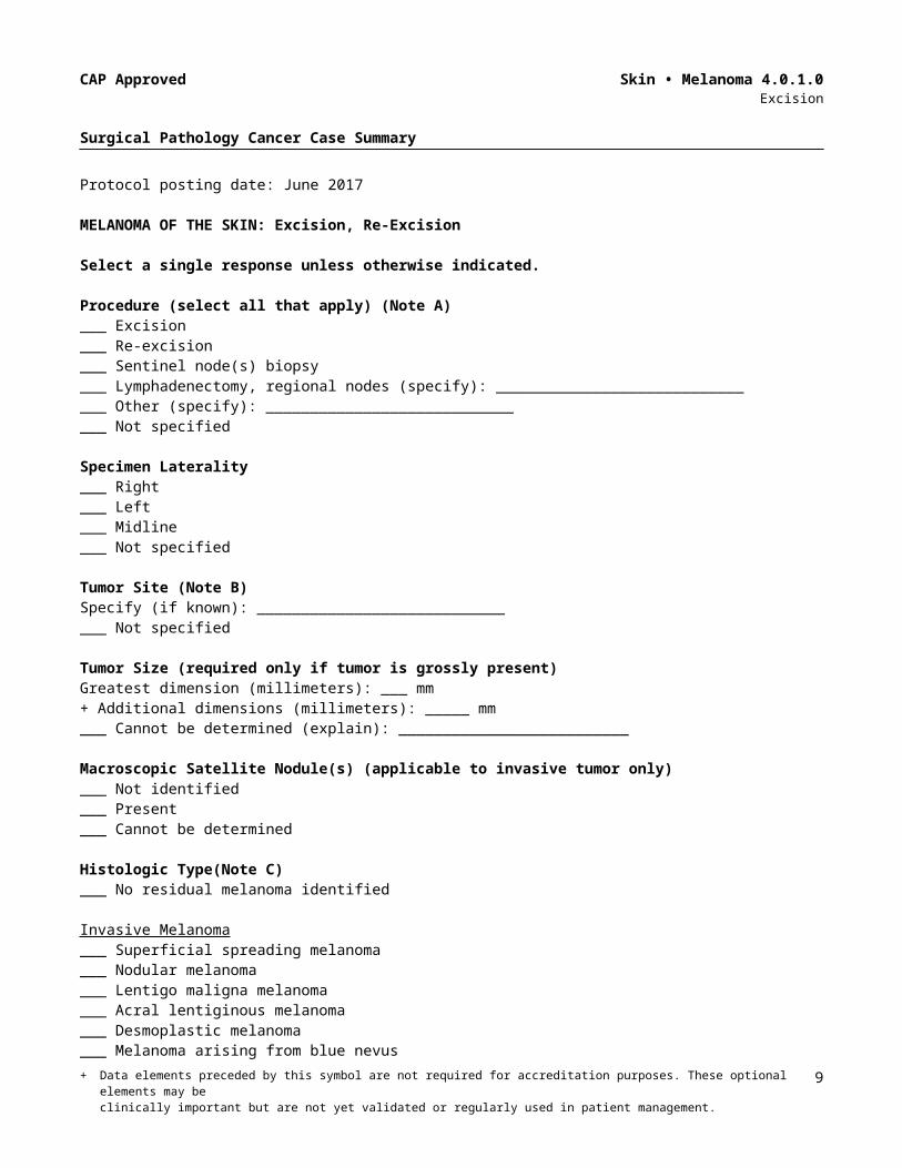
Procedure (select all that apply) (Note A)___ Excision___ Re-excision___ Sentinel node(s) biopsy___ Lymphadenectomy, regional nodes (specify): _______________________________ Other (specify): _______________________________ Not specified
Specimen Laterality___ Right___ Left___ Midline___ Not specified
Tumor Site (Note B)Specify (if known): _______________________________ Not specified
Tumor Size (required only if tumor is grossly present)Greatest dimension (millimeters): ___ mm+ Additional dimensions (millimeters): _____ mm___ Cannot be determined (explain): __________________________
Macroscopic Satellite Nodule(s) (applicable to invasive tumor only)___ Not identified___ Present ___ Cannot be determined
Histologic Type(Note C)___ No residual melanoma identified
Invasive Melanoma___ Superficial spreading melanoma___ Nodular melanoma___ Lentigo maligna melanoma ___ Acral lentiginous melanoma___ Desmoplastic melanoma___ Melanoma arising from blue nevus ___ Melanoma arising in a giant congenital nevus ___ Melanoma of childhood ___ Nevoid melanoma___ Persistent melanoma ___ Melanoma, not otherwise classified___ Other histologic type not listed (specify): ________________________
+ Data elements preceded by this symbol are not required for accreditation purposes. These optional elements may be clinically important but are not yet validated or regularly used in patient management.
7
CAP Approved Skin • Melanoma 4.0.1.0Excision
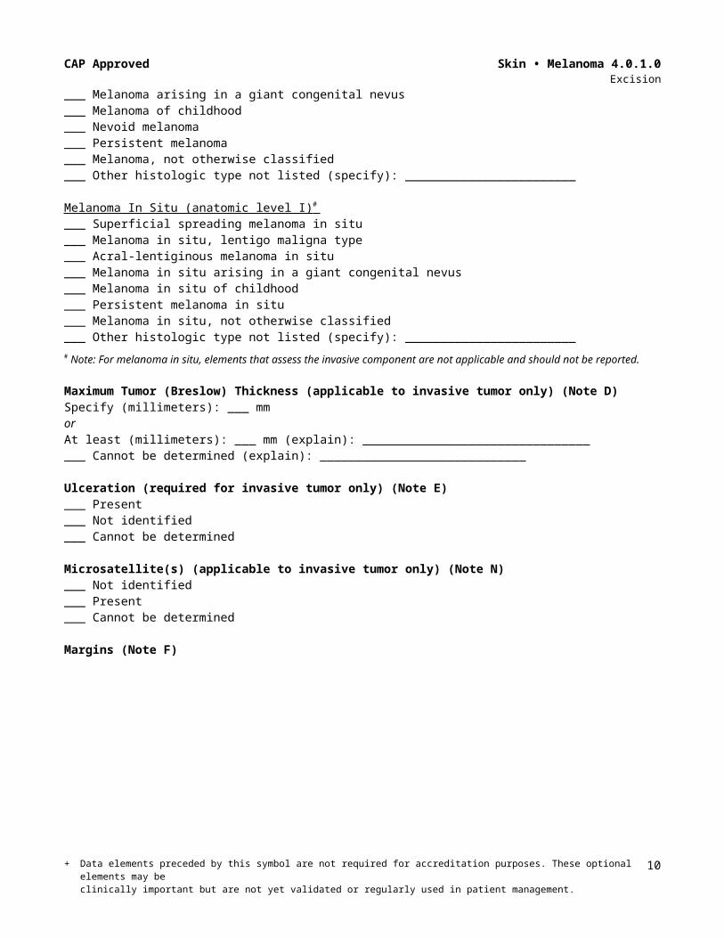
Melanoma In Situ (a natomic level I) # ___ Superficial spreading melanoma in situ___ Melanoma in situ, lentigo maligna type___ Acral-lentiginous melanoma in situ___ Melanoma in situ arising in a giant congenital nevus ___ Melanoma in situ of childhood ___ Persistent melanoma in situ___ Melanoma in situ, not otherwise classified___ Other histologic type not listed (specify): ________________________# Note: For melanoma in situ, elements that assess the invasive component are not applicable and should not be reported.
Maximum Tumor (Breslow) Thickness (applicable to invasive tumor only) (Note D)Specify (millimeters): ___ mmorAt least (millimeters): ___ mm (explain): ___________________________________ Cannot be determined (explain): _____________________________
Ulceration (required for invasive tumor only) (Note E)___ Present___ Not identified___ Cannot be determined
Microsatellite(s) (applicable to invasive tumor only) (Note N)___ Not identified ___ Present ___ Cannot be determined
Margins (Note F)
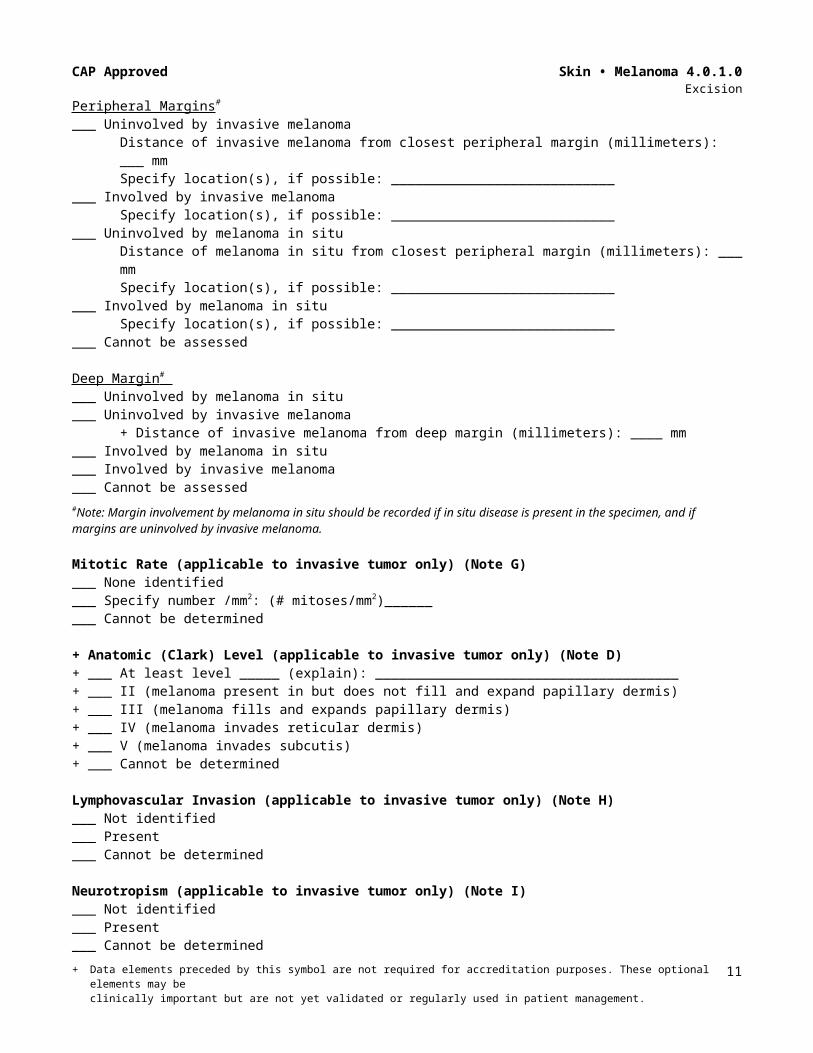
Peripheral Margins#
___ Uninvolved by invasive melanoma Distance of invasive melanoma from closest peripheral margin (millimeters): ___ mmSpecify location(s), if possible: ____________________________
___ Involved by invasive melanomaSpecify location(s), if possible: ____________________________
___ Uninvolved by melanoma in situ Distance of melanoma in situ from closest peripheral margin (millimeters): ___ mm Specify location(s), if possible: ____________________________
___ Involved by melanoma in situSpecify location(s), if possible: ____________________________
___ Cannot be assessed
Deep Margin # ___ Uninvolved by melanoma in situ___ Uninvolved by invasive melanoma
+ Distance of invasive melanoma from deep margin (millimeters): ____ mm___ Involved by melanoma in situ___ Involved by invasive melanoma___ Cannot be assessed#Note: Margin involvement by melanoma in situ should be recorded if in situ disease is present in the specimen, and if margins are uninvolved by invasive melanoma.
+ Data elements preceded by this symbol are not required for accreditation purposes. These optional elements may be clinically important but are not yet validated or regularly used in patient management.
8
CAP Approved Skin • Melanoma 4.0.1.0Excision
Mitotic Rate (applicable to invasive tumor only) (Note G)___ None identified___ Specify number /mm2: (# mitoses/mm2)_________ Cannot be determined
+ Anatomic (Clark) Level (applicable to invasive tumor only) (Note D)+ ___ At least level _____ (explain): ______________________________________+ ___ II (melanoma present in but does not fill and expand papillary dermis)+ ___ III (melanoma fills and expands papillary dermis)+ ___ IV (melanoma invades reticular dermis)+ ___ V (melanoma invades subcutis)+ ___ Cannot be determined
Lymphovascular Invasion (applicable to invasive tumor only) (Note H)___ Not identified___ Present___ Cannot be determined
Neurotropism (applicable to invasive tumor only) (Note I)___ Not identified___ Present___ Cannot be determined
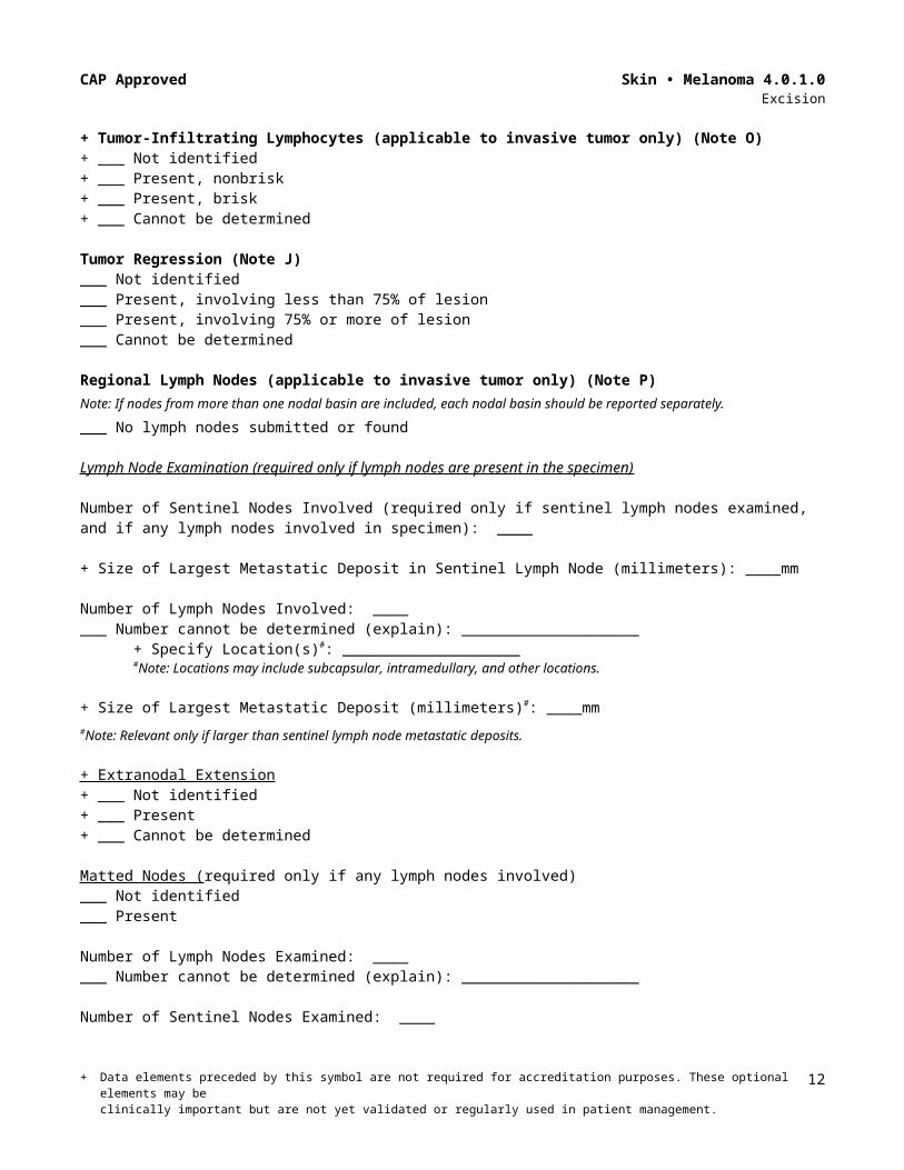
+ Tumor-Infiltrating Lymphocytes (applicable to invasive tumor only) (Note O)+ ___ Not identified+ ___ Present, nonbrisk+ ___ Present, brisk+ ___ Cannot be determined
Tumor Regression (Note J)___ Not identified___ Present, involving less than 75% of lesion___ Present, involving 75% or more of lesion___ Cannot be determined
Regional Lymph Nodes (applicable to invasive tumor only) (Note P)Note: If nodes from more than one nodal basin are included, each nodal basin should be reported separately.___ No lymph nodes submitted or found
Lymph Node Examination (required only if lymph nodes are present in the specimen)
Number of Sentinel Nodes Involved (required only if sentinel lymph nodes examined, and if any lymph nodes involved in specimen): ____
+ Size of Largest Metastatic Deposit in Sentinel Lymph Node (millimeters): ____mm
Number of Lymph Nodes Involved: _______ Number cannot be determined (explain): ____________________
+ Specify Location(s)#: ____________________ #Note: Locations may include subcapsular, intramedullary, and other locations.
+ Size of Largest Metastatic Deposit (millimeters)#: ____mm#Note: Relevant only if larger than sentinel lymph node metastatic deposits.
+ Extranodal Extension
+ Data elements preceded by this symbol are not required for accreditation purposes. These optional elements may be clinically important but are not yet validated or regularly used in patient management.
9
CAP Approved Skin • Melanoma 4.0.1.0Excision
+ ___ Not identified+ ___ Present+ ___ Cannot be determined
Matted Nodes (required only if any lymph nodes involved)___ Not identified___ Present
Number of Lymph Nodes Examined: _______ Number cannot be determined (explain): ____________________
Number of Sentinel Nodes Examined: ____
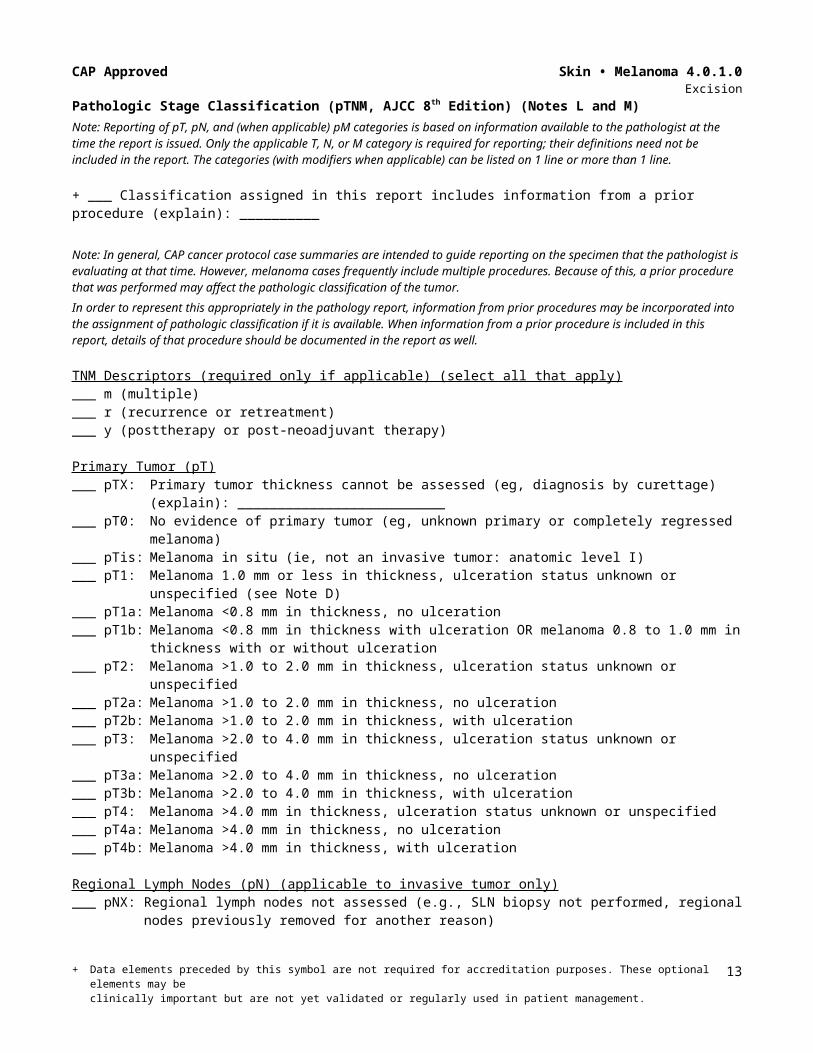
Pathologic Stage Classification (pTNM, AJCC 8th Edition) (Notes L and M)Note: Reporting of pT, pN, and (when applicable) pM categories is based on information available to the pathologist at the time the report is issued. Only the applicable T, N, or M category is required for reporting; their definitions need not be included in the report. The categories (with modifiers when applicable) can be listed on 1 line or more than 1 line.
+ ___ Classification assigned in this report includes information from a prior procedure (explain): __________
Note: In general, CAP cancer protocol case summaries are intended to guide reporting on the specimen that the pathologist is evaluating at that time. However, melanoma cases frequently include multiple procedures. Because of this, a prior procedure that was performed may affect the pathologic classification of the tumor. In order to represent this appropriately in the pathology report, information from prior procedures may be incorporated into the assignment of pathologic classification if it is available. When information from a prior procedure is included in this report, details of that procedure should be documented in the report as well.
TNM Descriptors (required only if applicable) (select all that apply)___ m (multiple)___ r (recurrence or retreatment)___ y (posttherapy or post-neoadjuvant therapy)
Primary Tumor (pT)___ pTX: Primary tumor thickness cannot be assessed (eg, diagnosis by curettage)
(explain): _____________________________ pT0: No evidence of primary tumor (eg, unknown primary or completely regressed melanoma)___ pTis: Melanoma in situ (ie, not an invasive tumor: anatomic level I)___ pT1: Melanoma 1.0 mm or less in thickness, ulceration status unknown or unspecified (see Note D)___ pT1a: Melanoma <0.8 mm in thickness, no ulceration___ pT1b: Melanoma <0.8 mm in thickness with ulceration OR melanoma 0.8 to 1.0 mm in thickness with or
without ulceration
___ pT2: Melanoma >1.0 to 2.0 mm in thickness, ulceration status unknown or unspecified___ pT2a: Melanoma >1.0 to 2.0 mm in thickness, no ulceration ___ pT2b: Melanoma >1.0 to 2.0 mm in thickness, with ulceration ___ pT3: Melanoma >2.0 to 4.0 mm in thickness, ulceration status unknown or unspecified___ pT3a: Melanoma >2.0 to 4.0 mm in thickness, no ulceration___ pT3b: Melanoma >2.0 to 4.0 mm in thickness, with ulceration___ pT4: Melanoma >4.0 mm in thickness, ulceration status unknown or unspecified___ pT4a: Melanoma >4.0 mm in thickness, no ulceration ___ pT4b: Melanoma >4.0 mm in thickness, with ulceration
Regional Lymph Nodes (pN) (applicable to invasive tumor only) ___ pNX: Regional lymph nodes not assessed (e.g., SLN biopsy not performed, regional nodes previously
removed for another reason)
+ Data elements preceded by this symbol are not required for accreditation purposes. These optional elements may be clinically important but are not yet validated or regularly used in patient management.
10
CAP Approved Skin • Melanoma 4.0.1.0Excision
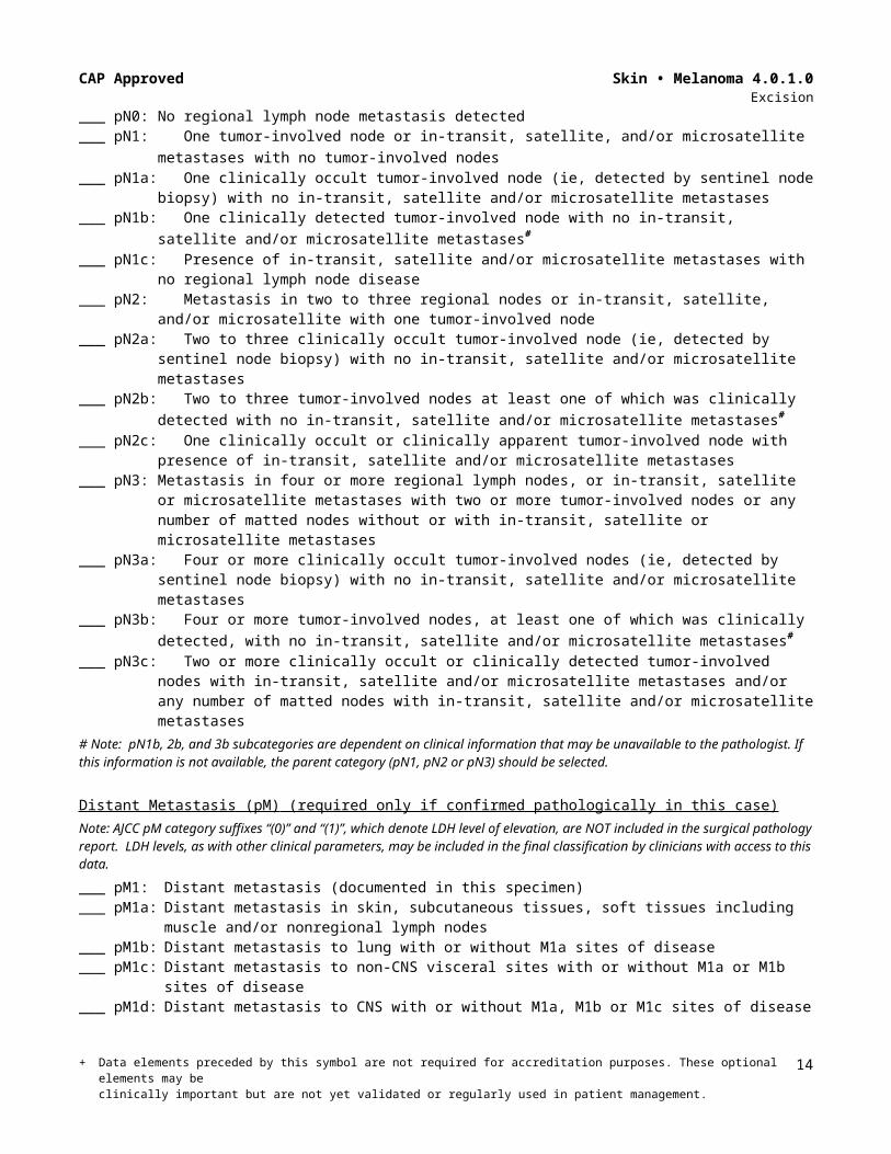
___ pN0: No regional lymph node metastasis detected___ pN1: One tumor-involved node or in-transit, satellite, and/or microsatellite metastases with no tumor-
involved nodes ___ pN1a: One clinically occult tumor-involved node (ie, detected by sentinel node biopsy) with no in-transit,
satellite and/or microsatellite metastases___ pN1b: One clinically detected tumor-involved node with no in-transit, satellite and/or microsatellite
metastases# ___ pN1c: Presence of in-transit, satellite and/or microsatellite metastases with no regional lymph node disease___ pN2: Metastasis in two to three regional nodes or in-transit, satellite, and/or microsatellite with one tumor-
involved node ___ pN2a: Two to three clinically occult tumor-involved node (ie, detected by sentinel node biopsy) with no in-
transit, satellite and/or microsatellite metastases___ pN2b: Two to three tumor-involved nodes at least one of which was clinically detected with no in-transit,
satellite and/or microsatellite metastases#
___ pN2c: One clinically occult or clinically apparent tumor-involved node with presence of in-transit, satellite and/or microsatellite metastases
___ pN3: Metastasis in four or more regional lymph nodes, or in-transit, satellite or microsatellite metastases with two or more tumor-involved nodes or any number of matted nodes without or with in-transit, satellite or microsatellite metastases
___ pN3a: Four or more clinically occult tumor-involved nodes (ie, detected by sentinel node biopsy) with no in-transit, satellite and/or microsatellite metastases
___ pN3b: Four or more tumor-involved nodes, at least one of which was clinically detected, with no in-transit, satellite and/or microsatellite metastases#
___ pN3c: Two or more clinically occult or clinically detected tumor-involved nodes with in-transit, satellite and/or microsatellite metastases and/or any number of matted nodes with in-transit, satellite and/or microsatellite metastases
# Note: pN1b, 2b, and 3b subcategories are dependent on clinical information that may be unavailable to the pathologist. If this information is not available, the parent category (pN1, pN2 or pN3) should be selected.
Distant Metastasis (pM) (required only if confirmed pathologically in this case) Note: AJCC pM category suffixes “(0)” and “(1)”, which denote LDH level of elevation, are NOT included in the surgical pathology report. LDH levels, as with other clinical parameters, may be included in the final classification by clinicians with access to this data.___ pM1: Distant metastasis (documented in this specimen)___ pM1a: Distant metastasis in skin, subcutaneous tissues, soft tissues including muscle and/or nonregional
lymph nodes___ pM1b: Distant metastasis to lung with or without M1a sites of disease___ pM1c: Distant metastasis to non-CNS visceral sites with or without M1a or M1b sites of disease___ pM1d: Distant metastasis to CNS with or without M1a, M1b or M1c sites of disease
Specify site(s), if known: __________________________
+ Additional Pathologic Findings (select all that apply)+ ___ Associated nevus (specify type): _______________________+ ___ Other (specify): ____________________________
+ Ancillary StudiesNote: For molecular genetic reporting, the CAP Melanoma Biomarker Template should be used. Pending biomarker studies should be listed in the Comments section of this report.
+ Comment(s)
+ Data elements preceded by this symbol are not required for accreditation purposes. These optional elements may be clinically important but are not yet validated or regularly used in patient management.
11
Background Documentation Skin • Melanoma 4.0.1.0
Explanatory Notes
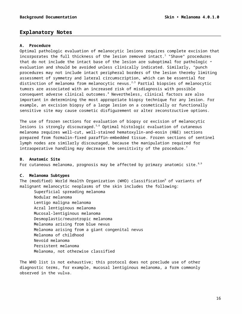
A. ProcedureOptimal pathologic evaluation of melanocytic lesions requires complete excision that incorporates the full thickness of the lesion removed intact.1 "Shave" procedures that do not include the intact base of the lesion are suboptimal for pathologic evaluation and should be avoided unless clinically indicated. Similarly, “punch” procedures may not include intact peripheral borders of the lesion thereby limiting assessment of symmetry and lateral circumscription, which can be essential for distinction of melanoma from melanocytic nevus.2,3 Partial biopsies of melanocytic tumors are associated with an increased risk of misdiagnosis with possible consequent adverse clinical outcomes.4 Nevertheless, clinical factors are also important in determining the most appropriate biopsy technique for any lesion. For example, an excision biopsy of a large lesion on a cosmetically or functionally sensitive site may cause cosmetic disfigurement or alter reconstructive options.
The use of frozen sections for evaluation of biopsy or excision of melanocytic lesions is strongly discouraged.5,6 Optimal histologic evaluation of cutaneous melanoma requires well-cut, well-stained hematoxylin-and-eosin (H&E) sections prepared from formalin-fixed paraffin-embedded tissue. Frozen sections of sentinel lymph nodes are similarly discouraged, because the manipulation required for intraoperative handling may decrease the sensitivity of the procedure.7
B. Anatomic SiteFor cutaneous melanoma, prognosis may be affected by primary anatomic site.8,9
C. Melanoma Subtypes The (modified) World Health Organization (WHO) classification9 of variants of malignant melanocytic neoplasms of the skin includes the following:
Superficial spreading melanomaNodular melanomaLentigo maligna melanomaAcral lentiginous melanoma Mucosal-lentiginous melanoma Desmoplastic/neurotropic melanoma Melanoma arising from blue nevusMelanoma arising from a giant congenital nevusMelanoma of childhoodNevoid melanomaPersistent melanomaMelanoma, not otherwise classified
The WHO list is not exhaustive; this protocol does not preclude use of other diagnostic terms, for example, mucosal lentiginous melanoma, a form commonly observed in the vulva.
Recurrent hot spot driver mutations in BRAF, NRAS, NF1, CKIT, and other genes have been identified in melanomas10 and in some instances (particularly BRAF mutations), may be important for selecting patients with advanced and/or unresectable metastatic disease who may benefit from treatment with targeted therapies.11 Recent studies have highlighted clinical and morphologic features predictive of the presence of some mutations, particularly BRAF mutations.12,13 However, the sensitivity and specificity of these features are insufficient for clinical purposes and formal mutation testing is recommended if this is indicated for clinical care.
12
Background Documentation Skin • Melanoma 4.0.1.0
D. Primary Tumor (Breslow) Thickness) and Anatomic (Clark) LevelsMaximum tumor thickness is measured with a calibrated ocular micrometer at a right angle to the adjacent normal skin. The upper point of reference is the upper edge of the granular layer of the epidermis of the overlying skin or, if the lesion is ulcerated overlying the entire dermal component, the base of the ulcer. The lower reference point is the deepest point of tumor invasion (ie, the leading edge of a single mass or an isolated group of cells deep to the main mass).
If the tumor is transected by the deep margin of the specimen, the depth may be indicated as “at least __ mm” with a comment explaining the limitation of thickness assessment. For example, “The maximum tumor thickness cannot be determined in this specimen because the deep plane of the biopsy transects the tumor.” Tumor thickness measurements should not be based on periadnexal extension (either periadnexal adventitial or extra-adventitial extension), except when it is the only focus of invasion. In that circumstance, Breslow thickness may be measured from the inner layer of the outer root sheath epithelium or inner luminal surface of sweat glands, to the furthest extent of infiltration into the periadnexal dermis. Microsatellites or foci of neurotropism or lymphovascular invasion should not be included in tumor thickness measurements.
In the 8th edition of the AJCC melanoma staging system,14 it is recommended that tumor thickness measurements be recorded to the nearest 0.1 mm, not the nearest 0.01 mm, because of the impracticality and imprecision of measurements, particularly for tumors >1 mm thick. Tumors ≤1 mm thick may be measured to the nearest 0.01 mm if practical, but should be reported to the nearest 0.1mm (eg, melanomas measured to be in the range of 0.75 mm to 0.84 mm are reported as 0.8 mm in thickness and hence T1b, and tumors 1.01 to 1.04 mm in thickness are reported as 1.0 mm).
While the principal T category tumor thickness ranges have been maintained in the AJCC 8th edition, T1 is now subcategorized by tumor thickness strata at a 0.8 mm threshold. Tumor mitotic rate as a dichotomous variable is no longer used as a staging category criterion for T1 tumors. T1a melanomas are now defined as non-ulcerated and <0.8 mm in thickness. T1b melanomas are defined as 0.8-1.0mm in thickness or ulcerated melanomas <0.8 mm in thickness.
Anatomic (Clark) levels are defined as follows:
I Intraepidermal tumor only (ie, melanoma in situ)II Tumor present in but does not fill and expand papillary dermisIII Tumor fills and expands papillary dermisIV Tumor invades into reticular dermisV Tumor invades subcutis
Anatomic (Clark) level of invasion remains an independent predictor of outcome and is recommended by the AJCC to be reported as a primary tumor characteristic.14 However, assessment of Clark levels is less reproducible among pathologists than is tumor thickness, and Clark levels are not used in the AJCC staging system for pT status. Accordingly, Clark levels are included in this checklist as an optional data item.
E. UlcerationPrimary tumor ulceration has been shown to be a dominant independent prognostic factor in invasive cutaneous melanoma,8,14 and if present, changes the pT stage from T1a to T1b, T2a to T2b, etc., depending on the thickness of the tumor. The presence or absence of ulceration must be confirmed on microscopic examination.14 Melanoma ulceration is defined as the combination of the following features: full-thickness epidermal defect (including absence of stratum corneum and basement membrane); evidence of reactive changes (ie, fibrin deposition, neutrophils); and thinning, effacement, or reactive hyperplasia of the surrounding epidermis in the absence of trauma or a recent surgical procedure. Ulcerated melanomas typically show invasion through the epidermis, whereas nonulcerated melanomas tend to lift the overlying epidermis.
Only nontraumatic (“tumorigenic”) ulceration should be recorded as ulceration. If ulceration is present related to a prior biopsy, the tumor should not be recorded as ulcerated for staging purposes. If a lesion has been recently biopsied or there is only focal loss of the epidermis, assessment of ulceration may be difficult or impossible; in this instance it may be difficult to determine whether the epidermal deficiency is due to true ulceration or to sectioning
13
Background Documentation Skin • Melanoma 4.0.1.0
artifact.14 Absence of fibrin, neutrophils, or granulation tissue from putative areas of ulceration would be clues that the apparent ulceration is actually due to sectioning of only part of the epidermis and this should not be designated as ulceration. If non-traumatic (“tumorigenic”) ulceration is present in either an initial partial biopsy or a re-excision specimen, then for staging purposes, the tumor should be recorded as ulcerated.
Ulceration may be present in an in situ melanoma but does not affect the staging.
A number of recent studies have demonstrated that the extent of ulceration (measured either as a percentage of the width of the dermal invasive component of the tumor or as a diameter) more accurately predicts outcome than the presence or absence of ulceration alone.15,16
F. MarginsMicroscopically measured distances between tumor and labeled peripheral (lateral) or deep margins are appropriately recorded for melanoma excision specimens. If a margin is involved by tumor, it should be stated whether the tumor is in situ or invasive.
G. Mitotic RateTumor mitotic rate (of the invasive component of a melanoma) is a strong independent predictor of outcome across its dynamic range in all thickness categories and should be assessed and recorded in all primary melanomas including in both initial and excision biopsies (the highest value in any specimen should be used for prognostic purposes). Although tumor mitotic rate is no longer used as a T1-category criterion in the 8 th edition of the AJCC melanoma staging system (due to the more significant prognostic significance of the new tumor thickness strata within T1 melanoma), mitotic rate will likely be an important parameter in prognostic models developed in the future that will provide personalized prediction of prognosis for individual patients. The method recommended for enumerating the tumor mitotic rate in the 8th edition of the AJCC staging system is provided below:
“The recommended approach to enumerating mitoses is to first find the regions in the dermis containing the most mitotic figures, the so-called "hot spot" or "dermal hot spot." After counting the mitoses in the initial high‐power field, the count is extended to immediately adjacent nonoverlapping fields until an area of tissue corresponding to 1 mm2 is assessed. If no hot spot is found and mitoses are sparse and/or randomly scattered throughout the lesion, then a representative mitosis is chosen and, beginning with that field, the count is then extended to immediately adjacent nonoverlapping fields until an area corresponding to 1 mm2 of tissue is assessed. The count then is expressed as the (whole) number of mitoses/mm2. If the invasive component of the tumor involves an area <1 mm2, the number of mitoses should be assessed and recorded as if they were found within square millimeter. For example, if the entire dermal component of a tumor occupies 0.5 mm2 and only one mitosis is identified, the mitotic rate should be recorded as 1/mm2 (not 2/mm2). The number of mitoses should be listed as a whole number per square millimeter. If no mitoses are identified, the mitotic rate may be recorded as “none identified” or “0/mm2.” Only mitotic figures in invasive melanoma cells should be counted. This methodology for determining the mitotic rate of a melanoma has been shown to have excellent interobserver reproducibility, including among pathologists with widely differing experience in the assessment of melanocytic tumors.17
To obtain accurate measurement, calibration of individual microscopes is recommended using a stage micrometer to determine the number of high-power fields that equates to a square millimeter.
The data that demonstrated the strong prognostic significance of mitotic rate were obtained from the melanoma pathology reports of routinely assessed H&E stained sections. It therefore is recommended that no additional sections be cut and examined in excess of those that would normally be used to report and diagnose the melanoma to determine the mitotic count (ie, no additional sections should be cut and examined for the sole purpose of determining the mitotic rate, including in situations in which no mitoses are identified on the initial, routinely examined sections). Immunohistochemical stains for identifying mitoses are not used for determining mitotic rate for staging and/ or reporting purposes. A possible exception is the use to dual immunohistochemistry (eg, MART1 and pHH3) to determine if a cell in mitosis is a melanocyte or not (macrophage, endothelial cell, etc).18
Although the AJCC recommends reporting “0” rather than “none identified” or “less than 1,” for the purposes of cancer registry reporting all of these terms should be considered equivalent.
14
Background Documentation Skin • Melanoma 4.0.1.0
H. Lymphovascular InvasionLymphovascular invasion is identified by the demonstration of melanoma cells within the lumina of blood vessels or lymphatics, or both.14 Immunohistochemistry for vascular endothelial cell markers CD31, CD34 or ERG or the lymphatic marker D2-40 may assist in the identification of the presence of intravascular or intralymphatic tumor by highlighting vascular lumina. Vascular invasion by melanoma correlates independently with worsened overall survival .7,19 The detection of LVI is increased in primary melanomas when double labeling of tumor cells and lymphatic endothelium is applied.20
I. NeurotropismNeurotropism is defined as the presence of melanoma cells abutting nerve sheaths usually circumferentially (perineural invasion) or within nerves (intraneural invasion).14 Occasionally, the tumor itself may form neuroid structures (termed ‘neural transformation’ and this is also regarded as neurotropism). Neurotropism is best identified at the periphery of the tumor; the presence of melanoma cells around nerves in the main tumor mass caused by “entrapment” of nerves in the expanding tumor does not represent neurotropism.
Neurotropism is most commonly identified in desmoplastic melanomas (sometimes termed desmoplastic neurotropic melanoma) but may occur in any melanoma subtype.9 Neurotropism may correlate with an increased risk for local recurrence.
J. Tumor RegressionCharacteristic features of regression include replacement of tumor cells by lymphocytic inflammation, as well as attenuation of the epidermis and nonlaminated dermal fibrosis with inflammatory cells, melanophagocytosis, and telangiectasia.
L. TNM and Stage GroupingsThe latest edition TNM Staging System of the American Joint Committee on Cancer (AJCC) and the International Union Against Cancer (UICC) is recommended by this protocol.14
Changes in the 8th edition AJCC Cancer Staging Manual of importance to practicing pathologists include: T1a melanoma now defined as non-ulcerated melanomas <0.8mm thick T1b melanomas now defined as melanoma between 0.8mm and 1.0mm in thickness OR ulcerated
melanomas <0.8mm thick Tumor mitotic rate no longer used as a T category criterion but remains an important prognostic factor and
should be reported in all invasive primary melanomas Recommendation to record tumor thickness to the nearest 0.1mm (not the nearest 0.01mm) Regarding regional lymph node metastasis, the previously empirically defined terms “microscopic” and
“macroscopic” replaced with “clinically occult” (ie, detected by sentinel node biopsy) and “clinically detected”
Non-nodal regional metastatic disease (ie, microsatellites, satellites and in transit metastases) now formally stratified by N category according to the number of tumor-involved nodes
Gross extranodal extension no longer used as an N-category criterion (but presence of “matted nodes” retained)
M1 now defined by both anatomic site of distant metastasis and serum LDH levels for all anatomic subsite categories of metastasis
New M1d designation added for distant metastasis to central nervous system pT1bN0M0 is now pathologic stage IA in contrast to cT1N0M0 which remains clinical stage IB disease N category now defines four stage subgroups and takes into account both T category elements and N
category elements
Pathologic staging includes microstaging of the primary melanoma and pathologic information about the regional lymph nodes after partial or complete lymphadenectomy.
In virtually all studies of cutaneous melanoma, tumor thickness has been shown to be a dominant prognostic factor,9,13,21 and it forms the basis for the stratification of pT. Although anatomic (Clark) levels are also commonly used to indicate depth of invasion of the primary tumor,9,21,22 they are less predictive of clinical outcome than mitotic activity or ulceration.20
15
Background Documentation Skin • Melanoma 4.0.1.0
By AJCC/UICC convention, the designation “T” refers to a primary tumor that has not been previously treated. Similarly by convention, clinical staging is performed after biopsy of the primary melanoma (including utilizing pathologic information on microstaging of the primary melanoma) with clinical or biopsy assessment of regional lymph nodes and distant sites.16 Pathologic staging uses information gained from pathologic evaluation of both the primary melanoma after biopsy and wide excision as well as pathological evaluation of the regional node basin after SLN biopsy (required for N categorization of all >T1 melanomas) and/or complete regional lymphadenectomy.14 In additional, for pathological staging, if information from any prior biopsy is known and is relevant for staging, this should be documented in the pathology report (in the staging section) and used for assigning T, N and M categories and staging purposes.
T Category ConsiderationsPathologic (microscopic) assessment of the primary tumor is required for accurate staging. Therefore, excision of the primary tumor, rather than incisional biopsy, is advised. The T classification of melanoma is based on the thickness of the primary tumor and presence or absence of ulceration (see also Notes D, and E).
N Category Considerations (see also Note P)The regional lymph nodes are the most common sites of metastasis. The widespread use of cutaneous lymphoscintigraphy, lymphatic mapping, and sentinel lymph node biopsies has greatly enhanced the ability to identify the presence of lymph node metastasis.23 By convention, the term regional lymph nodal metastasis refers to disease confined to one draining nodal basin or 2 contiguous draining nodal basins, as in patients with nodal disease in combinations of femoral/iliac, axillary/supraclavicular, cervical/supraclavicular, axillary/femoral, or bilateral axillary or femoral metastases. In some patients, lymphoscintigraphic imaging may define multiple regional lymph node basins and disease identified in any of such basins also constitutes regional disease. Metastasis to nondraining nodal basin(s) is considered M1 disease.
Sentinel Lymph Nodes Sentinel lymph node identification and evaluation may be included in the surgical approach to cutaneous melanoma.24 A sentinel lymph node is defined as any lymph node receiving direct lymphatic drainage from a primary tumor site. There may be more than 1 sentinel node for some tumors. The clinical rationale for sentinel lymph node identification and separate evaluation is based on the assumption that metastatic involvement of a sentinel node increases the likelihood that other, more distant nodes may also contain metastatic disease. Conversely, if sentinel nodes are negative, other regional nodes would be less likely to contain metastasis. M Category ConsiderationsThe category “MX” has been eliminated from the AJCC/UICC TMN system.14 Unless there is clinical or pathologic evidence of distant metastasis the stage is classified as clinical M0 (ie, no distant metastasis). pM should only be reported when metastases have been documented by pathologic examination, that is, pM1 disease. pMX and pM0 should not be reported by the pathologist.
TNM DescriptorsFor identification of special cases of TNM or pTNM classifications, the “y,” “r,” and “a” prefixes are used. Although they do not affect the stage grouping, they indicate cases needing separate analysis.
Post-therapy or post neoadjuvant therapy classification (yTNM) documents the extent of the disease for patients whose first course of therapy includes systemic or radiation treatment prior to surgical resection or when systemic therapy or radiation is primary treatment with no surgical resection. The extent of disease is classified using the same T, N, and M definitions and identified as post-treatment with a “yc” or “yp” prefix (ycT, ycN, ycTNM; ypT, ypN, ypTNM).
Recurrence or retreatment classification (rTNM) is used because information gleaned from therapeutic procedures and from extent of disease defined clinically may be prognostic for patients with recurrent cancer after a disease-free interval. It is important to understand that the rTNM classification does not change the original clinical or pathologic staging of the case.
16
Background Documentation Skin • Melanoma 4.0.1.0
Autopsy classification (aTNM) is used to stage cases of cancer not recognized during life and only recognized postmortem.
Additional Descriptors
Residual Tumor (R)Tumor remaining in a patient after therapy with curative intent (eg, surgical resection for cure) is categorized by a system known as R classification, as follows.
RX Presence of residual tumor cannot be assessedR0 No residual tumorR1 Microscopic residual tumorR2 Macroscopic residual tumor
For the surgeon, the R classification may be useful to indicate the known or assumed status of the completeness of a surgical excision. For the pathologist, the R classification is relevant to the status of the margins of a surgical resection specimen. That is, tumor involving the resection margin on pathologic examination may be assumed to correspond to residual tumor in the patient and may be classified as macroscopic or microscopic according to the findings at the specimen margin(s). For example, a T4 primary melanoma with involved microscopic margins on the definitive treatment/excision specimen would be reported as pT4N0M0R1.
Lymphovascular Invasion Lymphovascular invasion (LVI) indicates whether microscopic lymphatic or vascular invasion is identified and includes lymphatic invasion, blood vessel invasion, or invasion of a vessel for which it cannot be determined whether it is a blood or lymphatic vessel lLymphovascular invasion). By AJCC/UICC convention, LVI does not affect the T category indicating local extent of tumor, ie, foci of Lymphovascular invasion should not be included in the measurement of tumor (Breslow) thickness.25
M. Pretreatment Serum Lactate Dehydrogenase and Serum AlbuminData from numerous studies have indicated that an elevated serum level of lactate dehydrogenase (LDH) is an independent predictor of decreased survival in AJCC stage IV melanoma patients.
N. Microsatellite(s) A microsatellite(s) is defined as the presence of a microscopic cutaneous metastasis found adjacent or deep to a primary melanoma on pathological examination of the primary tumor site.14 The metastatic tumor cells must be discontinuous from the primary tumor. If the tissue between the apparently separate nodule and the primary tumor is only fibrous scarring and/or inflammation, this does not indicate a microsatellite, because the aforementioned changes may represent regression of the intervening tumor (but not separated only by fibrosis or inflammation because the features could signify regression of the intervening tumor). There is no minimum size threshold or distance from the primary tumor to define a microsatellite. Before diagnosing the presence of a microsatellite, it is generally recommended that multiple sections from the same tissue block being examined to verify that the microsatellite is indeed discontinuous from the primary tumor. For example, periadnexal extension of tumor or the irregular shape of the peripheral or deep extent of the tumor may result in tumor that is contiguous with the primary tumor appear discontiguous on single sections.
O. Tumor-Infiltrating Lymphocytes A paucity of tumor-infiltrating lymphocytes (TILs) is an adverse prognostic factor for cutaneous melanoma.21 Tumor-infiltrating lymphocytes may be assessed in a semiquantitative way, as defined below. To qualify as TILs, lymphocytes need to surround and disrupt tumor cells of the invasive component of the tumor.
TILs Not Identified: No lymphocytes present, or lymphocytes present but do not infiltrate tumor at all.
TILs Nonbrisk: Lymphocytes infiltrate melanoma only focally or not along the entire base of the invasive tumor.
TILs Brisk: Lymphocytes diffusely infiltrate the entire base of the invasive tumor (Figure 1, A) or show diffuse permeation of the invasive tumor (Figure 1, B).
17
Background Documentation Skin • Melanoma 4.0.1.0
Figure 1. Brisk tumor-infiltrating lymphocytes. A. Lymphocytes diffusely infiltrate the entire base of the invasive tumor. B. Lymphocytes diffusely infiltrate the entire invasive component of the melanoma.
P. Lymph NodesRemoval of sentinel lymph nodes may be performed for patients with clinically localized primary cutaneous melanomas with a thickness of 1 mm or greater, or in selected patients with thinner tumors with other adverse prognostic features.26,27 Frozen section analysis of sentinel lymph nodes is not advised.7 Review of the H&E-stained slides from multiple levels through serially sliced sentinel lymph nodes increases the sensitivity of detecting microscopic melanoma metastasis; routine analysis (H&E-stained sections of the cut surfaces of a simply bisected lymph node) may lead to a false-negative rate of 10% to 15%. The use of immunohistochemical stains (eg, for HMB-45 or MART-1) further increases the sensitivity of detection of microscopic melanoma metastases and should also be considered in the examination of sentinel lymph nodes. Although immunohistochemical staining should be used in conjunction with and not in place of standard H&E histologic examination, immunohistochemically identified micrometastases are accepted as representing greater than N0 disease by the 8th edition of the AJCC staging system (as in the 7th edition), ie, a lymph node in which any metastatic tumors cells are identified, irrespective of the number of cells or whether they were identified on H&E or immunostained sections, should be designated as a tumor-positive node.14
For histologic examination, whether for sentinel node analysis or for routine regional lymph node evaluation, the entire node, except tissue collected for consented research protocols, should be submitted. For routine evaluation, large lymph nodes may be bisected or sliced at 2-3 mm intervals, whereas smaller nodes (<5 mm) may be submitted whole.
Data from multiple studies28-30 indicated that the sentinel lymph node tumor burden and/or the microanatomical region/compartment of the sentinel node occupied by the metastasis may be useful in predicting patients who have additional disease in nonsentinel nodes as well as disease outcome. Because sentinel node tumor burden is considered a regional disease prognostic factor, it should be reported in all patients with a positive sentinel node but it is not used to determine N-category groupings in the 8th edition of the AJCC staging system. The current National Comprehensive Cancer Network (NCCN) guidelines31 also recommend recording the size and location of tumor present in a positive sentinel node.
18
Background Documentation Skin • Melanoma 4.0.1.0
References1. Sober AJ, Chuang TY, Duvic M, et al. Guidelines of care for primary cutaneous melanoma. J Am Acad
Dermatol. 2001;45(4):579-586.2. Stell VH, Norton HJ, Smith KS, Salo JC, White RL, Jr. Method of biopsy and incidence of positive margins in
primary melanoma. Ann Surg Oncol. 2007;14(2):893-898.3. Sober AJ, Balch CM. Method of biopsy and incidence of positive margins in primary melanoma. Ann Surg
Oncol. 2007;14(2):274-275.4. Ng JC, Swain S, Dowling JP, Wolfe R, Simpson P, Kelly JW. The impact of partial biopsy on histopathologic
diagnosis of cutaneous melanoma: experience of an Australian tertiary referral service. Arch Dermatol. 2010;146(3):234-239.
5. Smith-Zagone MJ, Schwartz MR. Frozen section of skin specimens. Arch Pathol Lab Med. 2005;129(12):1536-1543.
6. Prieto VG, Argenyi ZB, Barnhill RL, et al. Are en face frozen sections accurate for diagnosing margin status in melanocytic lesions? Am J Clin Pathol. 2003;120(2):203-208.
7. Scolyer RA, Thompson JF, McCarthy SW, Gershenwald JE, Ross MI, Cochran AJ. Intraoperative frozen-section evaluation can reduce accuracy of pathologic assessment of sentinel nodes in melanoma patients. J Am Coll Surg. 2005;201(5):821-823; author reply 823-824.
8. Balch CM, Soong SJ, Gershenwald JE, et al. Prognostic factors analysis of 17,600 melanoma patients: validation of the American Joint Committee on Cancer melanoma staging system. J Clin Oncol. 2001;19(16):3622-3634.
9. LeBoit PE BG, Weedon D, Sarasin A, eds. Pathology and Genetics of Skin Tumors. World Health Organization Classification of Tumors. Lyon, France: IARC Press; 2006:49-93.
10. The Cancer Genome Atlas Network. Genomic classification of cutaneous melanoma. Cell. 2015;161(7):1681-1696.
11. Flaherty KT, Infante JR, Daud A, et al. Combined BRAF and MEK inhibition in melanoma with BRAF V600 mutations. N Engl J Med. 2012;367(18):1694-1703.
12. Broekaert SM, Roy R, Okamoto I, et al. Genetic and morphologic features for melanoma classification. Pigment Cell Melanoma Res. 2010;23(6):763-770.
13. Viros A, Fridlyand J, Bauer J, Lasithiotakis K, Garbe C, Pinkel D, Bastian BC. Improving melanoma classification by integrating genetic and morphologic features. PLoS Med. 2008;5(6):e120.
14. Amin MB, Edge SB, Greene FL, et al. eds. AJCC Cancer Staging Manual. 8th ed. New York, NY: Springer; 2017.
15. Hout FE, Haydu LE, Murali R, Bonenkamp JJ, Thompson JF, Scolyer RA. Prognostic importance of the extent of ulceration in patients with clinically localized cutaneous melanoma. Ann Surg. 2012;255(6):1165-1170.
16. Grande Sarpa H, Reinke K, Shaikh L, Leong SP, Miller JR, 3rd, Sagebiel RW, Kashani-Sabet M. Prognostic significance of extent of ulceration in primary cutaneous melanoma. Am J Surg Pathol. 2006;30(11):1396-1400.
17. Scolyer RA, Shaw HM, Thompson JF, et al. Interobserver reproducibility of histopathologic prognostic variables in primary cutaneous melanomas. Am J Surg Pathol. 2003;27(12):1571-1576.
18. Tetzlaff MT, Curry JL, Ivan D, et al. Immunodetection of phosphohistone H3 as a surrogate of mitotic figure count and clinical outcome in cutaneous melanoma. Mod Pathol. 2013;26(9):1153-1160.
19. Petersson F, Diwan AH, Ivan D, Gershenwald JE, Johnson MM, Harrell R, Prieto VG. Immunohistochemical detection of lymphovascular invasion with D2-40 in melanoma correlates with sentinel lymph node status, metastasis and survival. J Cutan Pathol. 2009;36(11):1157-1163.
20. Feldmeyer L, Tetzlaff M, Fox P, et al. Prognostic Implication of Lymphovascular Invasion Detected by Double Immunostaining for D2-40 and MITF1 in Primary Cutaneous Melanoma. Am J Dermatopathol. 2016;38(7):484-491.
21. Crowson AN, Magro CM, Mihm MC. Prognosticators of melanoma, the melanoma report, and the sentinel lymph node. Mod Pathol. 2006;19(Suppl 2):S71-87.
22. Sobin LH WC. UICC TNM Classification of Malignant Tumours. New York: Wiley-Liss; 2002.23. Morton DL, Wen DR, Wong JH, Economou JS, Cagle LA, Storm FK, Foshag LJ, Cochran AJ. Technical
details of intraoperative lymphatic mapping for early stage melanoma. Arch Surg. 1992;127(4):392-399.24. Scheri RP, Essner R, Turner RR, Ye X, Morton DL. Isolated tumor cells in the sentinel node affect long-term
prognosis of patients with melanoma. Ann Surg Oncol. 2007;14(10):2861-2866.25. Balch CM, Gershenwald JE, Soong SJ, et al. Final version of 2009 AJCC melanoma staging and
classification. J Clin Oncol. 2009;27(36):6199-6206.
19
Background Documentation Skin • Melanoma 4.0.1.0
26. Wong SL, Balch CM, Hurley P, et al. Sentinel lymph node biopsy for melanoma: American Society of Clinical Oncology and Society of Surgical Oncology joint clinical practice guideline. J Clin Oncol. 2012;30(23):2912-2918.
27. Wong SL, Balch CM, Hurley P, et al. Sentinel lymph node biopsy for melanoma: American Society of Clinical Oncology and Society of Surgical Oncology joint clinical practice guideline. Ann Surg Oncol. 2012;19(11):3313-3324.
28. Gershenwald JE, Andtbacka RH, Prieto VG, et al. Microscopic tumor burden in sentinel lymph nodes predicts synchronous nonsentinel lymph node involvement in patients with melanoma. J Clin Oncol. 2008;26(26):4296-4303.
29. van Akkooi AC, Nowecki ZI, Voit C, et al. Sentinel node tumor burden according to the Rotterdam criteria is the most important prognostic factor for survival in melanoma patients: a multicenter study in 388 patients with positive sentinel nodes. Ann Surg. 2008;248(6):949-955.
30. Dewar DJ, Newell B, Green MA, Topping AP, Powell BW, Cook MG. The microanatomic location of metastatic melanoma in sentinel lymph nodes predicts nonsentinel lymph node involvement. J Clin Oncol. 2004;22(16):3345-3349.
31. Coit DG, Andtbacka R, Bichakjian CK, et al. Melanoma. J Natl Compr Canc Netw. 2009;7(3):250-275.
20