Embed Size (px)
Citation preview
INFECTION AND IMMUNITY, Apr. 2010, p. 1426–1436 Vol. 78, No. 40019-9567/10/$12.00 doi:10.1128/IAI.00989-09Copyright © 2010, American Society for Microbiology. All Rights Reserved.
Candida albicans �-Glucan Exposure Is Controlled by the FungalCEK1-Mediated Mitogen-Activated Protein Kinase Pathway That
Modulates Immune Responses Triggered through Dectin-1�†Marta Galan-Díez,1 David M. Arana,2 Diego Serrano-Gomez,1 Leonor Kremer,3 Jose M. Casasnovas,4
Mara Ortega,1 Alvaro Cuesta-Domínguez,1 Angel L. Corbí,5 Jesus Pla,2 and Elena Fernandez-Ruiz1*Unidad de Biología Molecular, Hospital Universitario de la Princesa,1 Departamento de Microbiología II, Facultad de Farmacia,
Universidad Complutense de Madrid,2 Departamento de Inmunología y Oncología,3 and Departamento de Estructura deMacromoleculas,4 Centro Nacional de Biotecnología, and Centro de Investigaciones Biologicas,
Consejo Superior de Investigaciones Científicas,5 Madrid, Spain
Received 31 August 2009/Returned for modification 20 September 2009/Accepted 14 January 2010
Innate immunity to Candida albicans depends upon the recognition of molecular patterns on the fungal cellwall. However, the masking of major components such as �-glucan seems to be a mechanism that fungi haveevolved to avoid immune cell recognition through the dectin-1 receptor. Although the role of C. albicansmitogen-activated protein kinase (MAPK) pathways as virulence determinants has been established previouslywith animal models, the mechanism involved in this behavior is largely unknown. In this study we demonstratethat a disruption of the C. albicans extracellular signal-regulated kinase (ERK)-like 1 (CEK1)-mediated MAPKpathway causes enhanced cell wall �-glucan exposure, triggering immune responses more efficiently than thewild type, as measured by dectin-1-mediated specific binding and human dendritic cell (hDC)- and macro-phage-mediated phagocytosis, killing, and activation of intracellular signaling pathways. At the molecular level,the disruption of CEK1 resulted in altered spleen tyrosine kinase (Syk), Raf-1, and ERK1/2 activations togetherwith I�B degradation on hDCs and increased dectin-1-dependent activator protein 1 (AP-1) activation ontransfected cells. In addition, concurring with these altered pathways, we detected increased reactive oxygenspecies production and cytokine secretion. In conclusion, the CEK1-mediated MAPK pathway is involved in�-glucan exposure in a fungal pathogen, hence influencing dectin-1-dependent immune cell recognition, thusestablishing this fungal intracellular signaling route as a promising novel therapeutic target.
Candida albicans is an opportunistic fungal pathogen thatlives as a commensal on mucosal surfaces. In immunocompro-mised individuals this microorganism can behave as a patho-gen, causing localized or disseminated candidiasis (41, 48). C.albicans is a polymorphic fungus able to change from the uni-cellular (yeast) to the mycelial (filamentous) form of growth, aprocess called dimorphic transition (3). Adaptation to a changingenvironment and coping with host defenses are essential forpathogen survival, and fungal mitogen-activated protein kinase(MAPK)-mediated signal transduction pathways are importantfor performing this function (45). A disruption of the CEK1-mediated pathway by the deletion of the CEK1 (Candida albicansextracellular signal-regulated kinase [ERK]-like 1) MAPK or theupstream HST7 MAPK kinase (MAPKK) genes causes defects ininvasive growth (32), and cek1 mutants are less virulent in someanimal models of candidiasis (10, 26). This route has also beenrelated to cell wall biogenesis, as cek1 mutants displayed hyper-sensitivity to agents disturbing the cell wall (15, 47). However, themolecular mechanisms mediating these effects are not clarified.
The C. albicans cell wall is a complex dynamic structurebased on a core structure of �-(1,3)-glucan covalently linked to
�-(1,6)-glucan and chitin and an outer layer or matrix com-posed mainly of mannose-glycosylated proteins (42). The fun-gal cell wall surface represents the interface between the hostand the infective pathogen. It is a valuable therapeutic target,as its highly conserved pathogen-associated molecular patterns(PAMPs) are recognized by different pathogen recognitionreceptors (PRRs), including Toll-like receptors (TLRs) (34)and C-type lectins (4, 23). Recognition by these PRRs medi-ates microbial uptake and killing as well as antigen presenta-tion and the production of proinflammatory cytokines (48).
Dectin-1 is a C-type lectin receptor (28), expressed predom-inantly by myeloid cells, that specifically binds �-(1,3)-glucan(5, 7), a potent proinflammatory molecule that is normallyhidden by the mannoprotein coat presumably to allow fungalescape from immune cell recognition (20, 58). As the mainnonopsonic receptor involved in fungal uptake (27), dectin-1ligation initiates intracellular signaling through the Syk- andCARD9-dependent pathways (25, 57), triggering different pro-tective responses (22, 33, 36, 44). A recent study demonstratedthat dectin-1 also signals through Raf-1 and that Syk- andRaf-1-dependent pathways converge at the level of NF-�Bactivation to control adaptive immunity to fungi (23). How-ever, the generation of protective cytokine responses appearsto require the simultaneous activation of TLR2 (6, 19). Al-though the role of dectin-1 in antifungal immunity in vivo is stillcontroversial (50, 54), there are strong evidences supporting itsinvolvement in the control of C. albicans infection (43). Mostof the in vitro studies of dectin-1 engagement have used iso-
* Corresponding author. Mailing address: Unidad de Biología Mo-lecular, Hospital Universitario de la Princesa, C/Diego de Leon 62,28006 Madrid, Spain. Phone and fax: 0034915202545. E-mail:[email protected].
† Supplemental material for this article may be found at http://iai.asm.org/.
� Published ahead of print on 25 January 2010.
1426
on April 12, 2019 by guest
http://iai.asm.org/
Dow
nloaded from
lated fungal components, particulate cell wall extracts (zymo-san), or heat-killed cells as stimuli, which could not reflect thetrue complexity of the response to intact live fungi. Moreover,little is known about the relevance of �-glucan recognition bydectin-1 for the activation of the host defense in primary hu-man cells, especially using whole live yeast cells.
In this study, we show for the first time that a disruption ofthe CEK1-mediated MAPK pathway leads to altered �-glucanexposure, triggering enhanced dectin-1-mediated immune re-sponses and further supporting a relevant role for this pathwayin C. albicans immune evasion mechanism.
MATERIALS AND METHODS
Candida albicans strains and growth conditions. Unless otherwise stated, cek1,hst7, and cek2 indicate homozygous Ura� strains CK43B-16 (10), CDH9 (32),and BEC73 (15, 38), respectively, while CAF2 (16) and RM100 (1) were used aswild-type (wt) strains. Yeast cells were grown in YPD rich medium (2% glucose,2% peptone, 1% yeast extract, 2% agar [if required]) at 30°C, and stationary-phase cells were used. Fluorescein isothiocyanate (FITC)-labeled yeast cells wereobtained as described previously (52). For UV inactivation, 2 � 108 cells in phos-phate-buffered saline (PBS) were exposed to single-dose UV radiation (1.2 � 105
�J/cm2) in a UV-DNA cross-linker. For heat inactivation, 2.5 � 107 cells were boiledin PBS (20 min at 98°C).
Cell lines. Human embryonic kidney 293 T (HEK293T) and mouse macro-phage (M�) RAW 264.7 cell lines (American Type Culture Collection [ATCC])were cultured in complete Dulbecco’s modified Eagle’s medium (DMEM) (10%heat-inactivated fetal calf serum [FCS], 1% penicillin-streptomycin, 4 mM glu-tamine; Gibco). The K562 cell line (ATCC) was cultured in complete RPMImedium (Gibco). K562 cells stably expressing dectin-1 (K562–dectin-1 cells) orDC-SIGN (see below) were maintained with 0.7 mg/ml neomycin.
Transmission electron microscopy (TEM). Stationary-phase yeast cells weregrown and harvested by centrifugation and washed in PBS, and pellets wereprefixed in a glutaraldehyde fixative (3% glutaraldehyde in 0.1 M phosphatebuffer [pH 7.4]) for 1 h. Next, a mix with an agar solution (1.5%) was carried out,followed by a new glutaraldehyde fixation step for 30 min. After extensivewashing with the phosphate buffer, postfixation was carried out with 1% osmiumtetroxide for 1 h. Pellets were embedded in Durcupan resin. Ultrathin sectionswere stained with 2% uranyl acetate. Samples were imaged with a Zeiss EM 900transmission microscope, and the images were recorded with a Show Scancharge-coupled-device (CCD) camera (TRS).
Generation of stable dectin-1 transfectants in K562 cells. The dectin-1–FlagcDNA was obtained by reverse transcription (RT)-PCR amplification from pe-ripheral blood mononuclear cell (PBMC) total RNA with specific primers for thefull coding region of human dectin-1 plus the Flag epitope tag (underlined in theprimer sequence) on the COOH terminus: sense primer 5�-CAG GGG CTCTCA AGA ACA ATG G-3� and antisense primer 5�-TTA CTT GTC ATC GTCGTC CTT GTA GTC CAT TGA AAA CTT CTT CTC ACA AAT ACT ATATGA GGG-3�. The resulting cDNA was cloned into the pcDNA3.1/V5-HisTOPO TA expression kit (Invitrogen). Plasmid pcDNA3.1/V5-His-Dectin-1-Flagwas transfected into K562 cells with Cell Line Nucleofector Kit V (AmaxaBiosystems), and cells were selected by using neomycin (0.8 mg/ml). K562–dectin-1–Flag cells were isolated by using a FACSAria cell sorter (BD-Bio-sciences) after staining with anti-Flag monoclonal antibody (MAb) (Sigma-Aldrich).
Generation of hM�s and hDCs. Human PBMCs were isolated from healthydonor buffy coats and purified as previously described (11, 51). Briefly, mono-cytes were purified by magnetic cell sorting using CD14 microbeads (MiltenyiBiotech) and cultured at 0.7 � 106 to 1 � 106 cells/ml for 7 days in RPMIcomplete medium supplemented with 1,000 U/ml granulocyte-macrophage col-ony-stimulating factor (GM-CSF) and 1,000 U/ml interleukin-4 (IL-4) (Immu-noTools) for human monocyte-derived dendritic cells (hDCs) or 1,000 U/mlGM-CSF for human monocyte-derived M�s (hM�s). The medium was replaced,and new cytokines were added every 2 days.
Soluble dectin-1 production. The cDNA for the complete extracellular regionof human dectin-1 (amino acids [aa] 71 to 247, corresponding to stalk andcarbohydrate recognition domains of the receptor) was cloned upstream of thehuman IgG1-Fc (Fc) genomic DNA in the mammalian expression vector pEF(30), bearing an amino-terminal Ig(�) chain signal peptide. The protein wastransiently expressed in HEK293T cells transfected by the calcium phosphatemethod. Culture cell supernatants containing Fc-tagged soluble proteins (sDectinFc)
were harvested, and protein secretion was quantified by enzyme-linked immu-nosorbent assay (ELISA) with anti-Fc region antibodies (Abs) (Dako). Solubleprotein was purified by protein A chromatography and high-performance liquidchromatography (HPLC).
Generation of anti-human dectin-1 monoclonal antibody MGD3. MAb MGD3was generated by the immunization of BALB/c mice with a fusion proteincontaining the coding region for the complete extracellular domain of humandectin-1 cloned upstream of the Flag epitope (as for sDectinFc). Splenic B cellsfrom immunized mice were then fused with murine plasmacytoma P3X63-Ag8.653 cells (ATCC) according to standard protocols (31). The MAbs wereinitially selected by using supernatants screened for anti-dectin-1 activity byELISA based on the fusion protein used for immunization. Subsequently, selec-tion was made by flow cytometry for the ability to recognize dectin-1 specificallyon stably transfected K562 cells and primary hDCs (see Fig. S5 in the supple-mental material for MAb specificity characterization). MAb MGD3 was purifiedby affinity chromatography with protein A-Sepharose (GE Healthcare) and wasanalyzed by surface plasmon resonance (SPR) using the biosensor Biacore 3000(GE Healthcare).
Candida albicans binding assay. Binding assays were performed as previouslydescribed (52). Briefly, cells were washed and blocked with staining PBS (PBSwith 2% bovine serum albumin [BSA], 1% fetal calf serum [FCS], and 50 �g/mlof poly-human Ig) for 15 min at room temperature. When indicated, cells werepretreated with laminarin (500 �g/ml; Sigma-Aldrich) or MAb MGD3 (5 �g/ml)before the addition of fluorescein isothiocyanate (FITC)-labeled C. albicans (10yeast cells:1 cell).
Phagocytic assays. The phagocytosis of yeast was performed by incubatinghM�s or hDCs in medium alone or with the indicated blocking Abs (5 �g/ml) for30 min at 37°C. Next, cells were washed, and media were replaced with freshRPMI medium containing C. albicans-FITC-conjugated yeast cells (5 yeastcells:1 phagocyte), followed by 15 min to up to 30 min of incubation at 37°C. Cellswere then immediately transferred onto ice and washed vigorously with coldPBS. Cell staining was performed as previously described (37). Briefly, yeast cellswere stained with polyclonal anti-C. albicans Ab (kindly provided by C. d’Enfert),followed by Alexa-647-labeled secondary Ab to differentiate noninternalizedyeast. After fixation and permeabilization, primary cells were stained with phal-loidin–Alexa-568, conjugated, mounted, and analyzed with a Leica TCS-SP con-focal microscope (Leica Microsystems). The phagocytic index was assessed bycounting the number of internalized yeast cells per 100 phagocytes.
Fungicidal assays with hDCs and hM�s. Killing assays were performed aspreviously described (2). Briefly, yeast cells and hDCs or hM�s were coincu-bated (1 yeast cell:20 phagocytes) for 4 h. After phagocyte lysis with water, serialdilutions were spread over YPD agar plates for determinations of CFU after 24 hof incubation (37°C). The killing percentage for each strain was expressed as thepercent reduction of CFU from hDC- or hM�-yeast cocultures versus simulta-neous culture containing yeast cells without phagocyte cells.
Western blot analysis. After overnight culture in serum-reduced medium, cellswere treated with lysis buffer (40 mM Tris [pH 7.6], 150 mM NaCl, 1% NP-40,1� EDTA-free protease inhibitor cocktail, and 1� PhosSTOP phosphataseinhibitor cocktail [Roche]), and cell lysates (40 �g) were resolved by SDS-PAGEunder reducing conditions. ERK1/2, Syk, and Raf-1 activation was detected byusing antibodies specific for anti-phospho-p44/42-MAPK/anti-p44/42-MAPK(Cell Signaling), for anti-phospho-Syk (Calbiochem)/anti-Syk MAb (Upstate),and for anti-phospho-Raf-1 (Tyr340/341)/anti-Raf-1 Abs (Upstate, Millipore).The levels of I�B-� were detected by using a polyclonal anti-I�B-� Ab (C-15;Santa Cruz).
Luciferase reporter assay. HEK293T cells were transfected with a mixture ofthe AP-1–Luc reporter plasmid (250 ng/105 cells) and pRL-Null (5 ng/105 cells)(Promega) bearing a promoterless Renilla luciferase gene used to normalize allthe firefly luciferase values obtained. The reporter plasmids were transfectedtogether with the receptor expression vector (pCDNA3.1 Dectin-1) or emptyvector (pCDNA3.1; Invitrogen) with Fugene HD reagent (Roche) according tothe manufacturer’s instructions. Forty-eight hours later, cells were cocultured (9to 12 h) with PBS, UV-inactivated yeast cells (50 yeast cells:1 cell), or zymosan(200 �g/ml). Luciferase activity was determined with the dual-luciferase reporterassay system (Promega) with a luminometer (Berthold Detection Systems) and isexpressed as relative light units. The histograms show means standard devia-tions (SD) for five independent experiments. Each experiment was carried out induplicate, and data are presented as values relative to data for untreated cells.
Measurement of ROS. A total of 106 hM�s or hDCs cultured in phenolred-free complete RPMI medium (Gibco) were cocultured with differentstimuli for 4 h. Intracellular reactive oxygen species (ROS) levels were mea-sured by incubating cells with 5 �M 2�,7�-dichlorodihydrofluorescein-diacetate(H2DCFDA; Molecular Probes) (30 min at 37°C). Cells were then washed with
VOL. 78, 2010 CEK1-MEDIATED PATHWAY MASKS �-GLUCAN 1427
on April 12, 2019 by guest
http://iai.asm.org/
Dow
nloaded from
cold PBS, and ROS production was examined by flow cytometry. Data are shownas the mean fluorescence intensities (MFIs) given by H2DCFDA in the presenceof the different C. albicans strains relative to the MFI in the presence of PBS.
Stimulation of cytokine production in hDCs. A total of 0.8 � 106 hDCs werecocultured with zymosan (200 �g/ml; Sigma-Aldrich) and UV-inactivated orheat-killed C. albicans strains (50 yeast cells:1 cell). For blocking, hDCs werepreincubated (30 min at 37°C) with anti-human dectin-1 MAb (MGD3; 5 �g/ml)or a functional-grade purified IgG2a isotype control (eBiosciences) before co-culture with UV-inactivated strains (10 yeast cells:1 cell). Controls included cellscultured in medium alone. Supernatants were analyzed for IL-10 and tumornecrosis factor alpha (TNF-�) by using commercial ELISA pair sets (R&D andImmunotools, respectively).
Immunofluorescence and flow cytometry. For immunofluorescence analysis,live yeast cells were allowed to adhere onto poly-L-lysine-coated coverslips (30min at 37°C), and after washing, cells were fixed with 3.7% paraformaldehyde (10min at room temperature). For C. albicans cell wall staining, live, heat-killed, orparaformaldehyde-fixed yeast cells (for immunofluorescence assays) were incu-bated with anti-�-(1,3)-glucan MAb (Biosupplies) or sDectinFc with an anti-FcMAb (Jackson ImmunoResearch) followed by FITC (for cytometry) or Alexa-488 (for immunofluorescence) secondary labeled antibody (Dako and MolecularProbes, respectively). Dectin-1 cell expression was determined with MAb MGD3followed by an FITC-labeled secondary Ab. Incubations were done at 4°C instaining PBS. Flow cytometry analyses were performed with a FACSCalibur flowcytometer using CellquestPro software (BD-Biosciences), and immunofluores-cence preparations were analyzed by confocal microscopy (described above) ora conventional Leica DMR photomicroscope with QFISH software (Leica Mi-crosystems).
Statistical analyses. The differences between groups were analyzed by theMann-Whitney U test. The levels of significance between groups were set at a Pvalue of 0.05, a P value of 0.001, and a P value of 0.0005. Unless otherwisestated, all experiments were performed at least five times, and the data are givenas mean values SD.
RESULTS
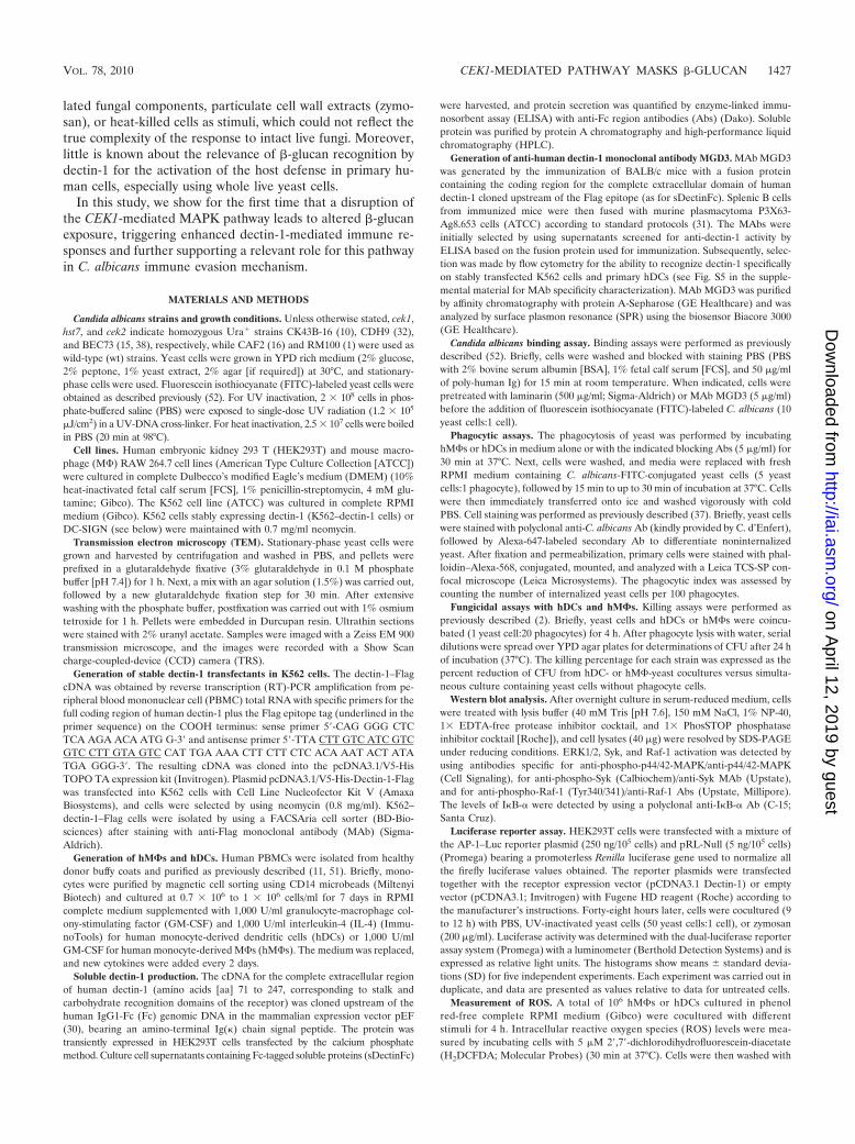
Disruption of the C. albicans CEK1-mediated MAPK path-way alters cell wall morphology. In the fungal pathogen C.albicans, MAPK pathways are mechanisms by which differenttypes of stress (oxidative, temperature, pH, and others) aresensed and an appropriate response is developed (Fig. 1A).The cek1 mutant is defective in the Cek1 MAPK, which par-
ticipates in cell wall construction (15) and becomes activatedduring growth-associated cell wall remodeling (47). In order toidentify cell wall alterations on the cek1 mutant compared towild-type (wt) strain CAF2, transmission electron microscopy(TEM) analysis was initially performed. Although cell wallthicknesses were not significantly different between them (datanot shown), TEM micrographs revealed a less-electron-densecell wall on the cek1 mutant compared to wt strain CAF2 (Fig.1B and C). This differential cell wall density on the cek1 mutantcould account for its hypersensitivity to agents disturbing thecell wall (15) and might modify fungal detection by humanimmune cells.
The C. albicans CEK1-mediated MAPK pathway controls�-glucan exposure and dectin-1-mediated fungal recognition.Considering the C. albicans cell wall structure, we hypothe-sized that the less electron-dense cell wall of cek1 mutants onTEM micrographs could reflect enhanced �-glucan exposure.Flow cytometry measurements revealed that �-glucan expo-sure is significantly higher in live cek1 deletion mutants than inwt CAF2 yeast cells (Fig. 2A). Moreover, the recognition ofthe cek1 mutant by soluble Fc-tagged dectin-1 recombinantprotein (sDectinFc) was also considerably elevated (Fig. 2A),thus demonstrating that a CEK1 mutation leads to enhanced�-glucan exposure and dectin-1 recognition. Assays with heat-killed (HK) fungi, which show increased �-glucan exposure atthe cell surface (20, 58), were used as positive controls (Fig.2A). Confocal microscopy analysis indicated that �-glucan wasfound over the entire surface of the mutant, whereas wt fungishowed a restricted pattern corresponding to the mother-daughter septal regions (20) (Fig. 2B). A similarly increasedlevel of �-glucan staining was seen in the upstream MAPKKHst7 mutant (hst7), further confirming the implication of theCEK1-mediated MAPK route for �-glucan exposure (see Fig.S1A and S1B in the supplemental material). A tridimensionalanalysis of the yeast cell wall revealed an even staining over the
FIG. 1. Candida albicans-MAPK signal transduction pathways and cell wall morphology of C. albicans wild-type CAF2 and the cek1 deletionmutant. (A) The main elements of MAPK signal transduction pathways in C. albicans are schematized, and the physiological function of eachpathway is indicated. The CEK1-mediated MAPK pathway is highlighted. (B and C) Transmission electron micrographs of the cell wall of wt strainCAF2 (B) and the cek1 deletion mutant (C), which lacks Cek1 MAPK. Scale bar, 100 nm.
1428 GALAN-DIEZ ET AL. INFECT. IMMUN.
on April 12, 2019 by guest
http://iai.asm.org/
Dow
nloaded from
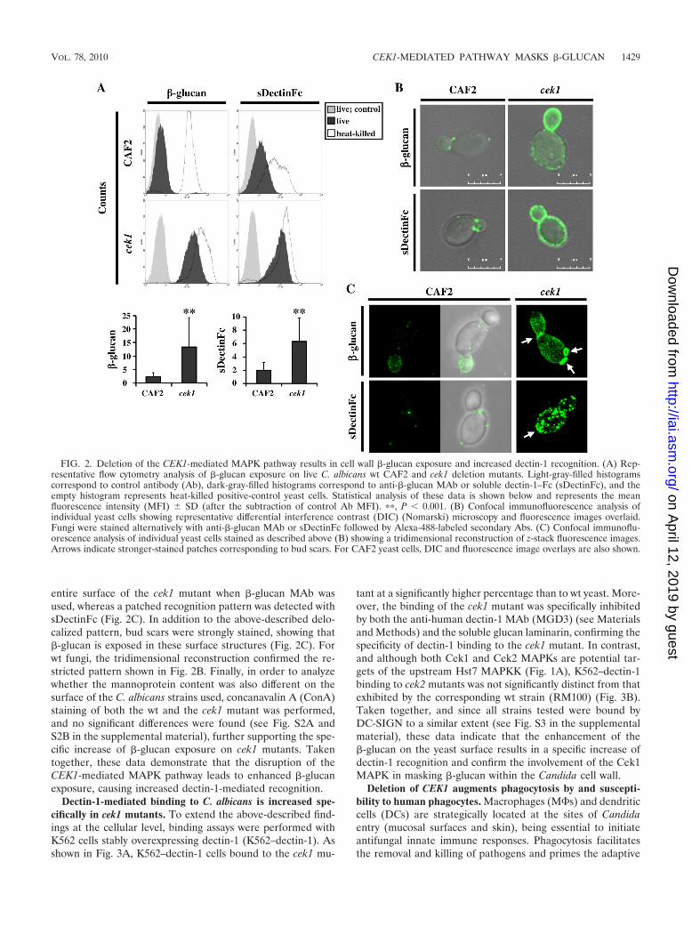
entire surface of the cek1 mutant when �-glucan MAb wasused, whereas a patched recognition pattern was detected withsDectinFc (Fig. 2C). In addition to the above-described delo-calized pattern, bud scars were strongly stained, showing that�-glucan is exposed in these surface structures (Fig. 2C). Forwt fungi, the tridimensional reconstruction confirmed the re-stricted pattern shown in Fig. 2B. Finally, in order to analyzewhether the mannoprotein content was also different on thesurface of the C. albicans strains used, concanavalin A (ConA)staining of both the wt and the cek1 mutant was performed,and no significant differences were found (see Fig. S2A andS2B in the supplemental material), further supporting the spe-cific increase of �-glucan exposure on cek1 mutants. Takentogether, these data demonstrate that the disruption of theCEK1-mediated MAPK pathway leads to enhanced �-glucanexposure, causing increased dectin-1-mediated recognition.
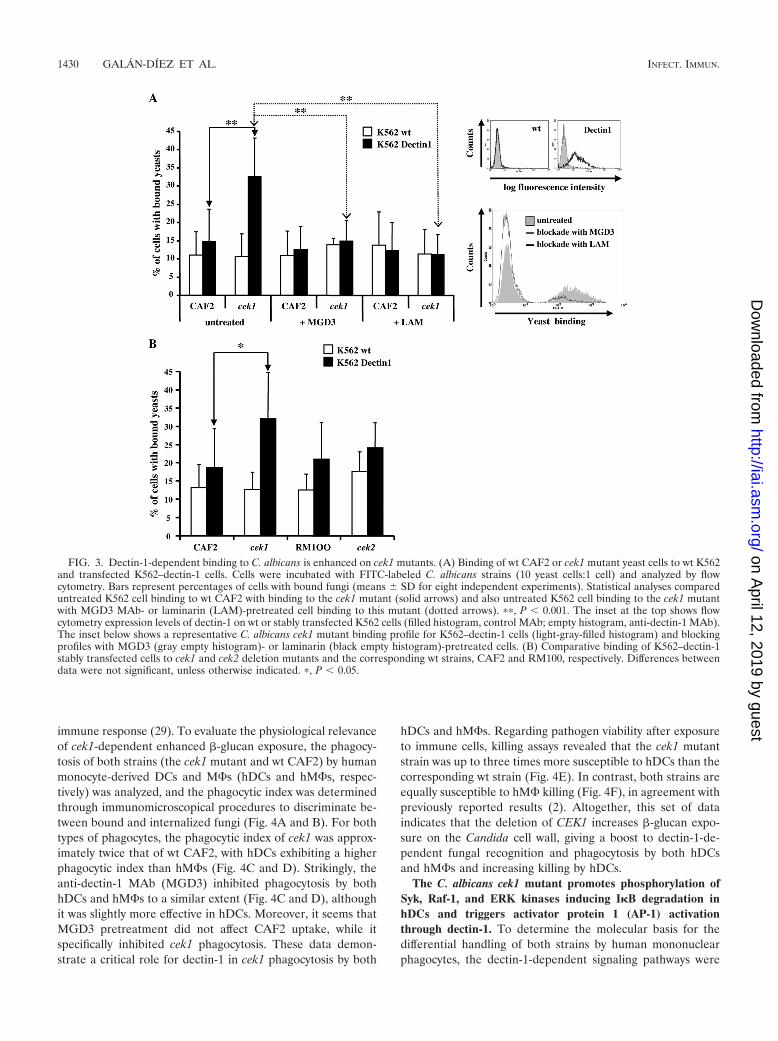
Dectin-1-mediated binding to C. albicans is increased spe-cifically in cek1 mutants. To extend the above-described find-ings at the cellular level, binding assays were performed withK562 cells stably overexpressing dectin-1 (K562–dectin-1). Asshown in Fig. 3A, K562–dectin-1 cells bound to the cek1 mu-
tant at a significantly higher percentage than to wt yeast. More-over, the binding of the cek1 mutant was specifically inhibitedby both the anti-human dectin-1 MAb (MGD3) (see Materialsand Methods) and the soluble glucan laminarin, confirming thespecificity of dectin-1 binding to the cek1 mutant. In contrast,and although both Cek1 and Cek2 MAPKs are potential tar-gets of the upstream Hst7 MAPKK (Fig. 1A), K562–dectin-1binding to cek2 mutants was not significantly distinct from thatexhibited by the corresponding wt strain (RM100) (Fig. 3B).Taken together, and since all strains tested were bound byDC-SIGN to a similar extent (see Fig. S3 in the supplementalmaterial), these data indicate that the enhancement of the�-glucan on the yeast surface results in a specific increase ofdectin-1 recognition and confirm the involvement of the Cek1MAPK in masking �-glucan within the Candida cell wall.
Deletion of CEK1 augments phagocytosis by and suscepti-bility to human phagocytes. Macrophages (M�s) and dendriticcells (DCs) are strategically located at the sites of Candidaentry (mucosal surfaces and skin), being essential to initiateantifungal innate immune responses. Phagocytosis facilitatesthe removal and killing of pathogens and primes the adaptive
FIG. 2. Deletion of the CEK1-mediated MAPK pathway results in cell wall �-glucan exposure and increased dectin-1 recognition. (A) Rep-resentative flow cytometry analysis of �-glucan exposure on live C. albicans wt CAF2 and cek1 deletion mutants. Light-gray-filled histogramscorrespond to control antibody (Ab), dark-gray-filled histograms correspond to anti-�-glucan MAb or soluble dectin-1–Fc (sDectinFc), and theempty histogram represents heat-killed positive-control yeast cells. Statistical analysis of these data is shown below and represents the meanfluorescence intensity (MFI) SD (after the subtraction of control Ab MFI). ��, P 0.001. (B) Confocal immunofluorescence analysis ofindividual yeast cells showing representative differential interference contrast (DIC) (Nomarski) microscopy and fluorescence images overlaid.Fungi were stained alternatively with anti-�-glucan MAb or sDectinFc followed by Alexa-488-labeled secondary Abs. (C) Confocal immunoflu-orescence analysis of individual yeast cells stained as described above (B) showing a tridimensional reconstruction of z-stack fluorescence images.Arrows indicate stronger-stained patches corresponding to bud scars. For CAF2 yeast cells, DIC and fluorescence image overlays are also shown.
VOL. 78, 2010 CEK1-MEDIATED PATHWAY MASKS �-GLUCAN 1429
on April 12, 2019 by guest
http://iai.asm.org/
Dow
nloaded from
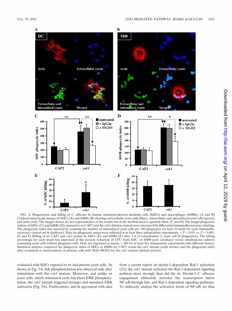
immune response (29). To evaluate the physiological relevanceof cek1-dependent enhanced �-glucan exposure, the phagocy-tosis of both strains (the cek1 mutant and wt CAF2) by humanmonocyte-derived DCs and M�s (hDCs and hM�s, respec-tively) was analyzed, and the phagocytic index was determinedthrough immunomicroscopical procedures to discriminate be-tween bound and internalized fungi (Fig. 4A and B). For bothtypes of phagocytes, the phagocytic index of cek1 was approx-imately twice that of wt CAF2, with hDCs exhibiting a higherphagocytic index than hM�s (Fig. 4C and D). Strikingly, theanti-dectin-1 MAb (MGD3) inhibited phagocytosis by bothhDCs and hM�s to a similar extent (Fig. 4C and D), althoughit was slightly more effective in hDCs. Moreover, it seems thatMGD3 pretreatment did not affect CAF2 uptake, while itspecifically inhibited cek1 phagocytosis. These data demon-strate a critical role for dectin-1 in cek1 phagocytosis by both
hDCs and hM�s. Regarding pathogen viability after exposureto immune cells, killing assays revealed that the cek1 mutantstrain was up to three times more susceptible to hDCs than thecorresponding wt strain (Fig. 4E). In contrast, both strains areequally susceptible to hM� killing (Fig. 4F), in agreement withpreviously reported results (2). Altogether, this set of dataindicates that the deletion of CEK1 increases �-glucan expo-sure on the Candida cell wall, giving a boost to dectin-1-de-pendent fungal recognition and phagocytosis by both hDCsand hM�s and increasing killing by hDCs.
The C. albicans cek1 mutant promotes phosphorylation ofSyk, Raf-1, and ERK kinases inducing I�B degradation inhDCs and triggers activator protein 1 (AP-1) activationthrough dectin-1. To determine the molecular basis for thedifferential handling of both strains by human mononuclearphagocytes, the dectin-1-dependent signaling pathways were
FIG. 3. Dectin-1-dependent binding to C. albicans is enhanced on cek1 mutants. (A) Binding of wt CAF2 or cek1 mutant yeast cells to wt K562and transfected K562–dectin-1 cells. Cells were incubated with FITC-labeled C. albicans strains (10 yeast cells:1 cell) and analyzed by flowcytometry. Bars represent percentages of cells with bound fungi (means SD for eight independent experiments). Statistical analyses compareduntreated K562 cell binding to wt CAF2 with binding to the cek1 mutant (solid arrows) and also untreated K562 cell binding to the cek1 mutantwith MGD3 MAb- or laminarin (LAM)-pretreated cell binding to this mutant (dotted arrows). ��, P 0.001. The inset at the top shows flowcytometry expression levels of dectin-1 on wt or stably transfected K562 cells (filled histogram, control MAb; empty histogram, anti-dectin-1 MAb).The inset below shows a representative C. albicans cek1 mutant binding profile for K562–dectin-1 cells (light-gray-filled histogram) and blockingprofiles with MGD3 (gray empty histogram)- or laminarin (black empty histogram)-pretreated cells. (B) Comparative binding of K562–dectin-1stably transfected cells to cek1 and cek2 deletion mutants and the corresponding wt strains, CAF2 and RM100, respectively. Differences betweendata were not significant, unless otherwise indicated. �, P 0.05.
1430 GALAN-DIEZ ET AL. INFECT. IMMUN.
on April 12, 2019 by guest
http://iai.asm.org/
Dow
nloaded from
evaluated with hDCs exposed to wt and mutant yeast cells. Asshown in Fig. 5A, Syk phosphorylation was observed only afterstimulation with the cek1 mutant. Moreover, and unlike wtyeast cells, which stimulated early but short ERK phosphory-lation, the cek1 mutant triggered stronger and sustained ERKactivation (Fig. 5A). Furthermore, and in agreement with data
from a recent report on dectin-1-dependent Raf-1 activation(23), the cek1 mutant activated the Raf-1-dependent signalingpathway more strongly than did the wt. Dectin-1–C. albicansengagement ultimately activates the transcription factorNF-�B through Syk- and Raf-1-dependent signaling pathways.To indirectly analyze the activation levels of NF-�B, we thus
FIG. 4. Phagocytosis and killing of C. albicans by human monocyte-derived dendritic cells (hDCs) and macrophages (hM�s). (A and B)Confocal microscopy images of hDCs (A) and hM�s (B) showing extracellular yeast cells (blue), extracellular and internalized yeast cells (green),and actin (red). The images shown are not representative of the results but of the method used to quantify them. (C and D) The fungal phagocyticindices of hDCs (C) and hM�s (D) exposed to wt CAF2 and the cek1 deletion mutant were measured by differential immunofluorescence labeling.The phagocytic index was assessed by counting the number of internalized yeast cells per 100 phagocytes (at least 10 fields for each immunoflu-orescence carried out in duplicate). Data for phagocytic assays were collected in at least three independent experiments. �, P 0.05; ��, P 0.001.(E and F) Killing of wt CAF2 and cek1 strains by hDCs (E) and hM�s (F) after 4 h of coincubation (1 yeast cell:20 phagocytes). The killingpercentage for each strain was expressed as the percent reduction of CFU from hDC- or hM�-yeast cocultures versus simultaneous culturescontaining yeast cells without phagocyte cells. Data are expressed as means SD for at least five independent experiments with different donors.Statistical analyses compared the phagocytic index of hDCs or hM�s for CAF2 versus the cek1 mutant (solid arrows) and the phagocytic indexafter treatment or nontreatment of primary cells with MAb MGD3 for the cek1 mutant (dotted arrows).
VOL. 78, 2010 CEK1-MEDIATED PATHWAY MASKS �-GLUCAN 1431
on April 12, 2019 by guest
http://iai.asm.org/
Dow
nloaded from
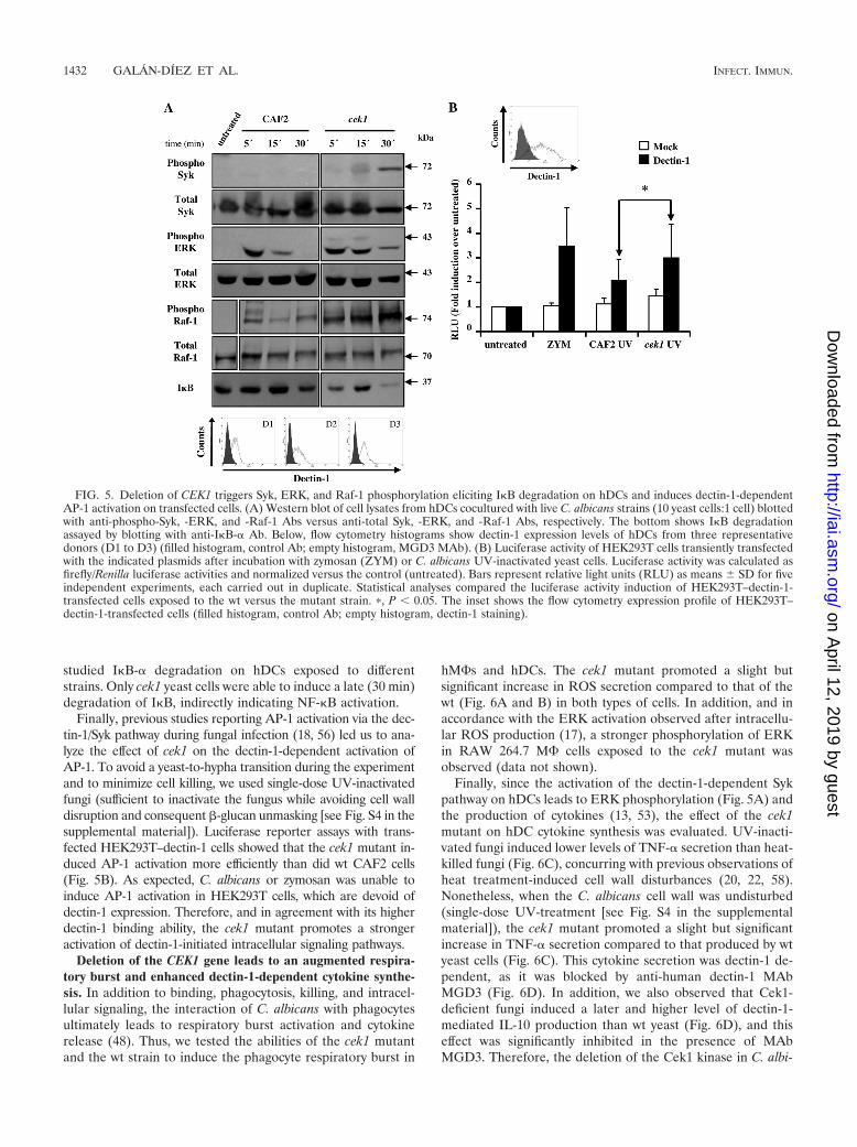
studied I�B-� degradation on hDCs exposed to differentstrains. Only cek1 yeast cells were able to induce a late (30 min)degradation of I�B, indirectly indicating NF-�B activation.
Finally, previous studies reporting AP-1 activation via the dec-tin-1/Syk pathway during fungal infection (18, 56) led us to ana-lyze the effect of cek1 on the dectin-1-dependent activation ofAP-1. To avoid a yeast-to-hypha transition during the experimentand to minimize cell killing, we used single-dose UV-inactivatedfungi (sufficient to inactivate the fungus while avoiding cell walldisruption and consequent �-glucan unmasking [see Fig. S4 in thesupplemental material]). Luciferase reporter assays with trans-fected HEK293T–dectin-1 cells showed that the cek1 mutant in-duced AP-1 activation more efficiently than did wt CAF2 cells(Fig. 5B). As expected, C. albicans or zymosan was unable toinduce AP-1 activation in HEK293T cells, which are devoid ofdectin-1 expression. Therefore, and in agreement with its higherdectin-1 binding ability, the cek1 mutant promotes a strongeractivation of dectin-1-initiated intracellular signaling pathways.
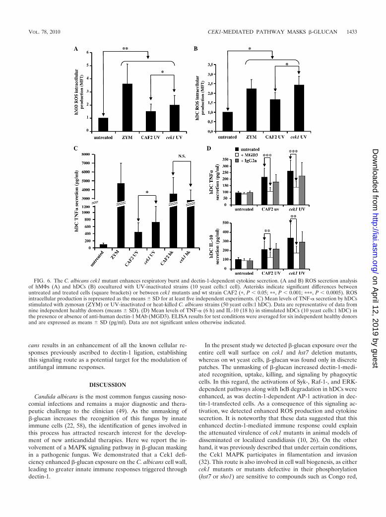
Deletion of the CEK1 gene leads to an augmented respira-tory burst and enhanced dectin-1-dependent cytokine synthe-sis. In addition to binding, phagocytosis, killing, and intracel-lular signaling, the interaction of C. albicans with phagocytesultimately leads to respiratory burst activation and cytokinerelease (48). Thus, we tested the abilities of the cek1 mutantand the wt strain to induce the phagocyte respiratory burst in
hM�s and hDCs. The cek1 mutant promoted a slight butsignificant increase in ROS secretion compared to that of thewt (Fig. 6A and B) in both types of cells. In addition, and inaccordance with the ERK activation observed after intracellu-lar ROS production (17), a stronger phosphorylation of ERKin RAW 264.7 M� cells exposed to the cek1 mutant wasobserved (data not shown).
Finally, since the activation of the dectin-1-dependent Sykpathway on hDCs leads to ERK phosphorylation (Fig. 5A) andthe production of cytokines (13, 53), the effect of the cek1mutant on hDC cytokine synthesis was evaluated. UV-inacti-vated fungi induced lower levels of TNF-� secretion than heat-killed fungi (Fig. 6C), concurring with previous observations ofheat treatment-induced cell wall disturbances (20, 22, 58).Nonetheless, when the C. albicans cell wall was undisturbed(single-dose UV-treatment [see Fig. S4 in the supplementalmaterial]), the cek1 mutant promoted a slight but significantincrease in TNF-� secretion compared to that produced by wtyeast cells (Fig. 6C). This cytokine secretion was dectin-1 de-pendent, as it was blocked by anti-human dectin-1 MAbMGD3 (Fig. 6D). In addition, we also observed that Cek1-deficient fungi induced a later and higher level of dectin-1-mediated IL-10 production than wt yeast (Fig. 6D), and thiseffect was significantly inhibited in the presence of MAbMGD3. Therefore, the deletion of the Cek1 kinase in C. albi-
FIG. 5. Deletion of CEK1 triggers Syk, ERK, and Raf-1 phosphorylation eliciting I�B degradation on hDCs and induces dectin-1-dependentAP-1 activation on transfected cells. (A) Western blot of cell lysates from hDCs cocultured with live C. albicans strains (10 yeast cells:1 cell) blottedwith anti-phospho-Syk, -ERK, and -Raf-1 Abs versus anti-total Syk, -ERK, and -Raf-1 Abs, respectively. The bottom shows I�B degradationassayed by blotting with anti-I�B-� Ab. Below, flow cytometry histograms show dectin-1 expression levels of hDCs from three representativedonors (D1 to D3) (filled histogram, control Ab; empty histogram, MGD3 MAb). (B) Luciferase activity of HEK293T cells transiently transfectedwith the indicated plasmids after incubation with zymosan (ZYM) or C. albicans UV-inactivated yeast cells. Luciferase activity was calculated asfirefly/Renilla luciferase activities and normalized versus the control (untreated). Bars represent relative light units (RLU) as means SD for fiveindependent experiments, each carried out in duplicate. Statistical analyses compared the luciferase activity induction of HEK293T–dectin-1-transfected cells exposed to the wt versus the mutant strain. �, P 0.05. The inset shows the flow cytometry expression profile of HEK293T–dectin-1-transfected cells (filled histogram, control Ab; empty histogram, dectin-1 staining).
1432 GALAN-DIEZ ET AL. INFECT. IMMUN.
on April 12, 2019 by guest
http://iai.asm.org/
Dow
nloaded from
cans results in an enhancement of all the known cellular re-sponses previously ascribed to dectin-1 ligation, establishingthis signaling route as a potential target for the modulation ofantifungal immune responses.
DISCUSSION
Candida albicans is the most common fungus causing noso-comial infections and remains a major diagnostic and thera-peutic challenge to the clinician (49). As the unmasking of�-glucan increases the recognition of this fungus by innateimmune cells (22, 58), the identification of genes involved inthis process has attracted research interest for the develop-ment of new anticandidal therapies. Here we report the in-volvement of a MAPK signaling pathway in �-glucan maskingin a pathogenic fungus. We demonstrated that a Cek1 defi-ciency enhanced �-glucan exposure on the C. albicans cell wall,leading to greater innate immune responses triggered throughdectin-1.
In the present study we detected �-glucan exposure over theentire cell wall surface on cek1 and hst7 deletion mutants,whereas on wt yeast cells, �-glucan was found only in discretepatches. The unmasking of �-glucan increased dectin-1-medi-ated recognition, uptake, killing, and signaling by phagocyticcells. In this regard, the activations of Syk-, Raf-1-, and ERK-dependent pathways along with I�B degradation in hDCs wereenhanced, as was dectin-1-dependent AP-1 activation in dec-tin-1-transfected cells. As a consequence of this signaling ac-tivation, we detected enhanced ROS production and cytokinesecretion. It is noteworthy that these data suggested that thisenhanced dectin-1-mediated immune response could explainthe attenuated virulence of cek1 mutants in animal models ofdisseminated or localized candidiasis (10, 26). On the otherhand, it was previously described that under certain conditions,the Cek1 MAPK participates in filamentation and invasion(32). This route is also involved in cell wall biogenesis, as eithercek1 mutants or mutants defective in their phosphorylation(hst7 or sho1) are sensitive to compounds such as Congo red,
FIG. 6. The C. albicans cek1 mutant enhances respiratory burst and dectin-1-dependent cytokine secretion. (A and B) ROS secretion analysisof hM�s (A) and hDCs (B) cocultured with UV-inactivated strains (10 yeast cells:1 cell). Asterisks indicate significant differences betweenuntreated and treated cells (square brackets) or between cek1 mutants and wt strain CAF2 (�, P 0.05; ��, P 0.001; ���, P 0.0005). ROSintracellular production is represented as the means SD for at least five independent experiments. (C) Mean levels of TNF-� secretion by hDCsstimulated with zymosan (ZYM) or UV-inactivated or heat-killed C. albicans strains (50 yeast cells:1 hDC). Data are representative of data fromnine independent healthy donors (means SD). (D) Mean levels of TNF-� (6 h) and IL-10 (18 h) in stimulated hDCs (10 yeast cells:1 hDC) inthe presence or absence of anti-human dectin-1 MAb (MGD3). ELISA results for test conditions were averaged for six independent healthy donorsand are expressed as means SD (pg/ml). Data are not significant unless otherwise indicated.
VOL. 78, 2010 CEK1-MEDIATED PATHWAY MASKS �-GLUCAN 1433
on April 12, 2019 by guest
http://iai.asm.org/
Dow
nloaded from

calcofluor white, caspofungin, or zymolyase (15, 46, 47). There-fore, it could be hypothesized that the hypersensitivity toagents perturbing the cell wall and the increased exposure of�-glucan shown by the cek1 mutant reflected an altered cellwall organization that could lead to a lower level of resistanceto candidacidal mechanisms. Accordingly, we found these mu-tants to be more susceptible to hDC-mediated uptake andkilling. Moreover, the phagocytic index was notably reduced inthe presence of an anti-dectin-1 MAb, suggesting a major roleof this receptor in cek1 uptake. Outstandingly, although thephagocytic index of the cek1 mutant by hM�s was twice that ofwt CAF2, the viabilities of both strains in response to hM�killing were similar. These monocyte-derived hM�s were notpreviously elicited; therefore, the M� cytocidal function couldbe only partially activated, thus explaining the reduced hM�phagocytic index compared to that of hDCs. Moreover, al-though dectin-1 confers the ability to phagocytose yeasts, thelevel of its expression on the surface of primary hM�s could belower than that on hDCs, since MGD3 blocking is less signif-icant. This lower level of surface expression together with theattenuated cytocidal function may not be sufficient to initiatethe specifically enhanced cek1 killing mechanisms observed forhDCs. In addition, M�s express higher levels of TLRs thanDCs (39) that could mask the specific cek1 killing mediated bydectin-1. The mosaic of PRRs that is expressed by each ofthese cell types ultimately determines the type of responseelicited following the recognition of C. albicans. However, fur-ther research into the specific regulation of yeast-killing mech-anisms will shed light on this. Nonetheless, a CEK1 disruptioncould result in additional phenotypes that cannot be excludedas a cause of their virulence defects. These data allow us tospeculate that dectin-1�/� mice should be more susceptiblethan wt mice to infection with Cek1-deficient strains. However,the outcome of experimental infections is influenced by severalmechanisms, including PRR recognition and cytokine produc-tion, as well as other factors such as adherence to host cells andthe growth rate of the mutants. Further in vivo studies toevaluate the relevance of �-glucan unmasking on the immuneresponse would thus be of interest. Regarding this issue, arecent study by Wheeler et al. (59) showed that �-glucan isprogressively unmasked during infection, further supporting amajor role of dectin-1 in protective antifungal immunity.
Dectin-1 engagement by the cell wall extract zymosan inDCs triggers Syk and ERK pathway activation (13, 44). Re-garding Syk phosphorylation, we observed activation only afterthe stimulation of hDCs with the cek1 mutant. Additionally,cek1-induced ERK activation was sustained in comparison tothat of wt yeast. These data concur with previous observationsshowing ERK phosphorylation coupled to dectin-1/Syk signal-ing (35, 53). We also showed that the cek1 mutant induced theRaf-1 signaling pathway (23) more strongly than the wt. Al-though Syk activation was induced exclusively through dec-tin-1, Raf-1 activation is induced through both dectin-1 andDC-SIGN (24), explaining the baseline activation of hDCsexposed to wt CAF2 cells. Finally, previous studies showed thatdectin-1 activates NF-�B (19, 25) and AP-1 (56) via Syk. It thusappears that the enhanced �-glucan exposure in the cek1 mu-tant induces increased dectin-1 recognition, leading to Syk/ERK/Raf-1 phosphorylation, which triggers NF-�B and AP-1activation and ultimately leads to ROS production and cyto-
kine synthesis. In this regard, the cek1 mutant elicited slightbut significant TNF-� secretion. In addition, significant IL-10production from hDCs was detected, which, at later stages ofthe infection, may be beneficial to resolving an inflammatoryprocess (13, 48). The weak cytokine response elicited by C.albicans wt cells may prevent the recruitment of effector cellsand the elimination of the pathogen, explaining its persistenceas a commensal. Nonetheless, the biological relevance of theslight increase in cytokine synthesis induced by the cek1 mutantis unclear, and this increase alone might not account for theloss of virulence, while the enhanced phagocytosis and killingobserved for hDCs may help the transition from commensal-ism to infection.
During C. albicans infection, both yeast and filamentousforms can be found in infected tissues, and DCs discriminatebetween them, eliciting a protective response against yeast andtolerance to hyphae through different PRRs (14). An inte-grated model of fungal recognition has been proposed, inwhich immune sensing of C. albicans requires the cooperativerecognition of mannans and glucans by PRRs (39). The out-come of an immune response to C. albicans thus depends onthe balance of the signals generated through TLR2, TLR4,dectin-1, MR, and DC-SIGN, among others, each of whichrecognizes a different PAMP of the fungal cell wall. BothTLR2 and TLR4 collaborate with dectin-1 to induce an in-flammatory response (6, 12, 19, 40). Herein we showed thatdectin-1-specific activation by C. albicans occurs only when�-glucan is exposed, concurring with data from previous stud-ies (20, 59). The enhanced uptake, killing, and cytokine syn-thesis observed for hDCs exposed to the cek1 mutant were thusinhibited by dectin-1 MAb MGD3, suggesting a dectin-1-de-pendent elicitation. Nonetheless, we cannot exclude the par-ticipation of other PRRs in this process (8, 9, 21, 24). Furtherstudies are needed to evaluate the complex cross talk betweendectin-1 and other PRRs in immunity to C. albicans, mainly inthe context of the in vivo recognition of intact fungi by primaryhuman immune cells. In this regard, we observed that re-sponses elicited by live or UV-inactivated fungi were weakerthan those triggered by heat-killed yeast (Fig. 6B), indicatingthe need for studies using whole live or suitable UV-inacti-vated C. albicans yeasts to avoid misleading conclusions (seeFig. S4 in the supplemental material). Moreover, convention-ally used isolated fungal components or zymosan could notreflect the true complexity of the response against intact livefungi. Therefore, the preservation of cell wall structure is ofoutstanding importance in elucidating the immune cell-fungusinteraction.
Finally, as we have demonstrated that increased cek1 mutant�-glucan exposure led to enhanced innate immune activation,the CEK1-mediated MAPK pathway can be proposed as avaluable antifungal target. Moreover, drug-induced �-glucanexposure could target fungi for recognition by natural anti-�-glucan antibodies, which are detected in patients with progres-sive fungal infection (42, 55), rendering the pathogen moresusceptible to the host immune system.
In conclusion, the present study demonstrated that theCEK1-mediated MAPK pathway has a key role in �-glucanmasking in C. albicans and that, given the high degree ofconservation in MAPK pathways, this phenomenon may be ageneral mechanism of fungi to evade host recognition. Addi-
1434 GALAN-DIEZ ET AL. INFECT. IMMUN.
on April 12, 2019 by guest
http://iai.asm.org/
Dow
nloaded from
tionally, our study highlights the value of fungal MAPK path-ways as potential therapeutic targets in modulating the hostimmune response to a pathogen.
ACKNOWLEDGMENTS
We thank F. Molina for help in using the confocal microscope, C.Santiago for assistance with soluble dectin-1 protein purification, M.Llorente for help with hybridoma production, M. Martín for biosensorassays, J. Gonzalez for TEM micrographs, and S. Chamorro, I. Olaza-bal, and P. Majano for their critical reading of the manuscript andhelpful discussions.
This work was partially supported by Ministerio de Ciencia e Inno-vacion (MICINN) grants PI05/1999 and PI08/1772, Fundacion de In-vestigacion Medica Mutua Madrilena, to E.F.-R.; Instituto de SaludCarlos III-FEDER, Spanish Network for the Research in InfectiousDiseases, grant REIPI RD06/0008 to E.F.-R. and A.L.-C.; grantsBIO2009-07788 and GEN2006-27775-C2-1-EPAT to J.P. and grantBFU2005-05972 to J.M.C. from the MICINN; and Consejo Superiorde Investigaciones Científicas grant CSIC-2009201016 to L.K. (ProteinTools Unit). M.G.-D. was supported by the Consejería de Educacionde la Comunidad de Madrid and Fondo Social Europeo (FSE) and isunder contract within the Fundacion de Investigacion Biomedica ofthe Hospital Universitario de la Princesa.
REFERENCES
1. Alonso-Monge, R., F. Navarro-Garcia, E. Roman, A. I. Negredo, B. Eisman,C. Nombela, and J. Pla. 2003. The Hog1 mitogen-activated protein kinase isessential in the oxidative stress response and chlamydospore formation inCandida albicans. Eukaryot. Cell 2:351–361.
2. Arana, D. M., R. Alonso-Monge, C. Du, R. Calderone, and J. Pla. 2007.Differential susceptibility of mitogen-activated protein kinase pathway mu-tants to oxidative-mediated killing by phagocytes in the fungal pathogenCandida albicans. Cell. Microbiol. 9:1647–1659.
3. Brown, A. J., and N. A. Gow. 1999. Regulatory networks controlling Candidaalbicans morphogenesis. Trends Microbiol. 7:333–338.
4. Brown, G. D. 2006. Dectin-1: a signalling non-TLR pattern-recognition re-ceptor. Nat. Rev. Immunol. 6:33–43.
5. Brown, G. D., and S. Gordon. 2001. Immune recognition. A new receptor forbeta-glucans. Nature 413:36–37.
6. Brown, G. D., J. Herre, D. L. Williams, J. A. Willment, A. S. Marshall, andS. Gordon. 2003. Dectin-1 mediates the biological effects of beta-glucans. J.Exp. Med. 197:1119–1124.
7. Brown, G. D., P. R. Taylor, D. M. Reid, J. A. Willment, D. L. Williams, L.Martinez-Pomares, S. Y. Wong, and S. Gordon. 2002. Dectin-1 is a majorbeta-glucan receptor on macrophages. J. Exp. Med. 196:407–412.
8. Cambi, A., K. Gijzen, J. M. de Vries, R. Torensma, B. Joosten, G. J. Adema,M. G. Netea, B. J. Kullberg, L. Romani, and C. G. Figdor. 2003. The C-typelectin DC-SIGN (CD209) is an antigen-uptake receptor for Candida albicanson dendritic cells. Eur. J. Immunol. 33:532–538.
9. Cambi, A., M. G. Netea, H. M. Mora-Montes, N. A. Gow, S. V. Hato, D. W.Lowman, B. J. Kullberg, R. Torensma, D. L. Williams, and C. G. Figdor.2008. Dendritic cell interaction with Candida albicans critically depends onN-linked mannan. J. Biol. Chem. 283:20590–20599.
10. Csank, C., K. Schroppel, E. Leberer, D. Harcus, O. Mohamed, S. Meloche,D. Y. Thomas, and M. Whiteway. 1998. Roles of the Candida albicansmitogen-activated protein kinase homolog, Cek1p, in hyphal developmentand systemic candidiasis. Infect. Immun. 66:2713–2721.
11. de la Rosa, G., M. Yanez-Mo, R. Samaneigo, D. Serrano-Gomez, L. Mar-tinez-Munoz, E. Fernandez-Ruiz, N. Longo, F. Sanchez-Madrid, A. L. Corbi,and P. Sanchez-Mateos. 2005. Regulated recruitment of DC-SIGN to cell-cell contact regions during zymosan-induced human dendritic cell aggrega-tion. J. Leukoc. Biol. 77:699–709.
12. Dennehy, K. M., G. Ferwerda, I. Faro-Trindade, E. Pyz, J. A. Willment, P. R.Taylor, A. Kerrigan, S. V. Tsoni, S. Gordon, F. Meyer-Wentrup, G. J. Adema,B. J. Kullberg, E. Schweighoffer, V. Tybulewicz, H. M. Mora-Montes, N. A.Gow, D. L. Williams, M. G. Netea, and G. D. Brown. 2008. Syk kinase isrequired for collaborative cytokine production induced through dectin-1 andToll-like receptors. Eur. J. Immunol. 38:500–506.
13. Dillon, S., S. Agrawal, K. Banerjee, J. Letterio, T. L. Denning, K. Oswald-Richter, D. J. Kasprowicz, K. Kellar, J. Pare, T. van Dyke, S. Ziegler, D.Unutmaz, and B. Pulendran. 2006. Yeast zymosan, a stimulus for TLR2 anddectin-1, induces regulatory antigen-presenting cells and immunological tol-erance. J. Clin. Invest. 116:916–928.
14. d’Ostiani, C. F., G. Del Sero, A. Bacci, C. Montagnoli, A. Spreca, A. Men-cacci, P. Ricciardi-Castagnoli, and L. Romani. 2000. Dendritic cells discrim-inate between yeasts and hyphae of the fungus Candida albicans. Implica-tions for initiation of T helper cell immunity in vitro and in vivo. J. Exp. Med.191:1661–1674.
15. Eisman, B., R. Alonso-Monge, E. Roman, D. Arana, C. Nombela, and J. Pla.2006. The Cek1 and Hog1 mitogen-activated protein kinases play comple-mentary roles in cell wall biogenesis and chlamydospore formation in thefungal pathogen Candida albicans. Eukaryot. Cell 5:347–358.
16. Fonzi, W. A., and M. Y. Irwin. 1993. Isogenic strain construction and genemapping in Candida albicans. Genetics 134:717–728.
17. Forman, H. J., and M. Torres. 2002. Reactive oxygen species and cell sig-naling: respiratory burst in macrophage signaling. Am. J. Respir. Crit. CareMed. 166:S4–S8.
18. Fujioka, S., J. Niu, C. Schmidt, G. M. Sclabas, B. Peng, T. Uwagawa, Z. Li,D. B. Evans, J. L. Abbruzzese, and P. J. Chiao. 2004. NF-kappaB and AP-1connection: mechanism of NF-kappaB-dependent regulation of AP-1 activ-ity. Mol. Cell. Biol. 24:7806–7819.
19. Gantner, B. N., R. M. Simmons, S. J. Canavera, S. Akira, and D. M.Underhill. 2003. Collaborative induction of inflammatory responses by dec-tin-1 and Toll-like receptor 2. J. Exp. Med. 197:1107–1117.
20. Gantner, B. N., R. M. Simmons, and D. M. Underhill. 2005. Dectin-1 me-diates macrophage recognition of Candida albicans yeast but not filaments.EMBO J. 24:1277–1286.
21. Gazi, U., and L. Martinez-Pomares. 2009. Influence of the mannose receptorin host immune responses. Immunobiology 214:554–561.
22. Gow, N. A., M. G. Netea, C. A. Munro, G. Ferwerda, S. Bates, H. M.Mora-Montes, L. Walker, T. Jansen, L. Jacobs, V. Tsoni, G. D. Brown, F. C.Odds, J. W. Van der Meer, A. J. Brown, and B. J. Kullberg. 2007. Immunerecognition of Candida albicans beta-glucan by dectin-1. J. Infect. Dis. 196:1565–1571.
23. Gringhuis, S. I., J. den Dunnen, M. Litjens, M. van der Vlist, B. Wevers,S. C. Bruijns, and T. B. Geijtenbeek. 2009. Dectin-1 directs T helper celldifferentiation by controlling noncanonical NF-kappaB activation throughRaf-1 and Syk. Nat. Immunol. 10:203–213.
24. Gringhuis, S. I., J. den Dunnen, M. Litjens, B. van Het Hof, Y. van Kooyk,and T. B. Geijtenbeek. 2007. C-type lectin DC-SIGN modulates Toll-likereceptor signaling via Raf-1 kinase-dependent acetylation of transcriptionfactor NF-kappaB. Immunity 26:605–616.
25. Gross, O., A. Gewies, K. Finger, M. Schafer, T. Sparwasser, C. Peschel, I.Forster, and J. Ruland. 2006. Card9 controls a non-TLR signalling pathwayfor innate anti-fungal immunity. Nature 442:651–656.
26. Guhad, F. A., H. E. Jensen, B. Aalbaek, C. Csank, O. Mohamed, D. Harcus,D. Y. Thomas, M. Whiteway, and J. Hau. 1998. Mitogen-activated proteinkinase-defective Candida albicans is avirulent in a novel model of localizedmurine candidiasis. FEMS Microbiol. Lett. 166:135–139.
27. Heinsbroek, S. E., P. R. Taylor, F. O. Martinez, L. Martinez-Pomares, G. D.Brown, and S. Gordon. 2008. Stage-specific sampling by pattern recognitionreceptors during Candida albicans phagocytosis. PLoS Pathog. 4:e1000218.
28. Hermanz-Falcon, P., I. Arce, P. Roda-Navarro, and E. Fernandez-Ruiz.2001. Cloning of human DECTIN-1, a novel C-type lectin-like receptor geneexpressed on dendritic cells. Immunogenetics 53:288–295.
29. Herre, J., A. S. Marshall, E. Caron, A. D. Edwards, D. L. Williams, E.Schweighoffer, V. Tybulewicz, C. Reis e Sousa, S. Gordon, and G. D. Brown.2004. Dectin-1 uses novel mechanisms for yeast phagocytosis in macro-phages. Blood 104:4038–4045.
30. Jimenez, D., P. Roda-Navarro, T. A. Springer, and J. M. Casasnovas. 2005.Contribution of N-linked glycans to the conformation and function of inter-cellular adhesion molecules (ICAMs). J. Biol. Chem. 280:5854–5861.
31. Kohler, G., and C. Milstein. 1975. Continuous cultures of fused cells secret-ing antibody of predefined specificity. Nature 256:495–497.
32. Leberer, E., D. Harcus, I. D. Broadbent, K. L. Clark, D. Dignard, K.Ziegelbauer, A. Schmidt, N. A. Gow, A. J. Brown, and D. Y. Thomas. 1996.Signal transduction through homologs of the Ste20p and Ste7p proteinkinases can trigger hyphal formation in the pathogenic fungus Candidaalbicans. Proc. Natl. Acad. Sci. U. S. A. 93:13217–13222.
33. LeibundGut-Landmann, S., O. Gross, M. J. Robinson, F. Osorio, E. C.Slack, S. V. Tsoni, E. Schweighoffer, V. Tybulewicz, G. D. Brown, J. Ruland,and C. Reis e Sousa. 2007. Syk- and CARD9-dependent coupling of innateimmunity to the induction of T helper cells that produce interleukin 17. Nat.Immunol. 8:630–638.
34. Medzhitov, R., and C. A. Janeway, Jr. 2002. Decoding the patterns of selfand nonself by the innate immune system. Science 296:298–300.
35. Meyer-Wentrup, F., A. Cambi, G. J. Adema, and C. G. Figdor. 2005. “Sweettalk”: closing in on C type lectin signaling. Immunity 22:399–400.
36. Meyer-Wentrup, F., C. G. Figdor, M. Ansems, P. Brossart, M. D. Wright,G. J. Adema, and A. B. van Spriel. 2007. Dectin-1 interaction with tet-raspanin CD37 inhibits IL-6 production. J. Immunol. 178:154–162.
37. Moreno-Ruiz, E., M. Galan-Diez, W. Zhu, E. Fernandez-Ruiz, C. d’Enfert,S. G. Filler, P. Cossart, and E. Veiga. 2009. Candida albicans internalizationby host cells is mediated by a clathrin-dependent mechanism. Cell. Micro-biol. 11:1179–1189.
38. Navarro-García, F., B. Eisman, S. M. Fiuza, C. Nombela, and J. Pla. 2005.The MAP kinase Mkc1p is activated under different stress conditions inCandida albicans. Microbiology 151:2737–2749.
39. Netea, M. G., G. D. Brown, B. J. Kullberg, and N. A. Gow. 2008. An
VOL. 78, 2010 CEK1-MEDIATED PATHWAY MASKS �-GLUCAN 1435
on April 12, 2019 by guest
http://iai.asm.org/
Dow
nloaded from
integrated model of the recognition of Candida albicans by the innate im-mune system. Nat. Rev. Microbiol. 6:67–78.
40. Netea, M. G., N. A. Gow, C. A. Munro, S. Bates, C. Collins, G. Ferwerda,R. P. Hobson, G. Bertram, H. B. Hughes, T. Jansen, L. Jacobs, E. T.Buurman, K. Gijzen, D. L. Williams, R. Torensma, A. McKinnon, D. M.MacCallum, F. C. Odds, J. W. Van der Meer, A. J. Brown, and B. J.Kullberg. 2006. Immune sensing of Candida albicans requires cooperativerecognition of mannans and glucans by lectin and Toll-like receptors. J. Clin.Invest. 116:1642–1650.
41. Odds, F. C. 1987. Candida infections: an overview. Crit. Rev. Microbiol.15:1–5.
42. Poulain, D., and T. Jouault. 2004. Candida albicans cell wall glycans, hostreceptors and responses: elements for a decisive crosstalk. Curr. Opin. Mi-crobiol. 7:342–349.
43. Reid, D. M., N. A. Gow, and G. D. Brown. 2009. Pattern recognition: recentinsights from dectin-1. Curr. Opin. Immunol. 21:30–37.
44. Rogers, N. C., E. C. Slack, A. D. Edwards, M. A. Nolte, O. Schulz, E.Schweighoffer, D. L. Williams, S. Gordon, V. L. Tybulewicz, G. D. Brown,and C. Reis e Sousa. 2005. Syk-dependent cytokine induction by dectin-1reveals a novel pattern recognition pathway for C type lectins. Immunity22:507–517.
45. Roman, E., D. M. Arana, C. Nombela, R. Alonso-Monge, and J. Pla. 2007.MAP kinase pathways as regulators of fungal virulence. Trends Microbiol.15:181–190.
46. Roman, E., F. Cottier, J. F. Ernst, and J. Pla. 2009. The Msb2 signalingmucin controls activation of Cek1 mitogen-activated protein kinase in Can-dida albicans. Eukaryot. Cell 8:1235–1249.
47. Roman, E., C. Nombela, and J. Pla. 2005. The Sho1 adaptor protein linksoxidative stress to morphogenesis and cell wall biosynthesis in the fungalpathogen Candida albicans. Mol. Cell. Biol. 25:10611–10627.
48. Romani, L. 2004. Immunity to fungal infections. Nat. Rev. Immunol. 4:1–23.49. Ruhnke, M. 2006. Epidemiology of Candida albicans infections and role of
non-Candida-albicans yeasts. Curr. Drug Targets 7:495–504.50. Saijo, S., N. Fujikado, T. Furuta, S. H. Chung, H. Kotaki, K. Seki, K. Sudo,
S. Akira, Y. Adachi, N. Ohno, T. Kinjo, K. Nakamura, K. Kawakami, and Y.
Iwakura. 2007. Dectin-1 is required for host defense against Pneumocystiscarinii but not against Candida albicans. Nat. Immunol. 8:39–46.
51. Sallusto, F., and A. Lanzavecchia. 1994. Efficient presentation of solubleantigen by cultured human dendritic cells is maintained by granulocyte/macrophage colony-stimulating factor plus interleukin 4 and downregulatedby tumor necrosis factor alpha. J. Exp. Med. 179:1109–1118.
52. Serrano-Gomez, D., R. T. Martinez-Nunez, E. Sierra-Filardi, N. Izquierdo,M. Colmenares, J. Pla, L. Rivas, J. Martinez-Picado, J. Jimenez-Barbero,J. L. Alonso-Lebrero, S. Gonzalez, and A. L. Corbi. 2007. AM3 modulatesdendritic cell pathogen recognition capabilities by targeting DC-SIGN. An-timicrob. Agents Chemother. 51:2313–2323.
53. Slack, E. C., M. J. Robinson, P. Hernanz-Falcon, G. D. Brown, D. L. Wil-liams, E. Schweighoffer, V. L. Tybulewicz, and C. Reis e Sousa. 2007. Syk-dependent ERK activation regulates IL-2 and IL-10 production by DCstimulated with zymosan. Eur. J. Immunol. 37:1600–1612.
54. Taylor, P. R., S. V. Tsoni, J. A. Willment, K. M. Dennehy, M. Rosas, H.Findon, K. Haynes, C. Steele, M. Botto, S. Gordon, and G. D. Brown. 2007.Dectin-1 is required for beta-glucan recognition and control of fungal infec-tion. Nat. Immunol. 8:31–38.
55. Torosantucci, A., C. Bromuro, P. Chiani, F. De Bernardis, F. Berti, C. Galli,F. Norelli, C. Bellucci, L. Polonelli, P. Costantino, R. Rappuoli, and A.Cassone. 2005. A novel glyco-conjugate vaccine against fungal pathogens. J.Exp. Med. 202:597–606.
56. Toyotome, T., Y. Adachi, A. Watanabe, E. Ochiai, N. Ohno, and K. Kamei.2008. Activator protein 1 is triggered by Aspergillus fumigatus beta-glucanssurface-exposed during specific growth stages. Microb. Pathog. 44:141–150.
57. Underhill, D. M., E. Rossnagle, C. A. Lowell, and R. M. Simmons. 2005.Dectin-1 activates Syk tyrosine kinase in a dynamic subset of macrophagesfor reactive oxygen production. Blood 106:2543–2550.
58. Wheeler, R. T., and G. R. Fink. 2006. A drug-sensitive genetic network masksfungi from the immune system. PLoS Pathog. 2:e35.
59. Wheeler, R. T., D. Kombe, S. D. Agarwala, and G. R. Fink. 2008. Dynamic,morphotype-specific Candida albicans beta-glucan exposure during infectionand drug treatment. PLoS Pathog. 4:e1000227.
Editor: G. S. Deepe, Jr.
1436 GALAN-DIEZ ET AL. INFECT. IMMUN.
on April 12, 2019 by guest
http://iai.asm.org/
Dow
nloaded from


![Candida albicans -Glucan Exposure Is Controlled by the ... · mediated pathway by the deletion of the CEK1 (Candida albicans extracellular signal-regulated kinase [ERK]-like 1) MAPK](https://img.dokumen.tips/doc/110x75/5cc532cc88c9936d678d3eab/candida-albicans-glucan-exposure-is-controlled-by-the-mediated-pathway.jpg)