Embed Size (px)
Citation preview
Hindawi Publishing CorporationJournal of Biomedicine and BiotechnologyVolume 2012, Article ID 782642, 10 pagesdoi:10.1155/2012/782642
Research Article
Camel Milk Modulates the Expression of Aryl HydrocarbonReceptor-Regulated Genes, Cyp1a1, Nqo1, and Gsta1, in Murinehepatoma Hepa 1c1c7 Cells
Hesham M. Korashy,1 Mohamed A. M. El Gendy,2
Abdulqader A. Alhaider,3 and Ayman O. El-Kadi2
1 Department of Pharmacology and Toxicology, College of Pharmacy, King Saud University, 11451 Riyadh, Saudi Arabia2 Faculty of Pharmacy & Pharmaceutical Sciences, University of Alberta, Edmonton, AB, Canada T6G 2E13 Department of Pharmacology, College of Medicine, King Saud University, 11461 Riyadh, Saudi Arabia
Correspondence should be addressed to Ayman O. El-Kadi, [email protected]
Received 6 September 2011; Revised 24 October 2011; Accepted 8 November 2011
Academic Editor: Ikhlas A. Khan
Copyright © 2012 Hesham M. Korashy et al. This is an open access article distributed under the Creative Commons AttributionLicense, which permits unrestricted use, distribution, and reproduction in any medium, provided the original work is properlycited.
There is a traditional belief in the Middle East that camel milk may aid in prevention and treatment of numerous cases of cancer yet,the exact mechanism was not investigated. Therefore, we examined the ability of camel milk to modulate the expression of a well-known cancer-activating gene, Cytochrome P450 1a1 (Cyp1a1), and cancer-protective genes, NAD(P)H:quinone oxidoreductase1 (Nqo1) and glutathione S-transferase a1 (Gsta1), in murine hepatoma Hepa 1c1c7 cell line. Our results showed that camel milksignificantly inhibited the induction of Cyp1a1 gene expression by 2,3,7,8-tetrachlorodibenzo-p-dioxin (TCDD), the most potentCyp1a1 inducer and known carcinogenic chemical, at mRNA, protein, and activity levels in a concentration-dependent manner.In addition, camel milk significantly decreased the xenobiotic responsive element (XRE)-dependent luciferase activity, suggestinga transcriptional mechanism is involved. Furthermore, this inhibitory effect of camel milk was associated with a proportionalincrease in heme oxygenase 1. On the other hand, camel milk significantly induced Nqo1 and Gsta1 mRNA expression level in aconcentration-dependent fashion. The RNA synthesis inhibitor, actinomycin D, completely blocked the induction of Nqo1 mRNAby camel milk suggesting the requirement of de novo RNA synthesis through a transcriptional mechanism. In conclusion, camelmilk modulates the expression of Cyp1a1, Nqo1, and Gsta1 at the transcriptional and posttranscriptional levels.
1. Introduction
The aryl hydrocarbon receptor (AhR), a cytosolic ligand-activated transcriptional factor, belongs to the basic-helix-loop-helix (bHLH)/Per-ARNT-Sim (PAS) family of tran-scription proteins, which are involved in regulation ofcell differentiation and proliferation [1, 2]. Mechanistically,AhR is located in the cytoplasm bound with heat shockprotein-90 (HSP90) and AhR interacting protein (AIP)forming inactive complex. Activation of AhR upon bindingwith its ligands, such as 2,3,7,8-tetrachlorodibenzo-p-dioxin(TCDD), a polycyclic aromatic hydrocarbon (PAH), causesdissociation of HSP90 and AIP from the activated ligand-receptor complex, subsequently leading to translocation of
the complex into nucleus. In the nucleus, the ligand-receptorcomplex dimerizes with AhR nuclear translocator (ARNT),which subsequently binds to xenobiotic-responsive element(XRE) located in the promoter region of so-called AhR-regulated genes resulting in promoting its transcription andprotein translation processes [3, 4].
The AhR-regulated gene batteries include phase I xeno-biotic metabolizing enzymes such as the cytochrome P4501A1 (CYP1A1), CYP1A2, CYP1B1 and phase II enzymessuch as NAD(P)H:quinone oxidoreductase 1 (NQO1), glu-tathione S-transferase A1 (GSTA1), uridine diphosphate glu-curonosyltransferase 1A6, and aldehyde dehydrogenase-3[3, 4]. Among these genes, CYP1A1 plays a particular rolein bioactivating procarcinogens into carcinogen and toxic
2 Journal of Biomedicine and Biotechnology
metabolites and hence is considered as cancer-activatinggene [5], whereas the NQO1 and GSTA1 catalyze reductionof several environmental contaminants and endogenouscompounds that maintain endogenous antioxidants, suchas ubiquinone and vitamin E, to protect tissues againstmutagens, carcinogens, and oxidative stress damage [6, 7].Accordingly, one of the strategies for protecting humancells and tissues from the toxic effects of carcinogenic andcytotoxic metabolites generally include attenuation of thecarcinogen-activating genes, CYP1A1 signaling pathways,and/or enhancing the adaptive mechanisms by increasing theexpression of detoxification and antioxidant genes, such asNQO1 and GSTA1 [8].
Chemoprevention by dietary constituents in the formof functional food has a well-established beneficial role inhealth promotion and emerged as a novel approach tocontrol cancers [9]. Camel’s milk is different from otherruminant milk, having low cholesterol, lactoferrin, low sugar,high minerals (sodium, potassium, iron, copper, zinc andmagnesium), high vitamin C, B2, A, and E, low protein,and high concentrations of insulin. Recent studies havereported that camel milk is the most active milk among otherspecies against E. coli, Staphylococcus aureus, Salmonellatyphimurium, and rotavirus [10, 11]. In addition, it hasbeen demonstrated that milk, in addition to secretory IgAand IgM, also contains numerous nonantibody compo-nents that possess antiviral activity, including lactoferrin[10].
Until recently, it is traditionally claimed that drinkingcamel milk may help to fight against serious diseases andcure numerous cases of cancer; however, this claim has neverbeen exposed to scientific scrutiny investigation. Therefore,the main objective of the current study was to explore thecapacity of camel milk to modulate the expression of Cyp1a1,Nqo1, and Gsta1 genes as target for cancer prevention inmurine hepatoma Hepa 1c1c7 cells.
2. Materials and Methods
2.1. Materials. 7-Ethoxyresorufin (7ER), Dulbecco’s Modi-fied Eagle’s Medium (DMEM), protease inhibitor cocktail,sulforaphane (SNF), resveratrol (RES), 3-(4,5-dimethyl-thiazol-2-yl)-2,5-diphenyltetrazolium bromide (MTT), andrabbit anti-goat IgG peroxidase secondary antibody werepurchased from Sigma-Aldrich (St. Louis, MO). 2,3,7,8-Tetrachlorodibenzo-p-dioxin, >99% pure, was purchasedfrom Cambridge Isotope Laboratories (Woburn, MA).Resorufin and 100 × vitamin supplements were purchasedfrom ICN Biomedicals Canada (Montreal, QC). TRIzoland Lipofectamine 2000 reagents were purchased fromInvitrogen (Carlsbad, CA). The High-Capacity cDNA reversetranscription kit and SYBR Green PCR Master Mix werepurchased from Applied Biosystems (Foster City, CA).Actinomycin D (Act-D) was purchased from Calbiochem(San Diego, CA). Dual-Luciferase Reporter Assay Systemwas obtained from Promega Corporation (Madison, WI).Chemiluminescence Western blotting detection reagentswere obtained from GE Healthcare Life Sciences (Piscataway,
NJ). Nitrocellulose membrane was purchased from Bio-Rad Laboratories (Hercules, CA). Cyp1a1 goat anti-mousepolyclonal primary antibody, glyceraldehyde-3-phosphatedehydrogenase (Gapdh) rabbit anti-goat polyclonal anti-body, and anti-rabbit IgG peroxidase secondary antibodieswere purchased from Santa Cruz Biotechnology (SantaCruz, CA). All other chemicals were purchased from FisherScientific Co. (Toronto, ON).
2.2. Milk Sample Collection and Preparation. Camel milkwas collected aseptically from five healthy domestic camels(Camelus dromedaries). The camel milk was collected fromfarm and desert living animals. The collection of milkwas usually conducted during the feeding time and wasperformed by experienced attendants. Milk was allowed toflow directly into sterile stainless steel containers and thentransferred to glass vials. Camel samples were transported tothe laboratory as soon as practical (<4 h) and were frozenat −80◦C. Aqueous portion (fat-free, skimmed milk) wasremoved from the lipids (cream) as described before [12].Briefly, aliquots of pooled milk samples were centrifuged at1400×g for 30 minutes at 4◦C, thereafter, the creamy layerconsisting largely of fat was removed by filtration through aglass wool plug in a Pasteur pipette. Camel milk was collectedand kept in −80◦C freezer until use.
2.3. Cell Culture and Treatments. Murine hepatoma Hepa1c1c7 cells (American Type Culture Collection, Manassas,VA) were maintained in DMEM, without phenol red supple-mented with 10% heat-inactivated fetal bovine serum, 20 μML-glutamine, 100 IU/mL penicillin, 10 μg/mL streptomycin,0.1 mM nonessential amino acids, and vitamin supplementsolution. Cells were grown in 75 cm2 tissue culture flasks at37◦C under a 5% CO2 humidified environment.
Hepa 1c1c7 cells were plated onto 96- and six-well cellculture plates in DMEM culture media for the mRNA,protein, and catalytic activity assays. In all experiments, thecells were pretreated for indicated time interval in serum-free media with various concentrations of camel milk in thepresence or absence of TCDD as indicated. Stock solutionsof TCDD were prepared in dimethyl sulfoxide (DMSO) andstored at −20◦C, in which the concentration of DMSO didnot exceed 0.05% (v/v).
2.4. Cytotoxicity of Camel Milk. The effects of camel milk(fat-free) on Hepa 1c1c7 cell viability were determined bymeasuring the capacity of reducing enzymes present in viablecells to convert MTT salt to formazan crystals as describedpreviously [13]. Twenty-four hours after incubating the cellswith the tested milk in the presence and absence of TCDDin a 96-well cell culture plate at 37◦C under a 4% CO2
humidified incubator, the media were removed and a 100 μLof serum-free medium containing 1.2 mM of MTT dissolvedin phosphate-buffered-saline (PBS), pH 7.4, was added toeach well. The plate was then incubated in a CO2 incubator at37◦C for 2 h. The media were then decanted off by invertingthe plate, and a 100 μL of isopropyl alcohol was added toeach well, with shaking for 1 h to dissolve the formazan
Journal of Biomedicine and Biotechnology 3
Table 1: Primers sequences used for Real-Time PCR reactions.
Gene Forward primer Reverse primer
Cyp1a1 5′-GGT TAA CCA TGA CCG GGA ACT-3′ 5′-TGC CCA AAC CAA AGA GAG TGA-3′Ho-1 5′-GTG ATG GAG CGT CCA CAG C-3′ 5′-TGG TGG CCT CCT TCA AGG-3′Nqo1 5′-GGA AGC TGC AGA CCT GGT GA-3′ 5′-CCT TTC AGA ATG GCT GGC A-3′Gsta1 5′-CCC CTT TCC CTC TGC TGA AG-3′ 5′-TGC AGC TTC ACT GAA TCT TGA AAG-3′β-actin 5′-TAT TGG CAA CGA GCG GTT CC-3′ 5′-GGC ATA GAG GTC TTT ACG GAT GTC-3′
crystals. The color intensity in each well was measured atwavelength of 550 using BIO-TEK Instruments EL 312emicroplate reader, Bio-Tek Instruments (Winooski, VT). Thepercentage of cell viability was calculated relative to controlwells designated as 100% viable cells.
2.5. Determination of Cyp1a1 Enzymatic Activity. Cyp1a1-dependent 7-ethoxyresorufin (7ER) O-deethylase (EROD)activity was performed on intact living Hepa 1c1c7 cellsusing 7ER as a substrate [14]. After incubation of the cellswith increasing concentrations of fat-free camel milk for24 h, media were aspirated and the cell monolayers wererinsed with PBS. Thereafter, 100 μL of 2 μM 7ER in assaybuffer (0.05 M Tris, 0.1 M NaCl, pH 7.8) was then addedto each well. Immediately, an initial fluorescence measure-ment (t = 0) at excitation/emission (545 nm/575 nm) wasrecorded from each well using Baxter 96-well fluorometer(Deerfield, IL). The plates were then replaced in the incu-bator, and additional set of fluorescence measurements ofthe wells were recorded every 5 min for 20 min interval. Theamount of resorufin formed in each well was determined bycomparison with a standard curve of known concentrations.The working solution was then aspirated, the cells wererinsed twice with PBS, and 50 μl of double de-ionized waterwas added to lyse the cells. After placing of the cell plates at−80◦C for 30 min, the cell lysates were allowed to thaw, andprotein levels were determined using a modified fluorescentassay [15]. The rate of resorufin formation was expressed aspmol/min/mg protein.
2.6. RNA Extraction and cDNA Synthesis. Total RNA wasisolated using TRIzol reagent (Invitrogen) according to themanufacturer’s instructions and quantified by measuringthe absorbance at 260 nm. RNA quality was determined bymeasuring the 260/280 ratio. Thereafter, first strand cDNAwas synthesized using the High-Capacity cDNA reversetranscription kit (Applied Biosystems) according to themanufacturer’s instructions. Briefly, 1 μg of total RNA fromeach sample was added to a mix of 2.0 μL of 10x reversetranscriptase buffer, 0.8 μL of 25x dNTP mix (100 mM),2.0 μL of 10x reverse transcriptase random primers, 1.0 μL ofMultiScribe reverse transcriptase, and 3.2 μL of nuclease-freewater. The final reaction mix was kept at 25◦C for 10 min,heated to 37◦C for 120 min, heated for 85◦C for 5 s, andfinally cooled to 4◦C [16].
2.7. Quantification of mRNA Expression by Real-Time Poly-merase Chain Reaction (RT-PCR). Quantitative analysis of
specific mRNA expression was performed by RT-PCR bysubjecting the resulting cDNA to PCR amplification using96-well optical reaction plates in the ABI Prism 7500 System(Applied Biosystems). The 25 μL reaction mix contained0.1 μL of 10 μM forward primer and 0.1 μL of 10 μM reverseprimer (40 nM final concentration of each primer), 12.5 μLof SYBR Green Universal Master Mix, 11.15 μL of nuclease-free water, and 1.25 μL of cDNA sample. The primers usedin the current study (Table 1) [17, 18] were purchased fromIntegrated DNA Technologies (IDT, Coralville, IA). Assaycontrols were incorporated onto the same plate, namely,no-template controls to test for the contamination of anyassay reagents. The RT-PCR data was analyzed using therelative gene expression (i.e., ΔΔCT) method, as describedin Applied Biosystems User Bulletin [19]. The data arepresented as the fold change in gene expression normalizedto the endogenous housekeeping gene (β-actin) and wasdetermined using the equation: fold change = 2−Δ(ΔCt), whereΔCt = Ct(target) − Ct(β-actin) and Δ(ΔCt) = ΔCt(treated) −ΔCt(untreated).
2.8. Protein Extraction and Western Blot Analysis. Twenty-four hours after incubating the cells with increasingconcentrations of camel milk (fat-free), the cells werewashed once with cold PBS and collected by scrapingin 100 μL of lysis buffer (50 mM HEPES, 0.5 M NaCl,1.5 mM MgCl2, 1 mM EDTA, 10% (v/v) glycerol, 1%Triton X-100, and 5 μL/mL of protease inhibitor cocktail).The lysates were incubated on ice for 1 h with intermit-tent vortexing every 10 min, followed by centrifugation at12,000×g for 10 min at 4◦C. The supernatant was thenstored at a −80◦C freezer for later use in the Western blotanalysis.
Western blot analysis was performed as described pre-viously [20]. For Cyp1a1 immuno-detection, 30 μg of pro-teins from each treatment group were diluted with sameamount (1 : 1) of 2X loading buffer (0.1 M Tris-HCl, pH6.8, 4% SDS, 1.5% bromophenol blue, 20% glycerol, 5% β-mercaptoethanol), boiled and loaded onto a 10% SDS-polyacrylamide gel. Samples were electrophoresed at 120 Vfor 2 h, and the separated proteins were transferred toTrans-Blot nitrocellulose membrane (0.45 μm) in a buffercontaining 25 mM Tri-HCl, 192 mM glycine, and 20% (v/v)methanol. Protein blots were blocked overnight at 4◦C ina solution containing 5% skim milk powder, 2% bovineserum albumin, and 0.5% Tween-20 in TBS solution (0.15 MNaCl, 3 mM KCl, 25 mM Tris-base). Thereafter, the blockingsolution was removed and the blots were rinsed three times
4 Journal of Biomedicine and Biotechnology
in a wash buffer (0.1% Tween-20 in TBS). Proteins weredetected by incubation with a primary polyclonal goat anti-mouse Cyp1a1 antibody for 2 h at 4◦C in TBS containing0.01% sodium azide and 0.05% Tween-20. The primaryantibody solution was removed and blots were rinsed threetimes with a wash buffer, followed by incubation withhorseradish peroxidase-conjugate rabbit anti-goat secondaryantibody for 1 h at room temperature followed by washingas previously described. Antibody detection was performedusing the enhanced chemiluminescence method. The inten-sity of Cyp1a1 bands was quantified, relative to the signalsobtained for Gapdh, using Java-based image-processingsoftware, ImageJ (W. Rasband (2005) National Institutes ofHealth, Bethesda, MD, http://rsb.info.nih.gov/ij/).
2.9. Transient Transfection and Luciferase Assay. Transienttransfection and luciferase assay were carried out as de-scribed previously [21]. Briefly, Hepa 1c1c7 cells (3 × 10⁴
cells per well) were plated onto 12-well cell culture plates.Each well was cotransfected with 1.5 μg of XRE-drivenluciferase reporter plasmid pGudLuc 1.1 and 0.1 μg of therenilla luciferase pRL-CMV vector, used for normalization.The pGudLuc 1.1 plasmid was provided as a gift fromDr. Michael S. Denison (University of California, Davis,CA), while pRL-CMV vector was obtained from PromegaCorporation (Madison,WI). Transfection procedure was car-ried out using Lipofectamine 2000 reagent according to themanufacturer’s instructions (Invitrogen), the luciferase assaywas performed according to the manufacturer’s instructions(Promega), and luciferase activity was reported as relativelight unit of firefly luciferase to renilla luciferase (Fluc/Rluc).
2.10. Statistical Analysis. All results are presented as mean ±SEM. The comparative analysis of the results from variousexperimental groups with their corresponding controls wasperformed using SigmaStat for Windows, Systat SoftwareInc., (San Jose, CA). One-way analysis of variance (ANOVA)followed by Student-Newman-Keul’s test was carried out toassess the significant difference between different groups.The differences were considered significant when P < 0.05.
3. Results
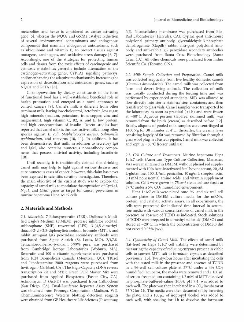
3.1. Effect of Camel Milk on Hepa 1c1c7 Cell Viability. Todetermine the cellular toxicity effects of camel milk (fat-free) in the presence and absence of TCDD on Hepa 1c1c7,cells were treated for 24 h with increasing concentrationsof camel milk (0, 15, 25, 50, 100, and 200 μL/mL) in thepresence and absence of TCDD (1 nM) and the cell viabilitywas determined by MTT assay. Figure 1 shows that neithercamel milk alone nor with TCDD were toxic to Hepa 1c1c7cells at all concentrations of camel milk used, with theexception of mixture of TCDD plus camel milk (200 μL/mL)that significantly decreased cell viability by approximately25%. Based on these results, concentrations of 0, 25, and100 μL/mL of fat-free camel milk in the presence andabsence of TCDD were chosen to be used in the subsequentexperiments.
00
20
15 25 50
40
60
80
100
100 200
120
Camel milk
Camel milk (µL/mL)
Camel milk +
+
TCDD
Cel
l via
bilit
y(%
of
con
trol
)
Figure 1: Effect of camel milk on Hepa 1c1c7 cell viability. Hepa1c1c7 cells were incubated for 24 h with various concentrationsof fat-free camel milk (0, 15, 25, 50, 100, and 200 μL/mL) in thepresence and absence of TCDD (1 nM). Thereafter, cell viability wasassessed using the MTT assay. Values are presented as percentageof the control (mean ± SEM, n = 8). ∗P < 0.05 compared withcontrol (concentration = 0 μL/mL, nontreated cells).
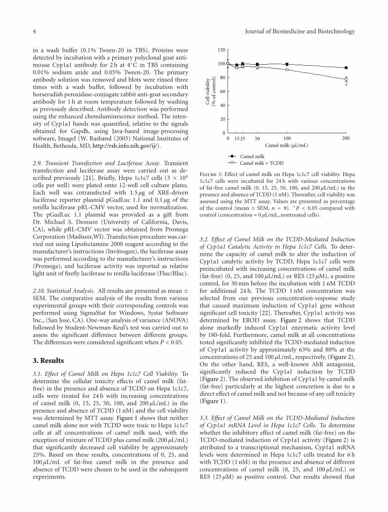
3.2. Effect of Camel Milk on the TCDD-Mediated Inductionof Cyp1a1 Catalytic Activity in Hepa 1c1c7 Cells. To deter-mine the capacity of camel milk to alter the induction ofCyp1a1 catalytic activity by TCDD, Hepa 1c1c7 cells werepreincubated with increasing concentrations of camel milk(fat-free) (0, 25, and 100 μL/mL) or RES (25 μM), a positivecontrol, for 30 min before the incubation with 1 nM TCDDfor additional 24 h. The TCDD 1 nM concentration wasselected from our previous concentration-response studythat caused maximum induction of Cyp1a1 gene withoutsignificant cell toxicity [22]. Thereafter, Cyp1a1 activity wasdetermined by EROD assay. Figure 2 shows that TCDDalone markedly induced Cyp1a1 enzymatic activity levelby 180-fold. Furthermore, camel milk at all concentrationstested significantly inhibited the TCDD-mediated inductionof Cyp1a1 activity by approximately 63% and 80% at theconcentrations of 25 and 100 μL/mL, respectively, (Figure 2).On the other hand, RES, a well-known AhR antagonist,significantly reduced the Cyp1a1 induction by TCDD(Figure 2). The observed inhibition of Cyp1a1 by camel milk(fat-free) particularly at the highest concretion is due to adirect effect of camel milk and not because of any cell toxicity(Figure 1).
3.3. Effect of Camel Milk on the TCDD-Mediated Inductionof Cyp1a1 mRNA Level in Hepa 1c1c7 Cells. To determinewhether the inhibitory effect of camel milk (fat-free) on theTCDD-mediated induction of Cyp1a1 activity (Figure 2) isattributed to a transcriptional mechanism, Cyp1a1 mRNAlevels were determined in Hepa 1c1c7 cells treated for 6 hwith TCDD (1 nM) in the presence and absence of differentconcentrations of camel milk (0, 25, and 100 μL/mL) orRES (25 μM) as positive control. Our results showed that
Journal of Biomedicine and Biotechnology 5
50
100
150
200
250
Camel milk (µL/mL)
TCDD (1 nM)
RES (25 µM)
Cyp
1a1
acti
vity
(pm
ol/m
in/m
g pr
otei
n)
∗∗
∗
−−
− − − −
−+ + + +
+
0 25 100
+
Figure 2: Effect of camel milk on the TCDD-mediated inductionof Cyp1a1 activity. Hepa 1c1c7 cells were treated for 24 withTCDD (1 nM) in the presence and absence of camel milk (0, 25,and 100 μL/mL) or the positive control, resveratrol (Res, 25 μM).Cyp1a1 activity was measured in intact living cells using ERODassay. Values are presented as mean ± SEM (n = 8). +P < 0.05compared with control (sterile water-treated cells), ∗P < 0.05compared with TCDD-treated cells.
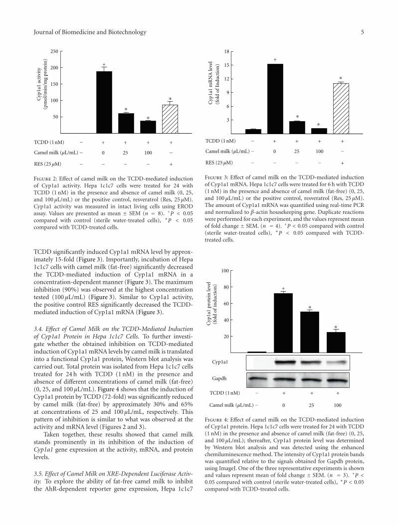
TCDD significantly induced Cyp1a1 mRNA level by approx-imately 15-fold (Figure 3). Importantly, incubation of Hepa1c1c7 cells with camel milk (fat-free) significantly decreasedthe TCDD-mediated induction of Cyp1a1 mRNA in aconcentration-dependent manner (Figure 3). The maximuminhibition (90%) was observed at the highest concentrationtested (100 μL/mL) (Figure 3). Similar to Cyp1a1 activity,the positive control RES significantly decreased the TCDD-mediated induction of Cyp1a1 mRNA (Figure 3).
3.4. Effect of Camel Milk on the TCDD-Mediated Inductionof Cyp1a1 Protein in Hepa 1c1c7 Cells. To further investi-gate whether the obtained inhibition on TCDD-mediatedinduction of Cyp1a1 mRNA levels by camel milk is translatedinto a functional Cyp1a1 protein, Western blot analysis wascarried out. Total protein was isolated from Hepa 1c1c7 cellstreated for 24 h with TCDD (1 nM) in the presence andabsence of different concentrations of camel milk (fat-free)(0, 25, and 100 μL/mL). Figure 4 shows that the induction ofCyp1a1 protein by TCDD (72-fold) was significantly reducedby camel milk (fat-free) by approximately 30% and 65%at concentrations of 25 and 100 μL/mL, respectively. Thispattern of inhibition is similar to what was observed at theactivity and mRNA level (Figures 2 and 3).
Taken together, these results showed that camel milkstands prominently in its inhibition of the induction ofCyp1a1 gene expression at the activity, mRNA, and proteinlevels.
3.5. Effect of Camel Milk on XRE-Dependent Luciferase Activ-ity. To explore the ability of fat-free camel milk to inhibitthe AhR-dependent reporter gene expression, Hepa 1c1c7
3
6
9
12
15
18
(fol
d of
In
duct
ion
) C
yp1a
1 m
RN
A le
vel
Camel milk (µL/mL)
TCDD (1 nM)
RES (25 µM)
−−
− − − −
−+ + + +
+
0 25 100
∗∗
∗
+
Figure 3: Effect of camel milk on the TCDD-mediated inductionof Cyp1a1 mRNA. Hepa 1c1c7 cells were treated for 6 h with TCDD(1 nM) in the presence and absence of camel milk (fat-free) (0, 25,and 100 μL/mL) or the positive control, resveratrol (Res, 25 μM).The amount of Cyp1a1 mRNA was quantified using real-time PCRand normalized to β-actin housekeeping gene. Duplicate reactionswere performed for each experiment, and the values represent meanof fold change ± SEM. (n = 4). +P < 0.05 compared with control(sterile water-treated cells), ∗P < 0.05 compared with TCDD-treated cells.
20
40
60
80
100
Cyp
1a1
prot
ein
leve
l(f
old
of in
duct
ion
)
∗
∗
TCDD (1 nM) − + + +
Camel milk (µL/mL)− 0 25 100
Cyp1a1
Gapdh
+
Figure 4: Effect of camel milk on the TCDD-mediated inductionof Cyp1a1 protein. Hepa 1c1c7 cells were treated for 24 with TCDD(1 nM) in the presence and absence of camel milk (fat-free) (0, 25,and 100 μL/mL); thereafter, Cyp1a1 protein level was determinedby Western blot analysis and was detected using the enhancedchemiluminescence method. The intensity of Cyp1a1 protein bandswas quantified relative to the signals obtained for Gapdh protein,using ImageJ. One of the three representative experiments is shownand values represent mean of fold change ± SEM. (n = 3). +P <0.05 compared with control (sterile water-treated cells), ∗P < 0.05compared with TCDD-treated cells.
6 Journal of Biomedicine and Biotechnology
2
4
6
8
10
∗
+
Camel milk (µL/mL)
TCDD (1 nM) −−
+
100−+
Luci
fera
se a
ctiv
ity
(fol
d of
Flu
c/R
luc)
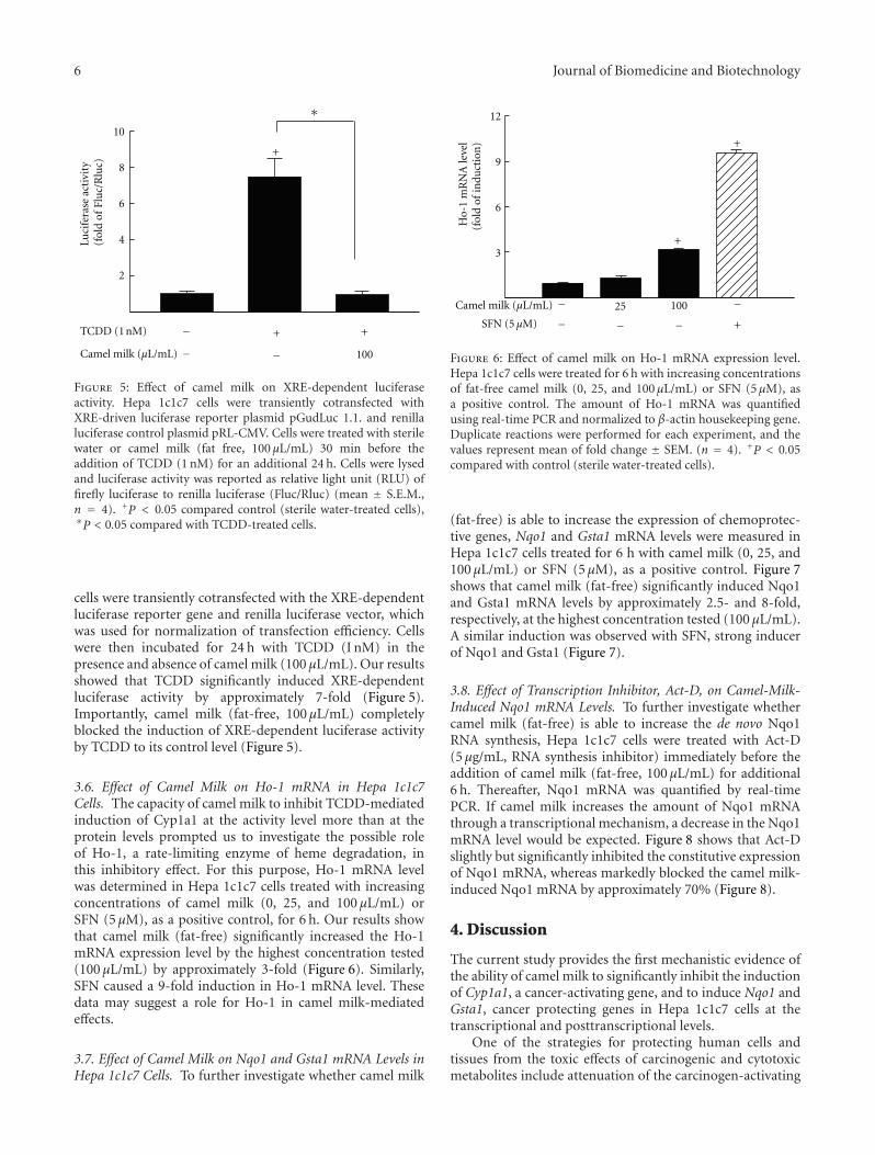
Figure 5: Effect of camel milk on XRE-dependent luciferaseactivity. Hepa 1c1c7 cells were transiently cotransfected withXRE-driven luciferase reporter plasmid pGudLuc 1.1. and renillaluciferase control plasmid pRL-CMV. Cells were treated with sterilewater or camel milk (fat free, 100 μL/mL) 30 min before theaddition of TCDD (1 nM) for an additional 24 h. Cells were lysedand luciferase activity was reported as relative light unit (RLU) offirefly luciferase to renilla luciferase (Fluc/Rluc) (mean ± S.E.M.,n = 4). +P < 0.05 compared control (sterile water-treated cells),∗P < 0.05 compared with TCDD-treated cells.
cells were transiently cotransfected with the XRE-dependentluciferase reporter gene and renilla luciferase vector, whichwas used for normalization of transfection efficiency. Cellswere then incubated for 24 h with TCDD (I nM) in thepresence and absence of camel milk (100 μL/mL). Our resultsshowed that TCDD significantly induced XRE-dependentluciferase activity by approximately 7-fold (Figure 5).Importantly, camel milk (fat-free, 100 μL/mL) completelyblocked the induction of XRE-dependent luciferase activityby TCDD to its control level (Figure 5).
3.6. Effect of Camel Milk on Ho-1 mRNA in Hepa 1c1c7Cells. The capacity of camel milk to inhibit TCDD-mediatedinduction of Cyp1a1 at the activity level more than at theprotein levels prompted us to investigate the possible roleof Ho-1, a rate-limiting enzyme of heme degradation, inthis inhibitory effect. For this purpose, Ho-1 mRNA levelwas determined in Hepa 1c1c7 cells treated with increasingconcentrations of camel milk (0, 25, and 100 μL/mL) orSFN (5 μM), as a positive control, for 6 h. Our results showthat camel milk (fat-free) significantly increased the Ho-1mRNA expression level by the highest concentration tested(100 μL/mL) by approximately 3-fold (Figure 6). Similarly,SFN caused a 9-fold induction in Ho-1 mRNA level. Thesedata may suggest a role for Ho-1 in camel milk-mediatedeffects.
3.7. Effect of Camel Milk on Nqo1 and Gsta1 mRNA Levels inHepa 1c1c7 Cells. To further investigate whether camel milk
3
6
9
12
(fol
d of
indu
ctio
n)
Camel milk (µL/mL) − −25 100
− − − +
+
+
SFN (5 µM)
Ho-
1 m
RN
A le
vel
Figure 6: Effect of camel milk on Ho-1 mRNA expression level.Hepa 1c1c7 cells were treated for 6 h with increasing concentrationsof fat-free camel milk (0, 25, and 100 μL/mL) or SFN (5 μM), asa positive control. The amount of Ho-1 mRNA was quantifiedusing real-time PCR and normalized to β-actin housekeeping gene.Duplicate reactions were performed for each experiment, and thevalues represent mean of fold change ± SEM. (n = 4). +P < 0.05compared with control (sterile water-treated cells).
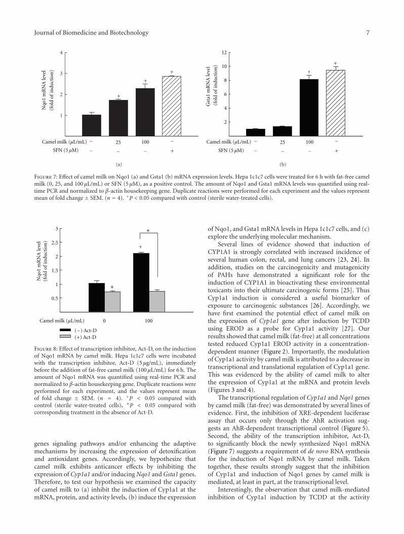
(fat-free) is able to increase the expression of chemoprotec-tive genes, Nqo1 and Gsta1 mRNA levels were measured inHepa 1c1c7 cells treated for 6 h with camel milk (0, 25, and100 μL/mL) or SFN (5 μM), as a positive control. Figure 7shows that camel milk (fat-free) significantly induced Nqo1and Gsta1 mRNA levels by approximately 2.5- and 8-fold,respectively, at the highest concentration tested (100 μL/mL).A similar induction was observed with SFN, strong inducerof Nqo1 and Gsta1 (Figure 7).
3.8. Effect of Transcription Inhibitor, Act-D, on Camel-Milk-Induced Nqo1 mRNA Levels. To further investigate whethercamel milk (fat-free) is able to increase the de novo Nqo1RNA synthesis, Hepa 1c1c7 cells were treated with Act-D(5 μg/mL, RNA synthesis inhibitor) immediately before theaddition of camel milk (fat-free, 100 μL/mL) for additional6 h. Thereafter, Nqo1 mRNA was quantified by real-timePCR. If camel milk increases the amount of Nqo1 mRNAthrough a transcriptional mechanism, a decrease in the Nqo1mRNA level would be expected. Figure 8 shows that Act-Dslightly but significantly inhibited the constitutive expressionof Nqo1 mRNA, whereas markedly blocked the camel milk-induced Nqo1 mRNA by approximately 70% (Figure 8).
4. Discussion
The current study provides the first mechanistic evidence ofthe ability of camel milk to significantly inhibit the inductionof Cyp1a1, a cancer-activating gene, and to induce Nqo1 andGsta1, cancer protecting genes in Hepa 1c1c7 cells at thetranscriptional and posttranscriptional levels.
One of the strategies for protecting human cells andtissues from the toxic effects of carcinogenic and cytotoxicmetabolites include attenuation of the carcinogen-activating
Journal of Biomedicine and Biotechnology 7
1
2
3
4
+
+
+
Camel milk (µL/mL) − −25 100
− − − +
Nqo
1 m
RN
A le
vel
(fol
d of
indu
ctio
n)
SFN (5 µM)
(a)
2
4
6
8
10
12
Camel milk (µL/mL) − −25 100
− − − +
+
+
Gst
a1 m
RN
A le
vel
(fol
d of
indu
ctio
n)
SFN (5 µM)
(b)
Figure 7: Effect of camel milk on Nqo1 (a) and Gsta1 (b) mRNA expression levels. Hepa 1c1c7 cells were treated for 6 h with fat-free camelmilk (0, 25, and 100 μL/mL) or SFN (5 μM), as a positive control. The amount of Nqo1 and Gsta1 mRNA levels was quantified using real-time PCR and normalized to β-actin housekeeping gene. Duplicate reactions were performed for each experiment and the values representmean of fold change ± SEM. (n = 4). +P < 0.05 compared with control (sterile water-treated cells).
0.5
1
1.5
2
2.5
3
Camel milk (µL/mL)
Nqo
1 m
RN
A le
vel
(fol
d of
indu
ctio
n)
0 100
(+) Act-D
+
∗
∗
(−) Act-D
Figure 8: Effect of transcription inhibitor, Act-D, on the inductionof Nqo1 mRNA by camel milk. Hepa 1c1c7 cells were incubatedwith the transcription inhibitor, Act-D (5 μg/mL), immediatelybefore the addition of fat-free camel milk (100 μL/mL) for 6 h. Theamount of Nqo1 mRNA was quantified using real-time PCR andnormalized to β-actin housekeeping gene. Duplicate reactions wereperformed for each experiment, and the values represent meanof fold change ± SEM. (n = 4). +P < 0.05 compared withcontrol (sterile water-treated cells), ∗P < 0.05 compared withcorresponding treatment in the absence of Act-D.
genes signaling pathways and/or enhancing the adaptivemechanisms by increasing the expression of detoxificationand antioxidant genes. Accordingly, we hypothesize thatcamel milk exhibits anticancer effects by inhibiting theexpression of Cyp1a1 and/or inducing Nqo1 and Gsta1 genes.Therefore, to test our hypothesis we examined the capacityof camel milk to (a) inhibit the induction of Cyp1a1 at themRNA, protein, and activity levels, (b) induce the expression
of Nqo1, and Gsta1 mRNA levels in Hepa 1c1c7 cells, and (c)explore the underlying molecular mechanism.
Several lines of evidence showed that induction ofCYP1A1 is strongly correlated with increased incidence ofseveral human colon, rectal, and lung cancers [23, 24]. Inaddition, studies on the carcinogenicity and mutagenicityof PAHs have demonstrated a significant role for theinduction of CYP1A1 in bioactivating these environmentaltoxicants into their ultimate carcinogenic forms [25]. ThusCyp1a1 induction is considered a useful biomarker ofexposure to carcinogenic substances [26]. Accordingly, wehave first examined the potential effect of camel milk onthe expression of Cyp1a1 gene after induction by TCDDusing EROD as a probe for Cyp1a1 activity [27]. Ourresults showed that camel milk (fat-free) at all concentrationstested reduced Cyp1a1 EROD activity in a concentration-dependent manner (Figure 2). Importantly, the modulationof Cyp1a1 activity by camel milk is attributed to a decrease intranscriptional and translational regulation of Cyp1a1 gene.This was evidenced by the ability of camel milk to alterthe expression of Cyp1a1 at the mRNA and protein levels(Figures 3 and 4).
The transcriptional regulation of Cyp1a1 and Nqo1 genesby camel milk (fat-free) was demonstrated by several lines ofevidence. First, the inhibition of XRE-dependent luciferaseassay that occurs only through the AhR activation sug-gests an AhR-dependent transcriptional control (Figure 5).Second, the ability of the transcription inhibitor, Act-D,to significantly block the newly synthesized Nqo1 mRNA(Figure 7) suggests a requirement of de novo RNA synthesisfor the induction of Nqo1 mRNA by camel milk. Takentogether, these results strongly suggest that the inhibitionof Cyp1a1 and induction of Nqo1 genes by camel milk ismediated, at least in part, at the transcriptional level.
Interestingly, the observation that camel milk-mediatedinhibition of Cyp1a1 induction by TCDD at the activity
8 Journal of Biomedicine and Biotechnology
levels is higher than those observed at the protein levels(Figure 2), suggest that a possible involvement of a post-translational mechanism, such as phosphorylation, protea-somal degradations, modulation of HO-1 gene expression,could be involved [22, 28]. Among these mechanisms,HO-1 gene expression, a rate-limiting enzyme in hemecatabolism, has been shown to alter cellular heme, theprosthetic group of CYP450, content and hence the enzymeactivity [29]. To test this hypothesis, we have examined theeffect of camel milk (fat-free) on the expression of Ho-1 mRNA level. Our results showed that the camel milkincreases the level of Ho-1 mRNA levels (Figure 6). Takentogether, we postulated here that the capacity of camelmilk to induce the expression of Ho-1 mRNA resulted ina decrease in Cyp1a1 activity levels through degrading itsheme content. This postulation is supported by our previ-ous observations that in mesoporphyrin, competitive Ho-1inhibitor or knockdown of Ho-1 using siRNA significantlyrestored the inhibition of Cyp1a1 activity by heavy metals[30, 31].
The chemo protective effect of camel milk was furthersupported, in addition to inhibition of Cyp1a1, by the abilityto upregulate antioxidant genes, particularly Nqo1 andGsta1 mRNA levels in a concentration-dependent manner(Figure 7). Our results are in agreement with previous stud-ies that reported the ability of camel milk to induce GSTlevels in healthy and schistosoma-infected mice [32]. Thusincreased expression of Nqo1 and Gsta1 by camel milkwill increase the levels of several antioxidant enzymes whichprevent the formation of highly reactive oxygen radicals andhence reduce DNA adduct and cell damage [33]. In addition,overexpression of NQO1 in several human solid tumorsand cancer cells has been shown to activate bioreductivechemotherapeutic agents in tumor cells that allow tumorcytotoxicity without corresponding toxicity to normal cells[34].
Although the potential mediators in camel milk involvedin the downregulation of Cyp1a1 and induction of Nqo1and Gsta1 were not examined in this study, several previousstudies have reported that camel milk contains considerablyhigher amounts of antioxidant vitamins, such as E and C,in comparison to cow milk [35], lysosomes [11], lactoferrins[11, 36], and immunoglobulines [36]. In addition, ongoingresearch in our laboratory has shown the presence of severalcompounds in camel milk that could be involved in milk-mediated effect (unpublished data). Among these mediators,lactoferrin, an iron-binding glycoprotein, is known to exertin vitro and in vivo antitumor activity [37]. In this context,it has been recently reported that lactoferrin inhibits thedevelopment of cancer through inhibiting CYP1A1 activa-tion in 7,12-dimethylbenz[a]anthracene- (DMBA-) inducedhamster buccal pouch carcinoma model. Taken togetherthe results obtained from our laboratory and previouslypublished reports, we speculate that lactoferrin could beresponsible for camel-milk-mediated effect. In addition, themain components of the camel milk have been alreadydetermined [38]. Ongoing research in our laboratory focuseson identifying and characterizing the most effective com-ponent of camel milk using liquid chromatography-tandem
mass spectrometry (LC-MS/MS) and one-dimensional gelelectrophoresis, where several proteins were found to berelatively abundant in camel milk (data not shown).
In conclusion, the results of current study suggest thatcamel milk could protect against or decrease the del-eterious effects of many environmental toxicants and car-cinogens such as PAHs, probably through modulation ofAhR-regulated genes of Cyp1a1, Nqo1, and Gsta1 at thetranscriptional and posttranscriptional mechanisms. Theseresults are of potential clinical significance to humans in thatit uncovers the molecular mechanism involved and couldexplain the anecdotal evidence for the successful use of camelmilk in the treatment and/or prevention of various medicalconditions.
Abbreviations
AhR: Aryl hydrocarbon receptorAct-D: Actinomycin DCYP1A1: Cytochrome P450 1A1DMSO: Dimethyl sulfoxide7ER: 7-ethoxyresorufinEROD: 7-ethoxyresorufin O-deethylaseGapdh: Glyceraldehyde-3-phosphate dehydrogenase.Gsta1: Glutathione S-transferase a1MTT: (3-(4,5-dimethylthiazol-2-yl)-2,5-diphenyl
tetrazolium bromide)Nqo1: NAD(P)H:quinone oxidoreductasePAHs: Polycyclic aromatic hydrocarbonsRES: ResveratrolSFN: SulforaphaneTCDD: 2,3,7,8-tetrachlorodibenzo-p-dioxinXRE: Xenobiotic responsive element.
Conflict of Interests
The authors declare that there is no conflict of interests.
Acknowledgments
The authors extend their appreciation to the Deanship ofScientific Research at King Saud University for funding thework through the research group project no. RGP-VPP-141.
References
[1] J. K. Kerzee and K. S. Ramos, “Constitutive and inducibleexpression of Cyp1a1 and Cyp1b1 in vascular smooth musclecells: role of the Ahr bHLH/PAS transcription factor,” Circula-tion Research, vol. 89, no. 7, pp. 573–582, 2001.
[2] M. L. Whitelaw, M. Gottlicher, J. A. Gustafsson, and L.Poellinger, “Definition of a novel ligand binding domain ofa nuclear bHLH receptor: co-localization of ligand and hsp90binding activities within the regulable inactivation domain ofthe dioxin receptor,” EMBO Journal, vol. 12, no. 11, pp. 4169–4179, 1993.
[3] J. P. Whitlock Jr., “Induction of cytochrome P4501A1,” AnnualReview of Pharmacology and Toxicology, vol. 39, pp. 103–125,1999.
Journal of Biomedicine and Biotechnology 9
[4] D. W. Nebert, A. L. Roe, M. Z. Dieter, W. A. Solis, Y. Yang, andT. P. Dalton, “Role of the aromatic hydrocarbon receptor and[Ah] gene battery in the oxidative stress response, cell cyclecontrol, and apoptosis,” Biochemical Pharmacology, vol. 59, no.1, pp. 65–85, 2000.
[5] D. W. Nebert, T. P. Dalton, A. B. Okey, and F. J. Gonzalez,“Role of aryl hydrocarbon receptor-mediated induction of theCYP1 enzymes in environmental toxicity and cancer,” Journalof Biological Chemistry, vol. 279, no. 23, pp. 23847–23850,2004.
[6] D. Ross, “Quinone reductases multitasking in the metabolicworld,” Drug Metabolism Reviews, vol. 36, no. 3-4, pp. 639–654, 2004.
[7] V. Vasiliou, D. Ross, and D. W. Nebert, “Update of theNAD(P)H:Quinone oxidoreductase (NQO) gene family,”Human Genomics, vol. 2, no. 5, pp. 329–335, 2006.
[8] M. Cuendet, C. P. Oteham, R. C. Moon, and J. M. Pezzuto,“Quinone reductase induction as a biomarker for cancerchemoprevention,” Journal of Natural Products, vol. 69, no. 3,pp. 460–463, 2006.
[9] N. Kontou, T. Psaltopoulou, D. Panagiotakos, M. A.Dimopoulos, and A. Linos, “The mediterranean diet in cancerprevention: a review,” Journal of Medicinal Food, vol. 14, no.10, pp. 1065–1078, 2011.
[10] C. Conesa, L. Sanchez, C. Rota et al., “Isolation of lactoferrinfrom milk of different species: calorimetric and antimicrobialstudies,” Comparative Biochemistry and Physiology B, vol. 150,no. 1, pp. 131–139, 2008.
[11] E. I. el Agamy, R. Ruppanner, A. Ismail, C. P. Champagne, andR. Assaf, “Antibacterial and antiviral activity of camel milkprotective proteins,” Journal of Dairy Research, vol. 59, no. 2,pp. 169–175, 1992.
[12] C. Saravanan, Z. Cao, J. Kumar et al., “Milk componentsinhibit Acanthamoeba-induced cytopathic effect,” Investiga-tive Ophthalmology & Visual Science, vol. 49, no. 3, pp. 1010–1015, 2008.
[13] H. M. Korashy and A. O. S. El-Kadi, “The role of arylhydrocarbon receptor and the reactive oxygen species in themodulation of glutathione transferase by heavy metals inmurine hepatoma cell lines,” Chemico-Biological Interactions,vol. 162, no. 3, pp. 237–248, 2006.
[14] S. W. Kennedy, A. Lorenzen, C. A. James, and B. T. Collins,“Ethoxyresorufin-O-deethylase and porphyrin analysis inchicken embryo hepatocyte cultures with fluorescence multi-well plate reader,” Analytical Biochemistry, vol. 211, no. 1, pp.102–112, 1993.
[15] A. Lorenzen and S. W. Kennedy, “A fluorescence-basedprotein assay for use with a microplate reader,” AnalyticalBiochemistry, vol. 214, no. 1, pp. 346–348, 1993.
[16] B. N. M. Zordoky, M. E. Aboutabl, and A. O. S. El-Kadi, “Modulation of cytochrome P450 gene expression andarachidonic acid metabolism during isoproterenol-inducedcardiac hypertrophy in rats,” Drug Metabolism and Disposition,vol. 36, no. 11, pp. 2277–2286, 2008.
[17] A. Anwar-Mohamed, O. S. Degenhardt, M. A. M. El Gendy,J. M. Seubert, S. R. Kleeberger, and A. O. S. El-Kadi, “Theeffect of Nrf2 knockout on the constitutive expression ofdrug metabolizing enzymes and transporters in C57Bl/6 micelivers,” Toxicology in Vitro, vol. 25, no. 4, pp. 785–795, 2011.
[18] A. Anwar-Mohamed, R. H. Elbekai, and A. O. S. El-Kadi,“MG-132 inhibits the TCDD-mediated induction of Cyp1a1at the catalytic activity but not the mRNA or protein levels inHepa 1c1c7 cells,” Toxicology Letters, vol. 182, no. 1-3, pp. 121–126, 2008.
[19] K. J. Livak and T. D. Schmittgen, “Analysis of relative geneexpression data using real-time quantitative PCR and the 2-ΔΔCT method,” Methods, vol. 25, no. 4, pp. 402–408, 2001.
[20] J. Sambrook, E. F. Fritsch, and T. Maniatatis, “MolecularCloning,” in A Laboratory Manual, N. Ford, Ed., Cold SpringHarbour Laboratory Press, Plainview, NY, USA, 1989.
[21] M. A. M. El Gendy, A. A. Soshilov, M. S. Denison, and A. O.S. El-Kadi, “Transcriptional and posttranslational inhibitionof dioxin-mediated induction of CYP1A1 by harmine andharmol,” Toxicology Letters, vol. 208, no. 1, pp. 51–61, 2012.
[22] H. M. Korashy and A. O. S. El-Kadi, “Regulatory mechanismsmodulating the expression of cytochrome P450 1A1 gene byheavy metals,” Toxicological Sciences, vol. 88, no. 1, pp. 39–51,2005.
[23] M. L. Slattery, W. Samowtiz, K. Ma et al., “CYP1A1, cigarettesmoking, and colon and rectal cancer,” American Journal ofEpidemiology, vol. 160, no. 9, pp. 842–852, 2004.
[24] P. P. Shah, K. Saurabh, M. C. Pant, N. Mathur, and D. Parmar,“Evidence for increased cytochrome P450 1A1 expression inblood lymphocytes of lung cancer patients,” Mutation Research- Fundamental and Molecular Mechanisms of Mutagenesis, vol.670, no. 1-2, pp. 74–78, 2009.
[25] T. Shimada and Y. Fujii-Kuriyama, “Metabolic activationof polycyclic aromatic hydrocarbons to carcinogens bycytochromes P450 1A1 and 1B1,” Cancer Science, vol. 95, no.1, pp. 1–6, 2004.
[26] T. D. Williams, J. S. Lee, D. L. Sheader, and J. K. Chipman,“The cytochrome P450 1A gene (CYP1A) from Europeanflounder (Platichthys flesus), analysis of regulatory regionsand development of a dual luciferase reporter gene system,”Marine Environmental Research, vol. 50, no. 1–5, pp. 1–6, 2000.
[27] B. Hasspieler, D. Haffner, M. Stelljes, and K. Adeli, “Toxi-cological assessment of industrial solvents using human cellbioassays: assessment of short-term cytotoxicity and long-term genotoxicity potential,” Toxicology and Industrial Health,vol. 22, no. 7, pp. 301–315, 2006.
[28] V. Werlinder, M. Backlund, A. Zhukov, and M. Ingelman-Sundberg, “Transcriptional and post-translational regulationof CYP1A1 by primaquine,” Journal of Pharmacology andExperimental Therapeutics, vol. 297, no. 1, pp. 206–214, 2001.
[29] G. Kikuchi, T. Yoshida, and M. Noguchi, “Heme oxygenaseand heme degradation,” Biochemical and Biophysical ResearchCommunications, vol. 338, no. 1, pp. 558–567, 2005.
[30] I. E. A. Amara, A. Anwar-Mohamed, and A. O. S. El-Kadi, “Mercury modulates the CYP1A1 at transcriptional andposttranslational levels in human hepatoma HepG2 cells,”Toxicology Letters, vol. 199, no. 3, pp. 225–233, 2010.
[31] A. Anwar-Mohamed and A. O. S. El-Kadi, “Arsenite down-regulates cytochrome P450 1A1 at the transcriptional andposttranslational levels in human HepG2 cells,” Free RadicalBiology and Medicine, vol. 48, no. 10, pp. 1399–1409, 2010.
[32] A. S. Maghraby, M. A. Mohamed, and A. M. Abdel-Salam,“Anti-schistosomal activity of colostral and mature camel milkon Schistosoma mansoni infected mice,” Asia Pacific Journal ofClinical Nutrition, vol. 14, no. 4, pp. 432–438, 2005.
[33] M. Mohora, “Role of Nad(P)h: quinone oxidoreductase in theregulation of intracellular redox state,” Romanian Journal ofInternal Medicine, vol. 38-39, pp. 33–50, 2000.
[34] S. Danson, T. H. Ward, J. Butler, and M. Ranson, “DT-diaphorase: a target for new anticancer drugs,” Cancer Treat-ment Reviews, vol. 30, no. 5, pp. 437–449, 2004.
[35] Z. Farah, R. Rettenmaier, and D. Atkins, “Vitamin content ofcamel milk,” International Journal for Vitamin and NutritionResearch, vol. 62, no. 1, pp. 30–33, 1992.
10 Journal of Biomedicine and Biotechnology
[36] G. Konuspayeva, B. Faye, G. Loiseau, and D. Levieux,“Lactoferrin and immunoglobulin contents in camel’s milk(Camelus bactrianus, Campus dromedarius, and Hybrids)from Kazakhstan,” Journal of Dairy Science, vol. 90, no. 1, pp.38–46, 2007.
[37] A. Roseanu, P. E. Florian, M. Moisei, L. E. Sima, R. W. Evans,and M. Trif, “Liposomalization of lactoferrin enhanced itsanti-tumoral effects on melanoma cells,” BioMetals, vol. 23,no. 3, pp. 485–492, 2010.
[38] M. S. Y. Haddadin, S. I. Gammoh, and R. K. Robinson,“Seasonal variations in the chemical composition of camelmilk in Jordan,” Journal of Dairy Research, vol. 75, no. 1, pp.8–12, 2008.

Submit your manuscripts athttp://www.hindawi.com
Hindawi Publishing Corporationhttp://www.hindawi.com Volume 2013
Journal of
Pharmaceutics
Hindawi Publishing Corporation http://www.hindawi.com Volume 2013Hindawi Publishing Corporation http://www.hindawi.com Volume 2013
The Scientific World Journal
BioMed Research International
Hindawi Publishing Corporationhttp://www.hindawi.com Volume 2013
Hindawi Publishing Corporationhttp://www.hindawi.com Volume 2013
ISRN Pain
Drug DeliveryJournal of
Hindawi Publishing Corporationhttp://www.hindawi.com Volume 2013
Hindawi Publishing Corporationhttp://www.hindawi.com Volume 2013
Advances in Pharmacological Sciences
Hindawi Publishing Corporationhttp://www.hindawi.com Volume 2013
AnesthesiologyResearch and Practice
ISRN Pharmaceutics
Volume 2013Hindawi Publishing Corporationhttp://www.hindawi.com
Hindawi Publishing Corporationhttp://www.hindawi.com Volume 2013
MediatorsinflaMMation
of
Hindawi Publishing Corporationhttp://www.hindawi.com Volume 2013
AntibioticsInternational Journal of
Volume 2013
ISRN Medicinal Chemistry
Hindawi Publishing Corporationhttp://www.hindawi.com
Hindawi Publishing Corporationhttp://www.hindawi.com Volume 2013
Medicinal ChemistryInternational Journal of
ISRN Toxicology
Volume 2013Hindawi Publishing Corporationhttp://www.hindawi.com
Hindawi Publishing Corporationhttp://www.hindawi.com
Volume 2013
ToxinsJournal of
Hindawi Publishing Corporationhttp://www.hindawi.com Volume 2013
Autoimmune Diseases
ToxicologyJournal of
Hindawi Publishing Corporationhttp://www.hindawi.com Volume 2013
ScientificaHindawi Publishing Corporationhttp://www.hindawi.com Volume 2013
ISRN Pharmacology
Hindawi Publishing Corporationhttp://www.hindawi.com Volume 2013
Hindawi Publishing Corporationhttp://www.hindawi.com Volume 2013
Emergency Medicine International