Embed Size (px)
Citation preview
Breast Screening Guidelines Update
Michael Policar, MD, MPHProfessor Emeritus of Ob, Gyn, and Repro SciencesUCSF School of [email protected]
National Reproductive Health ConferenceKansas CityJuly 18, 2018
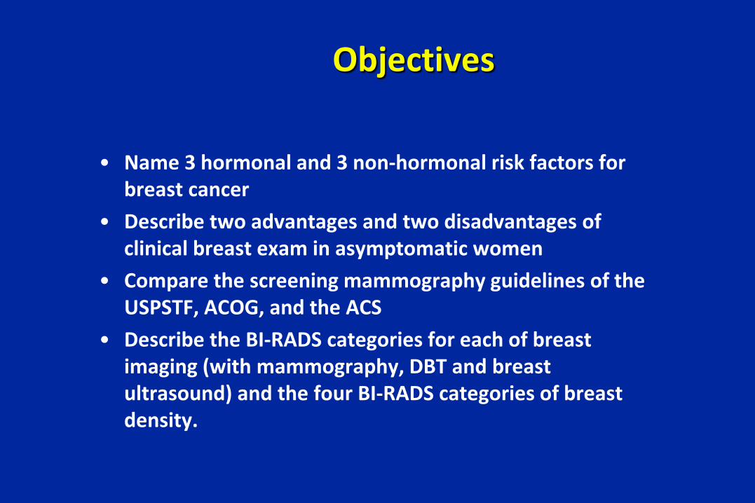
Objectives
• Name 3 hormonal and 3 non-hormonal risk factors for breast cancer
• Describe two advantages and two disadvantages of clinical breast exam in asymptomatic women
• Compare the screening mammography guidelines of the USPSTF, ACOG, and the ACS
• Describe the BI-RADS categories for each of breast imaging (with mammography, DBT and breast ultrasound) and the four BI-RADS categories of breast density.

• There are no relevant financial relationships with any commercial interests to disclose
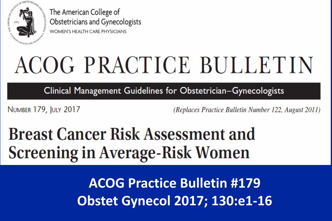
ACOG Practice Bulletin #179Obstet Gynecol 2017; 130:e1-16
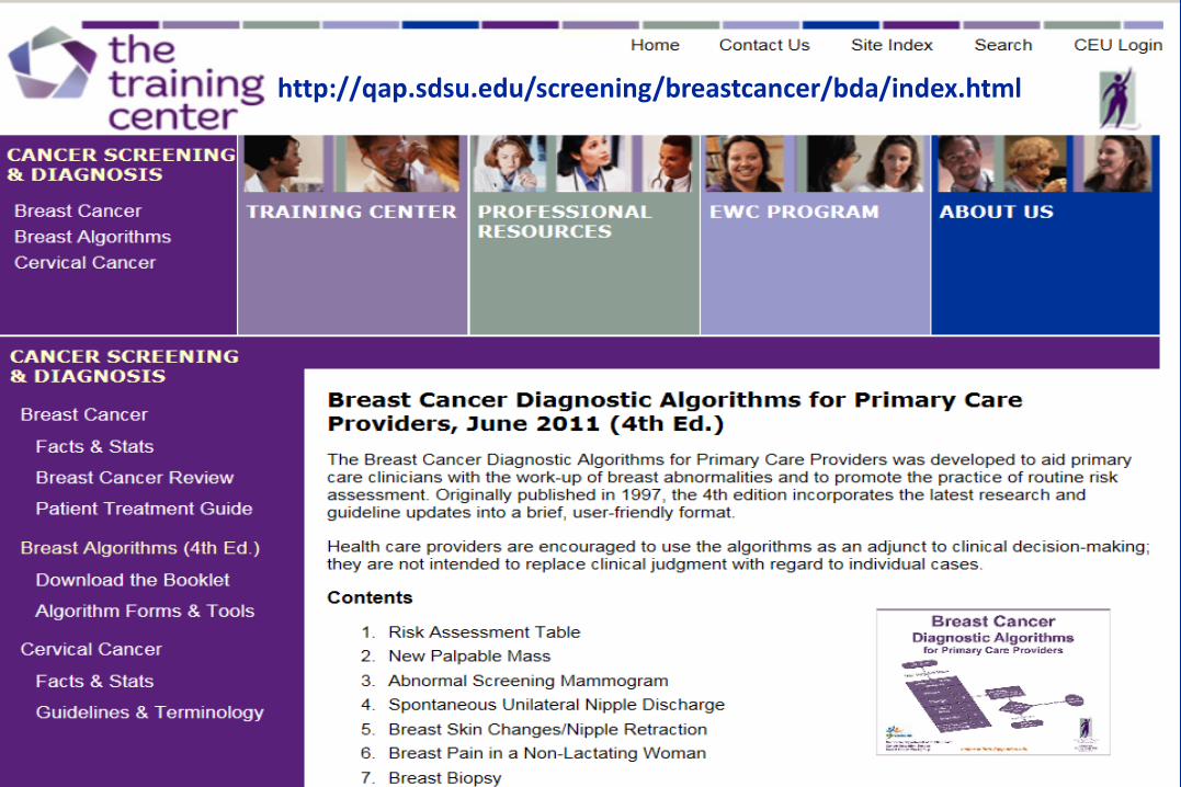
https://qap.sdsu.eduhttp://qap.sdsu.edu/screening/breastcancer/bda/index.html
https://www.nccn.org/professionals/physician_gls/pdf/breast-screening.pdf
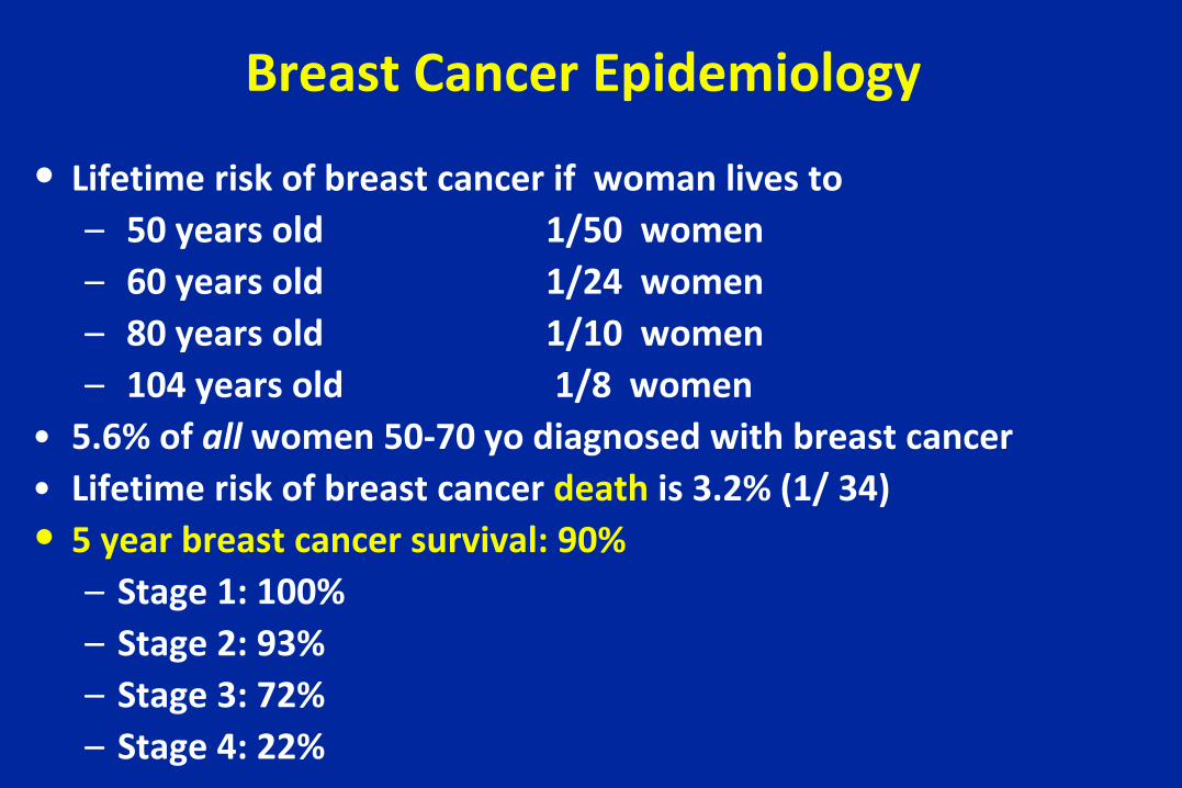
Breast Cancer Epidemiology
• Lifetime risk of breast cancer if woman lives to– 50 years old 1/50 women– 60 years old 1/24 women– 80 years old 1/10 women– 104 years old 1/8 women
• 5.6% of all women 50-70 yo diagnosed with breast cancer• Lifetime risk of breast cancer death is 3.2% (1/ 34)• 5 year breast cancer survival: 90%
– Stage 1: 100%– Stage 2: 93%– Stage 3: 72%– Stage 4: 22%
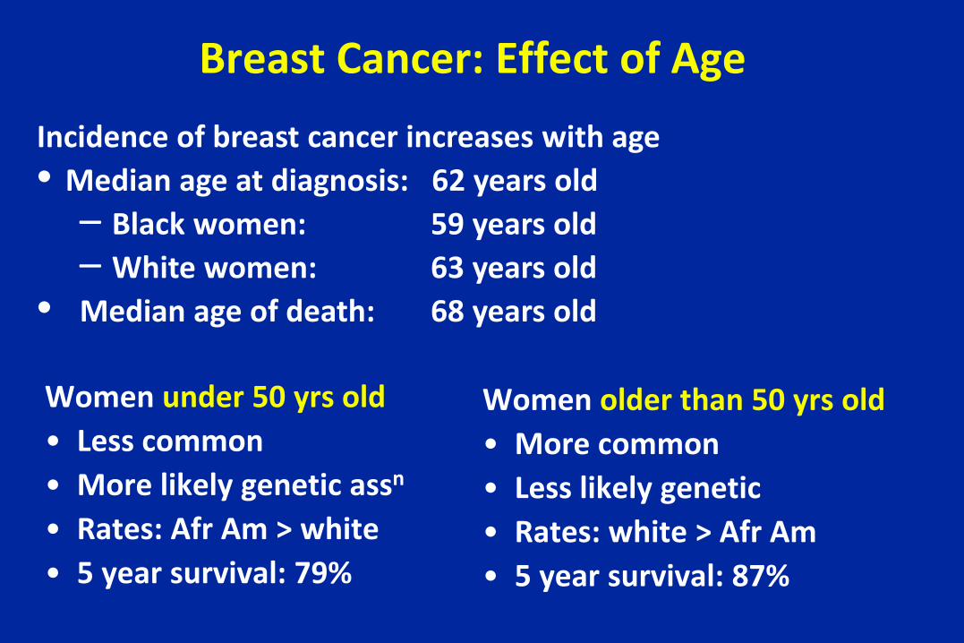
Breast Cancer: Effect of Age
Women under 50 yrs old• Less common• More likely genetic assn
• Rates: Afr Am > white • 5 year survival: 79%
Women older than 50 yrs old • More common• Less likely genetic • Rates: white > Afr Am• 5 year survival: 87%
Incidence of breast cancer increases with age• Median age at diagnosis: 62 years old
– Black women: 59 years old– White women: 63 years old
• Median age of death: 68 years old
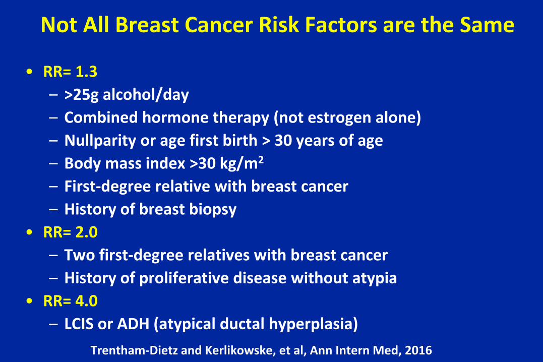
Not All Breast Cancer Risk Factors are the Same
• RR= 1.3 – >25g alcohol/day– Combined hormone therapy (not estrogen alone)– Nullparity or age first birth > 30 years of age– Body mass index >30 kg/m2
– First-degree relative with breast cancer– History of breast biopsy
• RR= 2.0– Two first-degree relatives with breast cancer– History of proliferative disease without atypia
• RR= 4.0 – LCIS or ADH (atypical ductal hyperplasia)
Trentham-Dietz and Kerlikowske, et al, Ann Intern Med, 2016
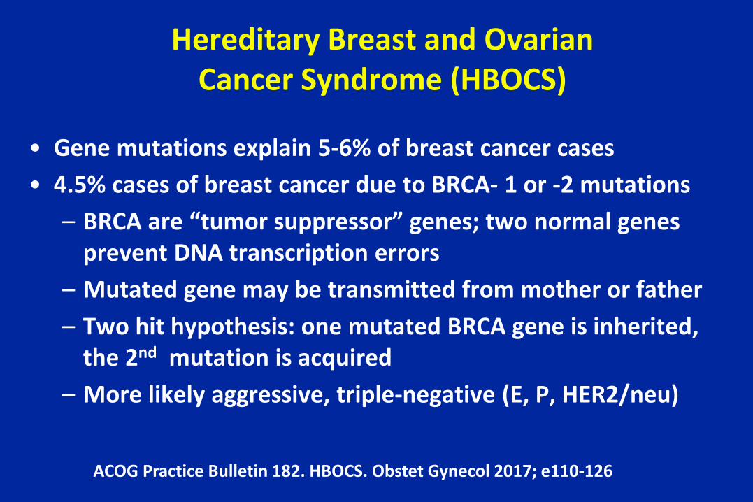
Hereditary Breast and Ovarian Cancer Syndrome (HBOCS)
• Gene mutations explain 5-6% of breast cancer cases• 4.5% cases of breast cancer due to BRCA- 1 or -2 mutations
– BRCA are “tumor suppressor” genes; two normal genes prevent DNA transcription errors
– Mutated gene may be transmitted from mother or father– Two hit hypothesis: one mutated BRCA gene is inherited,
the 2nd mutation is acquired– More likely aggressive, triple-negative (E, P, HER2/neu)
ACOG Practice Bulletin 182. HBOCS. Obstet Gynecol 2017; e110-126
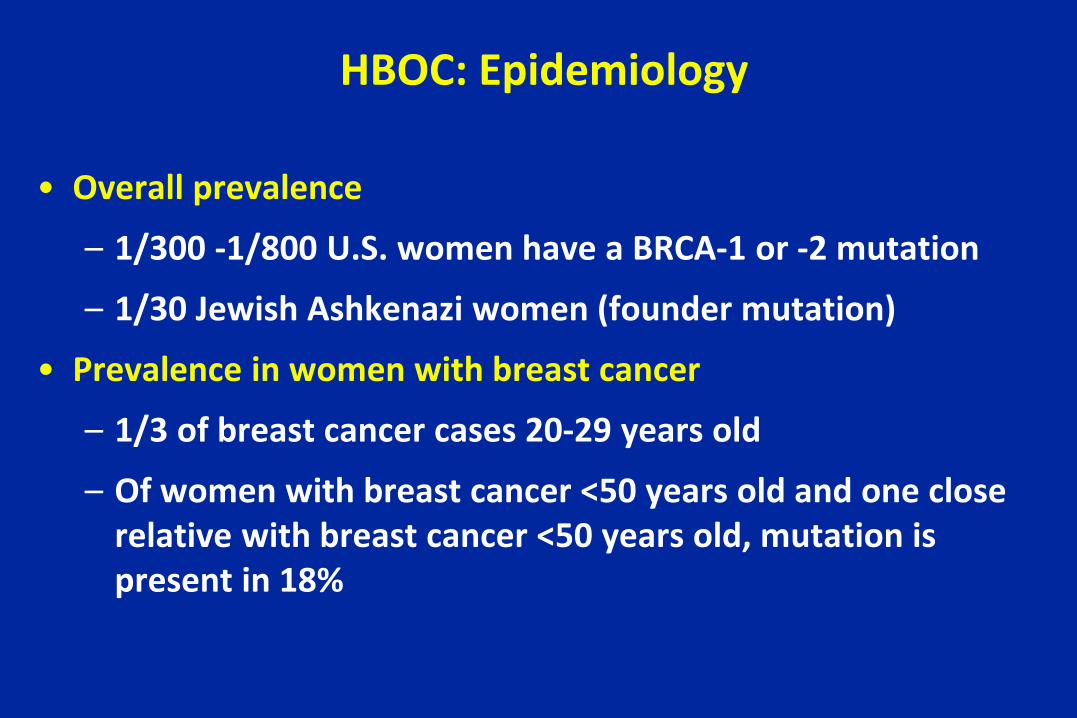
HBOC: Epidemiology
• Overall prevalence
– 1/300 -1/800 U.S. women have a BRCA-1 or -2 mutation
– 1/30 Jewish Ashkenazi women (founder mutation)
• Prevalence in women with breast cancer
– 1/3 of breast cancer cases 20-29 years old
– Of women with breast cancer <50 years old and one close relative with breast cancer <50 years old, mutation is present in 18%
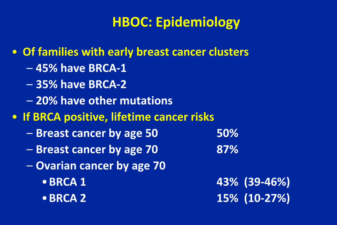
HBOC: Epidemiology
• Of families with early breast cancer clusters– 45% have BRCA-1– 35% have BRCA-2– 20% have other mutations
• If BRCA positive, lifetime cancer risks– Breast cancer by age 50 50%– Breast cancer by age 70 87%– Ovarian cancer by age 70
•BRCA 1 43% (39-46%)•BRCA 2 15% (10-27%)
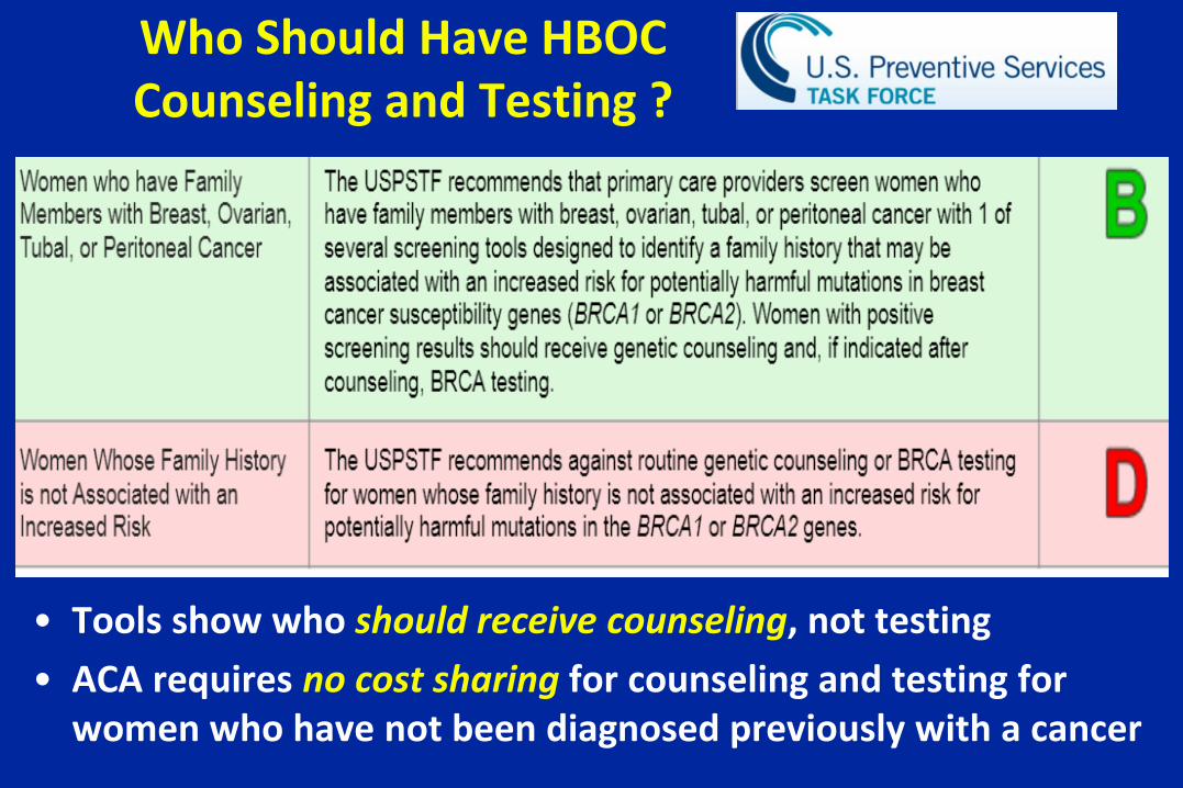
Who Should Have HBOC Counseling and Testing ?
• Tools show who should receive counseling, not testing• ACA requires no cost sharing for counseling and testing for
women who have not been diagnosed previously with a cancer
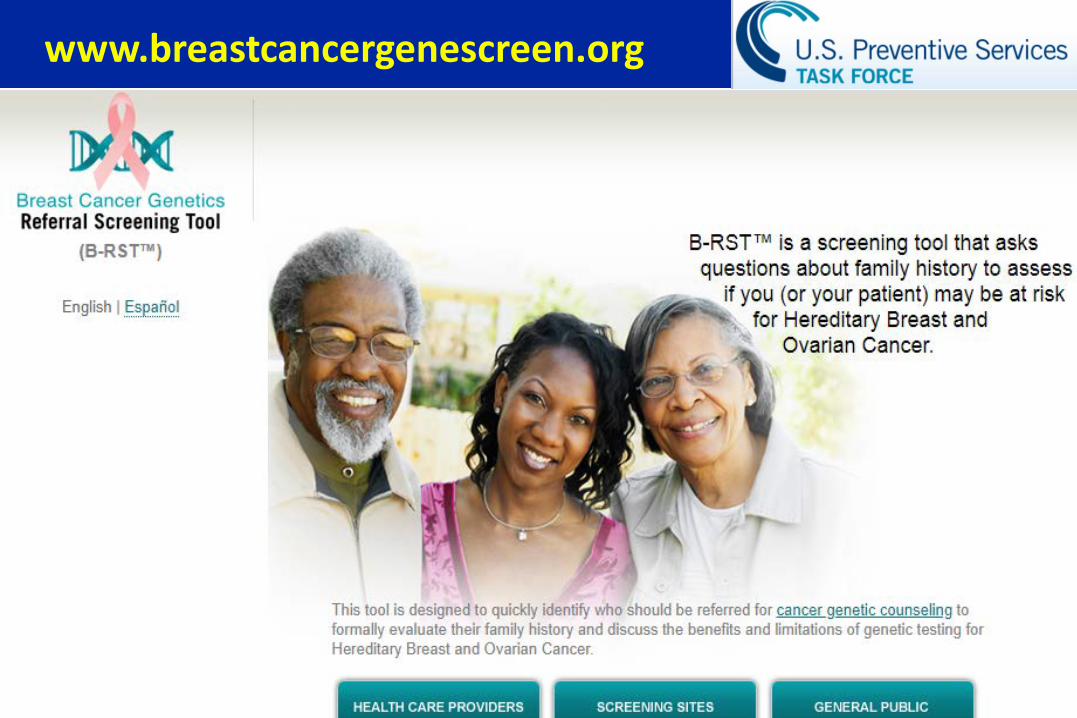
www.breastcancergenescreen.org
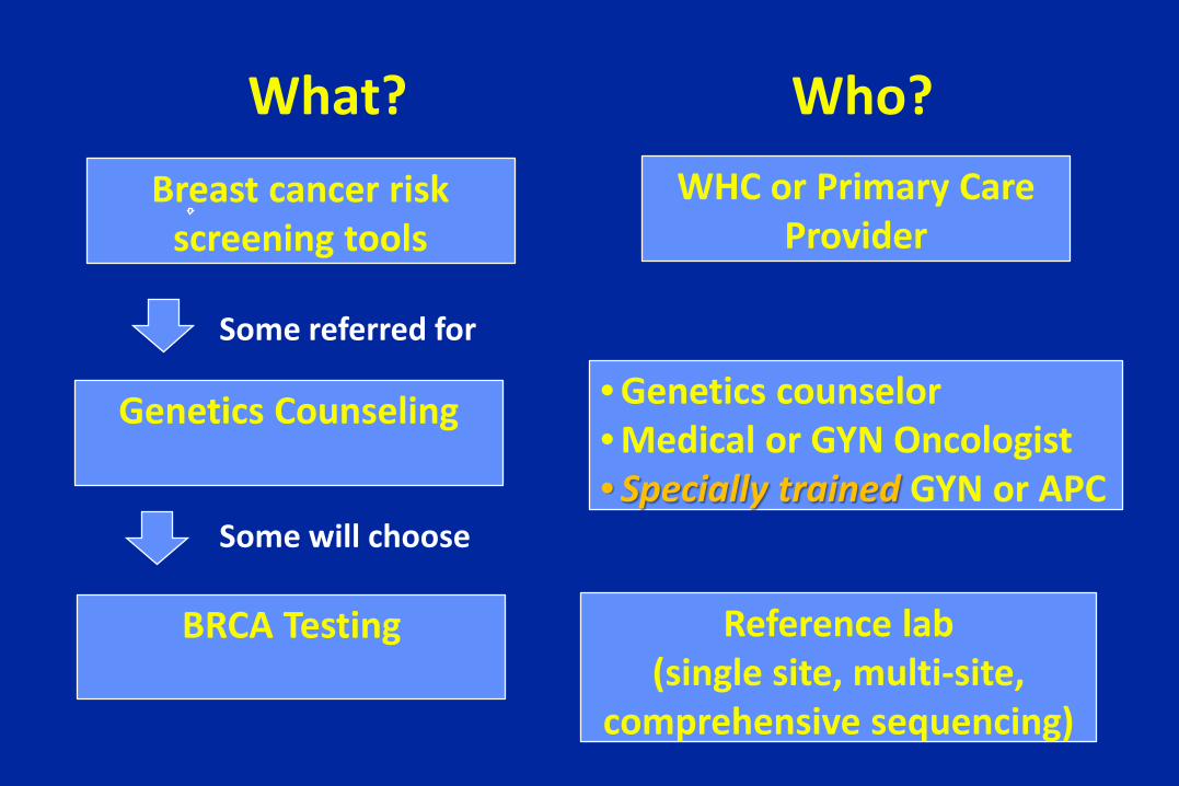
What?Breast cancer risk
screening toolsWHC or Primary Care
Provider
Genetics Counseling • Genetics counselor• Medical or GYN Oncologist• Specially trained GYN or APC
Reference lab(single site, multi-site,
comprehensive sequencing)
BRCA Testing
Who?
Some referred for
Some will choose
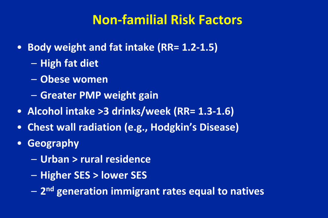
Non-familial Risk Factors
• Body weight and fat intake (RR= 1.2-1.5)– High fat diet – Obese women– Greater PMP weight gain
• Alcohol intake >3 drinks/week (RR= 1.3-1.6) • Chest wall radiation (e.g., Hodgkin’s Disease)• Geography
– Urban > rural residence– Higher SES > lower SES– 2nd generation immigrant rates equal to natives
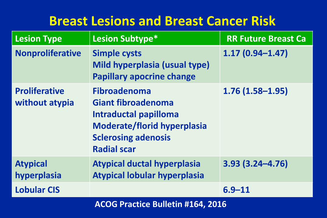
Breast Lesions and Breast Cancer RiskLesion Type Lesion Subtype* RR Future Breast CaNonproliferative Simple cysts
Mild hyperplasia (usual type)Papillary apocrine change
1.17 (0.94–1.47)
Proliferative without atypia
FibroadenomaGiant fibroadenomaIntraductal papillomaModerate/florid hyperplasia Sclerosing adenosisRadial scar
1.76 (1.58–1.95)
Atypical hyperplasia
Atypical ductal hyperplasiaAtypical lobular hyperplasia
3.93 (3.24–4.76)
Lobular CIS 6.9–11ACOG Practice Bulletin #164, 2016
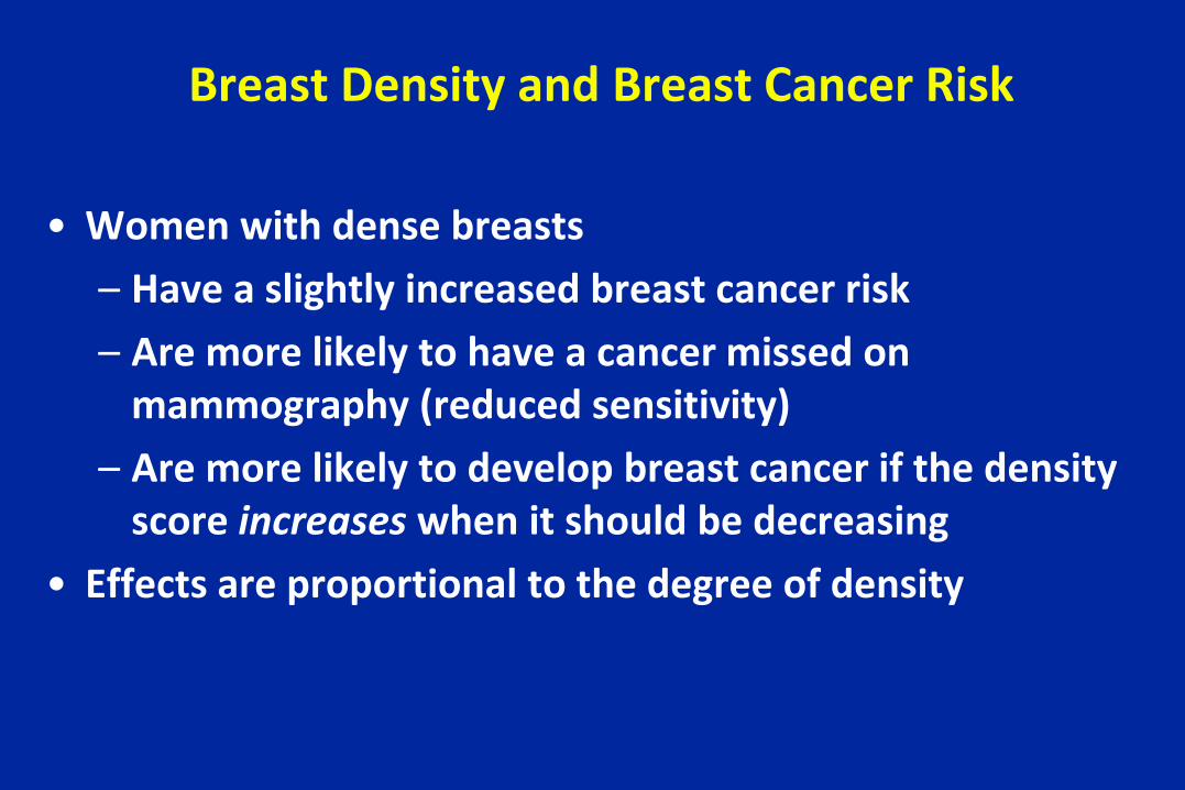
Breast Density and Breast Cancer Risk
• Women with dense breasts – Have a slightly increased breast cancer risk– Are more likely to have a cancer missed on
mammography (reduced sensitivity)– Are more likely to develop breast cancer if the density
score increases when it should be decreasing• Effects are proportional to the degree of density
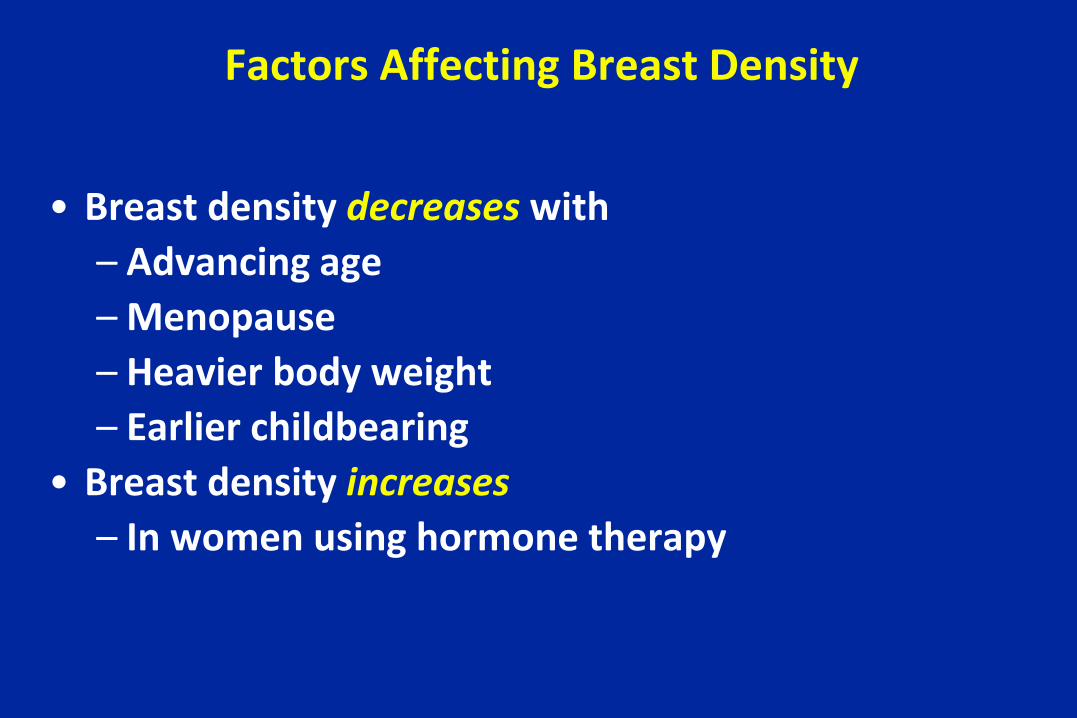
Factors Affecting Breast Density
• Breast density decreases with – Advancing age– Menopause – Heavier body weight– Earlier childbearing
• Breast density increases– In women using hormone therapy
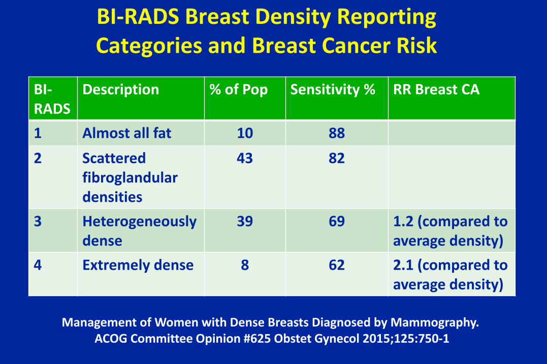
BI-RADS Breast Density Reporting Categories and Breast Cancer Risk
Management of Women with Dense Breasts Diagnosed by Mammography. ACOG Committee Opinion #625 Obstet Gynecol 2015;125:750-1
BI-RADS
Description % of Pop Sensitivity % RR Breast CA
1 Almost all fat 10 882 Scattered
fibroglandulardensities
43 82
3 Heterogeneously dense
39 69 1.2 (compared to average density)
4 Extremely dense 8 62 2.1 (compared to average density)
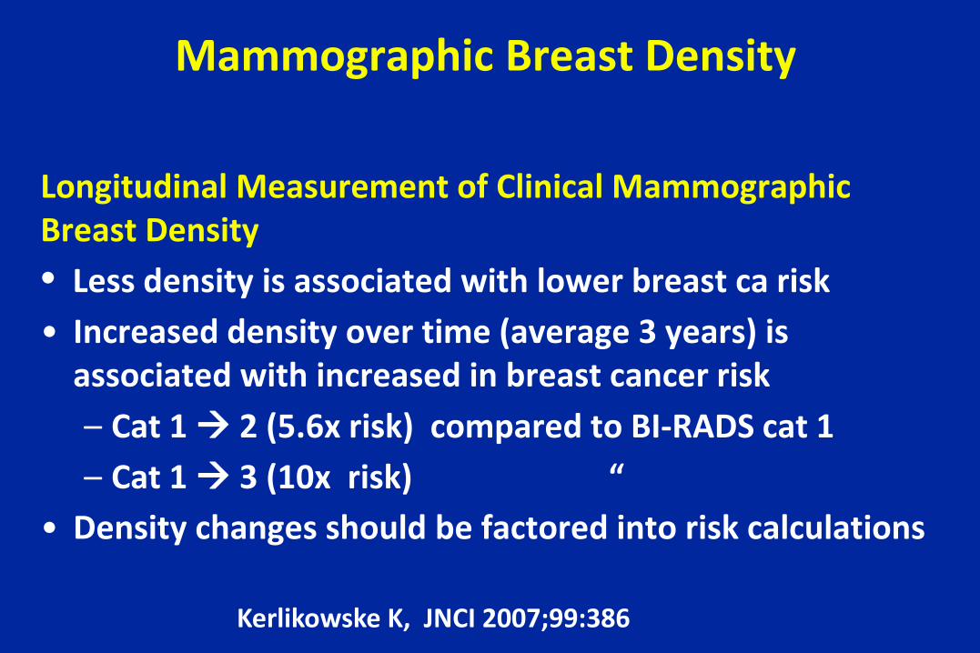
Mammographic Breast Density
Longitudinal Measurement of Clinical Mammographic Breast Density • Less density is associated with lower breast ca risk• Increased density over time (average 3 years) is
associated with increased in breast cancer risk– Cat 1 2 (5.6x risk) compared to BI-RADS cat 1– Cat 1 3 (10x risk) “
• Density changes should be factored into risk calculations
Kerlikowske K, JNCI 2007;99:386
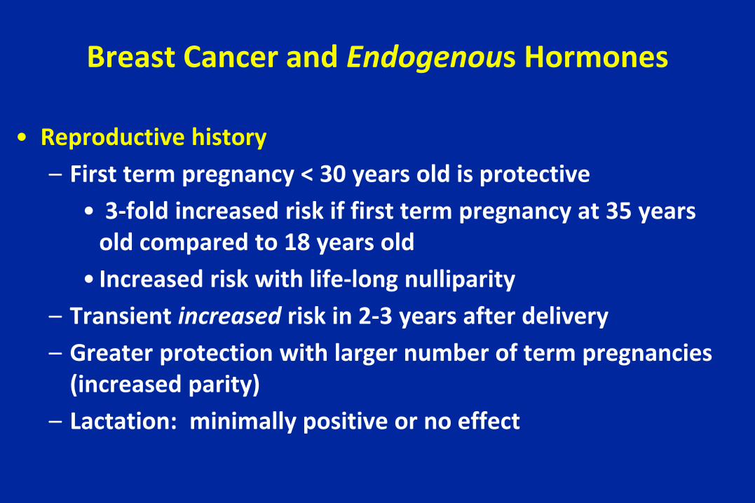
Breast Cancer and Endogenous Hormones
• Reproductive history– First term pregnancy < 30 years old is protective
• 3-fold increased risk if first term pregnancy at 35 years old compared to 18 years old
• Increased risk with life-long nulliparity– Transient increased risk in 2-3 years after delivery– Greater protection with larger number of term pregnancies
(increased parity) – Lactation: minimally positive or no effect
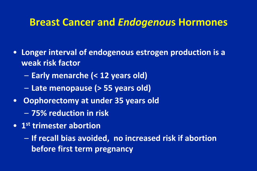
Breast Cancer and Endogenous Hormones
• Longer interval of endogenous estrogen production is a weak risk factor– Early menarche (< 12 years old)– Late menopause (> 55 years old)
• Oophorectomy at under 35 years old– 75% reduction in risk
• 1st trimester abortion– If recall bias avoided, no increased risk if abortion
before first term pregnancy
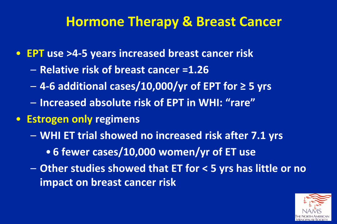
Hormone Therapy & Breast Cancer
• EPT use >4-5 years increased breast cancer risk– Relative risk of breast cancer =1.26– 4-6 additional cases/10,000/yr of EPT for ≥ 5 yrs– Increased absolute risk of EPT in WHI: “rare”
• Estrogen only regimens– WHI ET trial showed no increased risk after 7.1 yrs
• 6 fewer cases/10,000 women/yr of ET use– Other studies showed that ET for < 5 yrs has little or no
impact on breast cancer risk
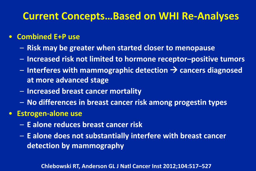
Current Concepts…Based on WHI Re-Analyses
• Combined E+P use– Risk may be greater when started closer to menopause– Increased risk not limited to hormone receptor–positive tumors– Interferes with mammographic detection cancers diagnosed
at more advanced stage– Increased breast cancer mortality– No differences in breast cancer risk among progestin types
• Estrogen-alone use– E alone reduces breast cancer risk– E alone does not substantially interfere with breast cancer
detection by mammography
Chlebowski RT, Anderson GL J Natl Cancer Inst 2012;104:517–527
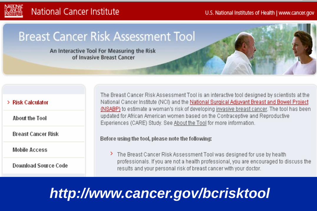
http://www.cancer.gov/bcrisktool
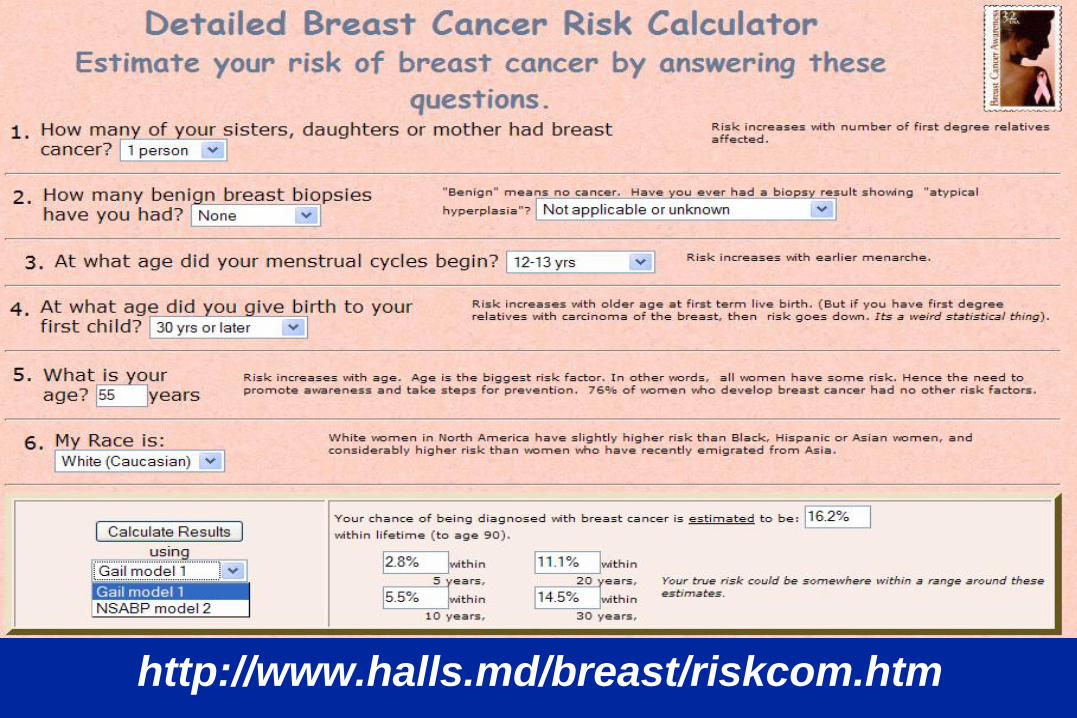
http://www.halls.md/breast/riskcom.htm
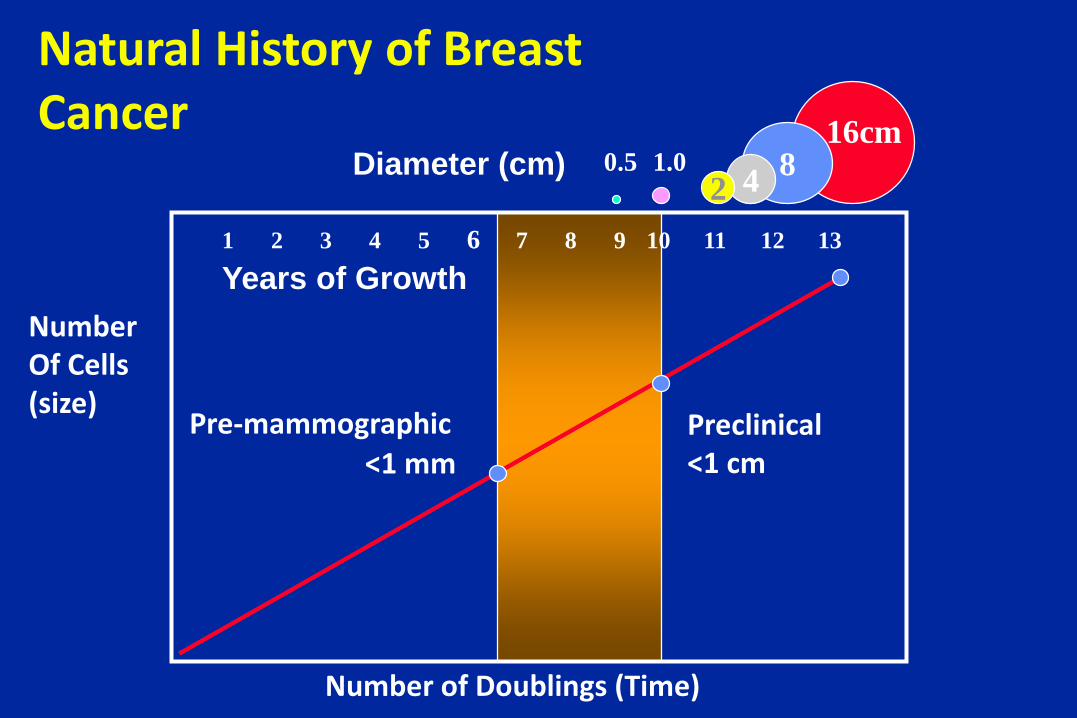
1 2 3 4 6 7 8 9 105 11 12 13
816cm
8421.00.5Diameter (cm)
Pre-mammographic<1 mm
Preclinical<1 cm
Years of Growth
Number of Doublings (Time)
NumberOf Cells(size)
Natural History of Breast Cancer
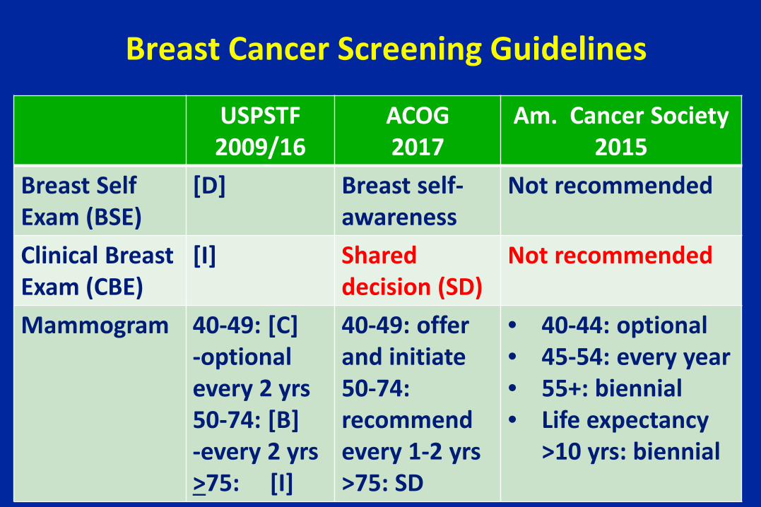
Breast Cancer Screening Guidelines
USPSTF2009/16
ACOG2017
Am. Cancer Society 2015
Breast Self Exam (BSE)
[D] Breast self-awareness
Not recommended
Clinical Breast Exam (CBE)
[I] Shared decision (SD)
Not recommended
Mammogram 40-49: [C]-optional every 2 yrs50-74: [B]-every 2 yrs>75: [I]
40-49: offer and initiate50-74: recommend every 1-2 yrs>75: SD
• 40-44: optional• 45-54: every year• 55+: biennial• Life expectancy
>10 yrs: biennial
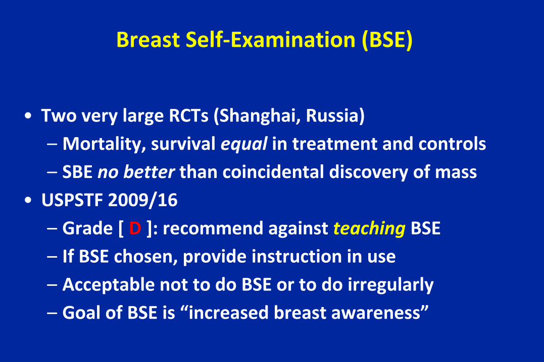
Breast Self-Examination (BSE)
• Two very large RCTs (Shanghai, Russia)– Mortality, survival equal in treatment and controls– SBE no better than coincidental discovery of mass
• USPSTF 2009/16– Grade [ D ]: recommend against teaching BSE– If BSE chosen, provide instruction in use– Acceptable not to do BSE or to do irregularly– Goal of BSE is “increased breast awareness”
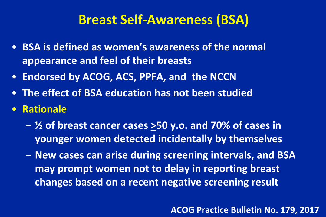
Breast Self-Awareness (BSA)
• BSA is defined as women’s awareness of the normal appearance and feel of their breasts
• Endorsed by ACOG, ACS, PPFA, and the NCCN• The effect of BSA education has not been studied• Rationale
– ½ of breast cancer cases >50 y.o. and 70% of cases in younger women detected incidentally by themselves
– New cases can arise during screening intervals, and BSA may prompt women not to delay in reporting breast changes based on a recent negative screening result
ACOG Practice Bulletin No. 179, 2017
Screening Clinical Breast Exam
The ACS does not recommend clinical breast
examination for breast cancer screening among
average-risk women at any age (Q)
2015
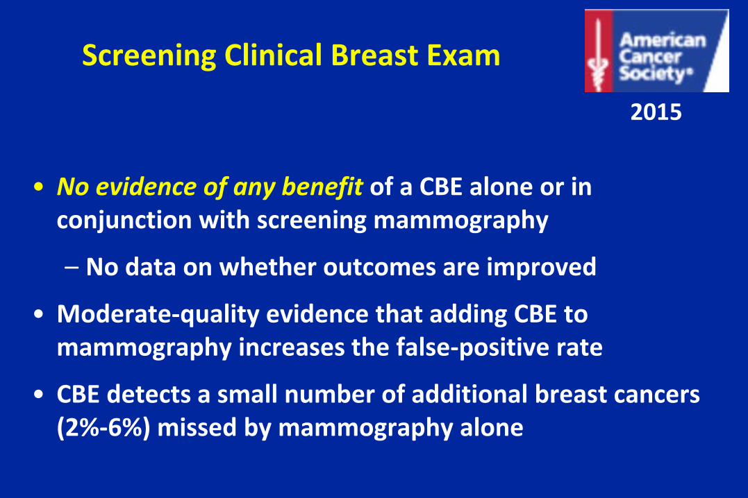
Screening Clinical Breast Exam
• No evidence of any benefit of a CBE alone or in conjunction with screening mammography
– No data on whether outcomes are improved
• Moderate-quality evidence that adding CBE to mammography increases the false-positive rate
• CBE detects a small number of additional breast cancers (2%-6%) missed by mammography alone
2015
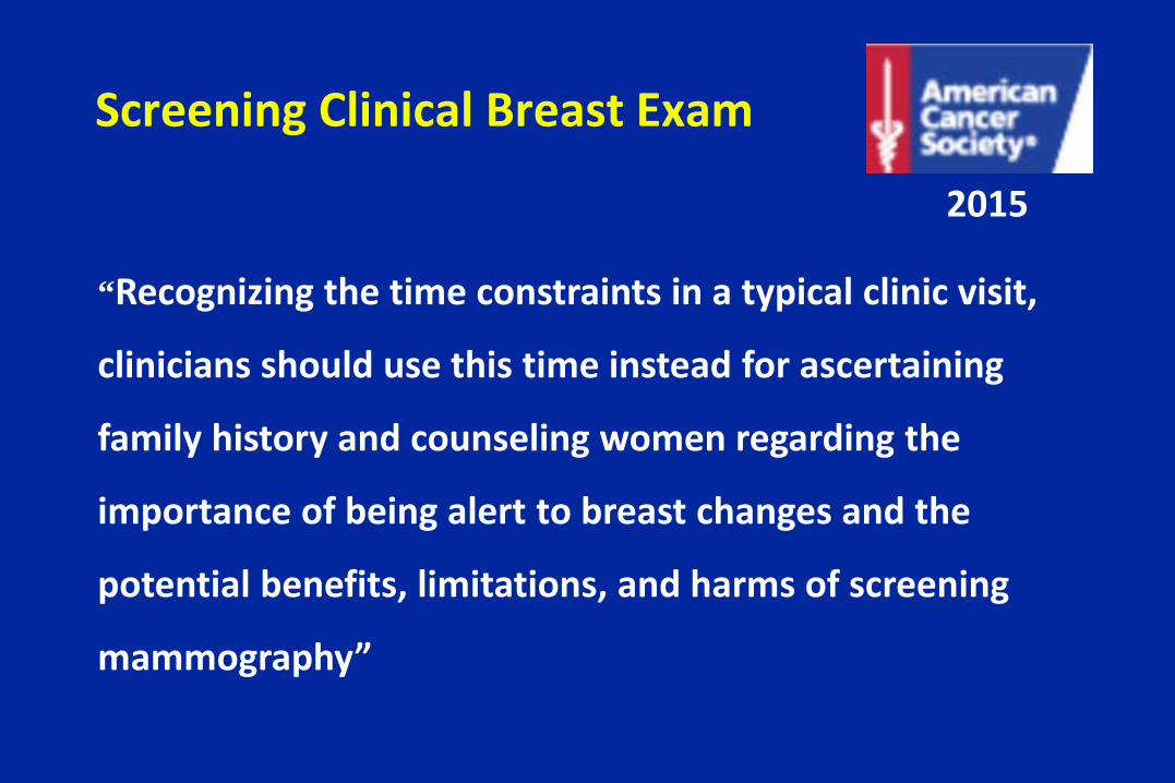
Screening Clinical Breast Exam
2015
“Recognizing the time constraints in a typical clinic visit,
clinicians should use this time instead for ascertaining
family history and counseling women regarding the
importance of being alert to breast changes and the
potential benefits, limitations, and harms of screening
mammography”
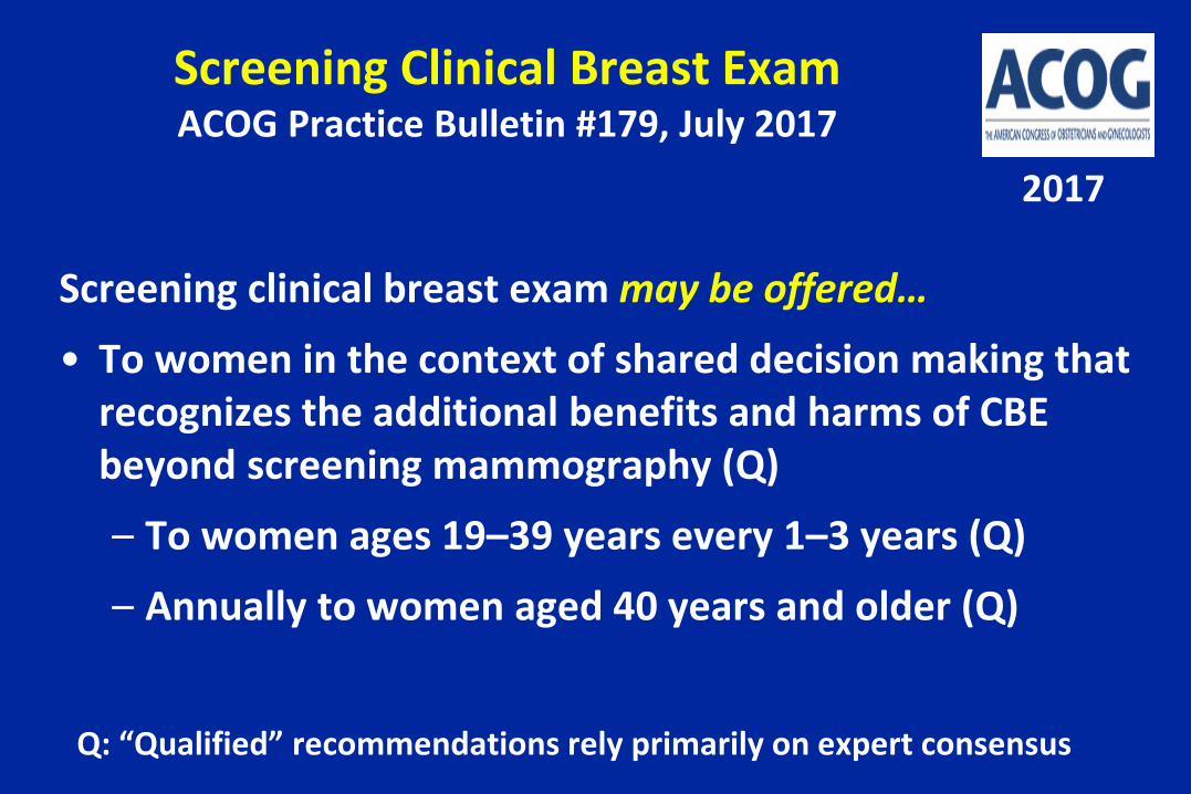
Screening Clinical Breast ExamACOG Practice Bulletin #179, July 2017
Screening clinical breast exam may be offered…
• To women in the context of shared decision making that recognizes the additional benefits and harms of CBE beyond screening mammography (Q)
– To women ages 19–39 years every 1–3 years (Q)
– Annually to women aged 40 years and older (Q)
Q: “Qualified” recommendations rely primarily on expert consensus
2017
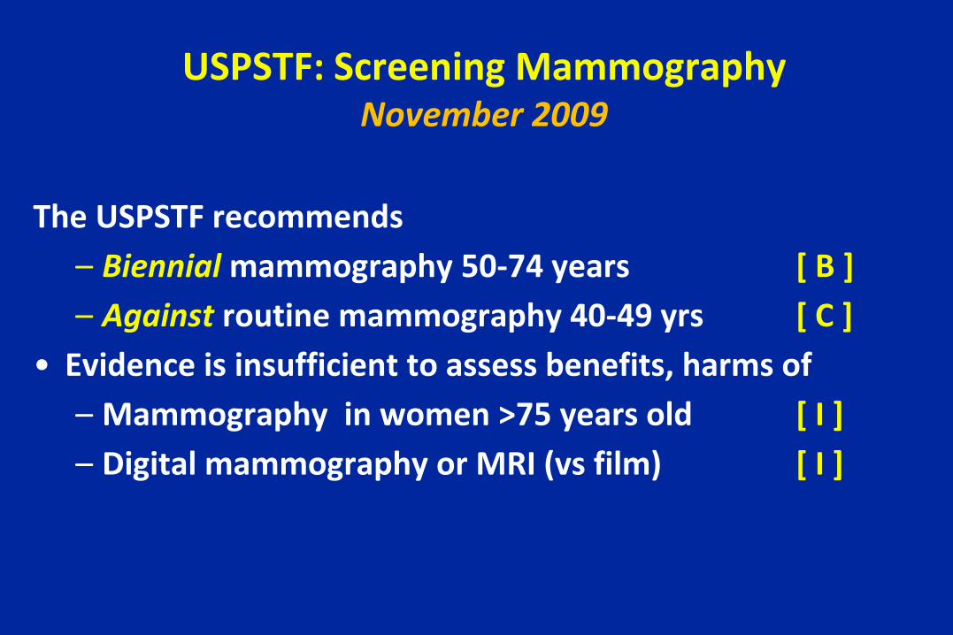
USPSTF: Screening MammographyNovember 2009
The USPSTF recommends– Biennial mammography 50-74 years [ B ]– Against routine mammography 40-49 yrs [ C ]
• Evidence is insufficient to assess benefits, harms of – Mammography in women >75 years old [ I ]– Digital mammography or MRI (vs film) [ I ]
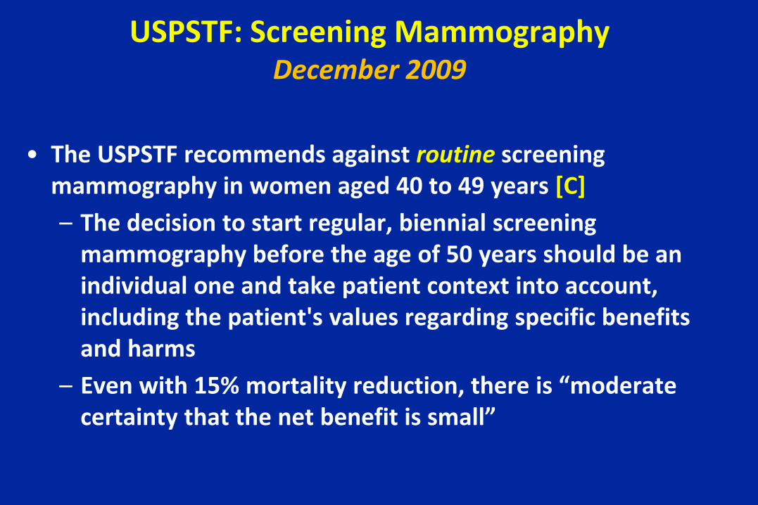
USPSTF: Screening MammographyDecember 2009
• The USPSTF recommends against routine screening mammography in women aged 40 to 49 years [C]– The decision to start regular, biennial screening
mammography before the age of 50 years should be an individual one and take patient context into account, including the patient's values regarding specific benefits and harms
– Even with 15% mortality reduction, there is “moderate certainty that the net benefit is small”
Meta-analysis of RCTs of screening mammography Ann of Intern Med 2009; 151:716
SAME
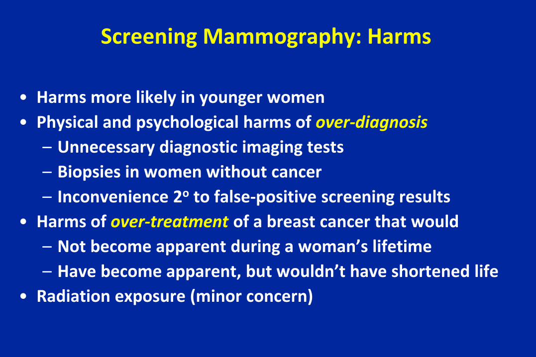
Screening Mammography: Harms
• Harms more likely in younger women• Physical and psychological harms of over-diagnosis
– Unnecessary diagnostic imaging tests– Biopsies in women without cancer– Inconvenience 2o to false-positive screening results
• Harms of over-treatment of a breast cancer that would – Not become apparent during a woman’s lifetime– Have become apparent, but wouldn’t have shortened life
• Radiation exposure (minor concern)
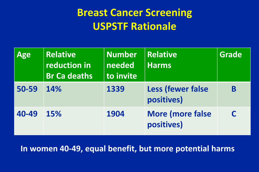
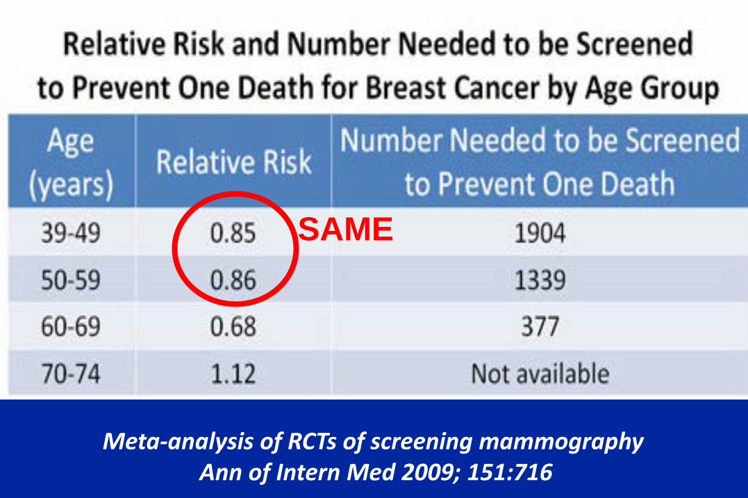
Breast Cancer ScreeningUSPSTF Rationale
Age Relativereduction in Br Ca deaths
Number needed to invite
RelativeHarms
Grade
50-59 14% 1339 Less (fewer false positives)
B
40-49 15% 1904 More (more false positives)
C
In women 40-49, equal benefit, but more potential harms
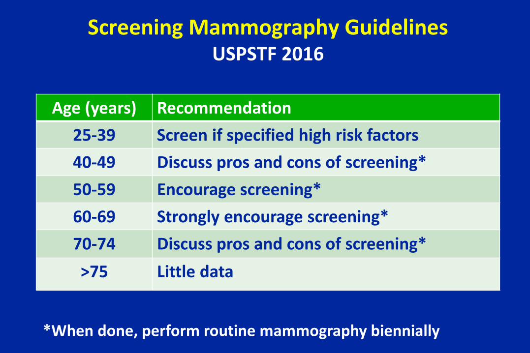
Screening Mammography GuidelinesUSPSTF 2016
Age (years) Recommendation25-39 Screen if specified high risk factors40-49 Discuss pros and cons of screening*50-59 Encourage screening*60-69 Strongly encourage screening*70-74 Discuss pros and cons of screening*>75 Little data
*When done, perform routine mammography biennially
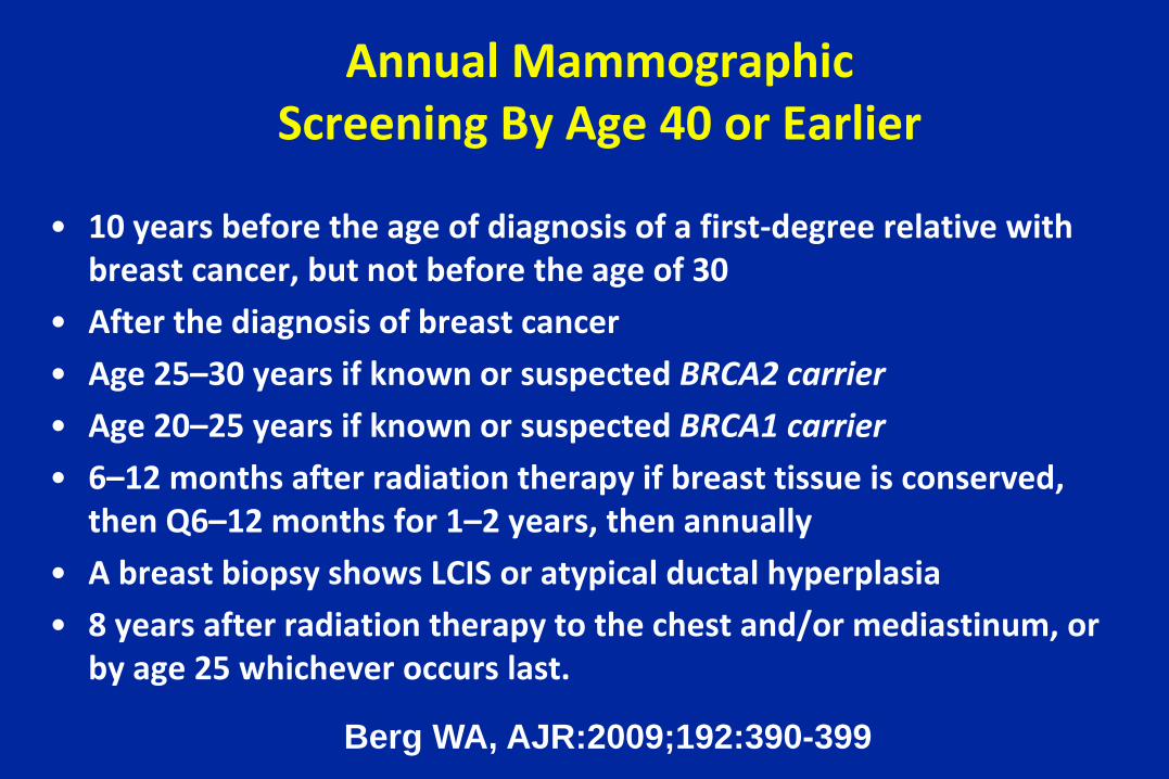
Annual MammographicScreening By Age 40 or Earlier
• 10 years before the age of diagnosis of a first-degree relative with breast cancer, but not before the age of 30
• After the diagnosis of breast cancer• Age 25–30 years if known or suspected BRCA2 carrier• Age 20–25 years if known or suspected BRCA1 carrier• 6–12 months after radiation therapy if breast tissue is conserved,
then Q6–12 months for 1–2 years, then annually• A breast biopsy shows LCIS or atypical ductal hyperplasia• 8 years after radiation therapy to the chest and/or mediastinum, or
by age 25 whichever occurs last.
Berg WA, AJR:2009;192:390-399
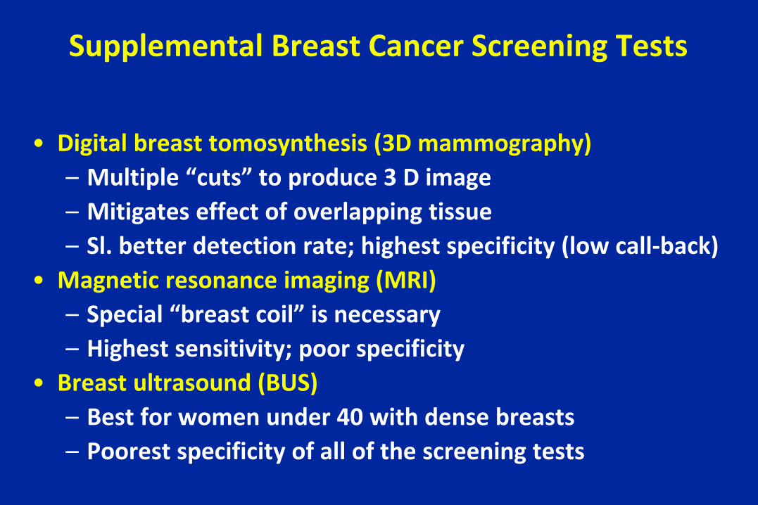
Supplemental Breast Cancer Screening Tests
• Digital breast tomosynthesis (3D mammography)– Multiple “cuts” to produce 3 D image– Mitigates effect of overlapping tissue– Sl. better detection rate; highest specificity (low call-back)
• Magnetic resonance imaging (MRI)– Special “breast coil” is necessary– Highest sensitivity; poor specificity
• Breast ultrasound (BUS)– Best for women under 40 with dense breasts– Poorest specificity of all of the screening tests
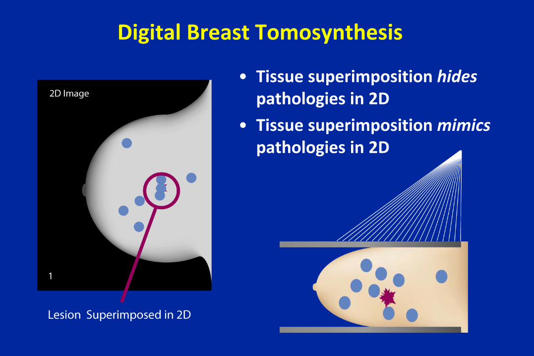
Digital Breast Tomosynthesis
• Tissue superimposition hidespathologies in 2D
• Tissue superimposition mimics pathologies in 2D
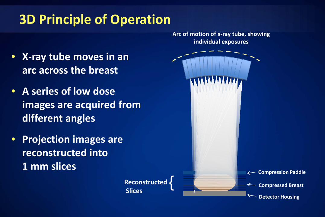
3D Principle of Operation
• X-ray tube moves in an arc across the breast
• A series of low dose images are acquired from different angles
• Projection images are reconstructed into 1 mm slices
Compression Paddle
Compressed Breast
Detector Housing
ReconstructedSlices
{
Arc of motion of x-ray tube, showing individual exposures
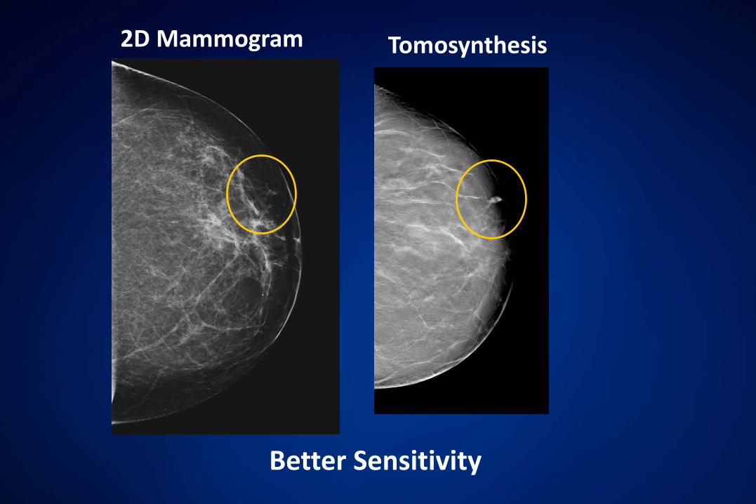
2D Mammogram Tomosynthesis
Better Sensitivity
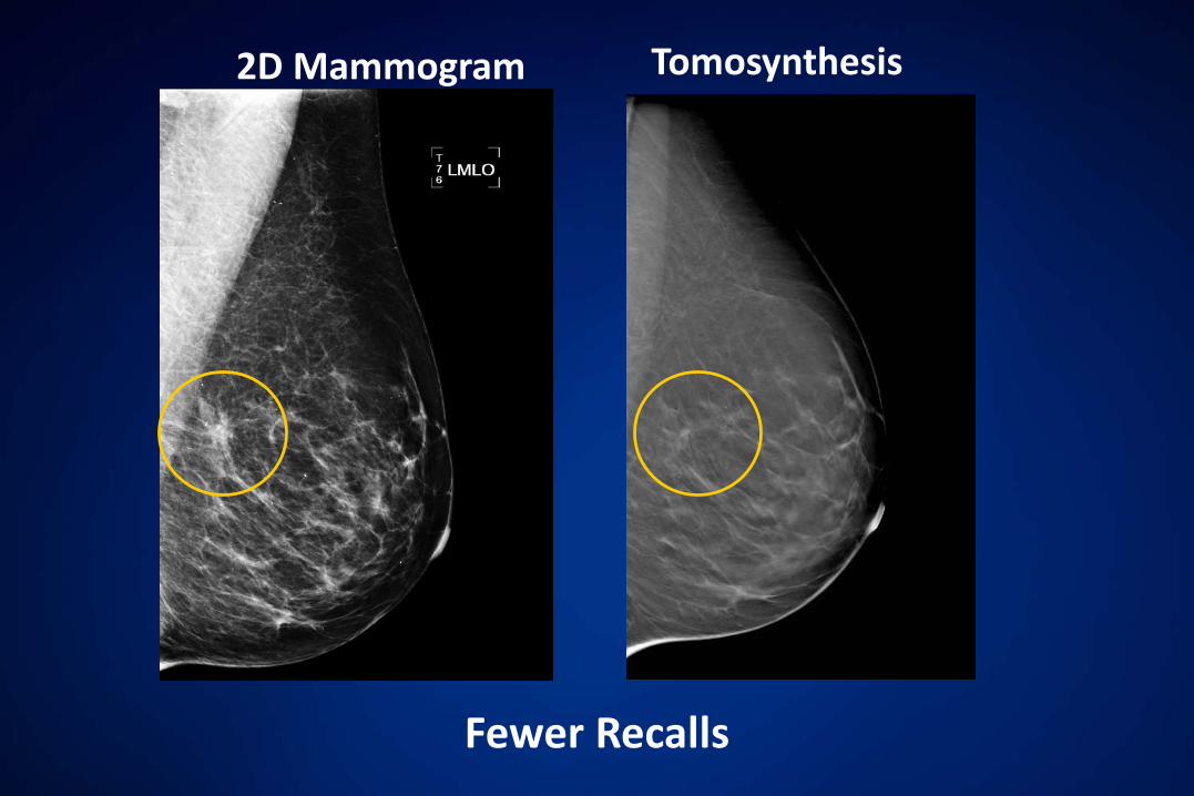
2D Mammogram Tomosynthesis
Fewer Recalls
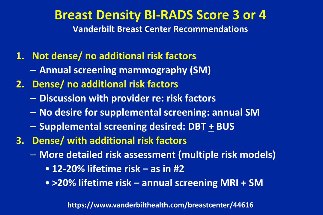
Breast Density BI-RADS Score 3 or 4Vanderbilt Breast Center Recommendations
1. Not dense/ no additional risk factors– Annual screening mammography (SM)
2. Dense/ no additional risk factors– Discussion with provider re: risk factors– No desire for supplemental screening: annual SM – Supplemental screening desired: DBT + BUS
3. Dense/ with additional risk factors– More detailed risk assessment (multiple risk models)
• 12-20% lifetime risk – as in #2• >20% lifetime risk – annual screening MRI + SM
https://www.vanderbilthealth.com/breastcenter/44616
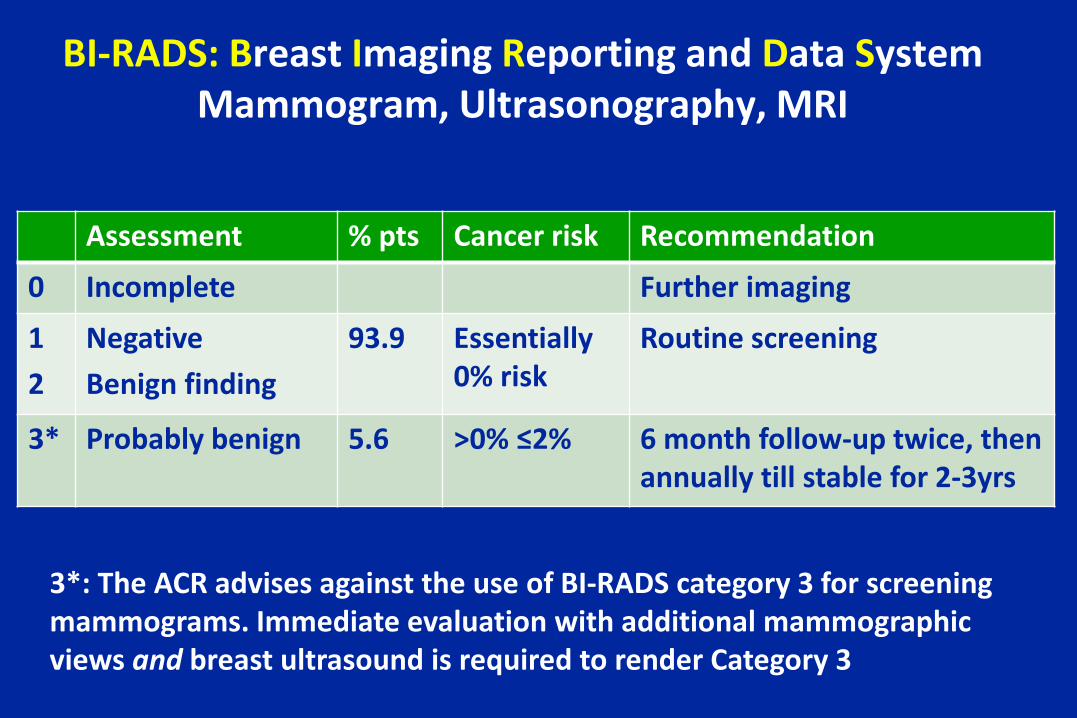
BI-RADS: Breast Imaging Reporting and Data SystemMammogram, Ultrasonography, MRI
Assessment % pts Cancer risk Recommendation
0 Incomplete Further imaging
12
NegativeBenign finding
93.9 Essentially 0% risk
Routine screening
3* Probably benign 5.6 >0% ≤2% 6 month follow-up twice, then annually till stable for 2-3yrs
3*: The ACR advises against the use of BI-RADS category 3 for screening mammograms. Immediate evaluation with additional mammographic views and breast ultrasound is required to render Category 3
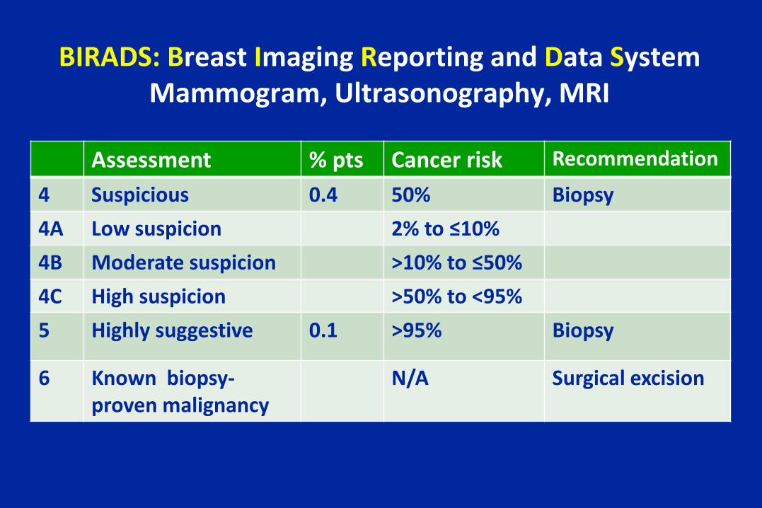
BIRADS: Breast Imaging Reporting and Data SystemMammogram, Ultrasonography, MRI
Assessment % pts Cancer risk Recommendation
4 Suspicious 0.4 50% Biopsy4A Low suspicion 2% to ≤10%4B Moderate suspicion >10% to ≤50%4C High suspicion >50% to <95%5 Highly suggestive 0.1 >95% Biopsy
6 Known biopsy-proven malignancy
N/A Surgical excision
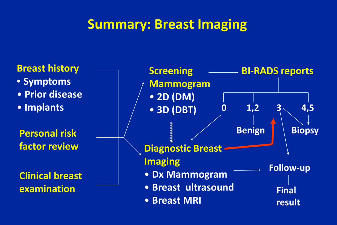
Summary: Breast Imaging
Breast history• Symptoms• Prior disease• Implants
Personal riskfactor review
Clinical breastexamination
ScreeningMammogram• 2D (DM)• 3D (DBT)
Diagnostic BreastImaging• Dx Mammogram• Breast ultrasound• Breast MRI
BI-RADS reports
4,5
Biopsy
1,2
Benign
0 3
Follow-up
Finalresult
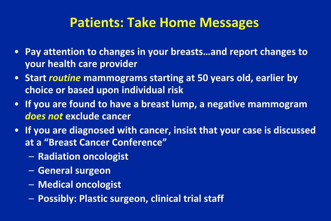
Patients: Take Home Messages
• Pay attention to changes in your breasts…and report changes to your health care provider
• Start routine mammograms starting at 50 years old, earlier by choice or based upon individual risk
• If you are found to have a breast lump, a negative mammogram does not exclude cancer
• If you are diagnosed with cancer, insist that your case is discussed at a “Breast Cancer Conference”– Radiation oncologist– General surgeon– Medical oncologist– Possibly: Plastic surgeon, clinical trial staff
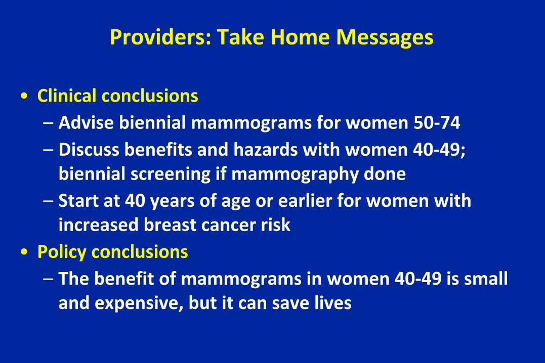
Providers: Take Home Messages
• Clinical conclusions– Advise biennial mammograms for women 50-74– Discuss benefits and hazards with women 40-49;
biennial screening if mammography done– Start at 40 years of age or earlier for women with
increased breast cancer risk• Policy conclusions
– The benefit of mammograms in women 40-49 is small and expensive, but it can save lives
Providers: Take Home Messages
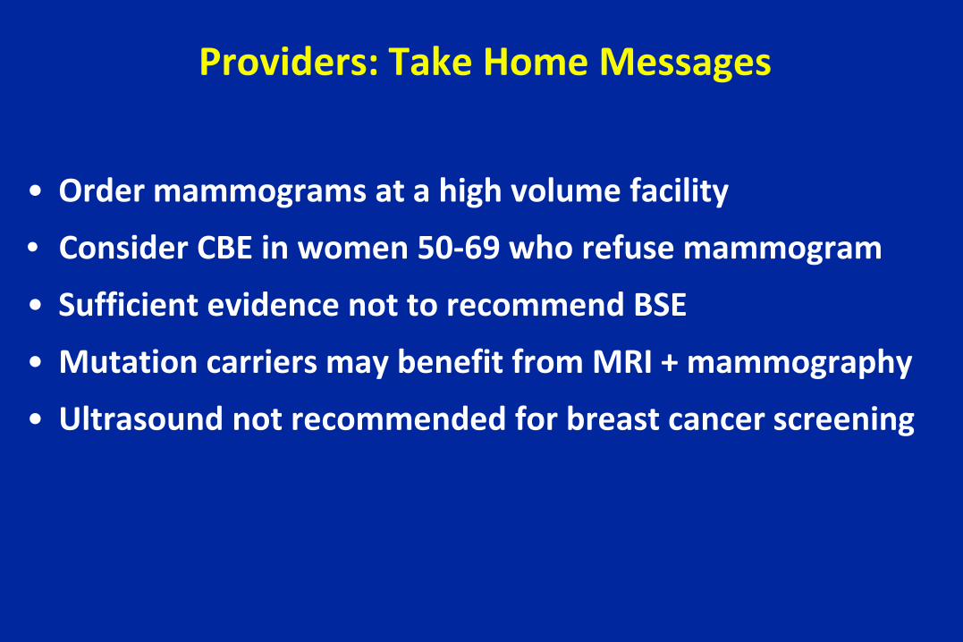
• Order mammograms at a high volume facility
• Consider CBE in women 50-69 who refuse mammogram
• Sufficient evidence not to recommend BSE
• Mutation carriers may benefit from MRI + mammography
• Ultrasound not recommended for breast cancer screening

Appendix
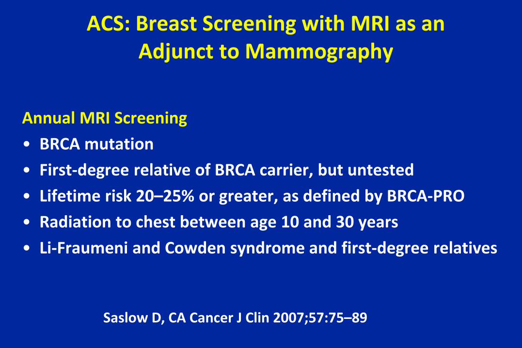
ACS: Breast Screening with MRI as an Adjunct to Mammography
Annual MRI Screening• BRCA mutation• First-degree relative of BRCA carrier, but untested• Lifetime risk 20–25% or greater, as defined by BRCA-PRO• Radiation to chest between age 10 and 30 years• Li-Fraumeni and Cowden syndrome and first-degree relatives
Saslow D, CA Cancer J Clin 2007;57:75–89
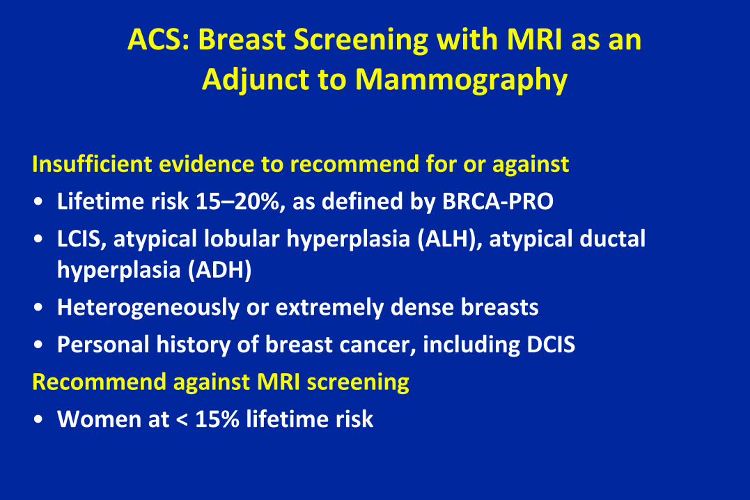
ACS: Breast Screening with MRI as an Adjunct to Mammography
Insufficient evidence to recommend for or against• Lifetime risk 15–20%, as defined by BRCA-PRO• LCIS, atypical lobular hyperplasia (ALH), atypical ductal
hyperplasia (ADH)• Heterogeneously or extremely dense breasts• Personal history of breast cancer, including DCISRecommend against MRI screening• Women at < 15% lifetime risk
References
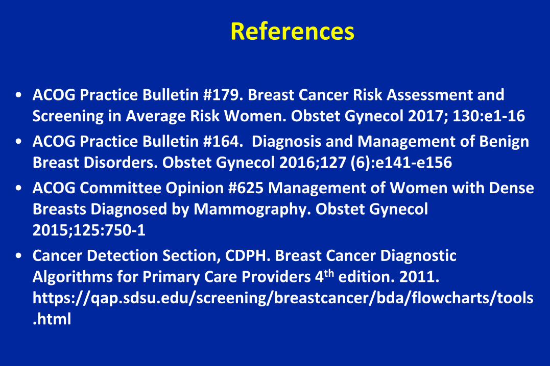
• ACOG Practice Bulletin #179. Breast Cancer Risk Assessment and Screening in Average Risk Women. Obstet Gynecol 2017; 130:e1-16
• ACOG Practice Bulletin #164. Diagnosis and Management of Benign Breast Disorders. Obstet Gynecol 2016;127 (6):e141-e156
• ACOG Committee Opinion #625 Management of Women with Dense Breasts Diagnosed by Mammography. Obstet Gynecol 2015;125:750-1
• Cancer Detection Section, CDPH. Breast Cancer Diagnostic Algorithms for Primary Care Providers 4th edition. 2011. https://qap.sdsu.edu/screening/breastcancer/bda/flowcharts/tools.html

Hereditary Breast Cancer Risk Assessment
• ACOG Committee Opinion No. 634. Hereditary cancer syndromes and risk assessment. Obstet Gynecol 2015; 125:1538–43.
• Lu KH, et al. American Society of Clinical Oncology Expert Statement: collection and use of a cancer family history for oncology providers. American Society of Clinical Oncology. J ClinOncol 2014;32:833–40
• Lancaster JM, et. al. SGO statement on risk assessment for inherited gynecologic cancer predispositions. Gynecol Oncol 2015;136:3–7.
• Moyer VA. Risk assessment, genetic counseling, and genetic testing for BRCA-related cancer in women: USPSTF recommendation statement. Ann Intern Med 2014;160:271–81.
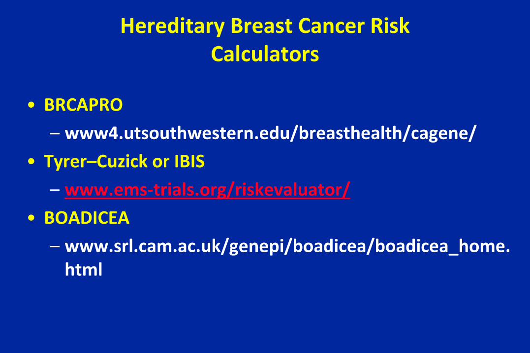
Hereditary Breast Cancer Risk Calculators
• BRCAPRO– www4.utsouthwestern.edu/breasthealth/cagene/
• Tyrer–Cuzick or IBIS– www.ems-trials.org/riskevaluator/
• BOADICEA– www.srl.cam.ac.uk/genepi/boadicea/boadicea_home.
html


![Screening for Breast Cancer[1]](https://img.dokumen.tips/doc/110x75/577d2c841a28ab4e1eac7094/screening-for-breast-cancer1.jpg)