Embed Size (px)
Citation preview
®Answers you can trust. Care you can count on.
B R E A S TIMAGING
Digital Mammography,Breast Ultrasound, Biopsies,
MRI & PET/CT
AN OVERVIEW OF BREAST IMAGINGBreast cancer is the second leading cause ofcancer death in women today. It is estimated that one in seven women in the U.S. will develop breast cancer in her lifetime. Yet research shows the � ve-year relative survival rate for those who detect their breast cancer early is 82 percent.
Our Breast Imaging Program gives your doctoraccess to innovative technology, specialized expertise, and superior-quality images to uncoverthe critical information needed to accuratelydiagnose and e� ectively treat breast cancer.
The Most Advanced Imaging TechnologyWith our comprehensive mix of imagingmodalities, we have the appropriate equipment and technology to get the job done — accurately,e� ciently and skillfully.
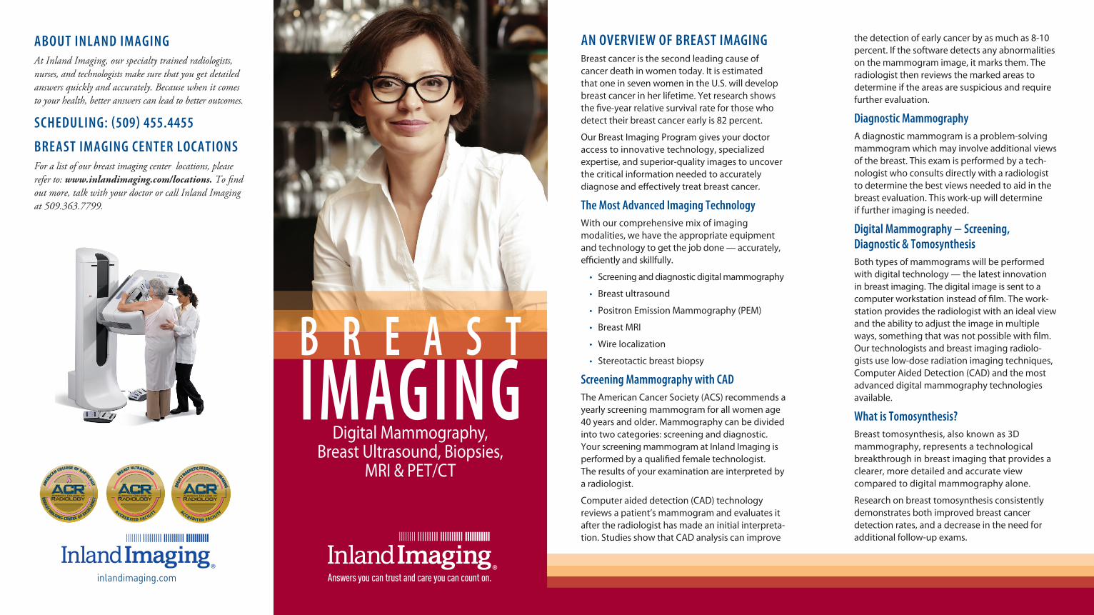
• Screening and diagnostic digital mammography
• Breast ultrasound
• Positron Emission Mammography (PEM)
• Breast MRI
• Wire localization
• Stereotactic breast biopsy
Screening Mammography with CADThe American Cancer Society (ACS) recommends a yearly screening mammogram for all women age 40 years and older. Mammography can be divided into two categories: screening and diagnostic. Your screening mammogram at Inland Imaging is performed by a quali� ed female technologist.The results of your examination are interpreted by a radiologist.
Computer aided detection (CAD) technology reviews a patient’s mammogram and evaluates it after the radiologist has made an initial interpreta-tion. Studies show that CAD analysis can improve
the detection of early cancer by as much as 8-10 percent. If the software detects any abnormalities on the mammogram image, it marks them. The radiologist then reviews the marked areas todetermine if the areas are suspicious and require further evaluation.
Diagnostic MammographyA diagnostic mammogram is a problem-solvingmammogram which may involve additional viewsof the breast. This exam is performed by a tech-nologist who consults directly with a radiologist to determine the best views needed to aid in the breast evaluation. This work-up will determineif further imaging is needed.
Digital Mammography − Screening, Diagnostic & TomosynthesisBoth types of mammograms will be performedwith digital technology — the latest innovationin breast imaging. The digital image is sent to a computer workstation instead of � lm. The work-station provides the radiologist with an ideal view and the ability to adjust the image in multiple ways, something that was not possible with � lm. Our technologists and breast imaging radiolo-gists use low-dose radiation imaging techniques, Computer Aided Detection (CAD) and the most advanced digital mammography technologies available.
What is Tomosynthesis?Breast tomosynthesis, also known as 3Dmammography, represents a technologicalbreakthrough in breast imaging that provides a clearer, more detailed and accurate viewcompared to digital mammography alone.
Research on breast tomosynthesis consistentlydemonstrates both improved breast cancerdetection rates, and a decrease in the need foradditional follow-up exams.
ABOUT INLAND IMAGINGAt Inland Imaging, our specialty trained radiologists, nurses, and technologists make sure that you get detailed answers quickly and accurately. Because when it comes to your health, better answers can lead to better outcomes.
SCHEDULING: (509) 455.4455
BREAST IMAGING CENTER LOCATIONSFor a list of our breast imaging center locations, please refer to: www.inlandimaging.com/locations. To � nd out more, talk with your doctor or call Inland Imagingat 509.363.7799.
inlandimaging.com
3D Breast Cancer DetectionConventional digital mammography produces one image of overlapping tissue, making it di� cult to detect abnormalities. Breast tomosynthesis takes multiple images of the entire breast,allowing Inland Imaging’s breast imagingradiologists to see through layers of tissue to examine areas of concern from a variety of angles, one thin slice at a time.
*3D mammography screening research� ndings included:
• A 41% increase in the detection of invasive breast cancers
• A 29% increase in the detection of all breast cancers
• A 15% decrease in women recalled for additional imaging
• A 40% decrease in “false positives”
Breast UltrasoundA breast ultrasound is useful in helping to deter-mine if a suspicious area is a fluid-filled cyst or a solid mass that requires further testing. The tool may also be used to guide biopsies.
Breast BiopsiesA breast biopsy is a tissue sampling technique used to con� rm or rule out the presence of breast cancer. Breast biopsies can be surgical or non-surgical; Inland Imaging specializes in non-surgical breast biopsies. Utilizing these methods bene� ts patients by decreasing recovery time and reducing scarring compared with surgical excisional biopsy. Inland Imaging utilizes two primary non-surgical methods to obtain samples: ultrasound-guided core-needle breast biopsy and stereotacticbreast biopsy.
*Source: Journal of the American Medical Association, June 25, 2014, Breast Cancer Screening Using Tomosynthesis in Combination with Digital Mammography.
Ultrasound-guided core-needle biopsy iscommonly used to evaluate suspicious masses within the breast, whether they can be felt duringa clinical examination. An ultrasound probe is placed over the site and a radiologist guides a biopsy needle directly into the mass. Localanesthesia is used during this procedure.
Stereotactic biopsy uses a dedicated biopsy table combined with digital mammography to deter-mine the exact biopsy location. Tissue samples are then extracted using a vacuum assisted biopsy instrument. A local anesthesia is used during this procedure so patients have minimal discomfort during and after procedure. Normal activity can usually resume the following day.
Breast Wire LocalizationUsing the appropriate imaging modality, a local-ization wire is placed in the breast to accurately target an abnormality for surgical removal. Duringsurgery, surgeons can electronically access theimages showing the placement of the wire. Imaging tests are done on the surgically excised specimen to verify removal of the abnormality and wire. Results are immediately phoned to the surgeon.
Breast MRIMagnetic resonance breast imaging (breast MRI) has been approved by the Food and Drug Admin-istration (FDA) since 1991 for use as a supplement to mammography to help diagnose breast cancer.Unlike mammography, which uses low-dose X-raysto image the breast, MRI uses powerful magnetic � elds and radio waves to create images of the breast.
Biopsies may also be performed using breast MRI. MRI-guided breast biopsy is a fast, safe and easy way to � nd and biopsy breast abnormalitieswithout putting women through unnecessarysurgery. Our new state-of-the-art breast MRIsystem optimizes patient comfort, reducesunnecessary movement during the exam, and produces superior images. It also o� ers improved access for biopsies.
Patients undergoing a breast MRI exam lie facedown on the MRI table which is speciallycon� gured so that the breasts are positionedto hang freely through two openings calledbreast coils. After images have been acquiredand assessed with CAD, a radiologist reads andinterprets the images.
Positron Emission Mammography (PEM)PEM, an advanced application of Positron Emission Tomography (PET), captures localized images of
the breast, producing sharp, detailed images of breast lesions – as small as 1.5 mm. This informa-tion is valuable to aid in developing treatment options and surgical plans.
PEM or MRI?The breast MRI is a standard in diagnostic imag-ing, but there may be circumstances in which a Positron Emission Mammography (PEM) test is a better option. To determine if a PEM test is an ap-propriate alternative, your physician will consider the following:
• PEM is a good option for a patient who has a large body habitus and cannot � t in an MRI machine, has a pacemaker or other metallic implants, or is claustrophobic.
• PEM can be helpful in patients with breast implants.
• PEM is less susceptible to the hormonal e� ects of the menstrual cycle.
• PEM can be useful to problem solve complex MRI � ndings.
• The higher sensitivity of PEM makes it an e� ective tool in the evaluation of DCIS (ductal carcinoma in situ), a common form of non- invasive breast cancer.
The Region’s Breast Imaging Experts• Each of our 10 breast imaging radiologists is board-certi� ed.
• Our physicians are knowledgeable and experienced in their � eld, and they keep current of the latest technological developments and clinical research.
• Our team of breast imaging experts includes more than 40 dedicated technologists.
Breast MRI images captured using our high-resolution breast MRI and biopsy table — recently upgraded with the Sentinelle Vanguard MRI coil.
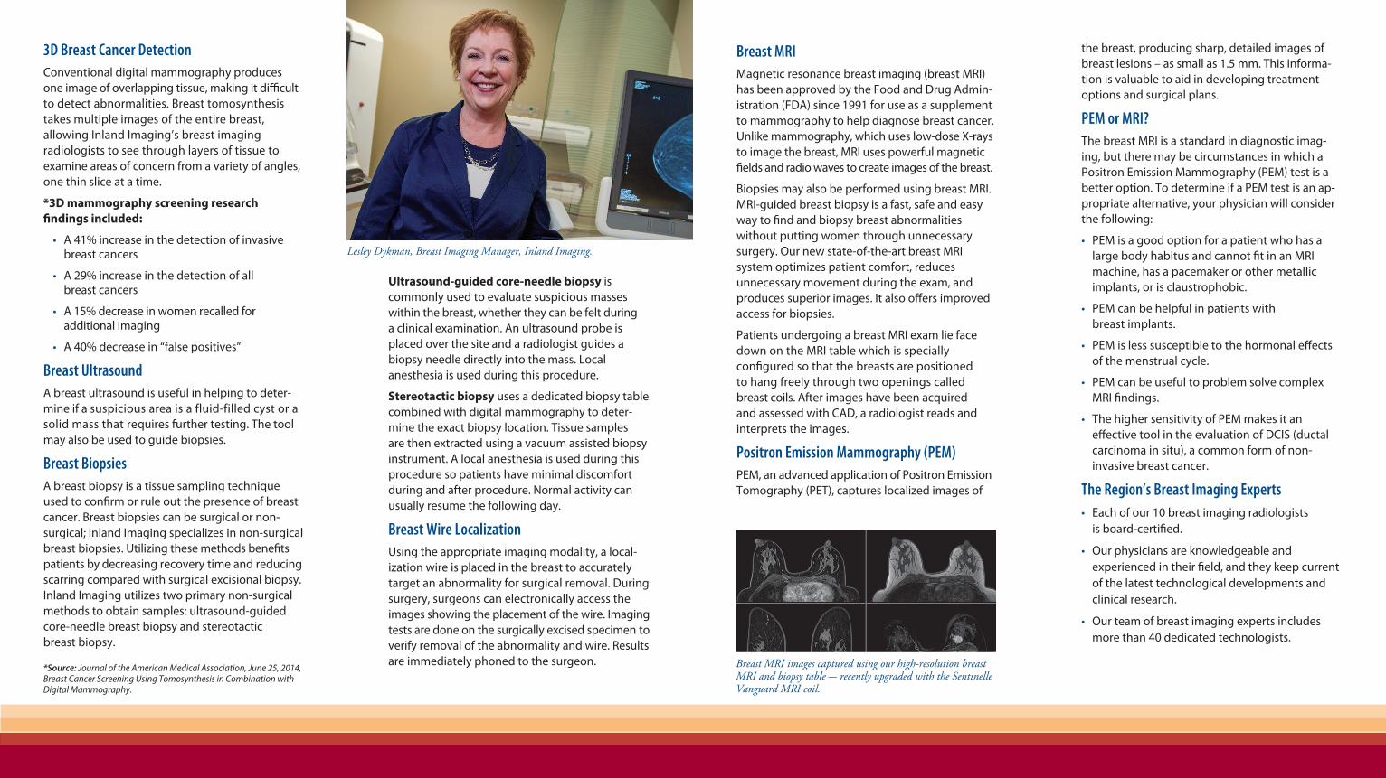
Lesley Dykman, Breast Imaging Manager, Inland Imaging.