Embed Size (px)
Citation preview

Breast: Difficulties in Core Biopsies
Anna Marie Mulligan, MB, MSc, FRCPath University Health Network and University of Toronto
E-mail: [email protected]

No conflicts of interest
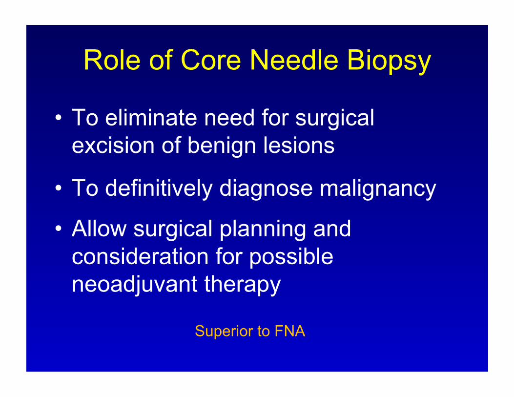
Role of Core Needle Biopsy
To eliminate need for surgical excision of benign lesions
To definitively diagnose malignancy
Allow surgical planning and consideration for possible neoadjuvant therapy
Superior to FNA

Advantages ↓ no. of surgeries in
women with IBC Less invasive Less expensive Less scarring on
subsequent imaging Confirm multiple foci
and determine appropriate surgical procedure
Challenges
Limited material
Disruption frequent
Sampling error

Accuracy Excellent agreement with open bx. Accuracy, sensitivity, specificity
– 0.98, 0.88, 0.98 (mostly palpable) – 0.98, 0.91, 1.00 (non-palpable) – ↑ when > 6 cores – ↑ with experienced radiologist
Original and review pathologist 96% concordance
Wei et al, Med Onc, 2010 Fajardo et al, Acad Rad, 2004
False Negatives 1-15% for malignancy
– Lower with 11G/vacuum
Causes: – Sampling/Suspicious mass – not explained
by histologic findings – Calcifications – not identified in the core
Calcs > mass DCIS > invasive
Key to success
Does my diagnosis correlate with the radiologic findings?
Know the indication for the biopsy !!!!!

Triple Test Clinical
Radiology Pathology
Must correlate!
Important for pathologist to appreciate imaging categories and pathology correlation
Multidisciplinary rounds
Specimen x-ray
Role of the Pathologist in the Evaluation of Calcifications
Calcifications Specimen radiograph mandatory for any biopsy performed for calcifications – Separate cores with calcs from those without
If calcs seen on mammo but none in specimen x-ray: – ability to make dx ↓ from 82 to 40%
Dx more likely in cores bearing calcs on specimen x-ray than those without – 84 vs 71%
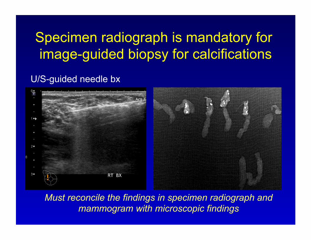
Specimen radiograph is mandatory for image-guided biopsy for calcifications
U/S-guided needle bx
Must reconcile the findings in specimen radiograph and mammogram with microscopic findings
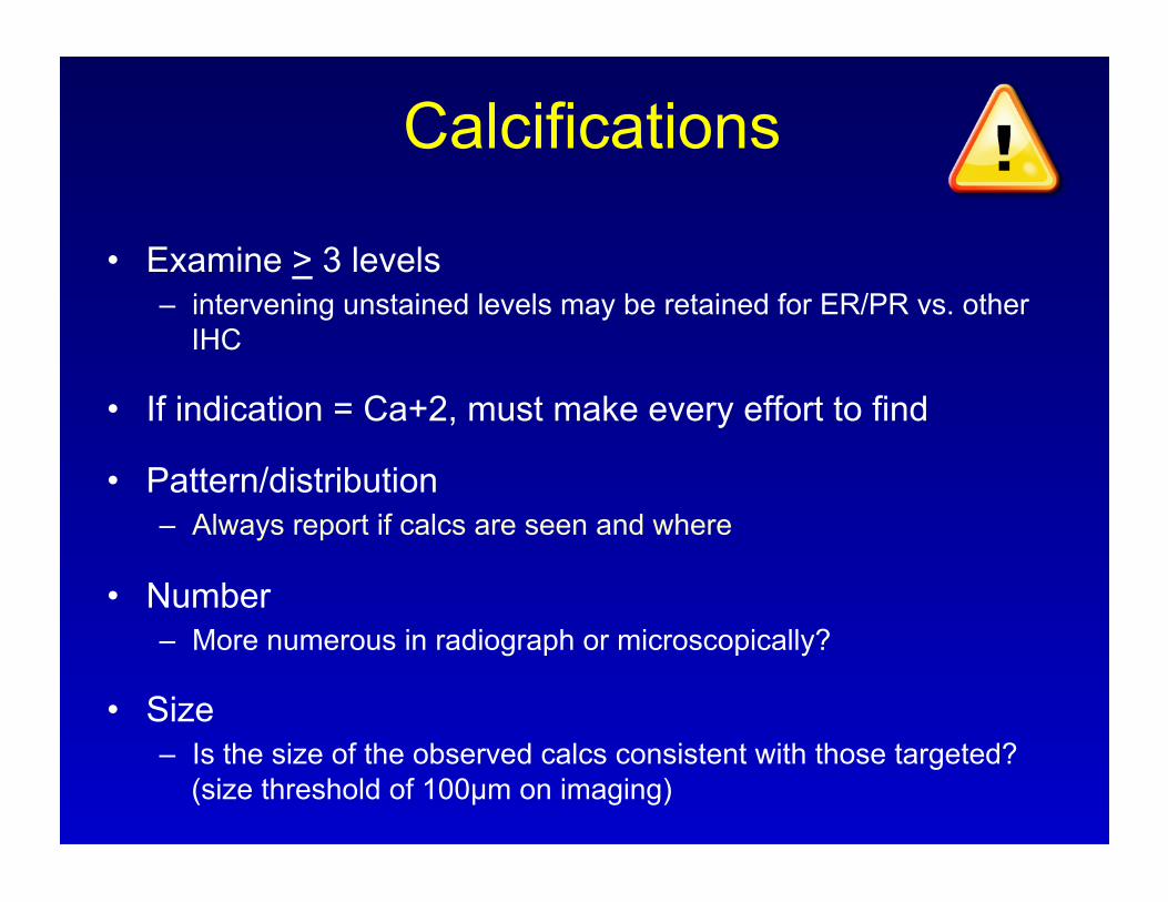
Calcifications
Examine > 3 levels – intervening unstained levels may be retained for ER/PR vs. other
IHC
If indication = Ca+2, must make every effort to find
Pattern/distribution – Always report if calcs are seen and where
Number – More numerous in radiograph or microscopically?
Size – Is the size of the observed calcs consistent with those targeted?
(size threshold of 100µm on imaging)

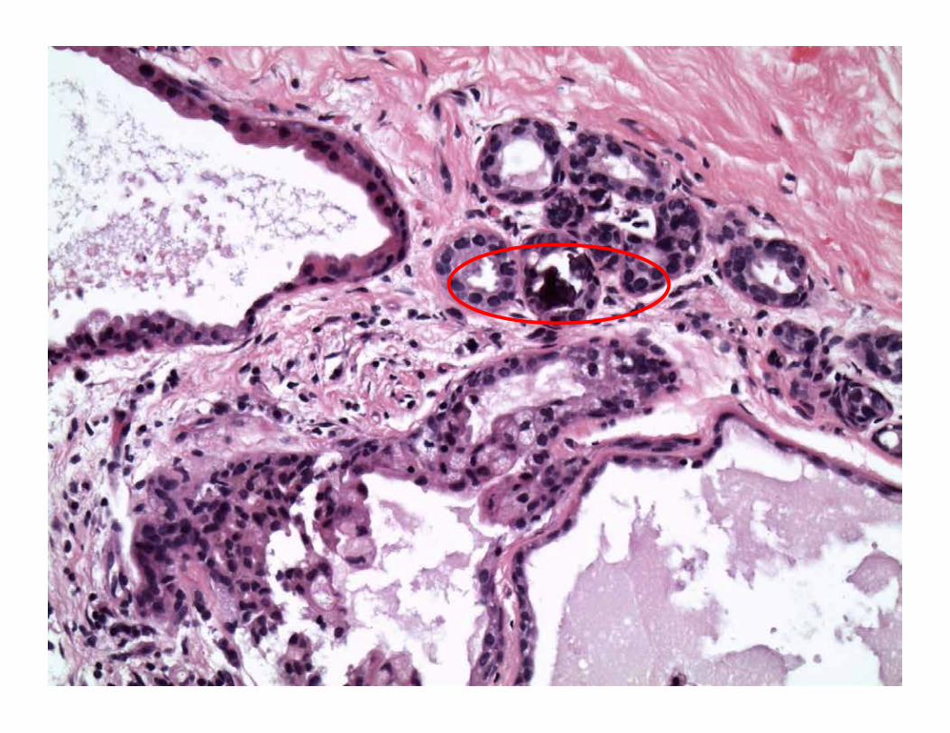
Audience participation 1
58 y.o woman with linear branching calcifications on mammography Stereotactic core biopsy Calcifications are reported as being present in the specimen x-ray
True or false
Pathology correlates with radiology Findings are benign
Patient returns to screening programme

False
Calcifications not in keeping with linear branching morphology
Cut deeper levels
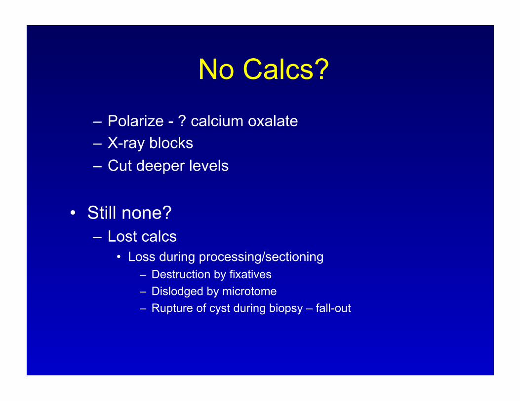
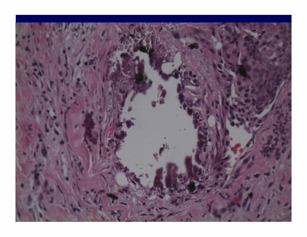
No Calcs? – Polarize - ? calcium oxalate – X-ray blocks – Cut deeper levels
Still none? – Lost calcs
Loss during processing/sectioning – Destruction by fixatives – Dislodged by microtome – Rupture of cyst during biopsy – fall-out

Microcalcifications: calcium oxalate
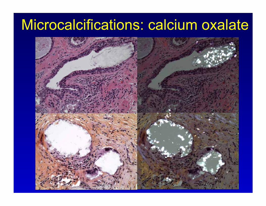
Microcalcifications: calcium oxalate
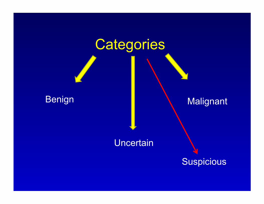
Categories
Benign Malignant
Uncertain
Suspicious
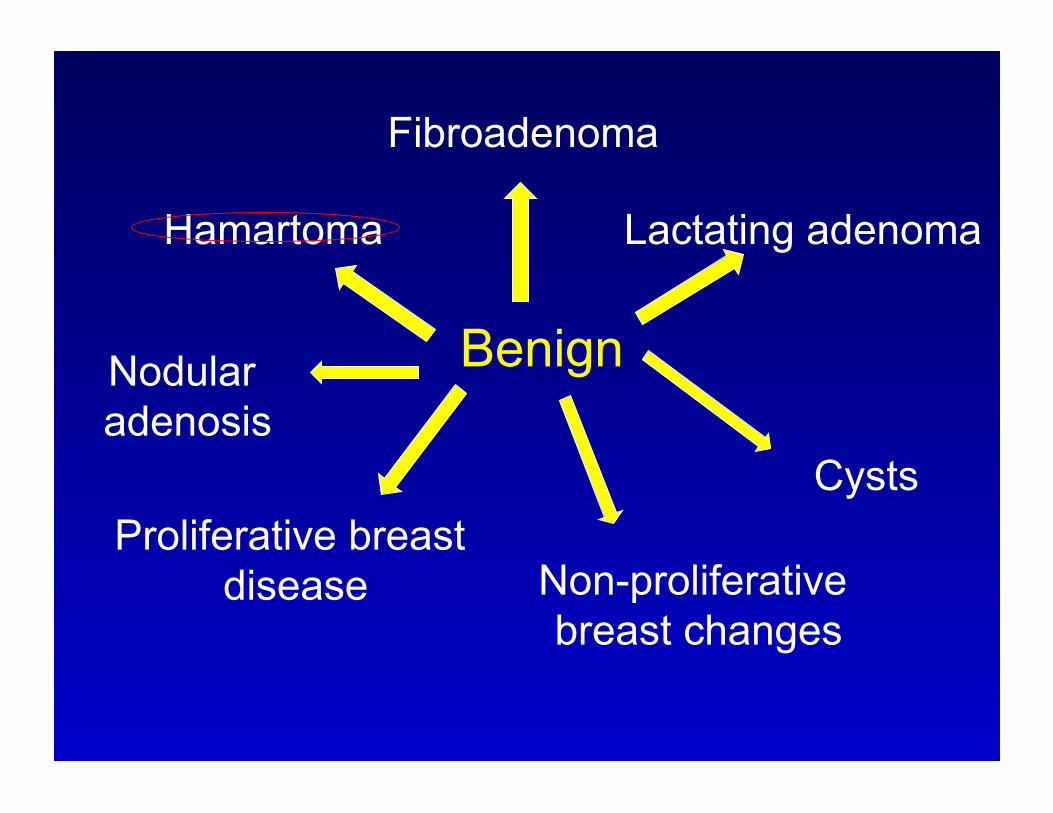
Benign
Lactating adenoma
Cysts
Nodular adenosis
Fibroadenoma
Hamartoma
Non-proliferative breast changes
Proliferative breast disease
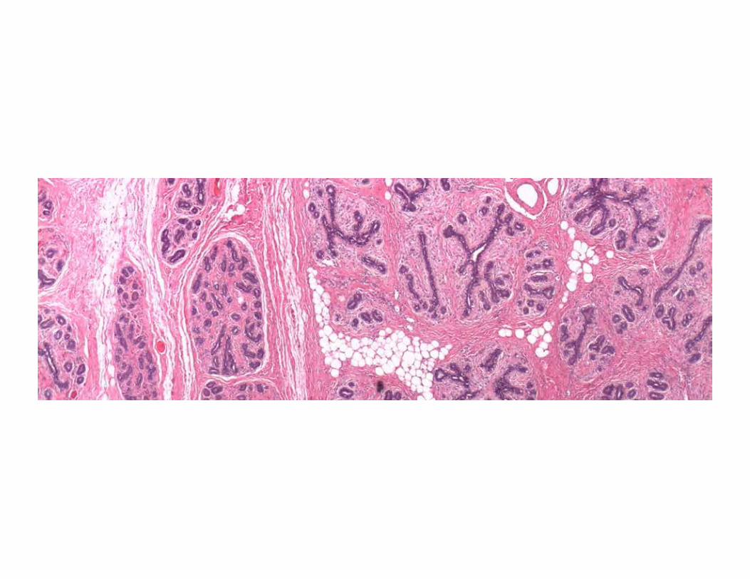
Hamartoma
Highlights importance of rad-path correlation
Circumscribed/encapsulated – Ducts, lobules, fibrous tissue, fat (variable)
Typical mammographic appearance – Obviating need for CNB in most
Hamartoma - CNB
“Benign fibrous and fatty breast tissue”
Was the lesion sampled?
Are the characteristic imaging features present?
If “yes” → consider hamartoma
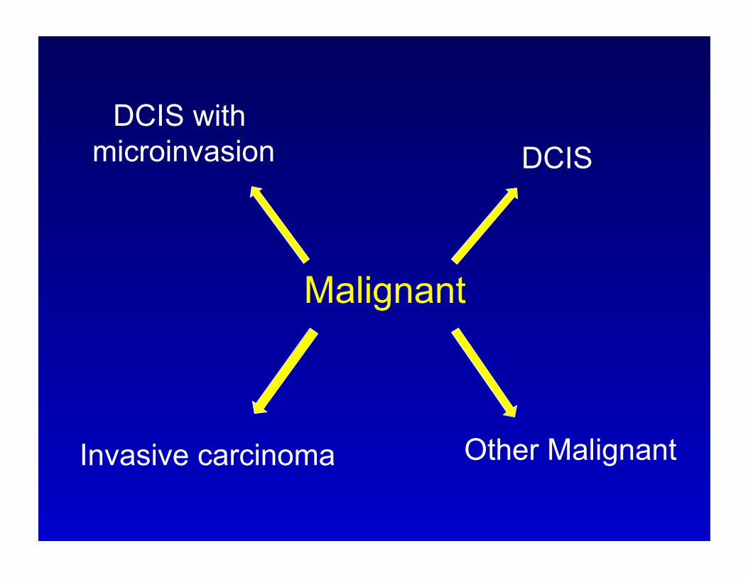
Malignant
Invasive carcinoma
DCIS DCIS with
microinvasion
Other Malignant
Pitfalls Beware:
– DCIS with obliterative sclerosis – Displacement of calcs – Displacement of atypical cells

Pitfalls Beware:
– DCIS with obliterative sclerosis – Displacement of calcs – Displacement of atypical cells
Pitfalls Beware:
– DCIS with obliterative sclerosis – Displacement of calcs – Displacement of atypical cells
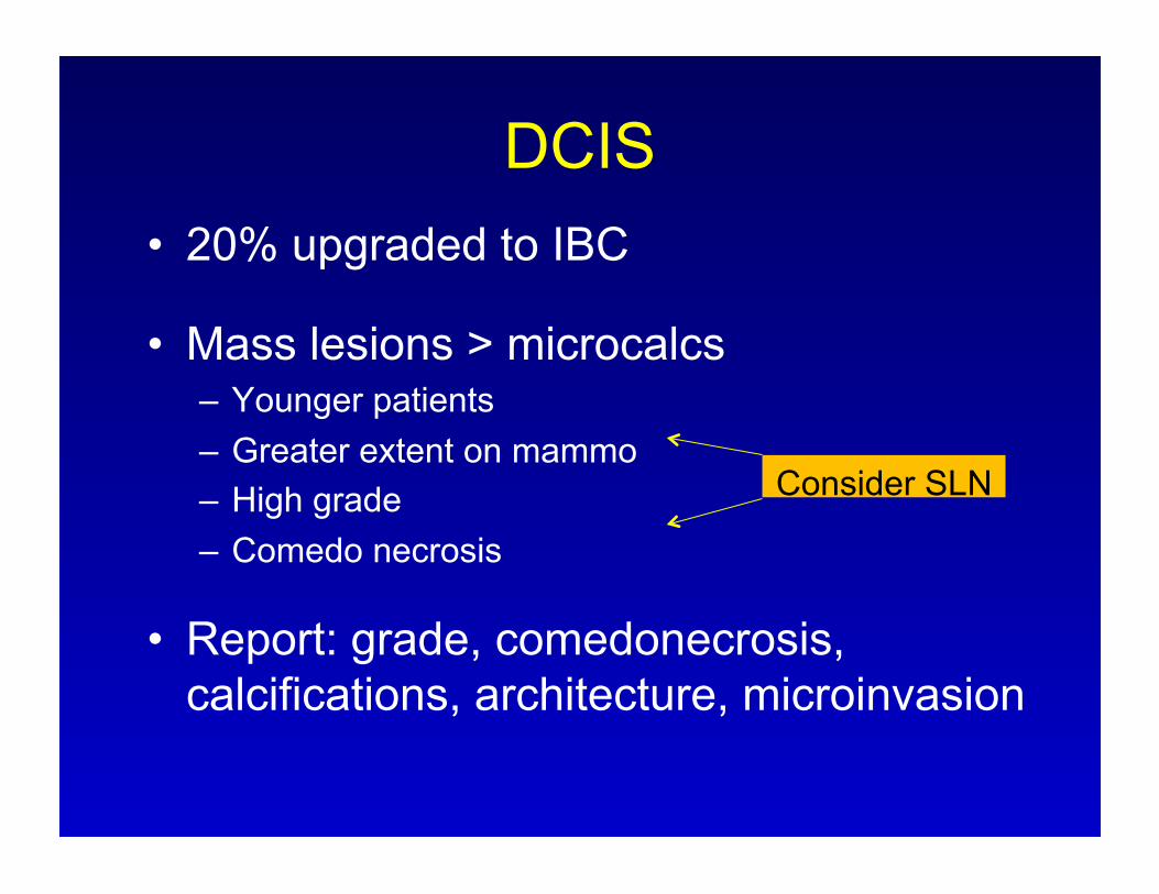
DCIS 20% upgraded to IBC
Mass lesions > microcalcs – Younger patients – Greater extent on mammo – High grade – Comedo necrosis
Report: grade, comedonecrosis, calcifications, architecture, microinvasion
Consider SLN
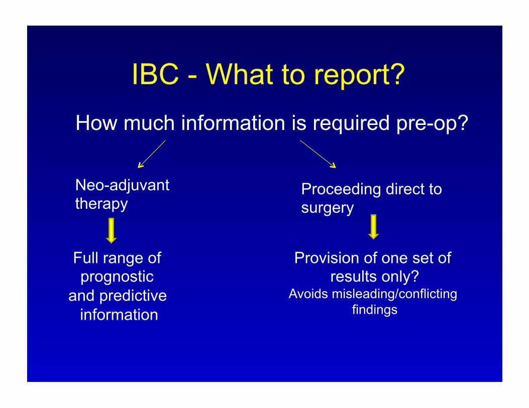
IBC - What to report? How much information is required pre-op?
Neo-adjuvant therapy
Proceeding direct to surgery
Full range of prognostic
and predictive information
Provision of one set of results only?
Avoids misleading/conflicting findings
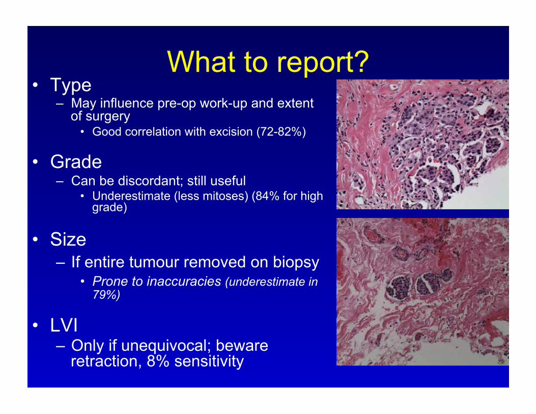
What to report? Type
– May influence pre-op work-up and extent of surgery Good correlation with excision (72-82%)
Grade – Can be discordant; still useful
Underestimate (less mitoses) (84% for high grade)
Size – If entire tumour removed on biopsy
Prone to inaccuracies (underestimate in 79%)
LVI – Only if unequivocal; beware
retraction, 8% sensitivity

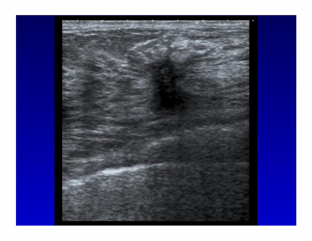
Audience Participation 2
65 year old woman, screening detected mass, 1.3 cm Ultrasound-guided core biopsy


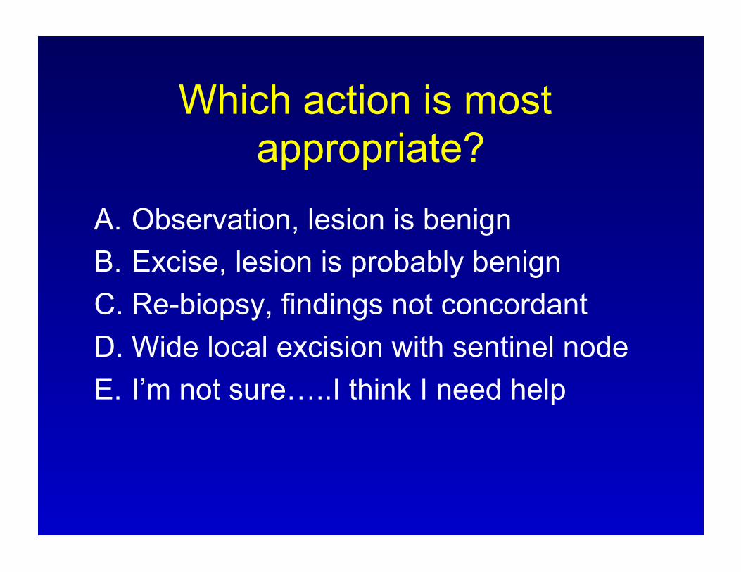
Which action is most appropriate?
A. Observation, lesion is benign B. Excise, lesion is probably benign C. Re-biopsy, findings not concordant D. Wide local excision with sentinel node E. I’m not sure…..I think I need help
Which action is most appropriate?
A. Observation, lesion is benign B. Excise, lesion is probably benign C. Re-biopsy, findings not concordant D. Wide local excision with sentinel node E. I’m not sure…..I think I need help
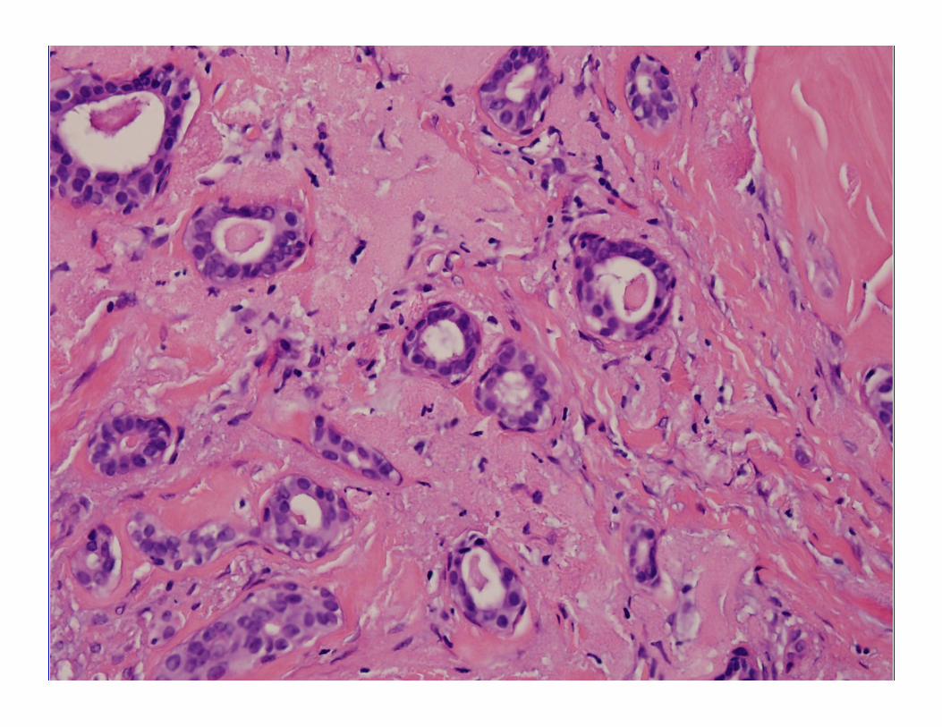
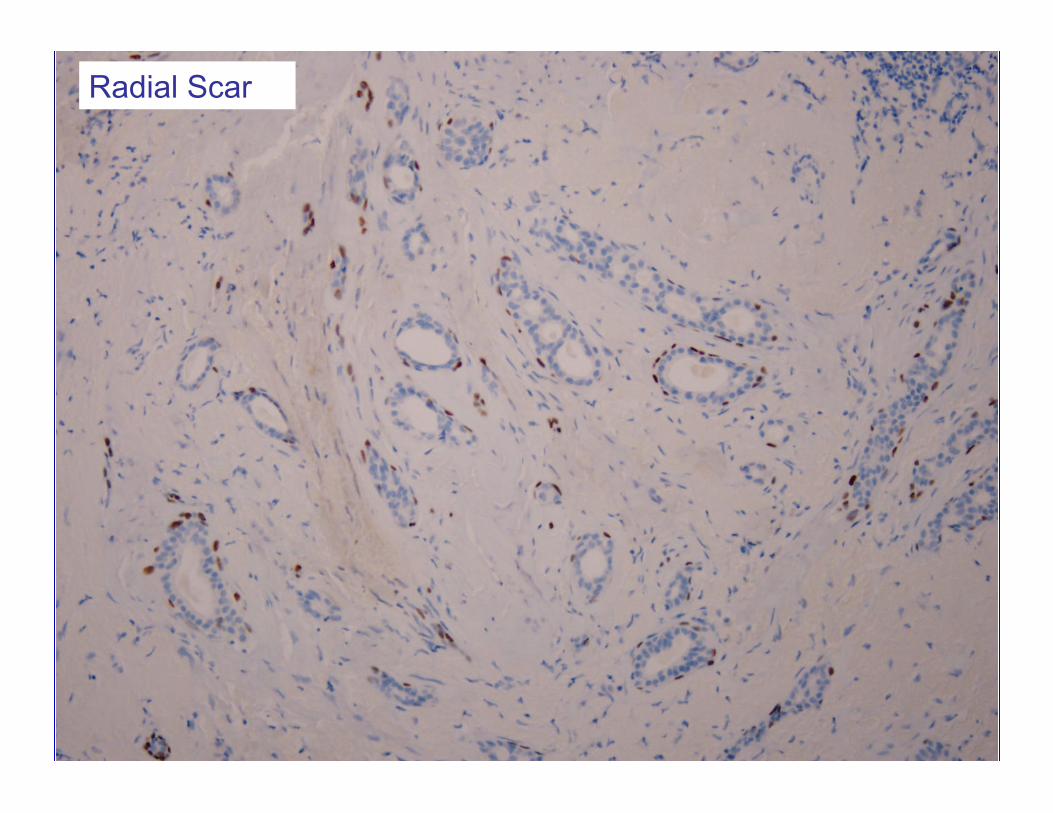
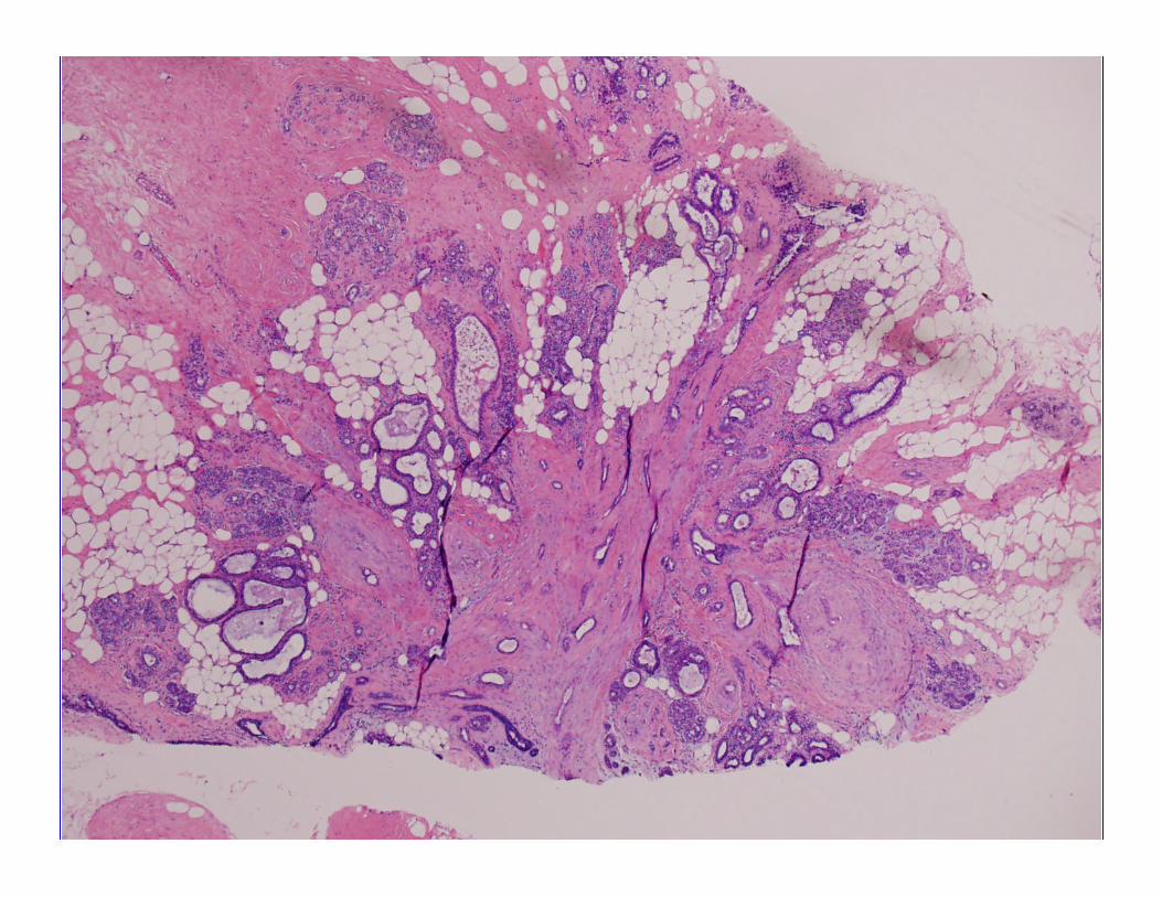
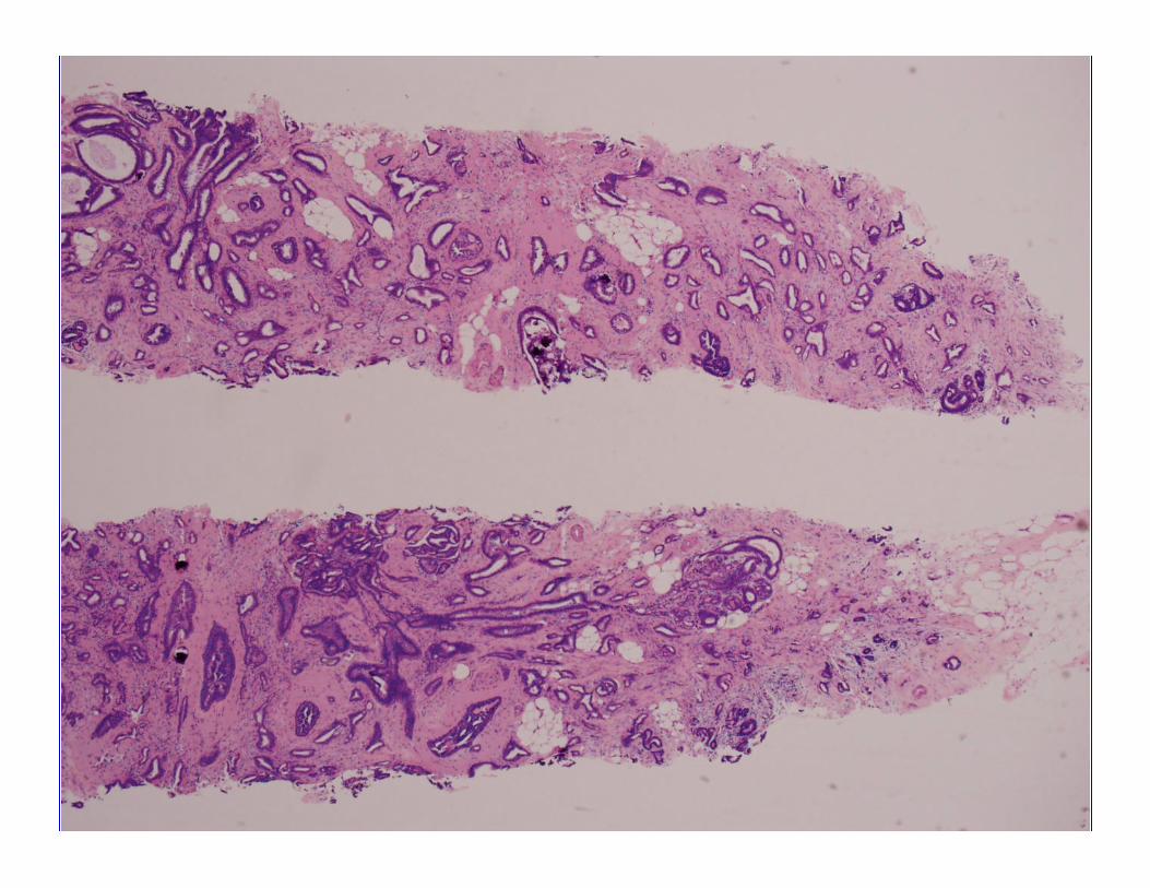
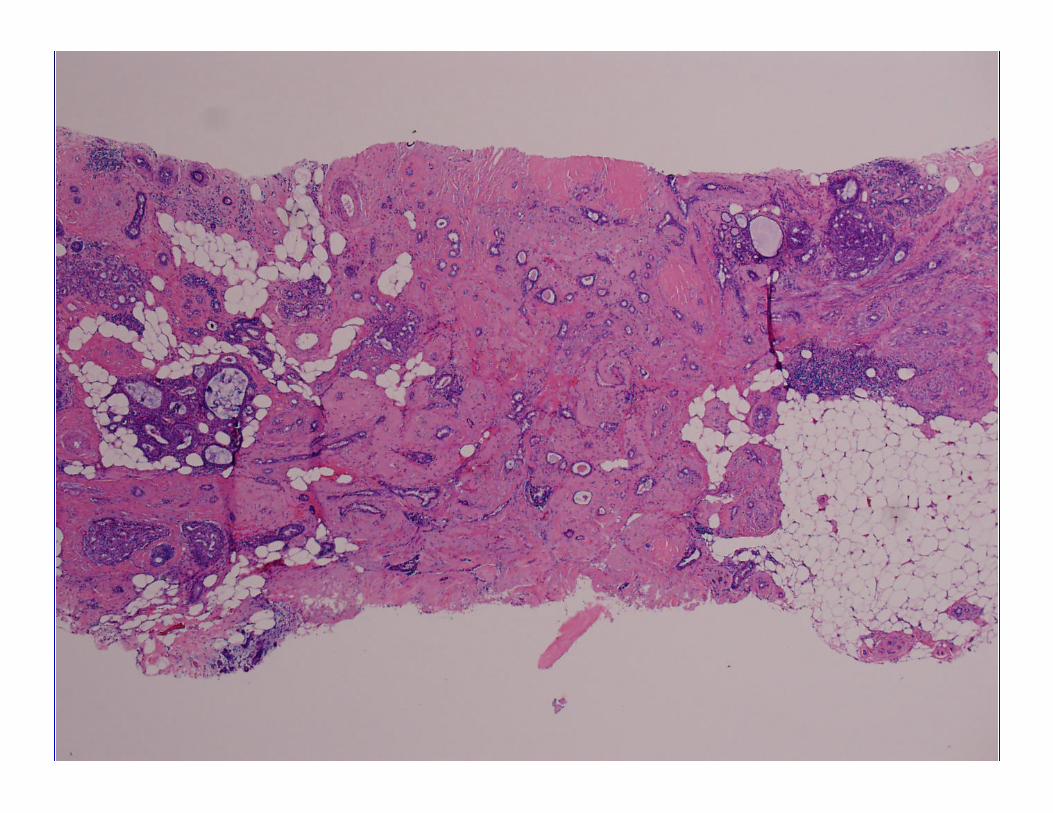
Radial Scar
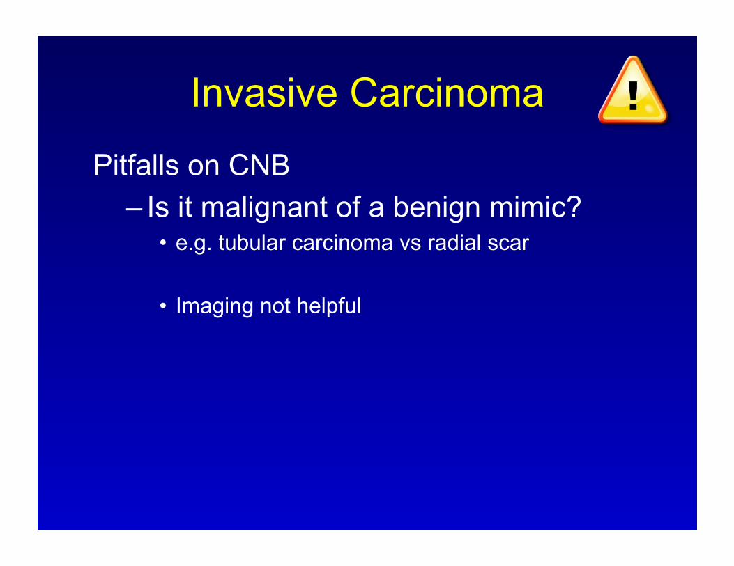
Invasive Carcinoma
Pitfalls on CNB – Is it malignant of a benign mimic?
e.g. tubular carcinoma vs radial scar
Imaging not helpful
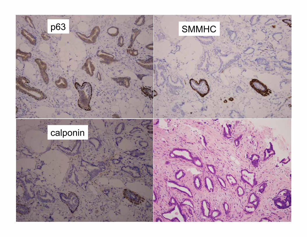
p63 SMMHC
calponin

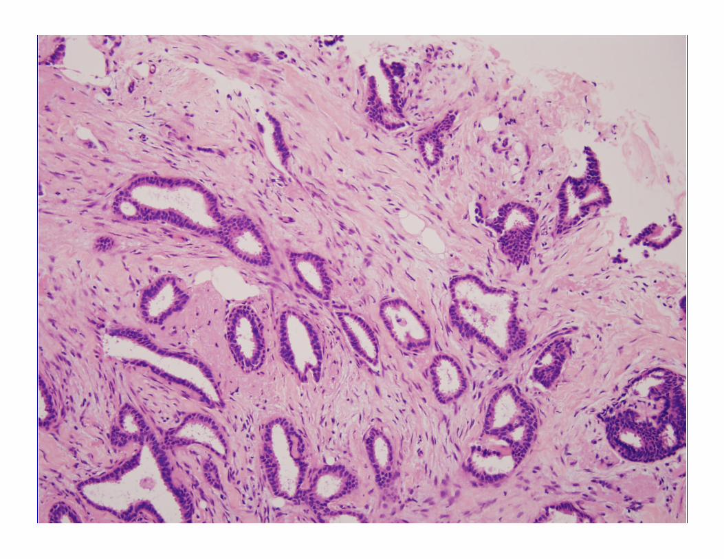
Radial Scar Pitfalls
– Entrapped distorted glands →Tubules resembling those of tubular ca.
– Rare absence of MEC IHC staining – Perineural invasion – Necrosis present in 10% with UEH
More challenging on CNB
Invasive Carcinoma
Pitfalls on CNB – Overcalling ILC – In the setting of a well-circumscribed
lesion SA, myoid hamartoma
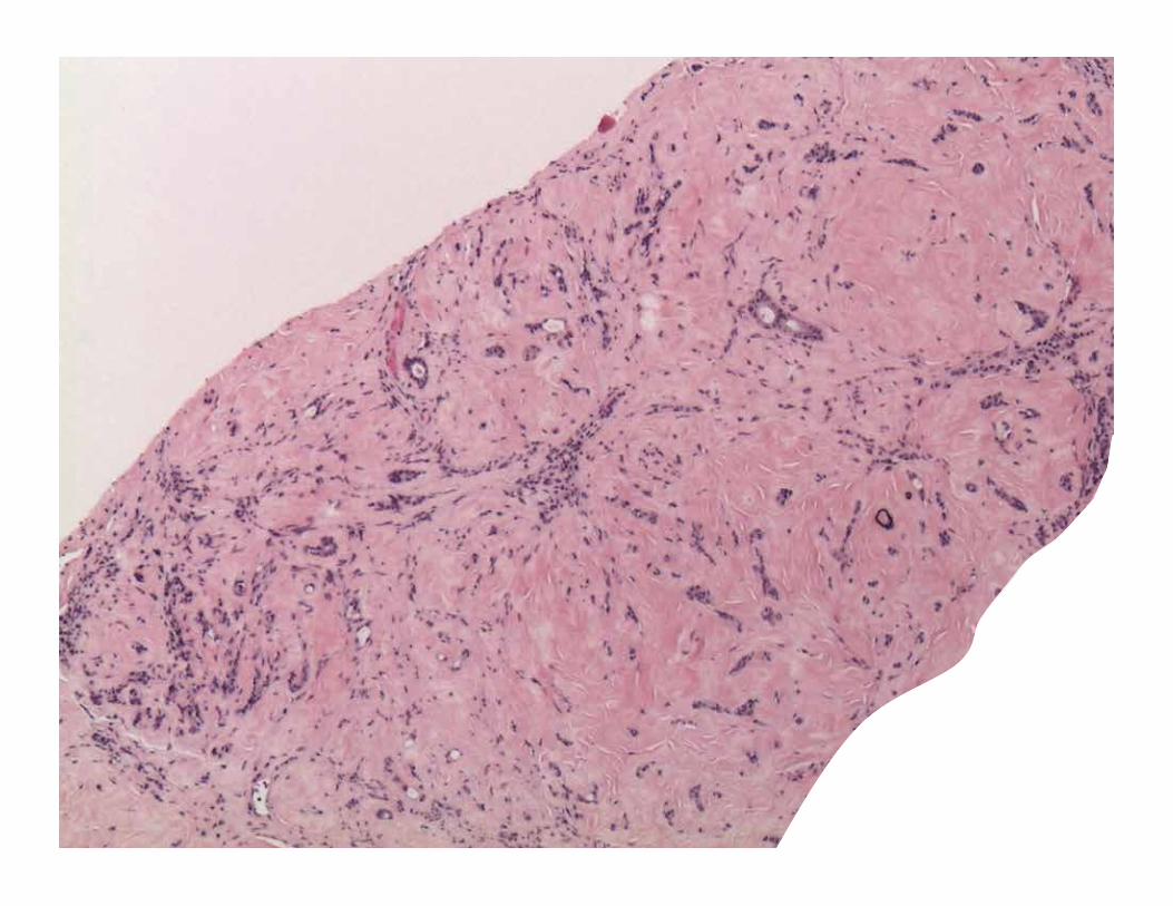
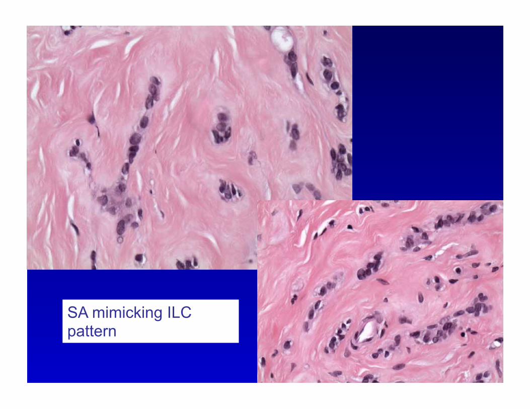
SA mimicking ILC pattern
Invasive Carcinoma
Pitfalls on CNB Under calling ILC
– Paucicellular infiltrate – Chronic inflammation/histiocytes

Stroma appears more cellular than usual

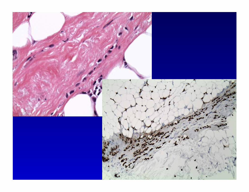
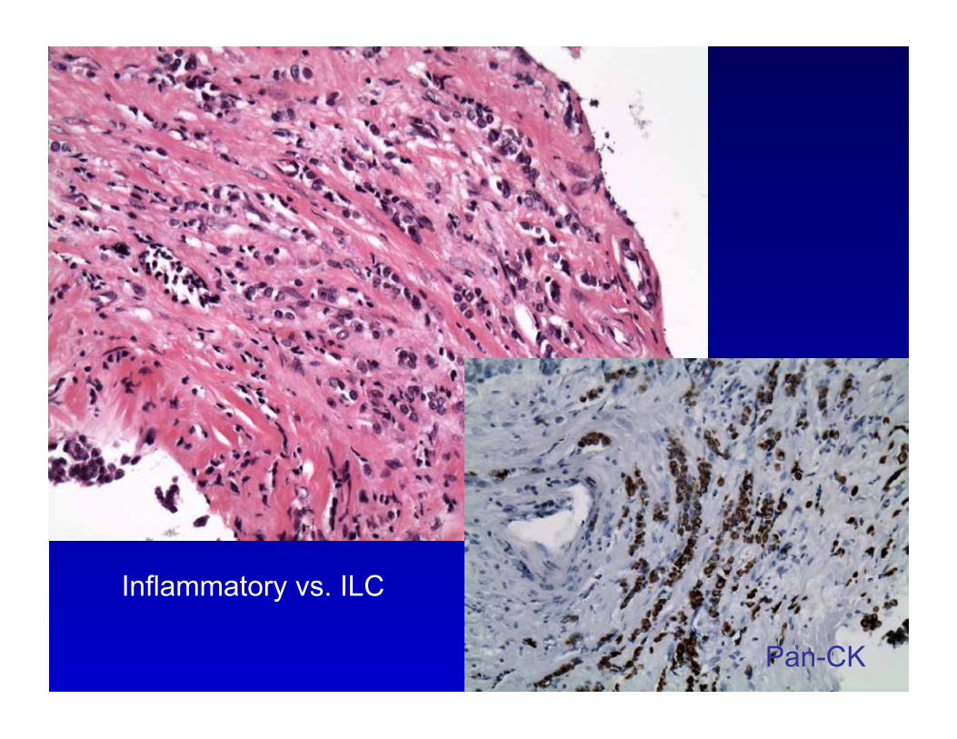
Inflammatory vs. ILC
Pan-CK

Uncertain
FELCS
ADH CCLs with
atypia
LN
Radial Scar
Papillary
Spindle cell lesions
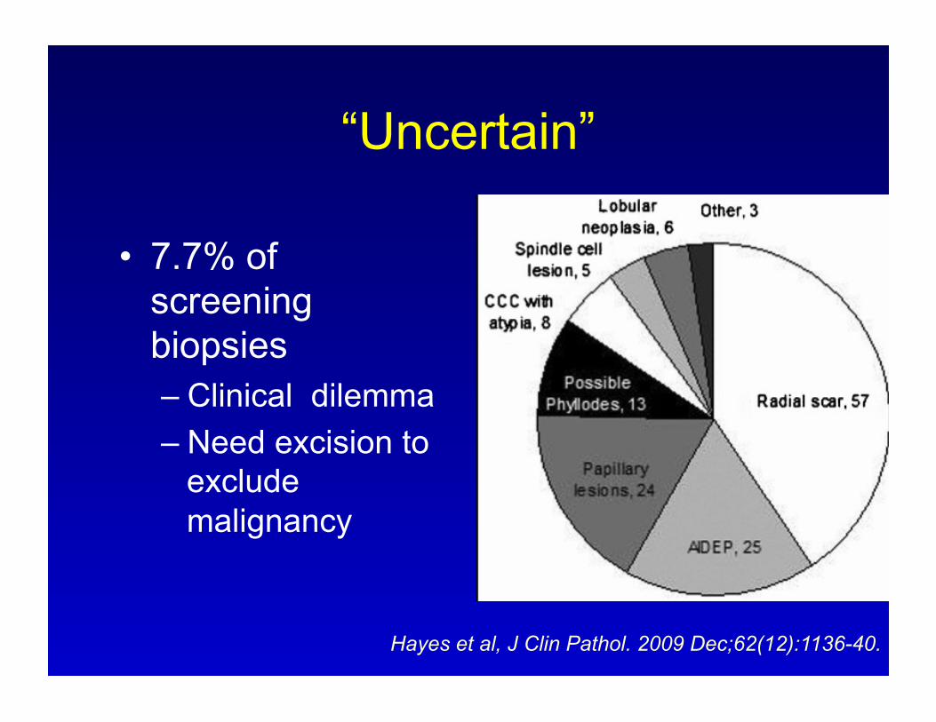
“Uncertain”
7.7% of screening biopsies – Clinical dilemma – Need excision to
exclude malignancy
Hayes et al, J Clin Pathol. 2009 Dec;62(12):1136-40.
Papillary Lesions
Heterogeneous – Finger-like projections – Central FV core – Lined by epithelium
IDP vs. IDP with atypia/DCIS vs. EPC
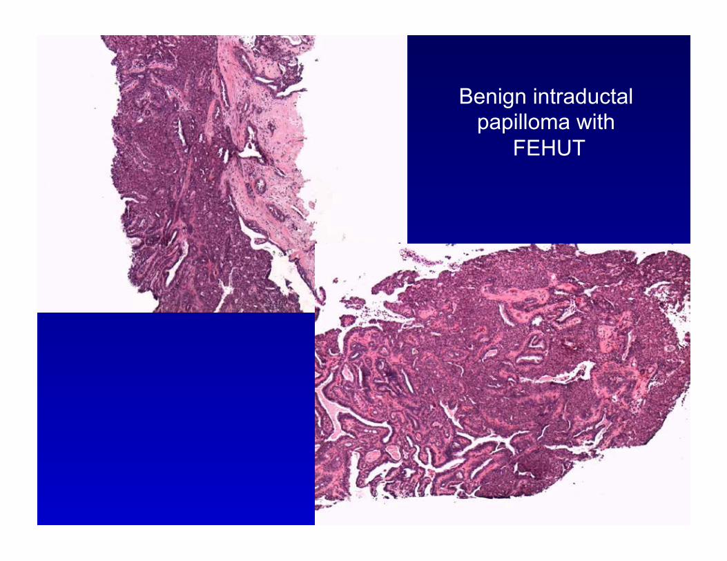
Benign intraductal papilloma with
FEHUT

SMM-HC

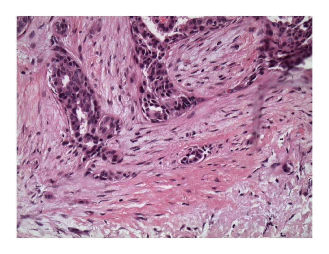
Intraductal Papilloma
Sclerosis – Obscure papillary nature
Entrapment – Mimics invasion
ID MECs Stroma hyalinized Underlying lesion is benign
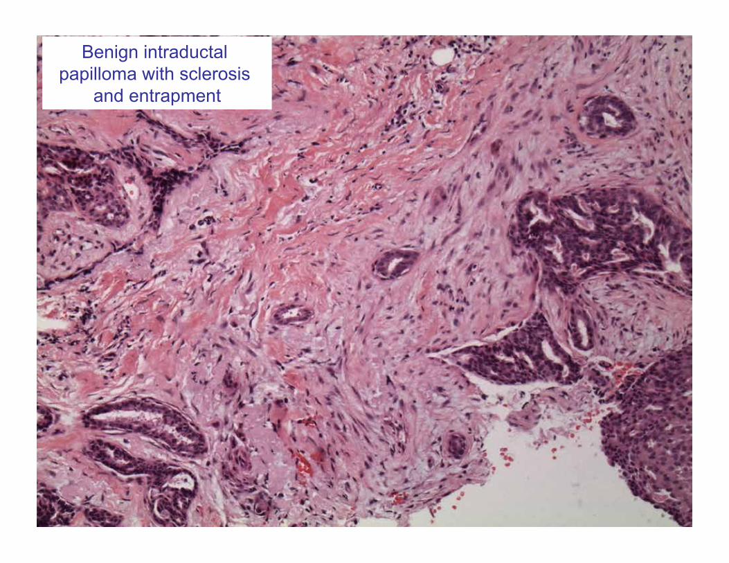
Benign intraductal papilloma with sclerosis
and entrapment

Atypia in Papilloma
IP with: – Low grade cytologic atypia – Architectural atypia
Extent < 3mm – If > 3mm → DCIS in IP
DDx UEH ER CK5
Page et al. Cancer.,1996 Jul 15;78(2):258-66. Grin et al. AJSP, 2009 Nov;33(11):1615-23
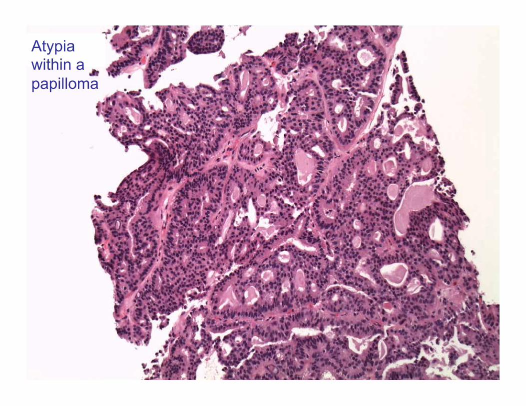
Atypia within a papilloma
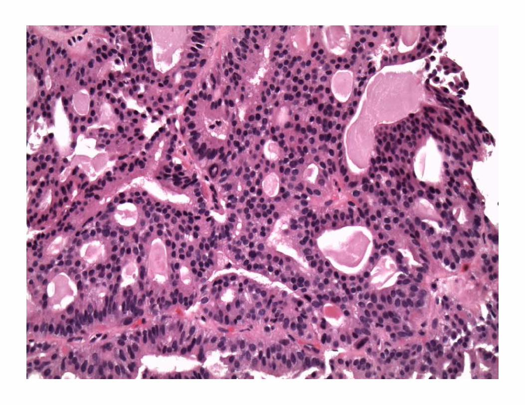
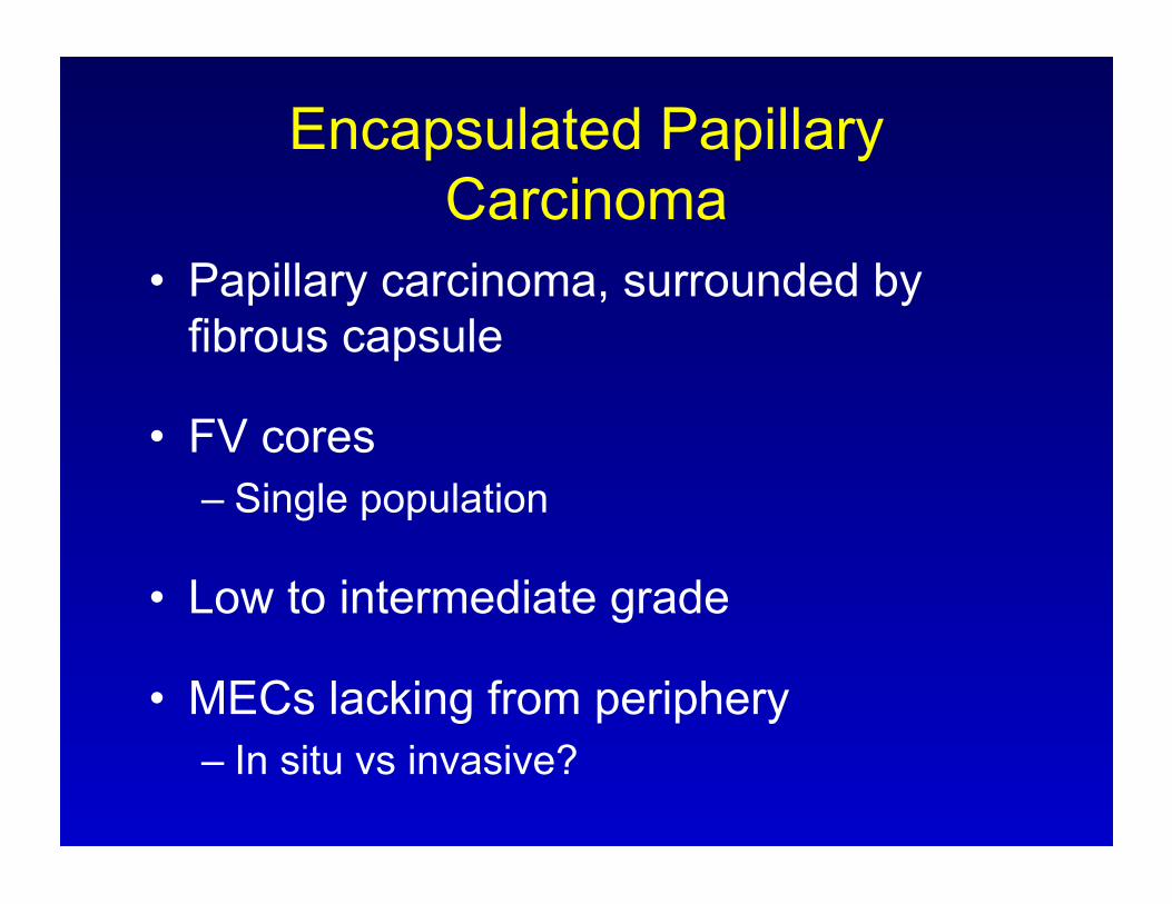
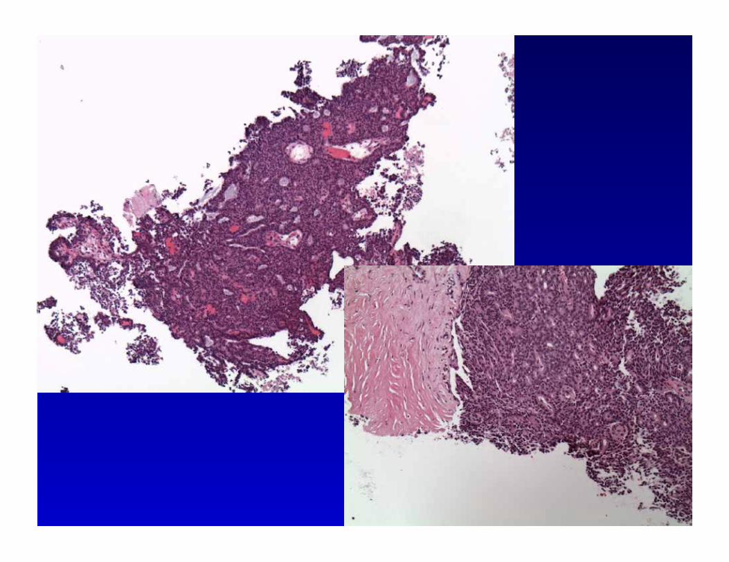
Encapsulated Papillary Carcinoma
Papillary carcinoma, surrounded by fibrous capsule
FV cores – Single population
Low to intermediate grade
MECs lacking from periphery – In situ vs invasive?

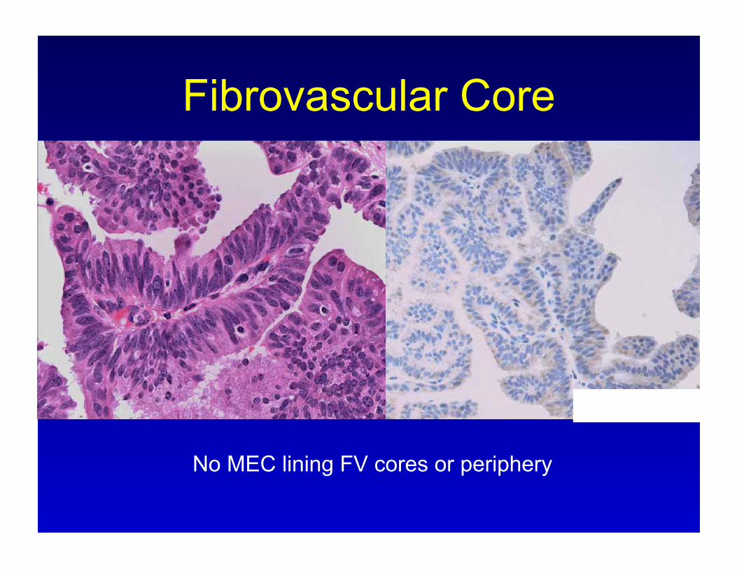
Fibrovascular Core
p63/SMMS
No MEC lining FV cores or periphery
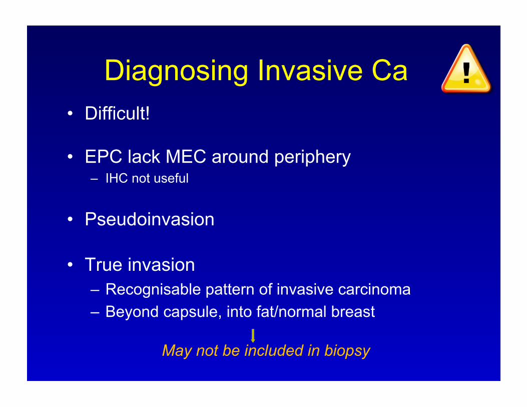
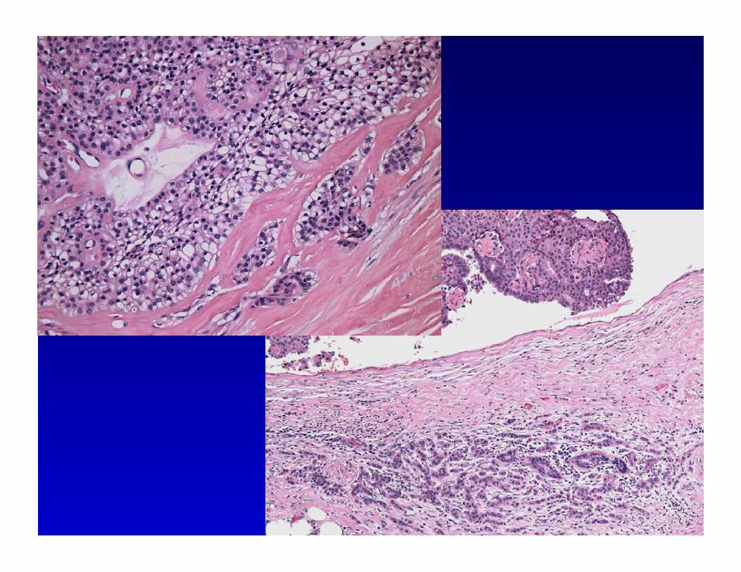
Diagnosing Invasive Ca Difficult!
EPC lack MEC around periphery – IHC not useful
Pseudoinvasion
True invasion – Recognisable pattern of invasive carcinoma – Beyond capsule, into fat/normal breast
May not be included in biopsy
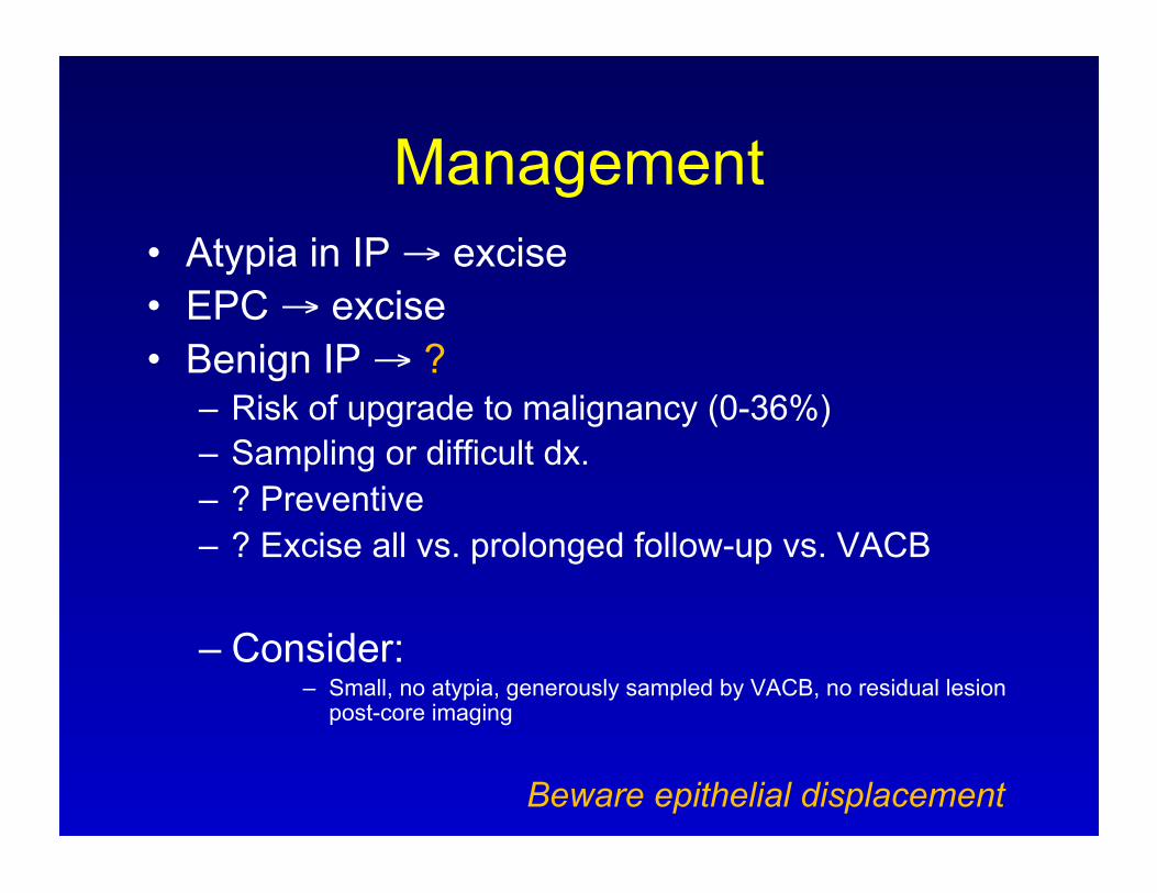
Management Atypia in IP → excise EPC → excise Benign IP → ?
– Risk of upgrade to malignancy (0-36%) – Sampling or difficult dx. – ? Preventive – ? Excise all vs. prolonged follow-up vs. VACB
– Consider: – Small, no atypia, generously sampled by VACB, no residual lesion
post-core imaging
Beware epithelial displacement
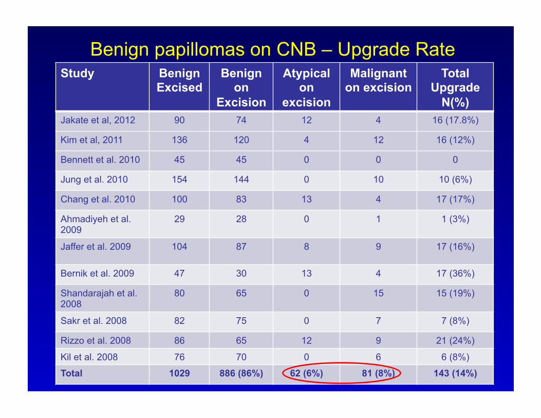
Study Benign Excised
Benign on
Excision
Atypical on
excision
Malignant on excision
Total Upgrade
N(%) Jakate et al, 2012 90 74 12 4 16 (17.8%)
Kim et al, 2011 136 120 4 12 16 (12%)
Bennett et al. 2010 45 45 0 0 0
Jung et al. 2010 154 144 0 10 10 (6%)
Chang et al. 2010 100 83 13 4 17 (17%)
Ahmadiyeh et al. 2009
29 28 0 1 1 (3%)
Jaffer et al. 2009 104 87 8 9 17 (16%)
Bernik et al. 2009 47 30 13 4 17 (36%)
Shandarajah et al. 2008
80 65 0 15 15 (19%)
Sakr et al. 2008 82 75 0 7 7 (8%)
Rizzo et al. 2008 86 65 12 9 21 (24%)
Kil et al. 2008 76 70 0 6 6 (8%)
Total 1029 886 (86%) 62 (6%) 81 (8%) 143 (14%)
Benign papillomas on CNB – Upgrade Rate
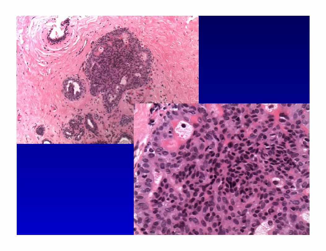
Audience Participation 3
47 year old woman, fine pleomorphic calcifications Stereotactic core biopsy performed Very little tissue obtained, mostly fat, with focal epithelial element….multiple additional levels cut
Most appropriate action
A. Benign, continue routine screening B. Re-biopsy patient C. Excise, it’s at least atypical D. Excise, it’s malignant E. I’m not sure…..help needed!!
Most appropriate action
A. Benign, continue routine screening B. Re-biopsy patient C. Excise, it’s at least atypical D. Excise, it’s malignant E. I’m not sure…..help needed!!
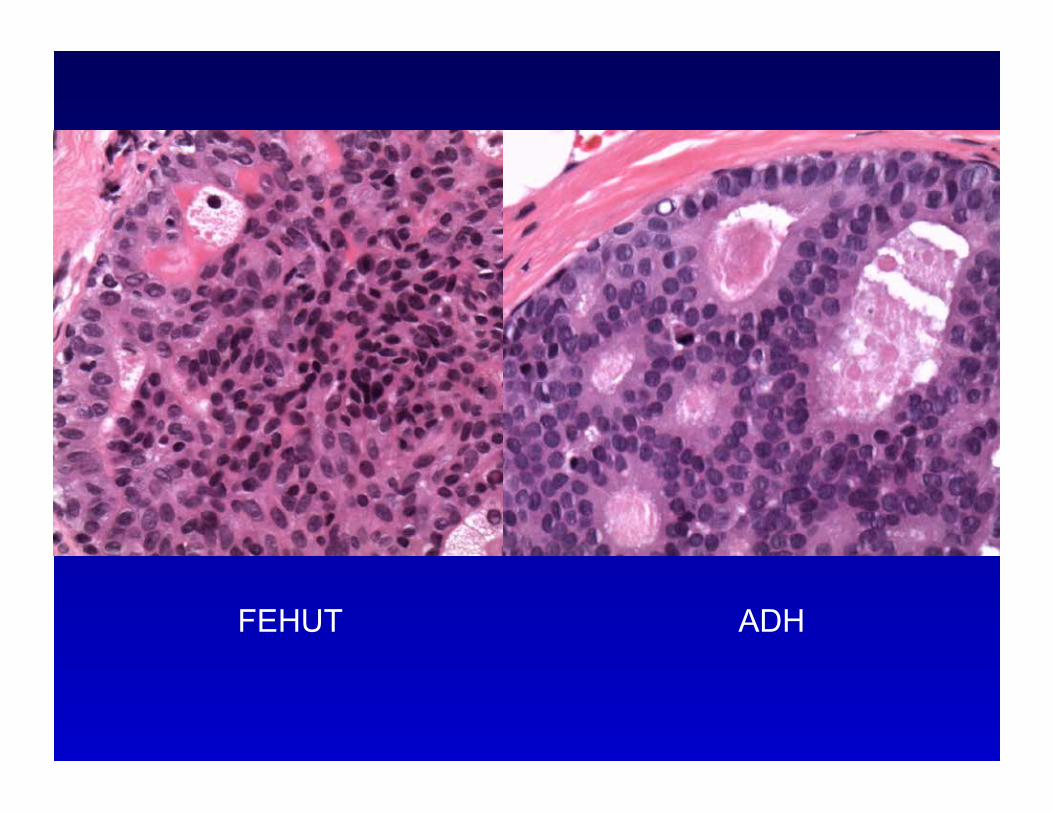
FEHUT ADH
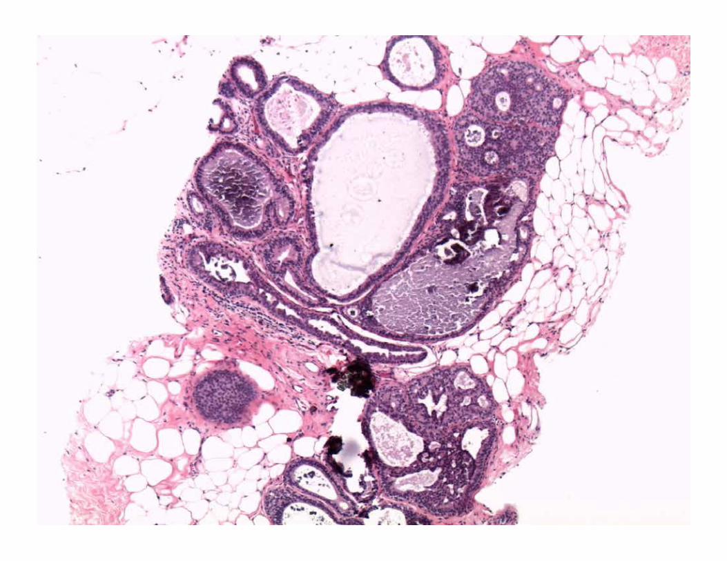
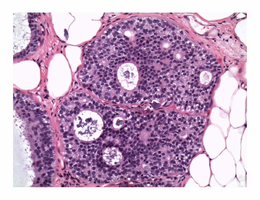
Atypical Ductal Hyperplasia
Core Biopsy Diagnosis
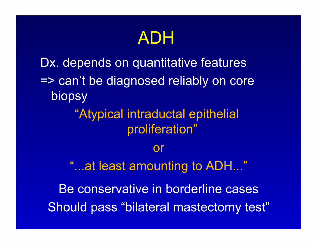
ADH Dx. depends on quantitative features => can’t be diagnosed reliably on core
biopsy “Atypical intraductal epithelial
proliferation” or
“...at least amounting to ADH...”
Be conservative in borderline cases Should pass “bilateral mastectomy test”
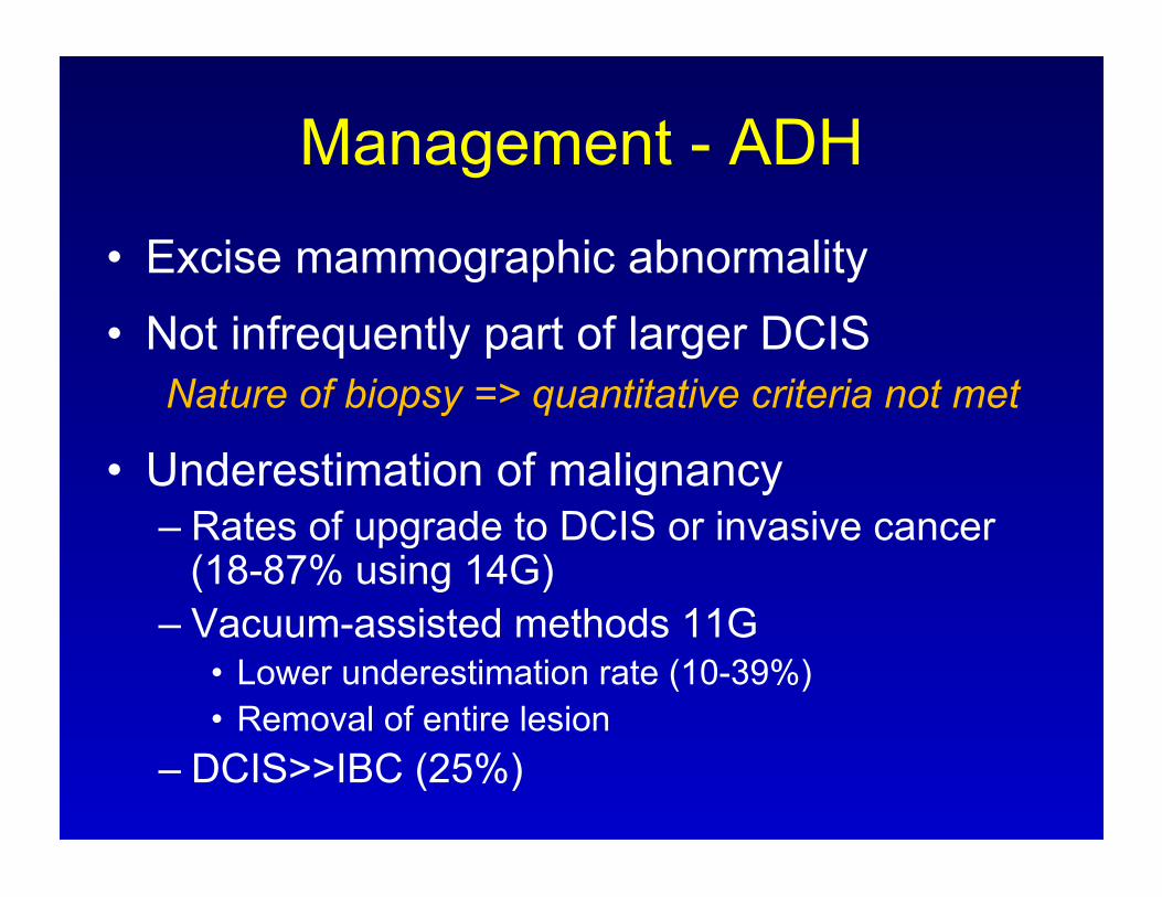
Management - ADH
Excise mammographic abnormality Not infrequently part of larger DCIS
Nature of biopsy => quantitative criteria not met
Underestimation of malignancy – Rates of upgrade to DCIS or invasive cancer
(18-87% using 14G) – Vacuum-assisted methods 11G
Lower underestimation rate (10-39%) Removal of entire lesion
– DCIS>>IBC (25%)

Audience Participation 4
53 year old woman Mammo: asymmetric density U/S: mass with microlobulation of margins U/S-guided core biopsy
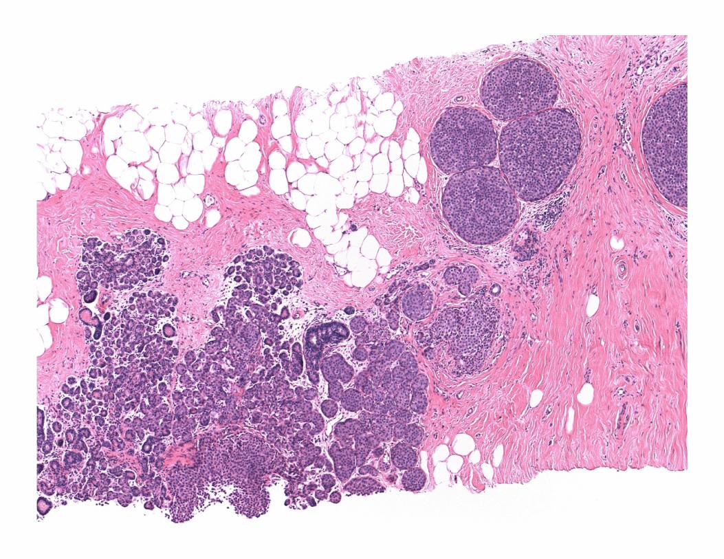
What next?
Patient follow-up, no surgery necessary Complete excision Complete excision with sentinel node Additional work-up of case None of the above
What next?
Patient follow-up, no surgery necessary Complete excision Complete excision with sentinel node Additional work-up of case None of the above

LCIS within sclerosing adenosis
E-cadherin
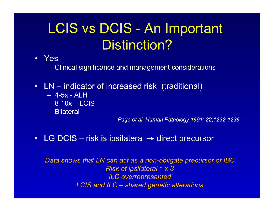
LCIS vs DCIS - An Important Distinction?
Yes – Clinical significance and management considerations
LN – indicator of increased risk (traditional) – 4-5x - ALH – 8-10x – LCIS – Bilateral
Page et al, Human Pathology 1991; 22;1232-1239 LG DCIS – risk is ipsilateral → direct precursor
Data shows that LN can act as a non-obligate precursor of IBC Risk of ipsilateral ↑ x 3 ILC overrepresented
LCIS and ILC – shared genetic alterations

Should LN be Excised?
Co-existent high risk lesion e.g. ADH
Rad-path discordance Indeterminate features
Variant LCIS
Liberman et al, AM J Roentgenol 1999;173:291-299 Reviewed in : Jacobs et al, Am J Surg Patholol 2002;26:1095-1110
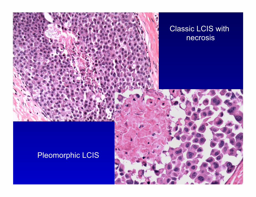
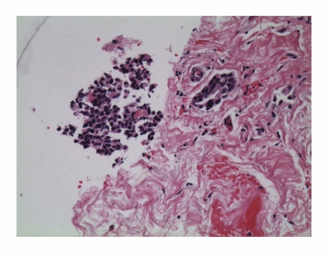
Classic LCIS with necrosis
Pleomorphic LCIS
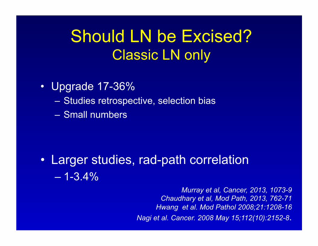
Should LN be Excised? Classic LN only
Upgrade 17-36% – Studies retrospective, selection bias – Small numbers
Larger studies, rad-path correlation – 1-3.4%
Murray et al, Cancer, 2013, 1073-9 Chaudhary et al, Mod Path, 2013, 762-71
Hwang et al. Mod Pathol 2008;21:1208-16 Nagi et al. Cancer. 2008 May 15;112(10):2152-8.
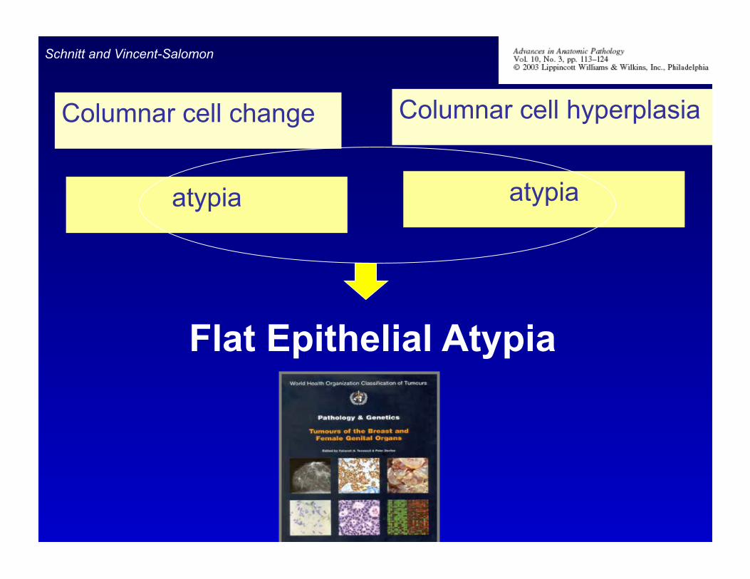
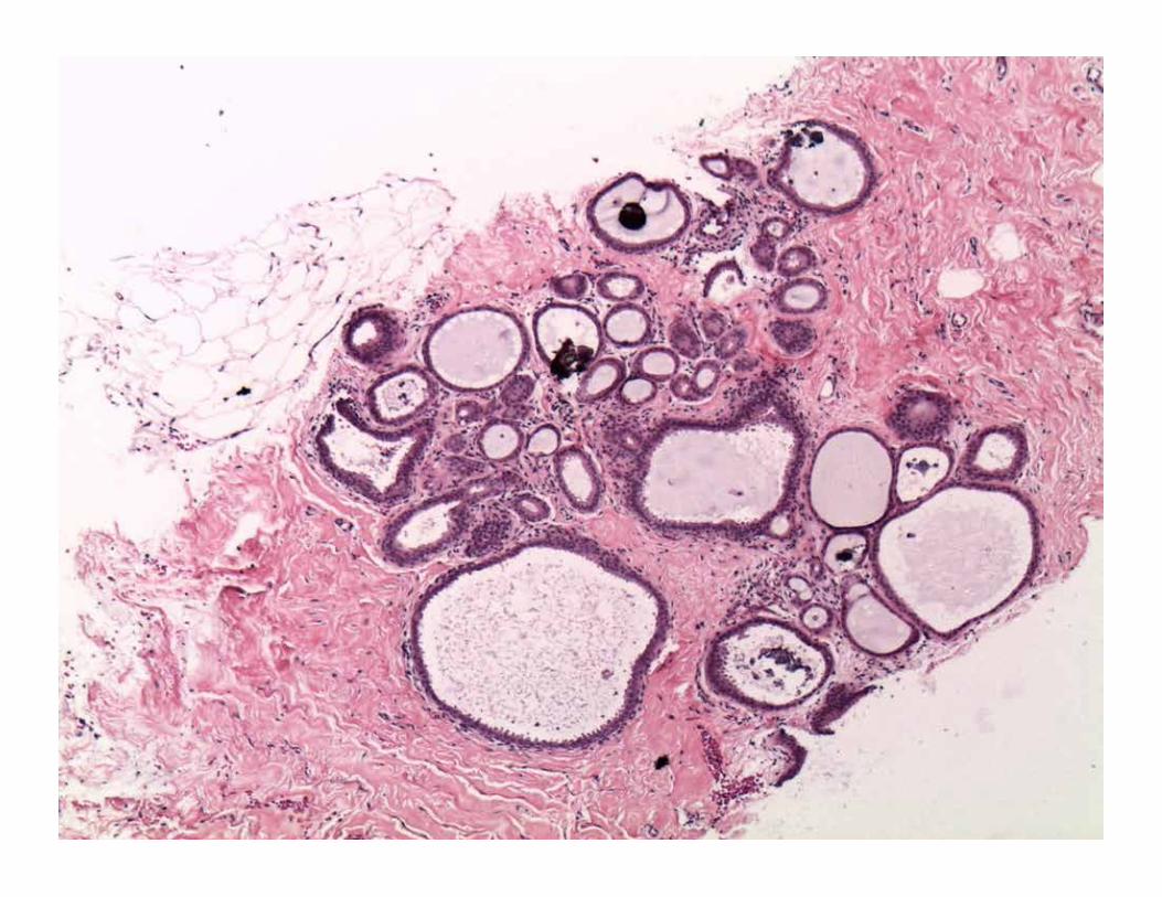
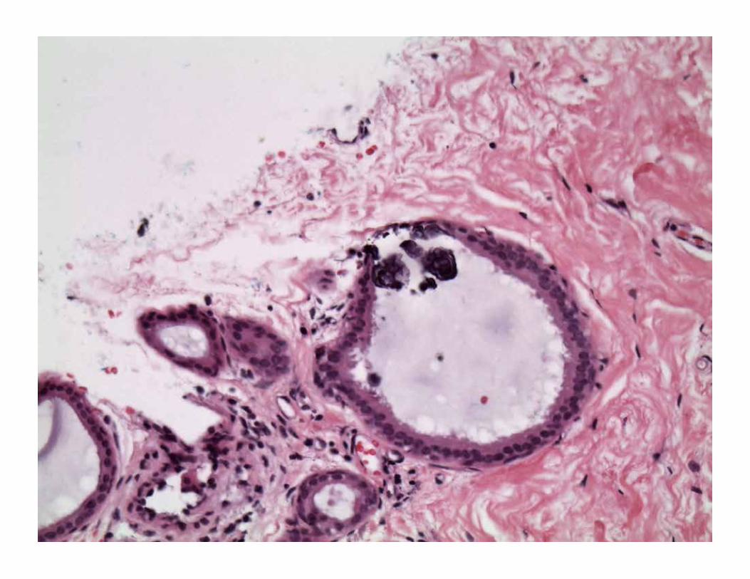
Columnar cell change
Schnitt and Vincent-Salomon
Columnar cell hyperplasia
atypia atypia
Flat Epithelial Atypia
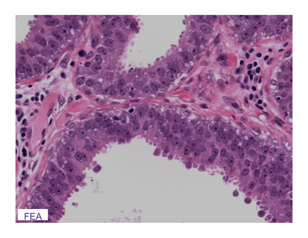
FEA

CCL - Management
Non-atypical – Excision not required If calcs are accounted for
Atypical lesions – Very low risk of progression – Red flag – Excise
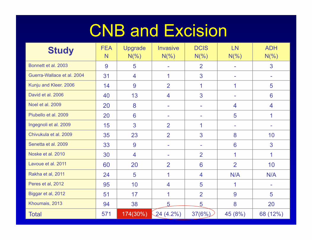
CNB and Excision Study FEA
N Upgrade
N(%) Invasive
N(%) DCIS N(%)
LN N(%)
ADH N(%)
Bonnett et al. 2003 9 5 - 2 - 3 Guerra-Wallace et al. 2004 31 4 1 3 - - Kunju and Kleer. 2006 14 9 2 1 1 5 David et al. 2006 40 13 4 3 - 6 Noel et al. 2009 20 8 - - 4 4 Piubello et al. 2009 20 6 - - 5 1 Ingegnoli et al. 2009 15 3 2 1 - - Chivukula et al. 2009 35 23 2 3 8 10 Senetta et al. 2009 33 9 - - 6 3 Noske et al. 2010 30 4 - 2 1 1 Lavoue et al. 2011 60 20 2 6 2 10 Rakha et al, 2011 24 5 1 4 N/A N/A Peres et al, 2012 95 10 4 5 1 - Biggar et al, 2012 51 17 1 2 9 5 Khoumais, 2013 94 38 5 5 8 20
Total 571 174(30%) 24 (4.2%) 37(6%) 45 (8%) 68 (12%)
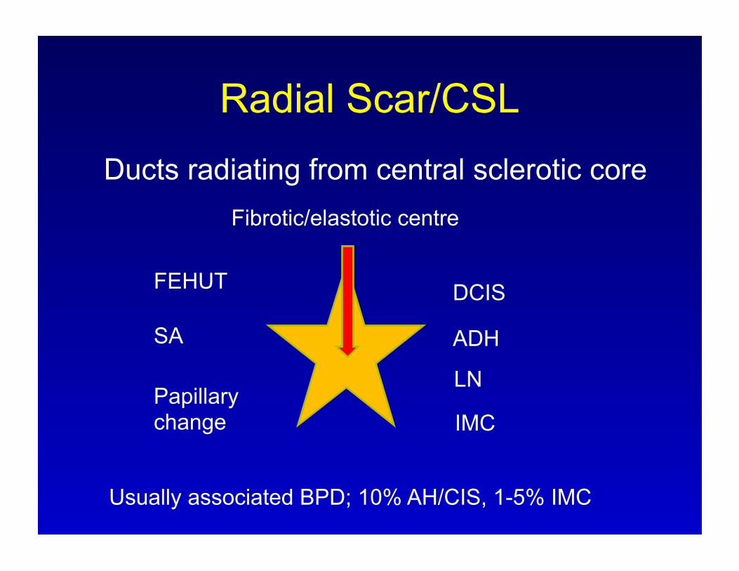
Radial Scar/CSL Ducts radiating from central sclerotic core
FEHUT
SA ADH
DCIS
LN Papillary change
Fibrotic/elastotic centre
Usually associated BPD; 10% AH/CIS, 1-5% IMC
IMC
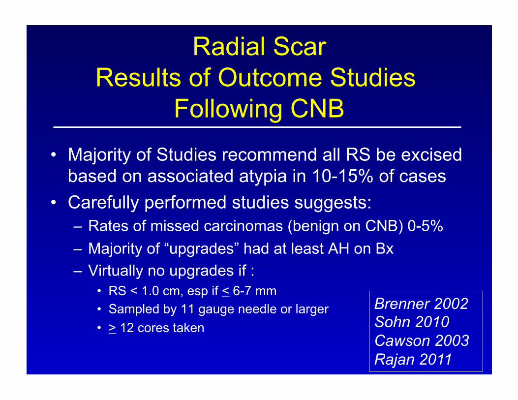
Radial Scar Results of Outcome Studies
Following CNB
Majority of Studies recommend all RS be excised based on associated atypia in 10-15% of cases Carefully performed studies suggests:
– Rates of missed carcinomas (benign on CNB) 0-5% – Majority of “upgrades” had at least AH on Bx – Virtually no upgrades if :
RS < 1.0 cm, esp if < 6-7 mm Sampled by 11 gauge needle or larger > 12 cores taken
Brenner 2002 Sohn 2010 Cawson 2003 Rajan 2011

RS Treatment Approach
Incidental Finding
No Further Treatment
Mammo Lesion of Interest
No Atypia Mammo < 6-7 mm Well sampled 11 or 8 gauge needle
No Atypia Limited Sample
Atypia Malignancy Mammo discordant
Excision
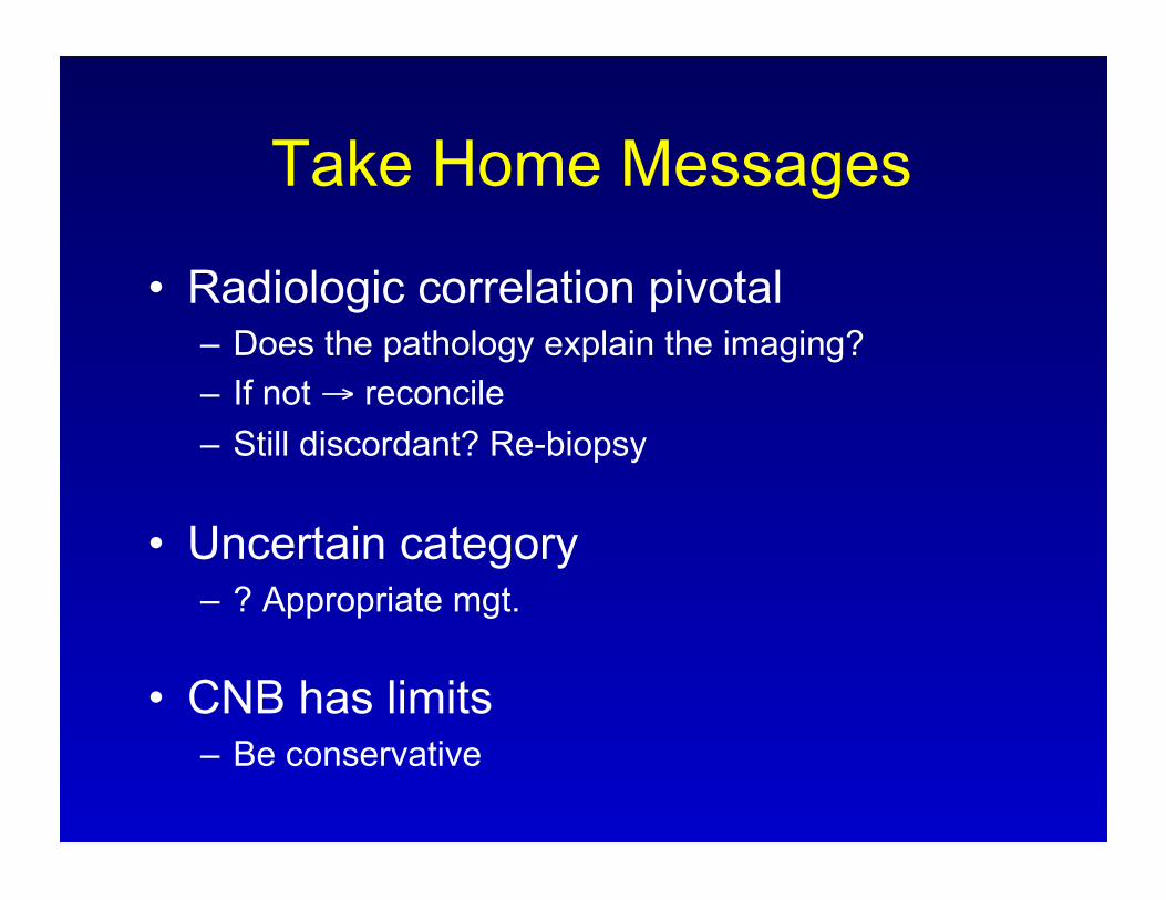
Take Home Messages
Radiologic correlation pivotal – Does the pathology explain the imaging? – If not → reconcile – Still discordant? Re-biopsy
Uncertain category – ? Appropriate mgt.
CNB has limits – Be conservative