Embed Size (px)
Citation preview
Breast Cancer in Men Early Detection,Diagnosis, and Staging Detection and Diagnosis
Catching cancer early often allows for more treatment options. Some early cancers mayhave signs and symptoms that can be noticed, but that is not always the case.
Can Breast Cancer in Men Be Found Early?●
Signs and Symptoms of Breast Cancer in Men●
Tests for Breast Cancer in Men●
Stages and Outlook (Prognosis)
After a cancer diagnosis, staging provides important information about the extent ofcancer in the body and anticipated response to treatment.
How Is Breast Cancer in Men Classified?●
Tests to Look for Breast Cancer Spread in Men●
Breast Cancer Stages in Men●
Breast Cancer Survival Rates in Men●
Can Breast Cancer in Men Be FoundEarly? Finding breast cancer early improves the chances that male breast cancer can betreated successfully. However, because breast cancer is so uncommon in men, there isunlikely to be any benefit in screening men in the general population for breast cancerwith mammograms or other tests.
Differences in early detection of breast cancers inmen and women
There are many similarities between breast cancer in men and women, but there aresome important differences that affect finding it early.
Breast size
The most obvious difference between the male and female breast is size. Because menhave very little breast tissue, it is easier for men and their health care professionals tofeel small masses (tumors). On the other hand, because men have so little breasttissue, cancers do not need to grow very far to reach the nipple, the skin covering thebreast, or the muscles underneath the breast. So even though breast cancers in mentend to be slightly smaller than in women when they are first found, more often havealready spread to nearby tissues or lymph nodes. The extent of spread is one of themost important factors in the prognosis (outcome) of a breast cancer.
Lack of awareness
Another difference is that breast cancer is common among women and rare amongmen. Women tend to be aware of this disease and its possible warning signs, but manymen do not think that they can get it at all. Some men ignore breast lumps or think theyare caused by an infection or some other reason, and don't get medical treatment untilthe mass has had a chance to grow. Some men are embarrassed when they find abreast lump and worry that someone might question their masculinity. This could alsodelay diagnosis and reduce a man's chances for successful treatment.
For men who are or may be at high risk
Careful breast exams might be useful for screening men with a strong family history ofbreast cancer and/or with BRCA mutations found by genetic testing. Screening men forbreast cancer has not been studied to know if it is helpful, and mammography (x-rays ofthe breast) and ultrasound is usually only done if a lump is found. Men who are at highrisk for breast cancer should discuss how to manage their risk with their doctor.
Genetic counseling and testing
If you have a strong family history of breast cancer (in men or women), ovarian cancer,
pancreatic cancer, and/or prostate cancer that might be caused by a BRCA mutation,and/or if someone else in your family is known to have a BRCA mutation, you mightwant to consider genetic testing to determine if you have inherited a mutated BRCAgene. If the test detects a mutated BRCA gene, you and your health care team canwatch carefully for early signs of cancer. Other cancers including prostate cancer,pancreatic cancer, and testicular cancer have been linked to BRCA mutations. .
Because breast cancer in men can be caused by BRCA mutations, men with breastcancer should also consider genetic testing.
If you are thinking about having genetic testing, it is strongly recommended that you talkfirst to a professional qualified to explain and interpret these tests, such as a geneticcounselor or a nurse or doctor with special training. It is very important to understandwhat genetic testing can and can't tell you, and to carefully weigh the benefits and risksof testing before having it done. Test results are not always clear cut, and even if theyare, it's not always clear what should be done about them. There may be otherconcerns as well, such as what the results might mean for other family members.
References●
National Cancer Institute Physician Data Query (PDQ). Genetics of Breast and OvarianCancer. 01/16/2018. Accessed athttp://www.cancer.gov/cancertopics/pdq/genetics/breast-and-ovarian/HealthProfessional on January 16, 2018.
Last Medical Review: April 27, 2018 Last Revised: April 27, 2018
American Cancer Society medical information is copyrighted material. For reprintrequests, please see our Content Usage Policy.
Signs and Symptoms of Breast Cancerin Men Possible symptoms of breast cancer to watch for include:
A lump or swelling, which is often (but not always) painless●
Skin dimpling or puckering●
Nipple retraction (turning inward)●
Redness or scaling of the nipple or breast skin●
Discharge from the nipple●
Sometimes a breast cancer can spread to lymph nodes under the arm or around thecollar bone and cause a lump or swelling there, even before the original tumor in thebreast is large enough to be felt.
These changes aren't always caused by cancer, but if you notice any breast changes,you should see a health care professional as soon as possible.
References●
Burstein HJ, Harris JR, Morrow M. Ch. 79 - Malignant tumors of the breast. In: DeVitaVT, Lawrence TS, Rosenberg SA, eds. DeVita, Hellman, and Rosenberg's Cancer:Principles and Practice of Oncology. 10th ed. Philadelphia, Pa: Lippincott Williams &Wilkins; 2015.
Morrow M. Chapter 3: Physical Exam of the Breast. In: Harris JR, Lippman ME, MorrowM, Osborne CK, eds. Diseases of the Breast. 5th ed. Philadelphia: Wolters KluwerHealth; 2014.
Wolff AC, Domchek SM, Davidson NE et al. Ch 91 - Cancer of the Breast. In:Niederhuber JE, Armitage JO, Doroshow JH, Kastan MB, Tepper JE, eds. Abeloff’sClinical Oncology. 5th ed. Philadelphia, Pa: Elsevier: 2014.
Last Medical Review: April 27, 2018 Last Revised: April 27, 2018
American Cancer Society medical information is copyrighted material. For reprintrequests, please see our Content Usage Policy.
Tests for Breast Cancer in Men
Medical history and physical exam
If there is a chance you have breast cancer, your doctor will want to get a completepersonal and family medical history. This may give some clues about the cause of anysymptoms you are having and if you might be at increased risk for breast cancer.
A complete breast exam will be done to find any lumps or suspicious areas and to feeltheir texture, size, and relationship to the skin and muscle. The doctor may alsoexamine the rest of your body to look for any evidence of possible spread, such asenlarged lymph nodes (especially under the arm).
Imaging tests for breast cancer in men
If you have signs or symptoms that could mean breast cancer or another breastdisease, your doctor might recommend one or more or the following imaging tests.
Diagnostic mammogram
A mammogram is a low dose x-ray exam of the breast that allows doctors calledradiologists to look for changes in breast tissue. It is called a diagnostic mammogramwhen it is done because problems are present.
A mammogram uses a machine designed to look only at breast tissue. The breast ispressed between 2 plates to flatten and spread the tissue. The compression only lasts afew seconds and may be uncomfortable briefly, but it is necessary to get a betterpicture. In some cases, special images known as cone or spot views with magnificationare taken to make a small area of abnormal breast tissue easier to evaluate.
The results of this test might suggest that a biopsy is needed to tell if the abnormal areais cancer. Mammography is often more accurate in men than women, since men do nothave dense breasts or other common breast changes that might interfere with the test.
Breast ultrasound
Breast ultrasound is often used to examine some types of breast changes.
Breast ultrasound uses sound waves to make a computer picture of the inside of thebreast. A gel is put on the skin of the breast, and a wand-like instrument called atransducer is moved over the skin. The transducer sends out sound waves and picksup the echoes as they bounce off body tissues. The echoes are made into a picture ona computer screen. You might feel some pressure as the transducer is moved acrossthe breast, but it should not be painful.
This test does not expose you to radiation.
Breast ultrasound is often used to look at breast changes that are found during a
mammogram or physical exam. It is useful because it can often tell the differencebetween fluid-filled cysts (which are unlikely to be cancer) and solid masses (whichmight need further testing to be sure they're not cancer).
In someone with a breast tumor, ultrasound can also be used to check if the lymphnodes under the arm are enlarged. If they are, ultrasound can be used to guide aneedle to take a sample (a biopsy) to look for cancer cells there and in the breast tissue.
Nipple discharge test
Fluid leaking from the nipple is called nipple discharge. It can look clear, cloudy orbloody. If you have nipple discharge, you should have it checked by your doctor. If thereis blood in this fluid, you might need more tests. One test collects some of the fluid tolook at it in the lab to see if cancer cells are there. This test is often not helpful, since abreast cancer can still be there even when no cancer cells are found in the nippledischarge. Other tests, such as a mammogram or breast ultrasound, may be morehelpful. If you have a breast mass, you will probably need a biopsy, even if the nippledischarge does not contain cancer cells or blood.
Breast Biopsy
When other tests show that you might have breast cancer, you will probably need tohave a biopsy. Needing a breast biopsy doesn’t necessarily mean you have cancer.Most biopsy results are not cancer, but a biopsy is the only way to find out. During abiopsy, a doctor will remove cells from the suspicious area so they can be looked at inthe lab to see if cancer cells are present. It typically takes at least a few days for you tofind out the results.
If your doctor thinks you don’t need a biopsy, but you still feel there’s something wrongwith your breast, follow your instincts. Don’t be afraid to talk to your doctor about this orgo to another doctor for a second opinion. A biopsy is the only sure way to diagnosebreast cancer.
There are different types of breast biopsies. The type you have depends on yoursituation.
Fine needle aspiration biopsy (FNA): This type of biopsy is often used to look forcancer spread in the nearby lymph nodes. The doctor uses a very thin, hollow needleattached to a syringe to withdraw (aspirate) a small amount of tissue or fluid from asuspicious area. A local anesthetic (numbing medicine) may or may not be used. Thebiopsy sample is then checked to see if there are cancer cells in it.
If the area to be biopsied can be felt, the needle can be guided into it while the doctor isfeeling it. If the lump can't be felt easily, the doctor might watch the needle on anultrasound screen as it moves into the area. This is called an ultrasound-guidedbiopsy.
An FNA biopsy is the easiest type of biopsy to have, but it can sometimes miss a cancerif the needle does not go into the cancer cells.
If the results of the FNA biopsy do not give a clear diagnosis, or your doctor still hasconcerns, you might need to have a second biopsy or a different type of biopsy.
Core needle biopsy (CNB): This is the most common type of biopsy used to make abreast cancer diagnosis. The doctor uses a wide, hollow needle to take out pieces ofbreast tissue from a suspicious area. The needle used in this technique is larger thanthat used for FNA and allows the doctor to remove larger cylinders (cores) of tissue.Several cylinders are often removed. The biopsy is done with local numbing medicineand with the doctor either feeling the abnormal area or using an imaging test (likeultrasound or MRI) to find the spot to biopsy.
In addition to the standard CNB, there are two other types of CNBs:
Stereotactic core needle biopsy●
Vacuum-assisted core biopsy●
If the results of the CNB do not give a clear diagnosis, or your doctor still has concerns,you might need to have a second biopsy or a different type of biopsy.
Surgical (open) biopsy: Most breast cancer can be diagnosed with a needle biopsy.Rarely, surgery is needed to remove all or part of the lump for testing. Most often, thesurgeon removes the entire mass or abnormal area, as well as a surrounding margin ofnormal-appearing breast tissue.
There are 2 types of surgical biopsies:
An incisional biopsy removes only part of the suspicious area, enough to make adiagnosis.
●
An excisional biopsy removes the entire tumor or abnormal area, with or withouttrying to take out an edge of normal breast tissue (depending on the reason for thebiopsy).
●
Lymph node biopsy: The doctor may also need to biopsy the lymph nodes under thearm to check them for cancer spread. This might be done at the same time as biopsy ofthe breast tumor, or during surgery to remove the breast tumor. This is done by needle
biopsy, or with a sentinel lymph node biopsy and/or an axillary lymph node dissection.
References●
Last Medical Review: April 27, 2018 Last Revised: April 27, 2018
American Cancer Society medical information is copyrighted material. For reprintrequests, please see our Content Usage Policy.
How Is Breast Cancer in MenClassified? Breast cancer is classified in different ways, based on the results of lab tests afterbiopsy or surgery. Breast cancer is given a type, based on the type of cells it startedfrom; a grade, based on how the cells look and how quickly they grow; and otherclassifications based on the results of tests for different hormone receptors or genes inthe cancer cells.
Breast cancer type
The tissue removed during the biopsy (or during surgery) is first looked at in the lab tosee if cancer is present and whether it is a carcinoma or some other type of cancer (likea sarcoma). If there is enough tissue, the pathologist may be able to determine if thecancer is in situ (not invasive) or invasive. The biopsy is also used to determine thecancer's type, such as invasive ductal carcinoma or invasive lobular carcinoma.
Breast cancer grade
Cancer cells are given a grade when they are removed from the breast and checked inthe lab. The grade is based on how much the cancer cells look like normal breast cells.
For invasive cancers, a lower grade number (1) usually means the cancer isslower-growing, and less likely to spread. A higher number (3) means a faster-growing cancer that’s more likely to spread. The grade is used to help predict youroutcome (prognosis) and help figure out what treatments might work best. Sometimeswords such as "well differentiated," "moderately differentiated," and "poorly
differentiated" are used to describe the grade instead of numbers:
Grade 1 or well differentiated: The cells are slower-growing, and look more likenormal breast tissue.
●
Grade 2 or moderately differentiated: The cells are growing at a speed of andlook like cells somewhere between grades 1 and 3.
●
Grade 3 or poorly differentiated: The cancer cells look very different from normalcells and will probably grow and spread faster.
●
Our information about pathology reports can help you understand details about yourbreast cancer.
Ductal carcinoma in situ is also graded, but the grade is based only on howabnormal the cancer cells look. Areas of necrosis (dead or dying cancer cells) arealso noted. If there is necrosis, it means the tumor is growing quickly. SeeUnderstanding Your Pathology Report: Ductal Carcinoma In Situ for more on how DCISis described.
Tests to classify breast cancers
Estrogen receptor (ER) and progesterone receptor (PR)
Receptors are proteins in or on cells that can attach to certain substances in the blood.Normal breast cells and some breast cancer cells have receptors that attach to thehormones estrogen and progesterone, and depend on these hormones to grow.Cancers are called hormone receptor-positive or hormone receptor-negative based onwhether or not they have these receptors (proteins). Knowing the hormone receptorstatus is important in deciding treatment options. Keeping these receptors fromattaching to the hormones can help keep the cancer from growing and spreading. Thereare drugs that can be used to do this.
Breast cancer cells may have one, both, or none of these receptors:
ER-positive (ER+) breast cancers have estrogen receptors.●
PR-positive (PR+) breast cancers have progesterone receptors.●
HER2/neu status
In a small number of breast cancers in men, the cells have too much of a growth-promoting protein called HER2/neu (often just shortened to HER2). Tumors with
increased levels of HER2/neu are referred to as HER2-positive. Cells become HER2-positive breast cancers by having too many copies of the HER2/neu gene (known asgene amplification). Cancer cells with greater than normal amounts of the HER2/neuprotein tend to grow and spread more aggressively than other breast cancers.
All newly-diagnosed breast cancers should be tested for HER2/neu because the outlookfor HER2-positive cancers is improved if drugs that target the HER2/neu protein, suchas trastuzumab (Herceptin®) and lapatinib (Tykerb®) are used as part of treatment. SeeTargeted Therapy for Breast Cancer in Menfor more information on drugs that targetthis protein.
The biopsy or surgery sample is usually tested in 1 of 2 ways:
Immunohistochemistry (IHC): In this test, special antibodies that identify theHER2/neu protein are applied to the sample, which cause it to change color ifabnormally high levels are present. The test results are reported as 0, 1+, 2+, or 3+.
●
Fluorescent in situ hybridization (FISH): This test uses fluorescent pieces ofDNA that specifically stick to copies of the HER2/neu gene in cells, which can thenbe counted under a special microscope.
●
Many breast cancer specialists think the FISH test gives more accurate results thanIHC, but it is more expensive and takes longer to get the results. Often the IHC test isused first.
If the results are 1+ (or 0), the cancer is considered HER2-negative. People withHER2-negative tumors are not treated with drugs that target HER2.
●
If the test comes back 3+, the cancer is HER2-positive. People with HER2-positivetumors may be treated with drugs that target HER2.
●
When the result is 2+, the HER2 status of the tumor is not clear and the tumor isthen tested with FISH. Some institutions also use FISH to confirm HER2 status thatis 3+ by IHC and some perform only FISH.
●
A newer type of test, known as chromogenic in situ hybridization (CISH), works similarlyto FISH, by using small DNA probes to count the number of HER2 genes in breastcancer cells. But this test doesn't require a special microscope and looks for colorchanges (not fluorescence) which may make it less expensive. Right now, it is not beingused as much as IHC or FISH.
Classifying breast cancer based on hormone receptors and HER2status
Doctors often divide invasive breast cancers into groups based on the presence of
hormone receptors (ER and PR) and whether or not the cancer has too much HER2.
Hormone receptor-positive: If the breast cancer cells contain either estrogen orprogesterone receptors, they can be called hormone receptor-positive (or just hormone-positive). Breast cancers in men that are hormone receptor-positive can be treated withhormone therapy drugs that lower estrogen levels, block estrogen receptors, or affectandrogen (male hormone) levels (see Hormone Therapy for Breast Cancer in Men).This includes cancers that are ER-negative but PR-positive. Hormone receptor-positivecancers tend to grow more slowly than those that are hormone receptor-negative (anddon’t have either estrogen or progesterone receptors). Patients with these cancers tendto have a better outlook in the short-term, but cancers that are hormone receptor-positive can sometimes come back many years after treatment. About 9 out of 10 malebreast cancers are hormone receptor-positive.
Hormone receptor-negative: If the breast cancer cells don’t have either estrogen orprogesterone receptors, they are said to be hormone receptor-negative (or justhormone-negative). Treatment with hormone therapy drugs is not helpful for thesecancers. These cancers tend to grow more quickly than hormone receptor-positivecancers. If they return after treatment, it is more often in the first few years.
HER2 positive: Cancers that have too much HER2 protein or gene are called HER2positive. These cancers can be treated with drugs that target HER2.
HER2 negative: Cancers that don’t have excess HER2 are called HER2 negative.These cancers do not respond to treatment with drugs that target HER2.
Triple-negative: If the breast cancer cells don’t have estrogen or progesteronereceptors and don’t have too much HER2, they are called triple-negative (HER2negative, ER ngative, and PR negative). Triple-negative breast cancers tend to growand spread more quickly than most other types of breast cancer. Because the tumorcells don’t have hormone receptors, hormone therapy is not helpful in treating thesecancers. Because they don’t have too much HER2, drugs that target HER2 aren’thelpful, either. Chemotherapy can still be useful, though.
Triple-positive: This term is used to describe cancers that are ER-positive, PR-positive,and have too much HER2. These cancers can be treated with hormone drugs as well asdrugs that target HER2.
Other lab tests
Tests of ploidy and cell proliferation rate
Finding out more information about the DNA in the breast cancer cells can help predicthow fast the cancer cells are dividing and growing.
The ploidy of cancer cells refers to how much DNA they contain.
If there's a normal amount of DNA in the cells, they are said to be diploid. Thesecancers tend to grow and spread more slowly.
●
If the amount is abnormal, then the cells are described as aneuploid. Thesecancers tend to be more aggressive and grow and spread faster.)
●
Tests of ploidy may help determine prognosis (outcome), but they rarely changetreatment and are considered optional. They are not usually recommended as part of aroutine breast cancer work-up.
Cell proliferation is how quickly a cancer cell copies its DNA and divides into 2 cells. Ifthe cancer cells are dividing more rapidly, it means the cancer is faster growing or moreaggressive. DNA is copied when the cell is getting ready to divide into 2 new cells. TheS-phase fraction is the percentage of cells in a sample that are copying their DNA. Therate of cancer cell division can also be estimated by a Ki-67 test. If the S-phase fractionor Ki-67 test is high, it means that the cancer cells are dividing more rapidly, which canindicate a more aggressive cancer.
Gene expression tests
Tests that look at the patterns of certain genes (sometimes referred to as geneexpression profiling) can help predict if some early-stage (stage 1 or 2) breast cancerare likely to come back after initial treatment. Doctors can use this information to knowwho will most likely benefit from chemotherapyafter breast surgery.
The Oncotype DX® and the MammaPrint® are examples of tests that look at differentsets of breast cancer genes. There are more tests in development. Tests like these arepart of what’s being called “personalized medicine” – learning more about your cancerto specifically tailor your treatment.
More information is needed to decide how useful this test is for breast cancer in men.But there is enough data that this test can help men with early stage breast cancermake decisions about chemotherapy after surgery. Ask your doctor if these tests mightbe appropriate.
References●
Agendia website: Mammaprint. Accessed at www.agendia.com/healthcare-professionals/breast-cancer/mammaprint/ on January 22, 2018.
Burstein HJ, Harris JR, Morrow M. Ch. 79 - Malignant tumors of the breast. In: DeVitaVT, Lawrence TS, Rosenberg SA, eds. DeVita, Hellman, and Rosenberg's Cancer:Principles and Practice of Oncology. 10th ed. Philadelphia, Pa: Lippincott Williams &Wilkins; 2015.
Fentiman IS. Review: The biology of male breast cancer. The Breast 38 (2018) 132-135.
Genomic Health, Inc. website: Oncotype DX. Accessed at www.genomichealth.com/en-US/OncotypeDX.aspx on January 18, 2018.
Jain S and Gradishar WJ. Chapter 61: Male Breast Cancer. In: Harris JR, Lippman ME,Morrow M, Osborne CK, eds. Diseases of the Breast. 5th ed. Philadelphia, Pa:Lippincott-Williams & Wilkins; 2014.
National Comprehensive Cancer Network (NCCN). Practice Guidelines in Oncology:Breast Cancer. Version 3.2017. Accessed at www.nccn.org on January 18, 2018.
Paik, S. Development and Clinical Utility of a 21-Gene Recurrence Score PrognosticAssay in Patients with Early Breast Cancer Treated with Tamoxifen. The Oncologist.2007;12(6): 631-635.
Rimawi MF and Osborne CK. Chapter 43: Adjuvant Systemic Therapy: EndocrineTherapy. In: Harris JR, Lippman ME, Morrow M, Osborne CK, eds. Diseases of theBreast. 5th ed. Philadelphia: Wolters Kluwer Health; 2014.
Stearns V and Davidson NE. Chapter 45: Adjuvant Chemo Endocrine Therapy. In: Harris JR, Lippman ME, Morrow M, Osborne CK, eds. Diseases of the Breast. 5th ed.Philadelphia: Wolters Kluwer Health; 2014.
Wolff AC, Domchek SM, Davidson NE et al. Ch 91 - Cancer of the Breast. In:Niederhuber JE, Armitage JO, Doroshow JH, Kastan MB, Tepper JE, eds. Abeloff’sClinical Oncology. 5th ed. Philadelphia, Pa: Elsevier: 2014.
Wolff AC, Hammond EH, Hicks DG et al. Recommendations for Human EpidermalGrowth Factor Receptor 2 Testing in Breast Cancer: American Society of ClinicalOncology/College of American Pathologists Clinical Practice Guideline Update. Journalof Clinical Oncology 2013 31:31, 3997-4013.
Last Medical Review: April 27, 2018 Last Revised: April 27, 2018
American Cancer Society medical information is copyrighted material. For reprintrequests, please see our Content Usage Policy.
Tests to Look for Breast Cancer Spreadin Men If you have been diagnosed with breast cancer, you might need more tests if yourdoctor thinks the cancer might have spread based on your symptoms, the results ofyour physical exam, or the size of your tumor. Your doctor will talk with you about which(if any) of these tests you will need.
Chest x-ray
This test may be done to see if the breast cancer has spread to the lungs.
Computed tomography (CT) scan
A CT scanuses x-rays taken from different angles, which are combined by a computerto make detailed pictures of the organs. This test is most often used to look at the chestand/or belly (abdomen) to see if breast cancer has spread to other organs. It can alsobe used to guide a biopsy needle into an area of concern.
Magnetic resonance imaging (MRI) scan
A MRI scanmakes detailed pictures using radio waves and strong magnets instead of x-rays. This test can be helpful in looking at your brain and spinal cord. MRIs can be moreuncomfortable than CT scans because they take longer and you often need to lie in anarrow tube while the test is done.
Ultrasound
For anultrasound, a wand that gives off sound waves is moved over the skin to takepictures of the inside of the body. A gel is often put on your skin first. This test can beused to diagnose breast cancer but it can also be used to look for cancer that hasspread to other parts of the body.
Abdominal ultrasound can be used to look for tumors in the liver or other abdominalorgans.
Bone scan
A bone scan can help show if a cancer has metastasized (spread) to the bones. It canshow all of the bones in the body at the same time and can find small areas of cancerspread not seen on plain x-rays.
Bone changes show up as "hot spots" on your skeleton. They attract the radioactivity.These areas may suggest metastatic cancer, but arthritis or other bone diseases canalso cause the same pattern. To distinguish between these conditions, your cancer careteam may use other imaging tests such as simple x-rays or CT,MRI or PET scans to geta better look at the abnormal areas or they may even take biopsy samples of the bone.
Positron emission tomography (PET) scan
For this test, a form of radioactive sugar is put into a vein and travels throughout thebody. Cancer cells absorb high amounts of this sugar. A special camera then takespictures that show the areas where the sugar collected throughout the body.
A PET scanis useful when your doctor thinks the cancer may have spread but doesn'tknow where. The picture is not as finely detailed as a CT or MRI scan, but it can providehelpful information about your whole body. Some machines can perform both a PETand CT scan at the same time (PET/CT scan). The radiologist can compare areas ofhigher radioactivity on the PET with the appearance of that area on the CT.
This test can be useful in looking for cancer that has spread to distant organs, but it isnot as helpful in looking for small deposits of cancer cells in the lymph nodes under thearm (axillary lymph nodes).
References●
National Comprehensive Cancer Network. NCCN Clinical Practice Guidelines inOncology. Breast Cancer. Version 3.2017. Accessed atwww.nccn.org/professionals/physician_gls/pdf/breast.pdf on January 16, 2018.
Niravath P, Osborne CK. Chapter 31: Evaluation of Patients for Metastasis Prior toPrimary Therapy. In: Harris JR, Lippman ME, Morrow M, Osborne CK, eds. Diseases ofthe Breast. 5th ed. Philadelphia, Pa: Lippincott Williams & Wilkins; 2014.
Last Medical Review: April 27, 2018 Last Revised: April 27, 2018
American Cancer Society medical information is copyrighted material. For reprintrequests, please see our Content Usage Policy.
Breast Cancer Stages in Men After someone is diagnosed with breast cancer, doctors will try to figure out if it hasspread, and if so, how far. This process is called staging. The stage of a cancerdescribes how much cancer is in the body. It helps determine how serious the cancer isand how best to treat it. Doctors also use a cancer's stage when talking about survivalstatistics.
The staging system used for breast cancer in men is the same as the one used forbreast cancer in women.
The earliest stage breast cancers are stage 0 (carcinoma in situ). It then ranges fromstage I (1) through IV (4). As a rule, the lower the number, the less the cancer hasspread. A higher number, such as stage IV, means cancer has spread more. And withina stage, an earlier letter means a lower stage.
How is the stage determined?
The staging system most often used for breast cancer is the American Joint Committeeon Cancer (AJCC) TNM system, which is based on 7 key pieces of information:
The extent (size) of the tumor (T): How large is the cancer? Has it grown intonearby areas?
●
The spread to nearby lymph nodes (N): Has the cancer spread to nearby lymphnodes? If so, how many?
●
The spread (metastasis) to distant sites (M): Has the cancer spread to distantorgans such as the lungs or liver?
●
Estrogen Receptor (ER) status: Does the cancer have the protein called anestrogen receptor?
●
Progesterone Receptor (PR) status: Does the cancer have the protein called aprogesterone receptor?
●
Her2/neu (Her2) status: Does the cancer make too much of a protein called Her2?●
Grade of the cancer (G): How much do the cancer cells look like normal cells?●
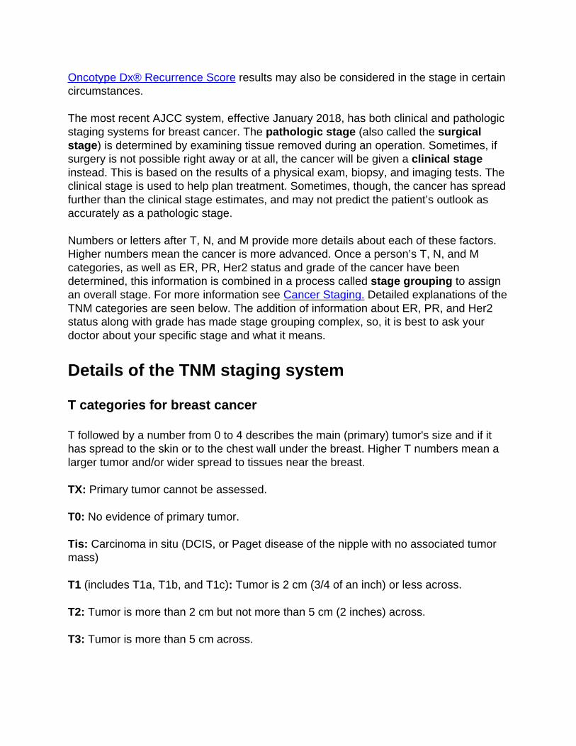
Oncotype Dx® Recurrence Score results may also be considered in the stage in certaincircumstances.
The most recent AJCC system, effective January 2018, has both clinical and pathologicstaging systems for breast cancer. The pathologic stage (also called the surgicalstage) is determined by examining tissue removed during an operation. Sometimes, ifsurgery is not possible right away or at all, the cancer will be given a clinical stageinstead. This is based on the results of a physical exam, biopsy, and imaging tests. Theclinical stage is used to help plan treatment. Sometimes, though, the cancer has spreadfurther than the clinical stage estimates, and may not predict the patient’s outlook asaccurately as a pathologic stage.
Numbers or letters after T, N, and M provide more details about each of these factors.Higher numbers mean the cancer is more advanced. Once a person’s T, N, and Mcategories, as well as ER, PR, Her2 status and grade of the cancer have beendetermined, this information is combined in a process called stage grouping to assignan overall stage. For more information see Cancer Staging. Detailed explanations of theTNM categories are seen below. The addition of information about ER, PR, and Her2status along with grade has made stage grouping complex, so, it is best to ask yourdoctor about your specific stage and what it means.
Details of the TNM staging system
T categories for breast cancer
T followed by a number from 0 to 4 describes the main (primary) tumor's size and if ithas spread to the skin or to the chest wall under the breast. Higher T numbers mean alarger tumor and/or wider spread to tissues near the breast.
TX: Primary tumor cannot be assessed.
T0: No evidence of primary tumor.
Tis: Carcinoma in situ (DCIS, or Paget disease of the nipple with no associated tumormass)
T1 (includes T1a, T1b, and T1c): Tumor is 2 cm (3/4 of an inch) or less across.
T2: Tumor is more than 2 cm but not more than 5 cm (2 inches) across.
T3: Tumor is more than 5 cm across.
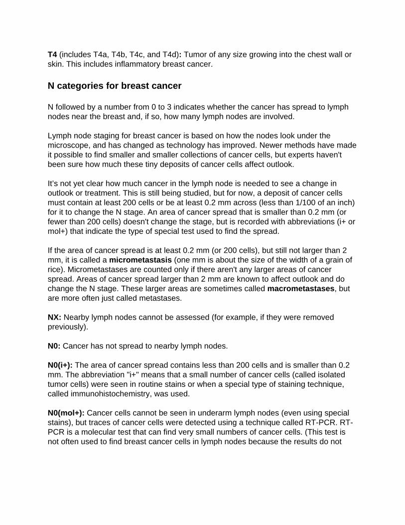
T4 (includes T4a, T4b, T4c, and T4d): Tumor of any size growing into the chest wall orskin. This includes inflammatory breast cancer.
N categories for breast cancer
N followed by a number from 0 to 3 indicates whether the cancer has spread to lymphnodes near the breast and, if so, how many lymph nodes are involved.
Lymph node staging for breast cancer is based on how the nodes look under themicroscope, and has changed as technology has improved. Newer methods have madeit possible to find smaller and smaller collections of cancer cells, but experts haven'tbeen sure how much these tiny deposits of cancer cells affect outlook.
It’s not yet clear how much cancer in the lymph node is needed to see a change inoutlook or treatment. This is still being studied, but for now, a deposit of cancer cellsmust contain at least 200 cells or be at least 0.2 mm across (less than 1/100 of an inch)for it to change the N stage. An area of cancer spread that is smaller than 0.2 mm (orfewer than 200 cells) doesn't change the stage, but is recorded with abbreviations (i+ ormol+) that indicate the type of special test used to find the spread.
If the area of cancer spread is at least 0.2 mm (or 200 cells), but still not larger than 2mm, it is called a micrometastasis (one mm is about the size of the width of a grain ofrice). Micrometastases are counted only if there aren't any larger areas of cancerspread. Areas of cancer spread larger than 2 mm are known to affect outlook and dochange the N stage. These larger areas are sometimes called macrometastases, butare more often just called metastases.
NX: Nearby lymph nodes cannot be assessed (for example, if they were removedpreviously).
N0: Cancer has not spread to nearby lymph nodes.
N0(i+): The area of cancer spread contains less than 200 cells and is smaller than 0.2mm. The abbreviation "i+" means that a small number of cancer cells (called isolatedtumor cells) were seen in routine stains or when a special type of staining technique,called immunohistochemistry, was used.
N0(mol+): Cancer cells cannot be seen in underarm lymph nodes (even using specialstains), but traces of cancer cells were detected using a technique called RT-PCR. RT-PCR is a molecular test that can find very small numbers of cancer cells. (This test isnot often used to find breast cancer cells in lymph nodes because the results do not
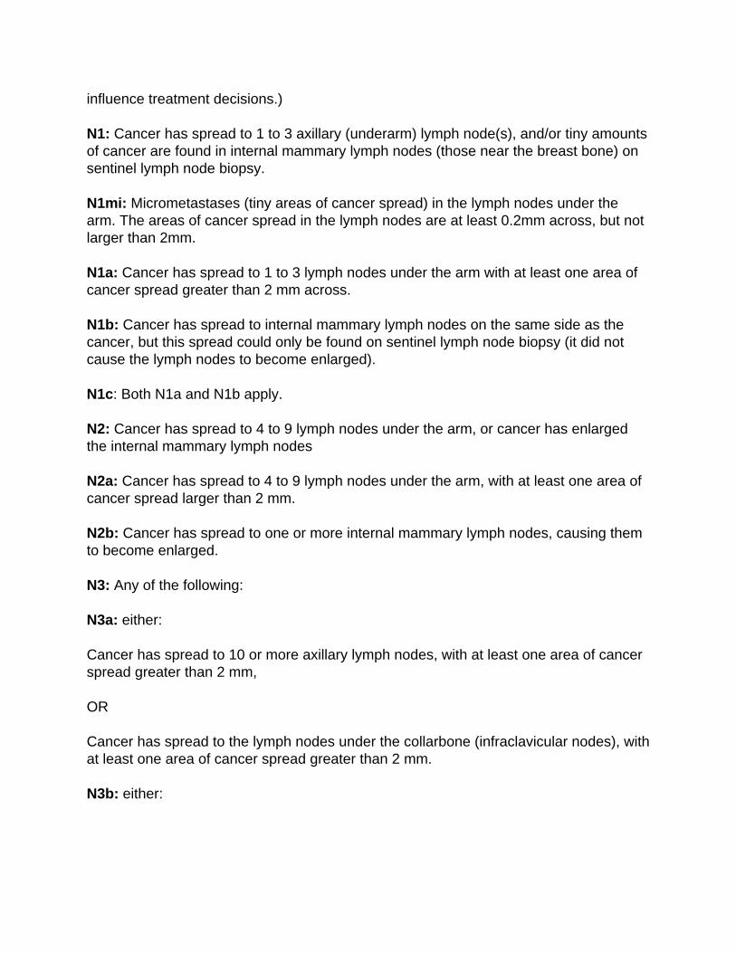
influence treatment decisions.)
N1: Cancer has spread to 1 to 3 axillary (underarm) lymph node(s), and/or tiny amountsof cancer are found in internal mammary lymph nodes (those near the breast bone) onsentinel lymph node biopsy.
N1mi: Micrometastases (tiny areas of cancer spread) in the lymph nodes under thearm. The areas of cancer spread in the lymph nodes are at least 0.2mm across, but notlarger than 2mm.
N1a: Cancer has spread to 1 to 3 lymph nodes under the arm with at least one area ofcancer spread greater than 2 mm across.
N1b: Cancer has spread to internal mammary lymph nodes on the same side as thecancer, but this spread could only be found on sentinel lymph node biopsy (it did notcause the lymph nodes to become enlarged).
N1c: Both N1a and N1b apply.
N2: Cancer has spread to 4 to 9 lymph nodes under the arm, or cancer has enlargedthe internal mammary lymph nodes
N2a: Cancer has spread to 4 to 9 lymph nodes under the arm, with at least one area ofcancer spread larger than 2 mm.
N2b: Cancer has spread to one or more internal mammary lymph nodes, causing themto become enlarged.
N3: Any of the following:
N3a: either:
Cancer has spread to 10 or more axillary lymph nodes, with at least one area of cancerspread greater than 2 mm,
OR
Cancer has spread to the lymph nodes under the collarbone (infraclavicular nodes), withat least one area of cancer spread greater than 2 mm.
N3b: either:
Cancer is found in at least one axillary lymph node (with at least one area of cancerspread greater than 2 mm) and has enlarged the internal mammary lymph nodes,
OR
Cancer has spread to 4 or more axillary lymph nodes (with at least one area of cancerspread greater than 2 mm), and tiny amounts of cancer are found in internal mammarylymph nodes on sentinel lymph node biopsy.
N3c: Cancer has spread to the lymph nodes above the collarbone (supraclavicularnodes) with at least one area of cancer spread greater than 2 mm.
M categories for breast cancer
M followed by a 0 or 1 indicates whether the cancer has spread to distant organs -- forexample, the lungs, liver, or bones.
MX: Distant spread (metastasis) cannot be assessed.
M0: No distant spread is found on x-rays (or other imaging tests) or by physical exam.
cM0(i+): Small numbers of cancer cells are found in blood or bone marrow (found onlyby special tests), or tiny areas of cancer spread (no larger than 0.2 mm) are found inlymph nodes away from the underarm, collarbone, or internal mammary areas.
M1: Cancer has spread to distant organs (most often to the bones, lungs, brain, orliver).
Examples using the new staging system
Example #1
If the cancer size is between 2 and 5 cm (T2) but it has not spread to the nearby lymphnodes (N0) or to distant organs (M0) AND is:
Grade 3●
Her2 negative●
ER positive●
PR positive●
The cancer stage is IB.
Example #2
If the cancer is larger than 5 cm (T3) and has spread to 4 to 9 lymph nodes under thearm or to any internal mammary lymph nodes (N2) but not to distant organs (M0) ANDis:
Grade 2●
Her2 positive●
ER positive●
PR positive●
The cancer stage is IB.
Example #3
If the cancer is larger than 5 cm (T3) and has spread to 4 to 9 lymph nodes under thearm or to any internal mammary lymph nodes (N2) but not to distant organs (M0) ANDis:
Grade 2●
Her2 negative●
ER negative●
PR negative●
The cancer stage is IIIB.
References●
American Joint Committee on Cancer. Breast. In: AJCC Cancer Staging Manual. 8th ed.New York, NY: Springer; 2017:589.
Genomic Health, Inc. website: Oncotype DX. Accessed at www.genomichealth.com/en-US/OncotypeDX.aspx on January 16, 2018.
National Comprehensive Cancer Network (NCCN). Practice Guidelines in Oncology:Breast Cancer. Version 3.2017. Accessed at www.nccn.org on January 16, 2018.
Paik, S. Development and Clinical Utility of a 21-Gene Recurrence Score PrognosticAssay in Patients with Early Breast Cancer Treated with Tamoxifen. The Oncologist.2007;12(6): 631-635.

Last Medical Review: April 27, 2018 Last Revised: April 27, 2018
American Cancer Society medical information is copyrighted material. For reprintrequests, please see our Content Usage Policy.
Breast Cancer Survival Rates in Men Survival rates tell you what portion of people with the same type and stage of cancerare still alive a certain amount of time (usually 5 years) after they were diagnosed. Theycan’t tell you how long you will live, but they may help give you a better understandingabout how likely it is that your treatment will be successful. Some people will want toknow the survival rates for their cancer type and stage, and some people won’t. If youdon’t want to know, you don’t have to.
What is a 5-year survival rate?
Statistics on the outlook for a certain type and stage of cancer are often given as 5-yearsurvival rates, but many people live longer – often much longer – than 5 years. The 5-year survival rate is the percentage of people who live at least 5 years after beingdiagnosed with cancer. For example, a 5-year survival rate of 90% means that anestimated 90 out of 100 people who have that cancer are still alive 5 years after beingdiagnosed.
Relative survival rates are a more accurate way to estimate the effect of cancer onsurvival. These rates compare men with breast cancer to men in the overallpopulation. For example, if the 5-year relative survival rate for a specific type of canceris 90%, it means that people who have that cancer are, on average, about 90% as likelyas people who don’t have that cancer to live for at least 5 years after being diagnosed.
But remember, the 5-year relative survival rates are estimates – your outlook can varybased on a number of your specific factors.
Cancer survival rates don’t tell the whole story
Survival rates are often based on previous outcomes of large numbers of people whohad the disease, but they can’t predict what will happen in any particular person’s case.
There are a number of limitations to remember:
The numbers below are among the most current available. But to get 5-yearsurvival rates, doctors have to look at people who were treated at least 5 years ago.As treatments are improving over time, men who are now being diagnosed withbreast cancer may have a better outlook than these statistics show.
●
The available statistics for breast cancer do not divide survival rates by all of thesub stages, such as IA and IB. The rates for these substages are likely to be closeto the rate for the overall stage. For example, the survival rate for stage IA is likelyto be slightly higher than that listed for stage I, while the survival rate for stage IBwould be expected to be slightly lower.
●
These statistics are based on the stage of the cancer when it was first diagnosed.They do not apply to cancers that come back later or spread, for example.
●
Many other factors can affect a person's outlook, such as age and health, thepresence of hormone receptors on the cancer cells, the treatment received, andhow well the cancer responds to treatment.
●
Your doctor can tell you how these numbers might apply to you, as he or she is familiarwith your particular situation.
Breast cancer survival rates, by stage
The outlook for men with breast cancer varies by thestage (extent) of the cancer. Ingeneral, the survival rates are better for men with earlier stage cancers. But remember,the outlook for each man is specific to his circumstances. The numbers below comefrom the National Cancer Institute's Surveillance Epidemiology and End Results (SEER)database. These statistics include only male breast cancer cases between 2007 and2013 and are based on an older version of AJCC staging.
It is also important to realize that these statistics are based on the stage of the cancerwhen it was first diagnosed. These do not apply to cancer after it has come back orspread.
Remember, these survival rates are only estimates – they can’t predict what will happento any individual. We understand that these statistics can be confusing and may leadyou to have more questions. Talk to your doctor to better understand your specificsituation.
Stage 5-year relative survival rate0 100%
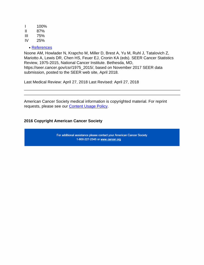
I 100%II 87%III 75%IV 25%
References●
Noone AM, Howlader N, Krapcho M, Miller D, Brest A, Yu M, Ruhl J, Tatalovich Z,Mariotto A, Lewis DR, Chen HS, Feuer EJ, Cronin KA (eds). SEER Cancer StatisticsReview, 1975-2015, National Cancer Institute. Bethesda, MD,https://seer.cancer.gov/csr/1975_2015/, based on November 2017 SEER datasubmission, posted to the SEER web site, April 2018.
Last Medical Review: April 27, 2018 Last Revised: April 27, 2018
American Cancer Society medical information is copyrighted material. For reprintrequests, please see our Content Usage Policy.
2016 Copyright American Cancer Society