Embed Size (px)
Citation preview

Natio
nal C
ance
r Ins
titut
e
What You Need To Know AboutTM
BrainTumors
U.S. DEPARTMENT OF HEALTH AND HUMAN SERVICES
National Institutes of Health

For more publicationsThis is only one of many free booklets for
people with cancer.
Here’s how to get other National CancerInstitute (NCI) booklets:
• Call the NCI Contact Centerat 1–800–4–CANCER (1–800–422–6237)
• Go to the NCI Web site at http://www.cancer.gov/publications
For materials in SpanishHere’s how to get NCI materials in Spanish:
• Call the NCI Contact Center at 1–800–422–6237
• Go to the NCI Web site at http://www.cancer.gov/espanol

U.S. DEPARTMENT OFHEALTH AND HUMAN SERVICESNational Institutes of HealthNational Cancer Institute
Contents
About This Booklet 1
The Brain 2
Tumor Grades and Types 5
Risk Factors 8
Symptoms 9
Diagnosis 10
Treatment 13
Second Opinion 23
Nutrition 25
Supportive Care 25
Rehabilitation 26
Follow-up Care 27
Sources of Support 28
Taking Part in Cancer Research 30
Dictionary 31
National Cancer Institute Services 43
National Cancer Institute Publications 44

About This Booklet
This National Cancer Institute (NCI) booklet isabout tumors* that begin in the brain (primary braintumors). Each year in the United States, more than35,000 people are told they have a tumor that started inthe brain.
This booklet tells about diagnosis, treatment, andsupportive care. Learning about medical care for braintumors can help you take an active part in makingchoices about your care.
1
*Words in italics are in the Dictionary on page 31. The Dictionaryexplains these terms. It also shows how to pronounce them.
This booklet is only about primary braintumors. Cancer that spreads to the brain fromanother part of the body is different from aprimary brain tumor.
Lung cancer, breast cancer, kidney cancer,melanoma, and other types of cancer commonlyspread to the brain. When this happens, thetumors are called metastatic brain tumors.
People with metastatic brain tumors havedifferent treatment options. Treatment dependsmainly on where the cancer started. Instead ofthis booklet, you may want to read the NCI factsheet Metastatic Cancer. The NCI Contact Centerat 1–800–4–CANCER (1–800–422–6237) cansend you this fact sheet, as well as otherinformation about metastatic brain tumors.
This booklet has lists of questions that you maywant to ask your doctor. Many people find it helpful totake a list of questions to a doctor visit. To helpremember what your doctor says, you can take notes orask whether you may use a tape recorder. You may alsowant to have a family member or friend go with youwhen you talk with the doctor—to take notes, askquestions, or just listen.
For the latest information about brain tumors,please visit our Web site at http://www.cancer.gov/cancertopics/types/brain. Also, the NCI ContactCenter can answer your questions about brain tumors.We can also send you NCI booklets and fact sheets.Call 1–800–4–CANCER (1–800–422–6237) orinstant message us through the LiveHelp service athttp://www.cancer.gov/help.
The Brain
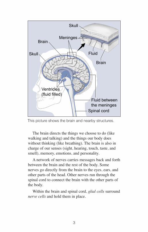
The brain is a soft, spongy mass of tissue. It isprotected by:
• The bones of the skull
• Three thin layers of tissue (meninges)
• Watery fluid (cerebrospinal fluid) that flowsthrough spaces between the meninges and throughspaces (ventricles) within the brain
2
The brain directs the things we choose to do (likewalking and talking) and the things our body doeswithout thinking (like breathing). The brain is also incharge of our senses (sight, hearing, touch, taste, andsmell), memory, emotions, and personality.
A network of nerves carries messages back and forthbetween the brain and the rest of the body. Somenerves go directly from the brain to the eyes, ears, andother parts of the head. Other nerves run through thespinal cord to connect the brain with the other parts ofthe body.
Within the brain and spinal cord, glial cells surroundnerve cells and hold them in place.
3
Ventricles(fluid filled)
Spinal cord
BrainMeninges
Skull
Skull
Fluid betweenthe meninges
Brain
Fluid
This picture shows the brain and nearby structures.
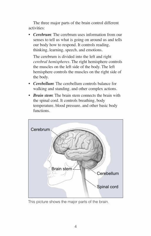
The three major parts of the brain control differentactivities:
• Cerebrum: The cerebrum uses information from oursenses to tell us what is going on around us and tellsour body how to respond. It controls reading,thinking, learning, speech, and emotions.
The cerebrum is divided into the left and rightcerebral hemispheres. The right hemisphere controlsthe muscles on the left side of the body. The lefthemisphere controls the muscles on the right side ofthe body.
• Cerebellum: The cerebellum controls balance forwalking and standing, and other complex actions.
• Brain stem: The brain stem connects the brain withthe spinal cord. It controls breathing, bodytemperature, blood pressure, and other basic bodyfunctions.
4
Cerebrum
Cerebellum
Spinal cord
Brain stem
This picture shows the major parts of the brain.

Tumor Grades and Types
When most normal cells grow old or get damaged,they die, and new cells take their place. Sometimes,this process goes wrong. New cells form when thebody doesn’t need them, and old or damaged cellsdon’t die as they should. The buildup of extra cellsoften forms a mass of tissue called a growth or tumor.
Primary brain tumors can be benign or malignant:
• Benign brain tumors do not contain cancer cells:
—Usually, benign tumors can be removed, and theyseldom grow back.
—Benign brain tumors usually have an obviousborder or edge. Cells from benign tumors rarelyinvade tissues around them. They don’t spread toother parts of the body. However, benign tumorscan press on sensitive areas of the brain andcause serious health problems.
—Unlike benign tumors in most other parts of thebody, benign brain tumors are sometimes lifethreatening.
—Benign brain tumors may become malignant.
• Malignant brain tumors (also called brain cancer)contain cancer cells:
—Malignant brain tumors are generally moreserious and often are a threat to life.
—They are likely to grow rapidly and crowd orinvade the nearby healthy brain tissue.
—Cancer cells may break away from malignantbrain tumors and spread to other parts of thebrain or to the spinal cord. They rarely spread toother parts of the body.
5

Tumor GradeDoctors group brain tumors by grade. The grade of
a tumor refers to the way the cells look under amicroscope:
• Grade I: The tissue is benign. The cells look nearlylike normal brain cells, and they grow slowly.
• Grade II: The tissue is malignant. The cells lookless like normal cells than do the cells in a Grade Itumor.
• Grade III: The malignant tissue has cells that lookvery different from normal cells. The abnormal cellsare actively growing (anaplastic).
• Grade IV: The malignant tissue has cells that lookmost abnormal and tend to grow quickly.
Cells from low-grade tumors (grades I and II) lookmore normal and generally grow more slowly thancells from high-grade tumors (grades III and IV).
Over time, a low-grade tumor may become a high-grade tumor. However, the change to a high-gradetumor happens more often among adults than children.
You may want to read the NCI fact sheet TumorGrade.
Types of Primary Brain TumorsThere are many types of primary brain tumors.
Primary brain tumors are named according to the typeof cells or the part of the brain in which they begin.For example, most primary brain tumors begin in glialcells. This type of tumor is called a glioma.
Among adults, the most common types are:
• Astrocytoma: The tumor arises from star-shapedglial cells called astrocytes. It can be any grade. Inadults, an astrocytoma most often arises in thecerebrum.
6

—Grade I or II astrocytoma: It may be called alow-grade glioma.
—Grade III astrocytoma: It’s sometimes called ahigh-grade or an anaplastic astrocytoma.
—Grade IV astrocytoma: It may be called aglioblastoma or malignant astrocytic glioma.
• Meningioma: The tumor arises in the meninges. Itcan be grade I, II, or III. It’s usually benign (grade I)and grows slowly.
• Oligodendroglioma: The tumor arises from cellsthat make the fatty substance that covers andprotects nerves. It usually occurs in the cerebrum.It’s most common in middle-aged adults. It can begrade II or III.
Among children, the most common types are:
• Medulloblastoma: The tumor usually arises in thecerebellum. It’s sometimes called a primitiveneuroectodermal tumor. It is grade IV.
• Grade I or II astrocytoma: In children, this low-grade tumor occurs anywhere in the brain. The mostcommon astrocytoma among children is juvenilepilocytic astrocytoma. It’s grade I.
• Ependymoma: The tumor arises from cells that linethe ventricles or the central canal of the spinal cord.It’s most commonly found in children and youngadults. It can be grade I, II, or III.
• Brain stem glioma: The tumor occurs in the lowestpart of the brain. It can be a low-grade or high-gradetumor. The most common type is diffuse intrinsicpontine glioma.
You can find more information about types of braintumors at http://www.cancer.gov/cancertopics/types/brain. Or, you can call the NCI Contact Centerat 1–800–4–CANCER (1–800–422–6237).
7
Risk Factors
When you’re told that you have a brain tumor, it’snatural to wonder what may have caused your disease.But no one knows the exact causes of brain tumors.Doctors seldom know why one person develops a braintumor and another doesn’t.
Researchers are studying whether people withcertain risk factors are more likely than others todevelop a brain tumor. A risk factor is something thatmay increase the chance of getting a disease.
Studies have found the following risk factors forbrain tumors:
• Ionizing radiation: Ionizing radiation from high-dose x-rays (such as radiation therapy from a largemachine aimed at the head) and other sources cancause cell damage that leads to a tumor. Peopleexposed to ionizing radiation may have an increasedrisk of a brain tumor, such as meningioma orglioma.
• Family history: It is rare for brain tumors to run ina family. Only a very small number of families haveseveral members with brain tumors.
Researchers are studying whether using cell phones,having had a head injury, or having been exposed tocertain chemicals at work or to magnetic fields areimportant risk factors. Studies have not shownconsistent links between these possible risk factors andbrain tumors, but additional research is needed.
8

Symptoms
The symptoms of a brain tumor depend on tumorsize, type, and location. Symptoms may be causedwhen a tumor presses on a nerve or harms a part of thebrain. Also, they may be caused when a tumor blocksthe fluid that flows through and around the brain, orwhen the brain swells because of the buildup of fluid.
These are the most common symptoms of braintumors:
• Headaches (usually worse in the morning)
• Nausea and vomiting
• Changes in speech, vision, or hearing
• Problems balancing or walking
• Changes in mood, personality, or ability toconcentrate
• Problems with memory
• Muscle jerking or twitching (seizures orconvulsions)
• Numbness or tingling in the arms or legs
Most often, these symptoms are not due to a braintumor. Another health problem could cause them. Ifyou have any of these symptoms, you should tell yourdoctor so that problems can be diagnosed and treated.
9
Diagnosis
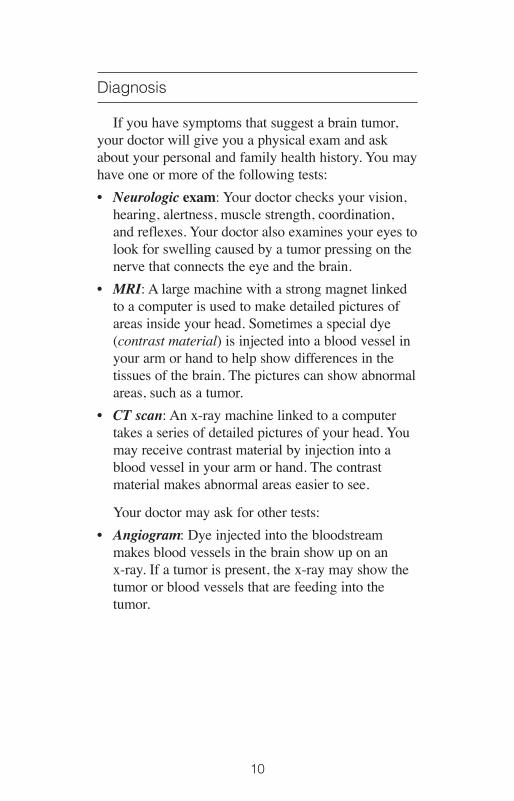
If you have symptoms that suggest a brain tumor,your doctor will give you a physical exam and askabout your personal and family health history. You mayhave one or more of the following tests:
• Neurologic exam: Your doctor checks your vision,hearing, alertness, muscle strength, coordination,and reflexes. Your doctor also examines your eyes tolook for swelling caused by a tumor pressing on thenerve that connects the eye and the brain.
• MRI: A large machine with a strong magnet linkedto a computer is used to make detailed pictures ofareas inside your head. Sometimes a special dye(contrast material) is injected into a blood vessel inyour arm or hand to help show differences in thetissues of the brain. The pictures can show abnormalareas, such as a tumor.
• CT scan: An x-ray machine linked to a computertakes a series of detailed pictures of your head. Youmay receive contrast material by injection into ablood vessel in your arm or hand. The contrastmaterial makes abnormal areas easier to see.
Your doctor may ask for other tests:
• Angiogram: Dye injected into the bloodstreammakes blood vessels in the brain show up on an x-ray. If a tumor is present, the x-ray may show thetumor or blood vessels that are feeding into thetumor.
10
• Spinal tap: Your doctor may remove a sample ofcerebrospinal fluid (the fluid that fills the spaces inand around the brain and spinal cord). Thisprocedure is performed with local anesthesia. Thedoctor uses a long, thin needle to remove fluid fromthe lower part of the spinal column. A spinal taptakes about 30 minutes. You must lie flat for severalhours afterward to keep from getting a headache. Alaboratory checks the fluid for cancer cells or othersigns of problems.
• Biopsy: The removal of tissue to look for tumorcells is called a biopsy. A pathologist looks at thecells under a microscope to check for abnormalcells. A biopsy can show cancer, tissue changes thatmay lead to cancer, and other conditions. A biopsy isthe only sure way to diagnose a brain tumor, learnwhat grade it is, and plan treatment.
11

Surgeons can obtain tissue to look for tumor cells intwo ways:
—Biopsy at the same time as treatment: Thesurgeon takes a tissue sample when you havesurgery to remove part or all of the tumor. See theSurgery section on page 16.
—Stereotactic biopsy: You may get local or generalanesthesia and wear a rigid head frame for thisprocedure. The surgeon makes a small incision inthe scalp and drills a small hole (a burr hole) intothe skull. CT or MRI is used to guide the needlethrough the burr hole to the location of the tumor.The surgeon withdraws a sample of tissue withthe needle. A needle biopsy may be used when atumor is deep inside the brain or in a part of thebrain that can’t be operated on.
However, if the tumor is in the brain stem or certainother areas, the surgeon may not be able to removetissue from the tumor without harming normal braintissue. In this case, the doctor uses MRI, CT, orother imaging tests to learn as much as possibleabout the brain tumor.
12

Treatment
People with brain tumors have several treatmentoptions. The options are surgery, radiation therapy,and chemotherapy. Many people get a combination oftreatments.
The choice of treatment depends mainly on thefollowing:
• The type and grade of brain tumor
• Its location in the brain
• Its size
• Your age and general health
For some types of brain cancer, the doctor alsoneeds to know whether cancer cells were found in thecerebrospinal fluid.
13
A person who needs a biopsy may want to askthe doctor the following questions:
• Why do I need a biopsy? How will the biopsyresults affect my treatment plan?
• What kind of biopsy will I have?
• How long will it take? Will I be awake? Will ithurt?
• What are the chances of infection or bleedingafter the biopsy? Are there any other risks?
• How soon will I know the results?
• If I do have a brain tumor, who will talk withme about treatment? When?

Your doctor can describe your treatment choices, theexpected results, and the possible side effects. Becausecancer therapy often damages healthy cells and tissues,side effects are common. Before treatment starts, askyour health care team about possible side effects andhow treatment may change your normal activities. Youand your health care team can work together todevelop a treatment plan that meets your medical andpersonal needs.
You may want to talk with your doctor about takingpart in a clinical trial, a research study of newtreatment methods. See the Taking Part in CancerResearch section on page 30.
Your doctor may refer you to a specialist, or youmay ask for a referral. Specialists who treat braintumors include neurologists, neurosurgeons, neuro-oncologists, medical oncologists, radiation oncologists,and neuroradiologists.
Your health care team may also include an oncologynurse, a registered dietitian, a mental health counselor,a social worker, a physical therapist, an occupationaltherapist, a speech therapist, and a physical medicinespecialist. Also, children may need tutors to help withschoolwork. (The Rehabilitation section on page 26has more information about therapists and tutors.)
14

15
You may want to ask your doctor thesequestions before you begin treatment:
• What type of brain tumor do I have?
• Is it benign or malignant?
• What is the grade of the tumor?
• What are my treatment choices? Which do yourecommend for me? Why?
• What are the expected benefits of each kind oftreatment?
• What can I do to prepare for treatment?
• Will I need to stay in the hospital? If so, forhow long?
• What are the risks and possible side effects ofeach treatment? How can side effects bemanaged?
• What is the treatment likely to cost? Will myinsurance cover it?
• How will treatment affect my normalactivities? What is the chance that I will haveto learn how to walk, speak, read, or write aftertreatment?
• Would a research study (clinical trial) beappropriate for me?
• Can you recommend other doctors who couldgive me a second opinion about my treatmentoptions?
• How often should I have checkups?

SurgerySurgery is the usual first treatment for most brain
tumors. Before surgery begins, you may be givengeneral anesthesia, and your scalp is shaved. Youprobably won’t need your entire head shaved.
Surgery to open the skull is called a craniotomy.The surgeon makes an incision in your scalp and uses aspecial type of saw to remove a piece of bone from theskull.
You may be awake when the surgeon removes partor all of the brain tumor. The surgeon removes as muchtumor as possible. You may be asked to move a leg,count, say the alphabet, or tell a story. Your ability tofollow these commands helps the surgeon protectimportant parts of the brain.
After the tumor is removed, the surgeon covers theopening in the skull with the piece of bone or with apiece of metal or fabric. The surgeon then closes theincision in the scalp.
Sometimes surgery isn’t possible. If the tumor is inthe brain stem or certain other areas, the surgeon maynot be able to remove the tumor without harmingnormal brain tissue. People who can’t have surgerymay receive radiation therapy or other treatment.
You may have a headache or be uncomfortable forthe first few days after surgery. However, medicine canusually control pain. Before surgery, you shoulddiscuss the plan for pain relief with your health careteam. After surgery, your team can adjust the plan ifyou need more relief.
16

You may also feel tired or weak. The time it takes toheal after surgery is different for everyone. You willprobably spend a few days in the hospital.
Other, less common problems may occur aftersurgery for a brain tumor. The brain may swell or fluidmay build up within the skull. The health care teamwill monitor you for signs of swelling or fluid buildup.You may receive steroids to help relieve swelling. Asecond surgery may be needed to drain the fluid. Thesurgeon may place a long, thin tube (shunt) in aventricle of the brain. (For some people, the shunt isplaced before performing surgery on the brain tumor.)The tube is threaded under the skin to another part of the body, usually the abdomen. Excess fluid iscarried from the brain and drained into the abdomen.Sometimes the fluid is drained into the heart instead.
Infection is another problem that may develop aftersurgery. If this happens, the health care team will giveyou an antibiotic.
Brain surgery may harm normal tissue. Braindamage can be a serious problem. It can causeproblems with thinking, seeing, or speaking. It can alsocause personality changes or seizures. Most of theseproblems lessen or disappear with time. But sometimesdamage to the brain is permanent. You may needphysical therapy, speech therapy, or occupationaltherapy. See the Rehabilitation section on page 26.
17

Radiation TherapyRadiation therapy kills brain tumor cells with high-
energy x-rays, gamma rays, or protons.
Radiation therapy usually follows surgery. Theradiation kills tumor cells that may remain in the area.Sometimes, people who can’t have surgery haveradiation therapy instead.
Doctors use external and internal types of radiationtherapy to treat brain tumors:
• External radiation therapy: You’ll go to a hospitalor clinic for treatment. A large machine outside thebody aims beams of radiation at the head. Becausecancer cells may invade normal tissue around atumor, the radiation may be aimed at the tumor andnearby brain tissue, or at the entire brain. Somepeople need radiation aimed at the spinal cord also.
18
You may want to ask your doctor thesequestions about surgery:
• Do you suggest surgery for me?
• How will I feel after the operation?
• What will you do for me if I have pain?
• How long will I be in the hospital?
• Will I have any long-term effects? Will my hairgrow back? Are there any side effects fromusing metal or fabric to replace the bone in theskull?
• When can I get back to my normal activities?
• What is my chance of a full recovery?

19
The treatment schedule depends on your age, and thetype and size of the tumor. Fractionated externalbeam therapy is the most common method ofradiation therapy used for people with brain tumors.Giving the total dose of radiation over several weekshelps to protect healthy tissue in the area of thetumor. Treatments are usually 5 days a week forseveral weeks. A typical visit lasts less than an hour,and each treatment takes only a few minutes.
Some treatment centers are studying other ways ofdelivering external beam radiation therapy:
—Intensity-modulated radiation therapy or 3-dimensional conformal radiation therapy:These types of treatment use computers to moreclosely target the brain tumor to lessen thedamage to healthy tissue.
—Proton beam radiation therapy: The source ofradiation is protons rather than x-rays. The doctoraims the proton beam at the tumor. The dose ofradiation to normal tissue from a proton beam isless than the dose from an x-ray beam.
—Stereotactic radiation therapy: Narrow beams ofx-rays or gamma rays are directed at the tumorfrom different angles. For this procedure, youwear a rigid head frame. The therapy may begiven during a single visit (stereotacticradiosurgery) or over several visits.
• Internal radiation therapy (implant radiationtherapy or brachytherapy): Internal radiation isn’tcommonly used for treating brain tumors and isunder study. The radiation comes from radioactivematerial usually contained in very small implantscalled seeds. The seeds are placed inside the brainand give off radiation for months. They don’t need tobe removed once the radiation is gone.
Some people have no or few side effects aftertreatment. Rarely, people may have nausea for severalhours after external radiation therapy. The health careteam can suggest ways to help you cope with thisproblem. Radiation therapy also may cause you tobecome very tired with each radiation treatment.Resting is important, but doctors usually advise peopleto try to stay as active as they can.
Also, external radiation therapy commonly causeshair loss from the part of the head that was treated.Hair usually grows back within a few months.Radiation therapy also may make the skin on the scalpand ears red, dry, and tender. The health care team cansuggest ways to relieve these problems.
Sometimes radiation therapy causes brain tissue toswell. You may get a headache or feel pressure. Thehealth care team watches for signs of this problem.They can provide medicine to reduce the discomfort.
Radiation sometimes kills healthy brain tissue.Although rare, this side effect can cause headaches,seizures, or even death.
Radiation may harm the pituitary gland and otherareas of the brain. For children, this damage couldcause learning problems or slow down growth anddevelopment. In addition, radiation increases the riskof secondary tumors later in life.
You may find it helpful to read the NCI bookletRadiation Therapy and You.
20

ChemotherapyChemotherapy, the use of drugs to kill cancer cells,
is sometimes used to treat brain tumors. Drugs may begiven in the following ways:
• By mouth or vein (intravenous): Chemotherapymay be given during and after radiation therapy. Thedrugs enter the bloodstream and travel throughoutthe body. They may be given in an outpatient part ofthe hospital, at the doctor’s office, or at home.Rarely, you may need to stay in the hospital.
The side effects of chemotherapy depend mainly onwhich drugs are given and how much. Commonside effects include nausea and vomiting, loss ofappetite, headache, fever and chills, and weakness.If the drugs lower the levels of healthy blood cells,you’re more likely to get infections, bruise or bleedeasily, and feel very weak and tired. Your healthcare team will check for low levels of blood cells.Some side effects may be relieved with medicine.
21
You may want to ask your doctor thesequestions about radiation therapy:
• Why do I need this treatment?
• When will the treatments begin? When willthey end?
• How will I feel during therapy? Are there sideeffects?
• What can I do to take care of myself duringtherapy?
• How will we know if the radiation is working?
• Will I be able to continue my normal activitiesduring treatment?

• In wafers that are put into the brain: For someadults with high-grade glioma, the surgeon implantsseveral wafers into the brain. Each wafer is aboutthe size of a dime. Over several weeks, the wafersdissolve, releasing the drug into the brain. The drugkills cancer cells. It may help prevent the tumorfrom returning in the brain after surgery to removethe tumor.
People who receive an implant (a wafer) thatcontains a drug are monitored by the health careteam for signs of infection after surgery. Aninfection can be treated with an antibiotic.
You may wish to read the NCI bookletChemotherapy and You.
22
You may want to ask your doctor thesequestions about chemotherapy:
• Why do I need this treatment?
• What will it do?
• Will I have side effects? What can I do aboutthem?
• When will treatment start? When will it end?
• How will treatment affect my normalactivities?

Second Opinion
Before starting treatment, you might want a secondopinion about your diagnosis and treatment plan. Somepeople worry that the doctor will be offended if theyask for a second opinion. Usually the opposite is true.Most doctors welcome a second opinion. And manyhealth insurance companies will pay for a secondopinion if you or your doctor requests it. Somecompanies require a second opinion.
If you get a second opinion, the doctor may agreewith your first doctor’s diagnosis and treatment plan.Or the second doctor may suggest another approach.Either way, you’ll have more information and perhapsa greater sense of control. You can feel more confidentabout the decisions you make, knowing that you’velooked at your options.
23

It may take some time and effort to gather yourmedical records and see another doctor. In many cases,it’s not a problem to take several weeks to get a secondopinion. The delay in starting treatment usually won’tmake treatment less effective. To make sure, youshould discuss this delay with your doctor. Somepeople with a brain tumor need treatment right away.
There are many ways to find a doctor for a secondopinion. You can ask your doctor, a local or statemedical society, a nearby hospital, or a medical schoolfor names of specialists.
Also, you can request a consultation with specialistsat the National Institutes of Health Clinical Center inBethesda, Maryland.
• Adults and children with a brain tumor:Specialists in the NCI Neuro-Oncology Branchprovide consultations. The telephone number is301–594–6767 or 866–251–9686.
• Children with a brain tumor: Specialists in theNCI Pediatric Neuro-Oncology Section of thePediatric Oncology Branch provide consultations.The telephone number is 301–496–8009 or877–624–4878.
The NCI Contact Center at 1–800–4–CANCER(1–800–422–6237) can tell you about nearbytreatment centers. Other sources can be found in NCI’sfact sheet How To Find a Doctor or Treatment FacilityIf You Have Cancer.
Nonprofit groups with an interest in brain tumorsmay be of help. Many such groups are listed in theNCI fact sheet National Organizations That OfferServices to People With Cancer and Their Families.
24

Nutrition
It’s important for you to take care of yourself byeating well. You need the right amount of calories tomaintain a good weight. You also need enough proteinto keep up your strength. Eating well may help youfeel better and have more energy.
Sometimes, especially during or soon aftertreatment, you may not feel like eating. You may beuncomfortable or tired. You may find that foods don’ttaste as good as they used to. In addition, the sideeffects of treatment (such as poor appetite, nausea,vomiting, or mouth blisters) can make it hard to eatwell. Your doctor, a registered dietitian, or anotherhealth care provider can suggest ways to deal withthese problems. Also, the NCI booklet Eating Hints hasmany useful ideas and recipes.
Supportive Care
A brain tumor and its treatment can lead to otherhealth problems. You may receive supportive care toprevent or control these problems.
You can have supportive care before, during, andafter cancer treatment. It can improve your comfort andquality of life during treatment.
Your health care team can help you with thefollowing problems:
• Swelling of the brain: Many people with braintumors need steroids to help relieve swelling of thebrain.
• Seizures: Brain tumors can cause seizures(convulsions). Certain drugs can help prevent orcontrol seizures.
25
• Fluid buildup in the skull: If fluid builds up in theskull, the surgeon may place a shunt to drain thefluid. Information about shunts is in the Surgery partof the Treatment section (page 17).
• Sadness and other feelings: It’s normal to feel sad,anxious, or confused after a diagnosis of a seriousillness. Some people find it helpful to talk abouttheir feelings. See the Sources of Support section onpage 28 for more information.
Many people with brain tumors receive supportivecare along with treatments intended to slow theprogress of the disease. Some decide not to haveantitumor treatment and receive only supportive care tomanage their symptoms.
You can get information about supportive care athttp://www.cancer.gov/cancerinfo/coping on the NCIWeb site and from the NCI Contact Center at1–800–4–CANCER (1–800–422–6237) or LiveHelp(http://www.cancer.gov/help).
Rehabilitation
Rehabilitation can be a very important part of thetreatment plan. The goals of rehabilitation depend onyour needs and how the tumor has affected your abilityto carry out daily activities.
Some people may never regain all the abilities theyhad before the brain tumor and its treatment. But yourhealth care team makes every effort to help you returnto normal activities as soon as possible.
26
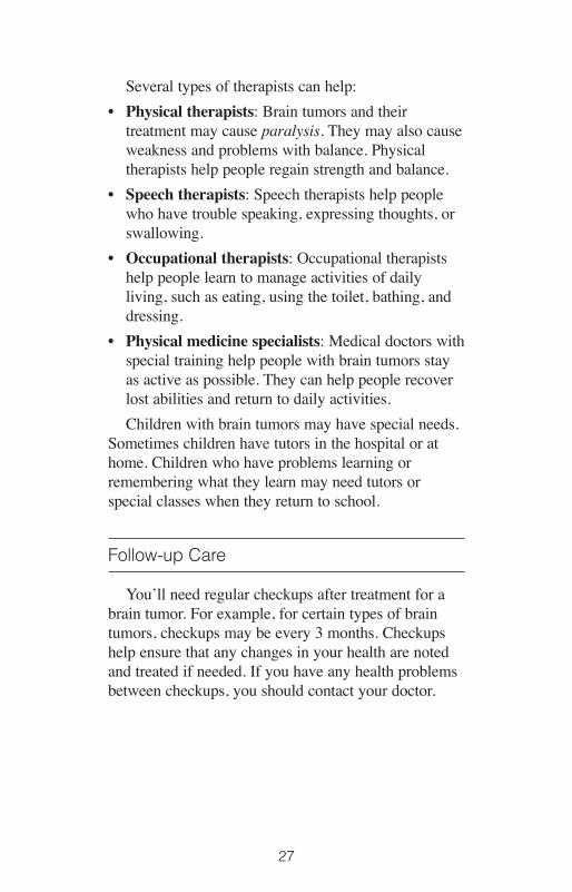
Several types of therapists can help:
• Physical therapists: Brain tumors and theirtreatment may cause paralysis. They may also causeweakness and problems with balance. Physicaltherapists help people regain strength and balance.
• Speech therapists: Speech therapists help peoplewho have trouble speaking, expressing thoughts, orswallowing.
• Occupational therapists: Occupational therapistshelp people learn to manage activities of dailyliving, such as eating, using the toilet, bathing, anddressing.
• Physical medicine specialists: Medical doctors withspecial training help people with brain tumors stayas active as possible. They can help people recoverlost abilities and return to daily activities.
Children with brain tumors may have special needs.Sometimes children have tutors in the hospital or athome. Children who have problems learning orremembering what they learn may need tutors orspecial classes when they return to school.
Follow-up Care
You’ll need regular checkups after treatment for abrain tumor. For example, for certain types of braintumors, checkups may be every 3 months. Checkupshelp ensure that any changes in your health are notedand treated if needed. If you have any health problemsbetween checkups, you should contact your doctor.
27

Your doctor will check for return of the tumor. Also,checkups help detect health problems that can resultfrom cancer treatment.
Checkups may include careful physical andneurologic exams, as well as MRI or CT scans. If youhave a shunt, your doctor checks to see that it’sworking well.
The NCI has publications to help answer questionsabout follow-up care and other concerns. You may findit helpful to read the NCI booklet Facing Forward:Life After Cancer Treatment. You may also want toread the NCI fact sheet Follow-up Care After CancerTreatment.
Sources of Support
Learning you have a brain tumor can change yourlife and the lives of those close to you. These changescan be hard to handle. It’s normal for you, your family,and your friends to need help coping with the feelingsthat such a diagnosis can bring.
Concerns about treatments and managing sideeffects, hospital stays, and medical bills are common.You may also worry about caring for your family,keeping your job, or continuing daily activities.
Here’s where you can go for support:
• Doctors, nurses, and other members of your healthcare team can answer questions about treatment,working, or other activities.
• Social workers, counselors, or members of theclergy can be helpful if you want to talk about yourfeelings or concerns. Often, social workers cansuggest resources for financial aid, transportation,home care, or emotional support.
28

• Support groups also can help. In these groups,people with brain tumors or their family membersmeet with other patients or their families to sharewhat they have learned about coping with thedisease and the effects of treatment. Groups mayoffer support in person, over the telephone, or on theInternet. You may want to talk with a member ofyour health care team about finding a support group.
• Information specialists at 1–800–4–CANCER(1–800–422–6237) and at LiveHelp(http://www.cancer.gov/help) can help you locateprograms, services, and publications. They can sendyou a list of organizations that offer services topeople with cancer.
For tips on coping, you may want to read the NCIbooklet Taking Time: Support for People With Cancer.
29
Taking Part in Cancer Research
Cancer research has led to real progress in thedetection and treatment of brain tumors. Continuingresearch offers hope that in the future even morepeople with brain tumors will be treated successfully.
Doctors all over the country are conducting manytypes of clinical trials (research studies in which peoplevolunteer to take part). Clinical trials are designed tofind out whether new approaches are safe andeffective.
Doctors are trying to find better ways to care foradults and children with brain tumors. They are testingnew drugs and combining drugs with radiation therapy.They are also studying how drugs may reduce the sideeffects of treatment.
Even if the people in a trial do not benefit directly,they may still make an important contribution byhelping doctors learn more about brain tumors and howto control them. Although clinical trials may pose somerisks, doctors do all they can to protect their patients.
If you’re interested in being part of a clinical trial,talk with your doctor. You may want to read the NCIbooklet Taking Part in Cancer Treatment ResearchStudies. It describes how treatment studies are carriedout and explains their possible benefits and risks.
The NCI Web site includes a section on clinicaltrials at http://www.cancer.gov/clinicaltrials. It hasgeneral information about clinical trials as well asdetailed information about specific ongoing studiesof brain tumors. NCI’s Information Specialists at1–800–4–CANCER (1–800–422–6237) and atLiveHelp at http://www.cancer.gov/help can answerquestions and provide information about clinical trials.
30

Dictionary
Definitions of thousands of terms are on the NCIWeb site in the NCI Dictionary of Cancer Terms. Youcan access it at http://www.cancer.gov/dictionary.
3-Dimensional conformal radiation therapy (3-dih-MEN-shuh-nul kun-FOR-mul RAY-dee-AY-shunTHAYR-uh-pee): A procedure that uses a computer tocreate a 3-dimensional picture of the tumor. Thisallows doctors to give the highest possible dose ofradiation to the tumor, while sparing the normal tissueas much as possible.
Anaplastic (an-ah-PLAS-tik): A term used to describecancer cells that divide rapidly and have little or noresemblance to normal cells.
Angiogram (AN-jee-oh-gram): An x-ray of bloodvessels. The person receives an injection of dye tooutline the vessels on the x-ray.
Antibiotic (an-tih-by-AH-tik): A drug used to treatinfections caused by bacteria and othermicroorganisms.
Astrocyte (AS-troh-site): A large, star-shaped cell thatholds nerve cells in place and helps them work the waythey should. It is a type of glial cell.
Astrocytoma (AS-troh-sy-TOH-muh): A tumor thatbegins in the brain or spinal cord in small, star-shapedcells called astrocytes.
Benign (beh-NINE): Not cancerous. Benign tumorsmay grow larger but do not spread to other parts of thebody.
Biopsy (BY-op-see): The removal of cells or tissues forexamination by a pathologist. The pathologist maystudy the tissue under a microscope or perform othertests on the cells or tissue.
31

Brachytherapy (BRAY-kee-THAYR-uh-pee): A typeof radiation therapy in which radioactive materialsealed in needles, seeds, wires, or catheters is placeddirectly into or near a tumor. Also called radiationbrachytherapy, internal radiation therapy, and implantradiation therapy.
Brain stem: The part of the brain that is connected tothe spinal cord.
Brain stem glioma (glee-OH-muh): A tumor locatedin the part of the brain that connects to the spinal cord(the brain stem). It may grow rapidly or slowly,depending on the grade of the tumor.
Burr hole: A small opening in the skull made with asurgical drill.
Cancer (KAN-ser): A term for diseases in whichabnormal cells divide without control. Cancer cells caninvade nearby tissues and can spread to other parts ofthe body through the blood and lymph systems.
Cell: The individual unit that makes up the tissues ofthe body. All living things are made up of one or morecells.
Cerebellum (ser-uh-BEL-um): The portion of the brainin the back of the head between the cerebrum and thebrain stem. The cerebellum controls balance forwalking and standing, and other complex motorfunctions.
Cerebral hemisphere (seh-REE-bral HEM-is-feer):One half of the cerebrum, the part of the brain thatcontrols muscle functions and also controls speech,thought, emotions, reading, writing, and learning. Theright hemisphere controls the muscles on the left sideof the body, and the left hemisphere controls themuscles on the right side of the body.
32

Cerebrospinal fluid (seh-REE-broh-SPY-nul): CSF.The fluid that flows in and around the hollow spaces ofthe brain and spinal cord, and between two of themeninges (the thin layers of tissue that cover andprotect the brain and spinal cord). Cerebrospinal fluidis made by tissue called the choroid plexus in theventricles (hollow spaces) in the brain.
Cerebrum (seh-REE-brum): The largest part of thebrain. It is divided into two hemispheres, or halves,called the cerebral hemispheres. Areas within thecerebrum control muscle functions and also controlspeech, thought, emotions, reading, writing, andlearning.
Chemotherapy (KEE-moh-THAYR-uh-pee):Treatment with drugs that kill cancer cells.
Clinical trial: A type of research study that tests howwell new medical approaches work in people. Thesestudies test new methods of screening, prevention,diagnosis, or treatment of a disease.
Contrast material: A dye or other substance that helpsto show abnormal areas inside the body. It is given byinjection into a vein, by enema, or by mouth. Contrastmaterial may be used with x-rays, CT scans, MRI, orother imaging tests.
Craniotomy (KRAY-nee-AH-toh-mee): An operationin which an opening is made in the skull.
CT scan: A series of detailed pictures of areas insidethe body taken from different angles. The pictures arecreated by a computer linked to an x-ray machine. Alsocalled CAT scan, computed tomography scan,computerized axial tomography scan, andcomputerized tomography.
33

Diffuse intrinsic pontine glioma (dih-FYOOS in-TRIN-sik PON-teen glee-OH-muh): A type of centralnervous system tumor that forms from glial(supportive) tissue of the brain and spinal cord. Diffuseintrinsic pontine glioma usually occurs in children. Itforms in the brain stem.
Ependymoma (eh-PEN-dih-MOH-muh): A type ofbrain tumor that begins in cells lining the spinal cordcentral canal (fluid-filled space down the center) or theventricles (fluid-filled spaces of the brain).Ependymomas may also form in the choroid plexus(tissue in the ventricles that makes cerebrospinal fluid).Also called ependymal tumor.
External radiation therapy (RAY-dee-AY-shunTHAYR-uh-pee): A type of radiation therapy that usesa machine to aim high-energy rays at the cancer fromoutside of the body. Also called external beamradiation therapy.
Gamma ray: A type of high-energy radiation that isdifferent from an x-ray.
General anesthesia (A-nes-THEE-zhuh): Drugs thatcause loss of feeling or awareness and put the personto sleep.
Glial cell (GLEE-ul): Any of the cells that hold nervecells in place and help them work the way they should.The types of glial cells include oligodendrocytes,astrocytes, microglia, and ependymal cells. Also calledneuroglia.
Glioblastoma (GLEE-oh-blas-TOH-muh): A fast-growing type of central nervous system tumor thatforms from glial (supportive) tissue of the brain andspinal cord and has cells that look very different fromnormal cells. Glioblastoma usually occurs in adults and
34

affects the brain more often than the spinal cord. Alsocalled GBM, glioblastoma multiforme, and grade IVastrocytoma.
Glioma (glee-OH-muh): A cancer of the brain thatbegins in glial cells (cells that surround and supportnerve cells).
Grade: The grade of a tumor depends on howabnormal the cancer cells look under a microscope andhow quickly the tumor is likely to grow and spread.Grading systems are different for each type of cancer.
Implant radiation therapy (RAY-dee-AY-shunTHAYR-uh-pee): A type of radiation therapy in whichradioactive material sealed in needles, seeds, wires, orcatheters is placed directly into or near a tumor. Alsocalled brachytherapy, radiation brachytherapy, andinternal radiation therapy.
Incision (in-SIH-zhun): A cut made in the body toperform surgery.
Intensity-modulated radiation therapy (in-TEN-sih-tee-MAH-juh-LAY-tid RAY-dee-AY-shun THAYR-uh-pee): A type of 3-dimensional radiation therapy thatuses computer-generated images to show the size andshape of the tumor. Thin beams of radiation of differentintensities are aimed at the tumor from many angles.This type of radiation therapy reduces the damage tohealthy tissue near the tumor.
Internal radiation therapy (in-TER-nul RAY-dee-AY-shun THAYR-uh-pee): A type of radiation therapy inwhich radioactive material sealed in needles, seeds,wires, or catheters is placed directly into or near atumor. Also called brachytherapy, radiationbrachytherapy, and implant radiation therapy.
Intravenous (IN-truh-VEE-nus): IV. Into or within avein. Intravenous usually refers to a way of giving adrug or other substance through a needle or tubeinserted into a vein.
35

Ionizing radiation (I-uh-NYZ-ing RAY-dee-AY-shun):A type of radiation made (or given off ) by x-rayprocedures, radioactive substances, rays that enter theEarth’s atmosphere from outer space, and othersources. At high doses, ionizing radiation increaseschemical activity inside cells and can lead to healthrisks, including cancer.
Juvenile pilocytic astrocytoma (JOO-veh-NILE PY-loh-SIH-tik AS-troh-sy-TOH-muh): A slow-growingtype of central nervous system tumor that forms fromglial (supportive) tissue of the brain and spinal cord.Juvenile pilocytic astrocytoma usually occurs inchildren and young adults. It forms in the brain moreoften than the spinal cord.
Local anesthesia (A-nes-THEE-zhuh): Drugs thatcause a temporary loss of feeling in one part of thebody. The patient remains awake but cannot feel thepart of the body treated with the anesthetic.
Malignant (muh-LIG-nunt): Cancerous. Malignanttumors can invade and destroy nearby tissue andspread to other parts of the body.
Medical oncologist (MEH-dih-kul on-KAH-loh-jist):A doctor who specializes in diagnosing and treatingcancer using chemotherapy, hormonal therapy, andbiological therapy. A medical oncologist often is themain health care provider for someone who has cancer.A medical oncologist also gives supportive care andmay coordinate treatment given by other specialists.
Medulloblastoma (MED-yoo-loh-blas-TOH-muh):A malignant brain tumor that begins in the lower partof the brain and that can spread to the spine or to otherparts of the body. Medulloblastomas are a type ofprimitive neuroectodermal tumor (PNET).
Meninges (meh-NIN-jees): The three thin layers oftissues that cover and protect the brain and spinal cord.
36

Meningioma (meh-NIN-jee-OH-muh): A type of slow-growing tumor that forms in the meninges (thin layersof tissue that cover and protect the brain and spinalcord). Meningiomas usually occur in adults.
Mental health counselor: A specialist who can talkwith patients and their families about emotional andpersonal matters, and can help them make decisions.
Metastatic (meh-tuh-STA-tik): Having to do withmetastasis, which is the spread of cancer from one partof the body to another.
MRI: Magnetic resonance imaging (mag-NEH-tikREH-zuh-nunts IH-muh-jing). A procedure in whichradio waves and a powerful magnet linked to acomputer are used to create detailed pictures of areasinside the body. These pictures can show the differencebetween normal and diseased tissue. MRI makes betterimages of organs and soft tissue than other scanningtechniques, such as computed tomography (CT) orx-ray. MRI is especially useful for imaging the brain,the spine, the soft tissue of joints, and the inside ofbones. Also called NMRI and nuclear magneticresonance imaging.
Nerve cell: A type of cell that receives and sendsmessages from the body to the brain and back to thebody. The messages are sent by a weak electricalcurrent. Also called neuron.
Neuro-oncologist (NOOR-oh-on-KAH-loh-jist): Adoctor who specializes in diagnosing and treating braintumors and other tumors of the nervous system.
Neurologic (NOOR-oh-LAH-jik): Having to do withnerves or the nervous system.
Neurologist (noo-RAH-loh-jist): A doctor whospecializes in the diagnosis and treatment of disordersof the nervous system.
37

Neuroradiologist (NOOR-oh-RAY-dee-AH-loh-jist):A doctor trained in radiology who specializes increating and interpreting pictures of the nervoussystem. The pictures are produced using forms ofradiation, such as x-rays, sound waves, or other typesof energy.
Neurosurgeon (NOOR-oh-SER-jun): A doctor whospecializes in surgery on the brain, spine, and otherparts of the nervous system.
Occupational therapist: A health professional trainedto help people who are ill or disabled learn to managetheir daily activities.
Oligodendroglioma (AH-lih-goh-DEN-droh-glee-OH-muh): A rare, slow-growing tumor that begins inoligodendrocytes (cells that cover and protect nervecells in the brain and spinal cord). Also calledoligodendroglial tumor.
Oncology nurse (on-KAH-loh-jee): A nurse whospecializes in treating and caring for people who havecancer.
Paralysis (puh-RAL-ih-siss): Loss of ability to moveall or part of the body.
Pathologist (puh-THAH-loh-jist): A doctor whoidentifies diseases by studying cells and tissues under amicroscope.
Physical medicine specialist (FIH-zih-kul MEH-dih-sin SPEH-shuh-list): A doctor who specializes inphysical medicine (the prevention and treatment ofdisease or injury with physical methods, such asexercise and machines). Also called physiatrist.
Physical therapist: A health professional who teachesexercises and physical activities that help conditionmuscles and restore strength and movement.
38

Pituitary gland (pih-TOO-ih-TAYR-ee): The mainendocrine gland. It produces hormones that controlother glands and many body functions, especiallygrowth.
Primitive neuroectodermal tumor (PRI-muh-tivNOOR-oh-EK-toh-DER-mul TOO-mer): PNET. Oneof a group of cancers that develop from the same typeof early cells, and share certain biochemical andgenetic features. Some primitive neuroectodermaltumors develop in the brain and central nervous system(CNS-PNET), and others develop in sites outside ofthe brain such as the limbs, pelvis, and chest wall(peripheral PNET).
Proton (PROH-ton): A small, positively chargedparticle of matter found in the atoms of all elements.Streams of protons generated by special equipment canbe used for radiation treatment.
Proton beam radiation therapy (PROH-ton beemRAY-dee-AY-shun THAYR-uh-pee): A type of high-energy, external radiation therapy that uses streams ofprotons (small, positively charged particles) that comefrom a special machine. Proton beam radiation isdifferent from x-ray radiation.
Radiation oncologist (RAY-dee-AY-shun on-KAH-loh-jist): A doctor who specializes in using radiation totreat cancer.
Radiation therapy (RAY-dee-AY-shun THAYR-uh-pee): The use of high-energy radiation from x-rays,gamma rays, neutrons, protons, and other sources tokill cancer cells and shrink tumors. Radiation maycome from a machine outside the body (external-beamradiation therapy), or it may come from radioactivematerial placed in the body near cancer cells (internalradiation therapy). Systemic radiation therapy uses aradioactive substance, such as a radiolabeled
39
monoclonal antibody, that travels in the blood totissues throughout the body. Also called irradiation andradiotherapy.
Radioactive (RAY-dee-oh-AK-tiv): Giving offradiation.
Registered dietitian (dy-eh-TIH-shun): A healthprofessional with special training in the use of diet andnutrition to keep the body healthy. A registereddietitian may help the medical team improve thenutritional health of a patient.
Risk factor: Something that may increase the chanceof developing a disease. Some examples of risk factorsfor cancer include age, a family history of certaincancers, use of tobacco products, certain eating habits,obesity, lack of exercise, exposure to radiation or othercancer-causing agents, and certain genetic changes.
Seizure (SEE-zhur): Convulsion; a sudden, involuntarymovement of the muscles.
Shunt: In medicine, a passage that is made to allowblood or other fluid to move from one part of the bodyto another. For example, a surgeon may implant a tubeto drain cerebrospinal fluid from the brain to theabdomen. A surgeon may also change normal bloodflow by making a passage that leads from one bloodvessel to another.
Side effect: A problem that occurs when treatmentaffects healthy tissues or organs. Some common sideeffects of cancer treatment are fatigue, pain, nausea,vomiting, decreased blood cell counts, hair loss, andmouth sores.
Social worker: A professional trained to talk withpeople and their families about emotional or physicalneeds, and to find them support services.
40

Speech therapist: A specialist who evaluates and treatspeople with communication and swallowing problems.Also called a speech pathologist.
Spinal tap (SPY-nul): A procedure in which a thinneedle called a spinal needle is put into the lower partof the spinal column to collect cerebrospinal fluid or togive drugs. Also called lumbar puncture.
Stereotactic biopsy (STAYR-ee-oh-TAK-tik BY-op-see): A biopsy procedure that uses a computer and a3-dimensional scanning device to find a tumor site andguide the removal of tissue for examination under amicroscope.
Stereotactic radiation therapy (STAYR-ee-oh-TAK-tik RAY-dee-AY-shun THAYR-uh-pee): A type ofexternal radiation therapy that uses special equipmentto position the patient and precisely deliver radiation toa tumor. The total dose of radiation is divided intoseveral smaller doses given over several days.Stereotactic radiation therapy is used to treat braintumors and other brain disorders. It is also beingstudied in the treatment of other types of cancer, suchas lung cancer. Also called stereotactic external-beamradiation therapy and stereotaxic radiation therapy.
Stereotactic radiosurgery (STAYR-ee-oh-TAK-tikRAY-dee-oh-SER-juh-ree): A type of external radiationtherapy that uses special equipment to position thepatient and precisely give a single large dose ofradiation to a tumor. It is used to treat brain tumors andother brain disorders that cannot be treated by regularsurgery. It is also being studied in the treatment ofother types of cancer. Also called radiation surgery,radiosurgery, and stereotaxic radiosurgery.
Steroid drug (STAYR-oyd): A type of drug used torelieve swelling and inflammation. Some steroid drugsmay also have antitumor effects.
41
Supportive care: Care given to improve the quality oflife of patients who have a serious or life-threateningdisease. The goal of supportive care is to prevent ortreat as early as possible the symptoms of a disease,side effects caused by treatment of a disease, andpsychological, social, and spiritual problems related toa disease or its treatment. Also called comfort care,palliative care, and symptom management.
Surgeon: A doctor who removes or repairs a part ofthe body by operating on the patient.
Surgery (SER-juh-ree): A procedure to remove orrepair a part of the body or to find out whether diseaseis present. An operation.
Tissue (TISH-oo): A group or layer of cells that worktogether to perform a specific function.
Tumor (TOO-mer): An abnormal mass of tissue thatresults when cells divide more than they should or donot die when they should. Tumors may be benign (notcancerous), or malignant (cancerous). Also calledneoplasm.
Ventricle (VEN-trih-kul): A fluid-filled cavity in theheart or brain.
X-ray: A type of high-energy radiation. In low doses,x-rays are used to diagnose diseases by makingpictures of the inside of the body. In high doses, x-raysare used to treat cancer.
42
National Cancer Institute Services
You may want more information for yourself, yourfamily, and your doctor. The NCI offers comprehensiveresearch-based information for patients and theirfamilies, health professionals, cancer researchers,advocates, and the public.
• Call the NCI Contact Center at1–800–4–CANCER (1–800–422–6237)
• Visit us at http://www.cancer.gov orhttp://www.cancer.gov/espanol
• Chat using LiveHelp, NCI's instant messagingservice, at http://www.cancer.gov/livehelp
• E-mail us at [email protected]
• Order publications at http://www.cancer.gov/publications or by calling 1–800–4–CANCER
• Get help with quitting smoking at1–877–44U–QUIT (1–877–448–7848)
43
National Cancer Institute Publications
NCI provides publications about cancer, includingthe booklets and fact sheets mentioned in this booklet.Many are available in both English and Spanish.
You may order these publications by telephone, onthe Internet, or by mail. You may also read them onlineand print your own copy.
• By telephone: People in the United States and itsterritories may order these and other NCIpublications by calling the NCI Contact Center at1–800–4–CANCER (1–800–422–6237).
• On the Internet: Many NCI publications may beviewed, downloaded, and ordered fromhttp://www.cancer.gov/publications on theInternet. People in the United States and itsterritories may use this Web site to order printedcopies. This Web site also explains how peopleoutside the United States can mail or fax theirrequests for NCI booklets.
• By mail: NCI publications may be ordered bywriting to the address below:
Publications Ordering ServiceNational Cancer InstituteP.O. Box 24128Baltimore, MD 21227
Clinical Trials• Taking Part in Cancer Treatment Research Studies
44

Finding a Doctor, Support Groups, or OtherOrganizations• How To Find a Doctor or Treatment Facility If You
Have Cancer (also in Spanish)
• Cancer Support Groups
• National Organizations That Offer Services toPeople With Cancer and Their Families (also inSpanish)
Diagnosis• Tumor Grade
Cancer Treatment and Supportive Care• Radiation Therapy and You (also in Spanish)
• Understanding Radiation Therapy: What To KnowAbout External Beam Radiation Therapy (also inSpanish)
• Understanding Radiation Therapy: What To KnowAbout Brachytherapy (A Type of Internal RadiationTherapy) (also in Spanish)
• Chemotherapy and You (also in Spanish)
• Eating Hints for Cancer Patients (also in Spanish)
• Pain Control (also in Spanish)
Coping with Cancer• Taking Time: Support for People with Cancer
• Managing Radiation Therapy Side Effects: What ToDo When You Feel Weak or Tired (Fatigue) (also inSpanish)
45

Life After Cancer Treatment• Facing Forward: Life After Cancer Treatment (also
in Spanish)
• Follow-up Care After Cancer Treatment
• Facing Forward: Ways You Can Make a Differencein Cancer
Advanced or Recurrent Cancer• Coping With Advanced Cancer
• When Cancer Returns
Complementary Medicine• Thinking about Complementary & Alternative
Medicine: A guide for people with cancer
• Complementary and Alternative Medicine in CancerTreatment (also in Spanish)
Caregivers• When Someone You Love Is Being Treated for
Cancer: Support for Caregivers
• When Someone You Love Has Advanced Cancer:Support for Caregivers
• Facing Forward: When Someone You Love HasCompleted Cancer Treatment
• Caring for the Caregiver: Support for CancerCaregivers
Possible Risk Factors• Cellular Telephone Use and Cancer Risk
• Magnetic Field Exposure and Cancer
• Secondhand Smoke
46

The National Cancer Institute The National Cancer Institute (NCI), part of the
National Institutes of Health, is the FederalGovernment’s principal agency for cancer researchand training. NCI conducts and supports basic andclinical research to find better ways to prevent,diagnose, and treat cancer. The Institute also supportseducation and training for cancer research andtreatment programs. In addition, NCI is responsiblefor communicating its research findings to themedical community and the public.
Copyright permissionYou must have permission to use or reproduce the
artwork in this booklet for other purposes. Theartwork was created by private sector illustrators,designers, and/or photographers, and they retain thecopyrights to artwork they develop under contract toNCI. In many cases, artists will grant you permission,but they may require a credit line and/or usage fees.To inquire about permission to reproduce NCIartwork, please write to:
Office of Communications and EducationNational Cancer Institute6116 Executive Boulevard, Room 3066 MSC 8323Rockville, MD 20892–8323
You do not need our permission to reproduce ortranslate NCI written text. The written text of thisNCI booklet is in the public domain, and it is notsubject to copyright restrictions. However, we wouldappreciate a credit line and a copy of your translationof this NCI booklet.

NIH Publication No.09-1558Revised February 2009
Printed May 2009