Embed Size (px)
Citation preview
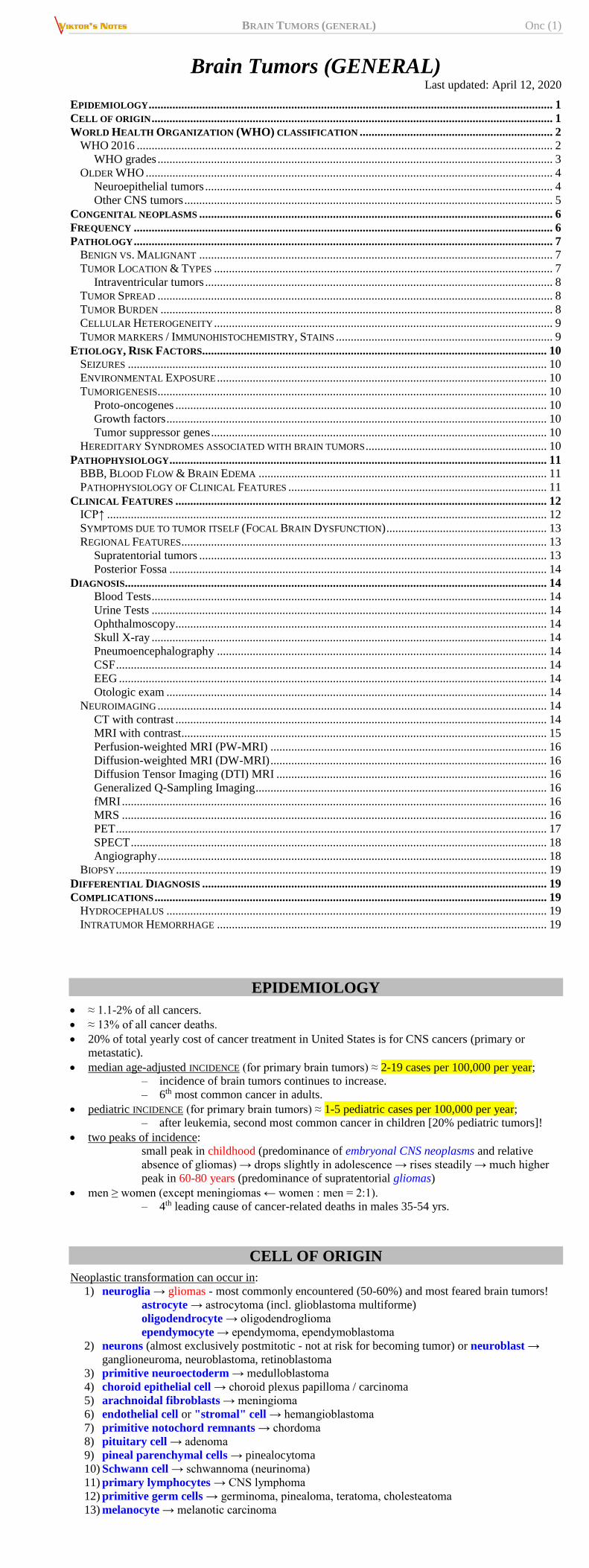
BRAIN TUMORS (GENERAL) Onc (1)
Brain Tumors (GENERAL) Last updated: April 12, 2020
EPIDEMIOLOGY ........................................................................................................................................ 1
CELL OF ORIGIN ....................................................................................................................................... 1 WORLD HEALTH ORGANIZATION (WHO) CLASSIFICATION ................................................................. 2
WHO 2016 ............................................................................................................................................ 2 WHO grades ..................................................................................................................................... 3
OLDER WHO ......................................................................................................................................... 4 Neuroepithelial tumors ..................................................................................................................... 4
Other CNS tumors ............................................................................................................................ 5
CONGENITAL NEOPLASMS ....................................................................................................................... 6 FREQUENCY ............................................................................................................................................. 6
PATHOLOGY ............................................................................................................................................. 7 BENIGN VS. MALIGNANT ....................................................................................................................... 7
TUMOR LOCATION & TYPES .................................................................................................................. 7 Intraventricular tumors ..................................................................................................................... 8
TUMOR SPREAD ..................................................................................................................................... 8
TUMOR BURDEN .................................................................................................................................... 8 CELLULAR HETEROGENEITY .................................................................................................................. 9
TUMOR MARKERS / IMMUNOHISTOCHEMISTRY, STAINS ......................................................................... 9
ETIOLOGY, RISK FACTORS.................................................................................................................... 10 SEIZURES ............................................................................................................................................. 10
ENVIRONMENTAL EXPOSURE ............................................................................................................... 10 TUMORIGENESIS................................................................................................................................... 10
Proto-oncogenes ............................................................................................................................. 10 Growth factors ................................................................................................................................ 10
Tumor suppressor genes ................................................................................................................. 10
HEREDITARY SYNDROMES ASSOCIATED WITH BRAIN TUMORS ............................................................. 10
PATHOPHYSIOLOGY ............................................................................................................................... 11 BBB, BLOOD FLOW & BRAIN EDEMA ................................................................................................. 11 PATHOPHYSIOLOGY OF CLINICAL FEATURES ....................................................................................... 11
CLINICAL FEATURES ............................................................................................................................. 12 ICP↑ .................................................................................................................................................... 12
SYMPTOMS DUE TO TUMOR ITSELF (FOCAL BRAIN DYSFUNCTION) ...................................................... 13
REGIONAL FEATURES ........................................................................................................................... 13 Supratentorial tumors ..................................................................................................................... 13
Posterior Fossa ............................................................................................................................... 14
DIAGNOSIS.............................................................................................................................................. 14 Blood Tests ..................................................................................................................................... 14 Urine Tests ..................................................................................................................................... 14 Ophthalmoscopy ............................................................................................................................. 14
Skull X-ray ..................................................................................................................................... 14 Pneumoencephalography ............................................................................................................... 14 CSF ................................................................................................................................................. 14 EEG ................................................................................................................................................ 14 Otologic exam ................................................................................................................................ 14
NEUROIMAGING ................................................................................................................................... 14 CT with contrast ............................................................................................................................. 14 MRI with contrast ........................................................................................................................... 15 Perfusion-weighted MRI (PW-MRI) ............................................................................................. 16 Diffusion-weighted MRI (DW-MRI) ............................................................................................. 16
Diffusion Tensor Imaging (DTI) MRI ........................................................................................... 16 Generalized Q-Sampling Imaging .................................................................................................. 16 fMRI ............................................................................................................................................... 16 MRS ............................................................................................................................................... 16 PET ................................................................................................................................................. 17
SPECT ............................................................................................................................................ 18 Angiography ................................................................................................................................... 18
BIOPSY ................................................................................................................................................. 19
DIFFERENTIAL DIAGNOSIS .................................................................................................................... 19 COMPLICATIONS .................................................................................................................................... 19
HYDROCEPHALUS ................................................................................................................................ 19
INTRATUMOR HEMORRHAGE ............................................................................................................... 19
EPIDEMIOLOGY
≈ 1.1-2% of all cancers.
≈ 13% of all cancer deaths.
20% of total yearly cost of cancer treatment in United States is for CNS cancers (primary or
metastatic).
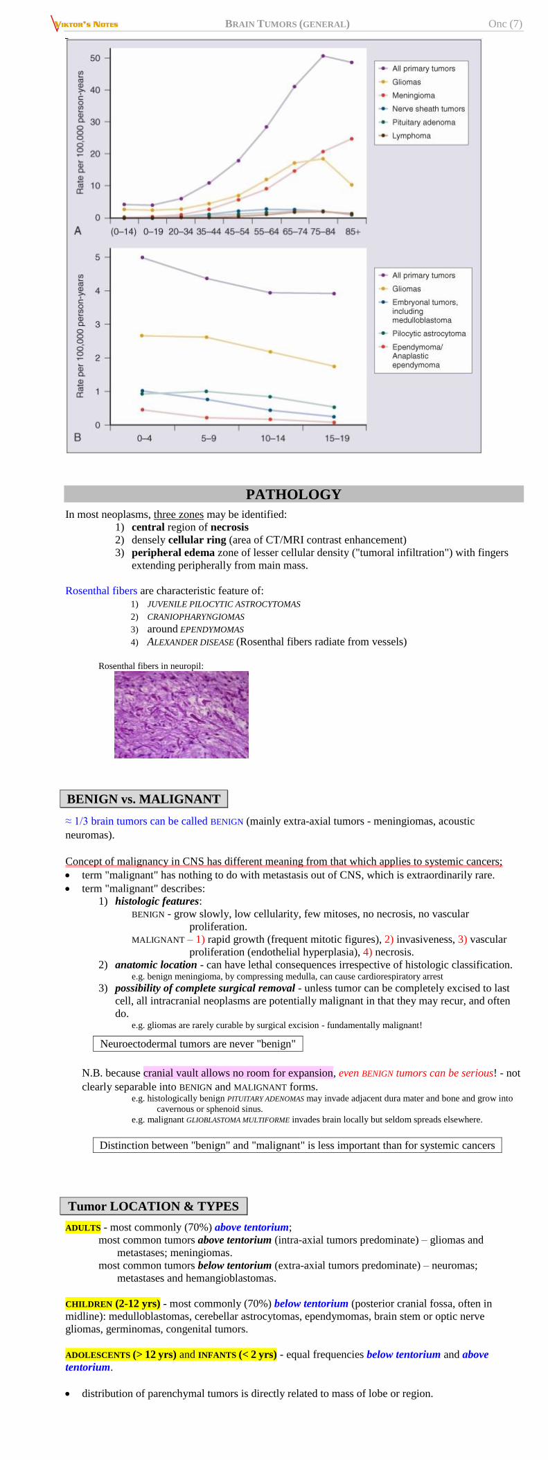
median age-adjusted INCIDENCE (for primary brain tumors) ≈ 2-19 cases per 100,000 per year;
– incidence of brain tumors continues to increase.
– 6th most common cancer in adults.
pediatric INCIDENCE (for primary brain tumors) ≈ 1-5 pediatric cases per 100,000 per year;
– after leukemia, second most common cancer in children [20% pediatric tumors]!
two peaks of incidence:
small peak in childhood (predominance of embryonal CNS neoplasms and relative
absence of gliomas) → drops slightly in adolescence → rises steadily → much higher
peak in 60-80 years (predominance of supratentorial gliomas)
men ≥ women (except meningiomas ← women : men = 2:1).
– 4th leading cause of cancer-related deaths in males 35-54 yrs.
CELL OF ORIGIN
Neoplastic transformation can occur in:
1) neuroglia → gliomas - most commonly encountered (50-60%) and most feared brain tumors!
astrocyte → astrocytoma (incl. glioblastoma multiforme)
oligodendrocyte → oligodendroglioma
ependymocyte → ependymoma, ependymoblastoma
2) neurons (almost exclusively postmitotic - not at risk for becoming tumor) or neuroblast →
ganglioneuroma, neuroblastoma, retinoblastoma
3) primitive neuroectoderm → medulloblastoma
4) choroid epithelial cell → choroid plexus papilloma / carcinoma
5) arachnoidal fibroblasts → meningioma
6) endothelial cell or "stromal" cell → hemangioblastoma
7) primitive notochord remnants → chordoma
8) pituitary cell → adenoma
9) pineal parenchymal cells → pinealocytoma
10) Schwann cell → schwannoma (neurinoma)
11) primary lymphocytes → CNS lymphoma
12) primitive germ cells → germinoma, pinealoma, teratoma, cholesteatoma
13) melanocyte → melanotic carcinoma

BRAIN TUMORS (GENERAL) Onc (2)
BAILEY & CUSHING schema of normal developing cells and neuroepithelial tumors derived from them:
WORLD HEALTH ORGANIZATION (WHO) CLASSIFICATION
First edition (1979)
Third edition (2000) – in addition to histological and immunohistochemical criteria is supplemented
by genetic results (genetic profiling).
N.B. genetic basis represents definitive criterion for tumor classification!
Fourth edition (2007)
Fourth revised edition (2016)
WHO 2016
CNS tumor diagnoses should consist of a histopathological name followed by the genetic features,
with the genetic features following a comma and as adjectives, as in: Diffuse astrocytoma, IDH-
mutant and Medulloblastoma, WNT-activated.
for those entities with more than one genetic determinant, the multiple necessary molecular
features are included in the name: Oligodendroglioma, IDH-mutant and 1p/19q-codeleted
for a tumor lacking a genetic mutation, the term wildtype can be used if an official “wildtype”
entity exists: Glioblastoma, IDH-wildtype; if formal wildtype diagnosis is not available, a tumor
lacking a diagnostic mutation is given an NOS designation.
BRAIN TUMORS (GENERAL) Onc (3)
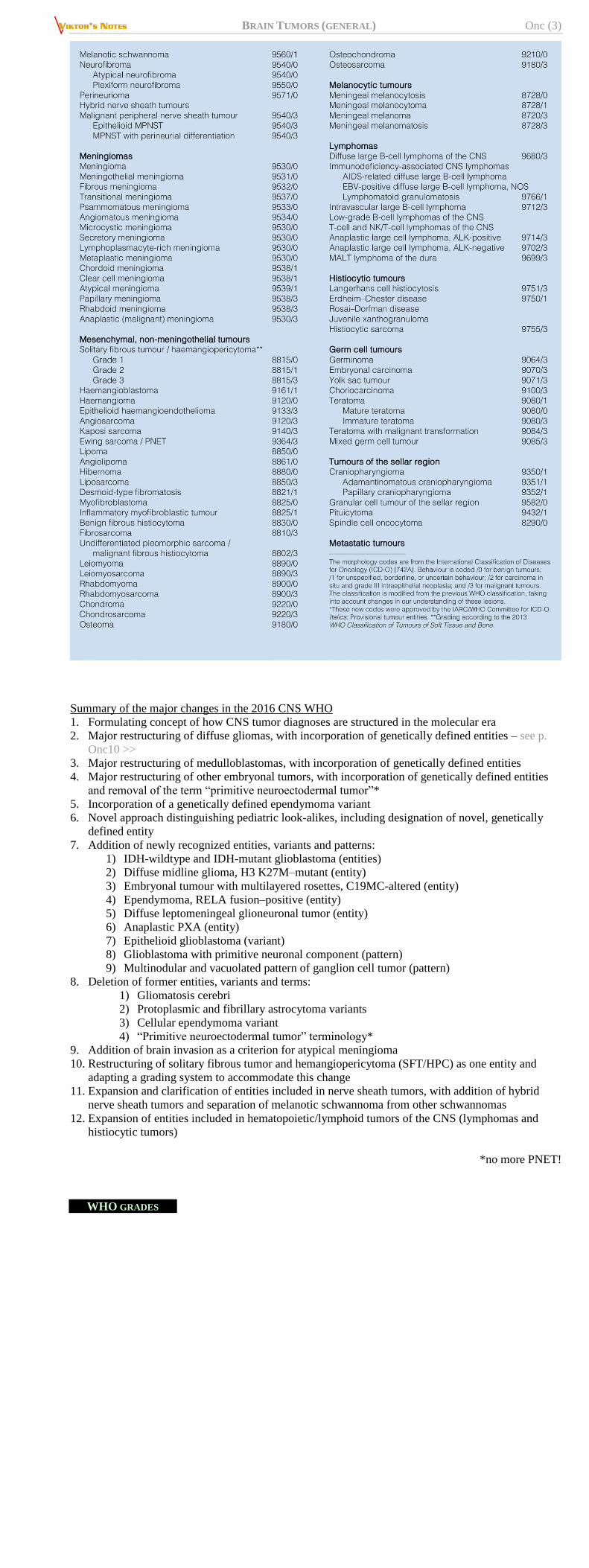
Summary of the major changes in the 2016 CNS WHO
1. Formulating concept of how CNS tumor diagnoses are structured in the molecular era
2. Major restructuring of diffuse gliomas, with incorporation of genetically defined entities – see p.
Onc10 >>
3. Major restructuring of medulloblastomas, with incorporation of genetically defined entities
4. Major restructuring of other embryonal tumors, with incorporation of genetically defined entities
and removal of the term “primitive neuroectodermal tumor”*
5. Incorporation of a genetically defined ependymoma variant
6. Novel approach distinguishing pediatric look-alikes, including designation of novel, genetically
defined entity
7. Addition of newly recognized entities, variants and patterns:
1) IDH-wildtype and IDH-mutant glioblastoma (entities)
2) Diffuse midline glioma, H3 K27M–mutant (entity)
3) Embryonal tumour with multilayered rosettes, C19MC-altered (entity)
4) Ependymoma, RELA fusion–positive (entity)
5) Diffuse leptomeningeal glioneuronal tumor (entity)
6) Anaplastic PXA (entity)
7) Epithelioid glioblastoma (variant)
8) Glioblastoma with primitive neuronal component (pattern)
9) Multinodular and vacuolated pattern of ganglion cell tumor (pattern)
8. Deletion of former entities, variants and terms:
1) Gliomatosis cerebri
2) Protoplasmic and fibrillary astrocytoma variants
3) Cellular ependymoma variant
4) “Primitive neuroectodermal tumor” terminology*
9. Addition of brain invasion as a criterion for atypical meningioma
10. Restructuring of solitary fibrous tumor and hemangiopericytoma (SFT/HPC) as one entity and
adapting a grading system to accommodate this change
11. Expansion and clarification of entities included in nerve sheath tumors, with addition of hybrid
nerve sheath tumors and separation of melanotic schwannoma from other schwannomas
12. Expansion of entities included in hematopoietic/lymphoid tumors of the CNS (lymphomas and
histiocytic tumors)
*no more PNET!
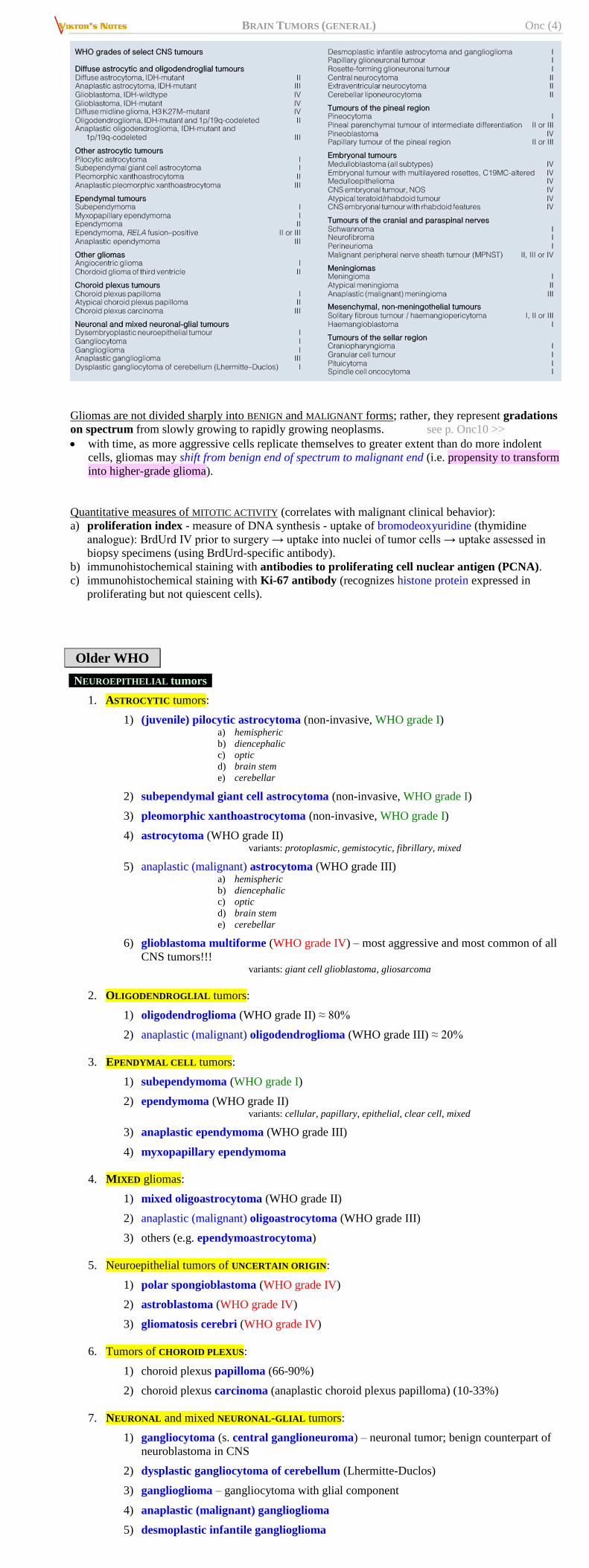
WHO GRADES
BRAIN TUMORS (GENERAL) Onc (4)
Gliomas are not divided sharply into BENIGN and MALIGNANT forms; rather, they represent gradations
on spectrum from slowly growing to rapidly growing neoplasms. see p. Onc10 >>
with time, as more aggressive cells replicate themselves to greater extent than do more indolent
cells, gliomas may shift from benign end of spectrum to malignant end (i.e. propensity to transform
into higher-grade glioma).
Quantitative measures of MITOTIC ACTIVITY (correlates with malignant clinical behavior):
a) proliferation index - measure of DNA synthesis - uptake of bromodeoxyuridine (thymidine
analogue): BrdUrd IV prior to surgery → uptake into nuclei of tumor cells → uptake assessed in
biopsy specimens (using BrdUrd-specific antibody).
b) immunohistochemical staining with antibodies to proliferating cell nuclear antigen (PCNA).
c) immunohistochemical staining with Ki-67 antibody (recognizes histone protein expressed in
proliferating but not quiescent cells).
Older WHO
NEUROEPITHELIAL tumors
1. ASTROCYTIC tumors:
1) (juvenile) pilocytic astrocytoma (non-invasive, WHO grade I) a) hemispheric
b) diencephalic
c) optic
d) brain stem
e) cerebellar
2) subependymal giant cell astrocytoma (non-invasive, WHO grade I)
3) pleomorphic xanthoastrocytoma (non-invasive, WHO grade I)
4) astrocytoma (WHO grade II) variants: protoplasmic, gemistocytic, fibrillary, mixed
5) anaplastic (malignant) astrocytoma (WHO grade III) a) hemispheric
b) diencephalic
c) optic
d) brain stem
e) cerebellar
6) glioblastoma multiforme (WHO grade IV) – most aggressive and most common of all
CNS tumors!!! variants: giant cell glioblastoma, gliosarcoma
2. OLIGODENDROGLIAL tumors:
1) oligodendroglioma (WHO grade II) ≈ 80%
2) anaplastic (malignant) oligodendroglioma (WHO grade III) ≈ 20%
3. EPENDYMAL CELL tumors:
1) subependymoma (WHO grade I)
2) ependymoma (WHO grade II) variants: cellular, papillary, epithelial, clear cell, mixed
3) anaplastic ependymoma (WHO grade III)
4) myxopapillary ependymoma
4. MIXED gliomas:
1) mixed oligoastrocytoma (WHO grade II)
2) anaplastic (malignant) oligoastrocytoma (WHO grade III)
3) others (e.g. ependymoastrocytoma)
5. Neuroepithelial tumors of UNCERTAIN ORIGIN:
1) polar spongioblastoma (WHO grade IV)
2) astroblastoma (WHO grade IV)
3) gliomatosis cerebri (WHO grade IV)
6. Tumors of CHOROID PLEXUS:
1) choroid plexus papilloma (66-90%)
2) choroid plexus carcinoma (anaplastic choroid plexus papilloma) (10-33%)
7. NEURONAL and mixed NEURONAL-GLIAL tumors:
1) gangliocytoma (s. central ganglioneuroma) – neuronal tumor; benign counterpart of
neuroblastoma in CNS
2) dysplastic gangliocytoma of cerebellum (Lhermitte-Duclos)
3) ganglioglioma – gangliocytoma with glial component
4) anaplastic (malignant) ganglioglioma
5) desmoplastic infantile ganglioglioma
BRAIN TUMORS (GENERAL) Onc (5)
variant: desmoplastic infantile astrocytoma
6) central neurocytoma – tumor of well-differentiated neurons
7) dysembryoplastic neuroepithelial tumor (DNET) – benign mixed glial-neuronal
tumor
8) olfactory neuroblastoma (esthesioneuroblastoma) variant: olfactory neuroepithelioma
8. PINEAL PARENCHYMA tumors:
1) pineocytoma (WHO grade I)
2) pineoblastoma (WHO grade IV)
3) pineal parenchymal tumor of intermediate differentiation (WHO grade II-III)
4) papillary tumor of pineal region (WHO grade II-III)
9. Tumors with NEUROBLASTIC or GLIOBLASTIC elements (s. EMBRYONAL TUMORS):
1) medulloepithelioma
2) primitive neuroectodermal tumors with multipotent differentiation:
a) medulloblastoma variants: melanocytic, desmoplastic, medullomyoblastoma
b) primitive neuroectodermal tumor (PNET)
3) neuroblastoma benign counterparts: ganglioneuroblastoma, ganglioneuroma (s. ganglioma)
4) retinoblastoma
5) ependymoblastoma
6) atypical teratoid/rhabdoid tumor
OTHER CNS tumors
1. Tumors of SELLAR REGION
1) pituitary adenoma
2) pituitary carcinoma
3) craniopharyngioma
2. HEMATOPOIETIC tumors
1) primary malignant lymphomas
2) plasmacytoma
3) granulocytic sarcoma
3. GERM CELL tumors see Intro (various topics) 2.jpg >>
1) germinoma
2) embryonal cell carcinoma
3) yolk sac tumor (endodermal sinus tumor)
4) choriocarcinoma
5) teratoma
6) mixed germ cell tumor
4. Tumors of MENINGES
1) meningioma variants: meningothelial, fibrous (fibroblastic), transitional (mixed), psammomatous,
angiomatous, microcystic, secretory, clear cell, chordoid, lymphoplasmacyte-rich, metaplastic
subtypes
2) atypical meningioma
3) anaplastic (malignant) meningioma
5. NON-MENINGOTHELIAL tumors of MENINGES
1) benign mesenchymal
a) osteocartilaginous tumors
b) lipoma
c) fibrous histiocytoma
2) malignant mesenchymal
a) chondrosarcoma
b) hemangiopericytoma
c) rhabdomyosarcoma
d) meningeal sarcomatosis
3) primary melanocytic lesions
a) diffuse melanosis
b) melanocytoma
c) malignant melanoma variant: meningeal melanomatosis
4) hemopoietic neoplasms
a) malignant lymphoma
b) plasmacytoma
c) granulocytic sarcoma d) tumors of uncertain histogenesis - hemangioblastoma (capillary
hemangioblastoma)
6. Tumors of CRANIAL / SPINAL NERVES
1) neurofibroma
2) schwannoma (neurinoma, neurilemoma) subtypes: cellular, plexiform, melanotic
3) malignant peripheral nerve sheath tumor variants: epithelioid, divergent mesenchymal or epithelial differentiation, melanotic
7. CYSTS and TUMOR-LIKE lesions
1) Rathke cleft cyst
2) epidermoid cyst
3) dermoid cyst
4) colloid cyst of 3rd ventricle
5) enterogenous cyst
6) neuroglial cyst
7) granular cell tumor (choristoma, pituicytoma)
8) hypothalamic neuronal hamartoma
9) nasal glial heterotopia
10) plasma cell granuloma
8. LOCAL EXTENSIONS from regional tumors (i.e. secondary intracranial tumors)
BRAIN TUMORS (GENERAL) Onc (6)
1) paraganglioma (chemodectoma)
2) chordoma
3) chondroma
4) chondrosarcoma
5) carcinoma
9. METASTATIC tumors (i.e. secondary intracranial tumors as blood-borne metastases)
10. UNCLASSIFIED tumors
CONGENITAL NEOPLASMS
1) craniopharyngioma
2) chordoma
3) hemangioblastoma
4) colloid cysts
5) germ cell tumors (germinoma, teratoma, etc)
6) dermoid, epidermoid
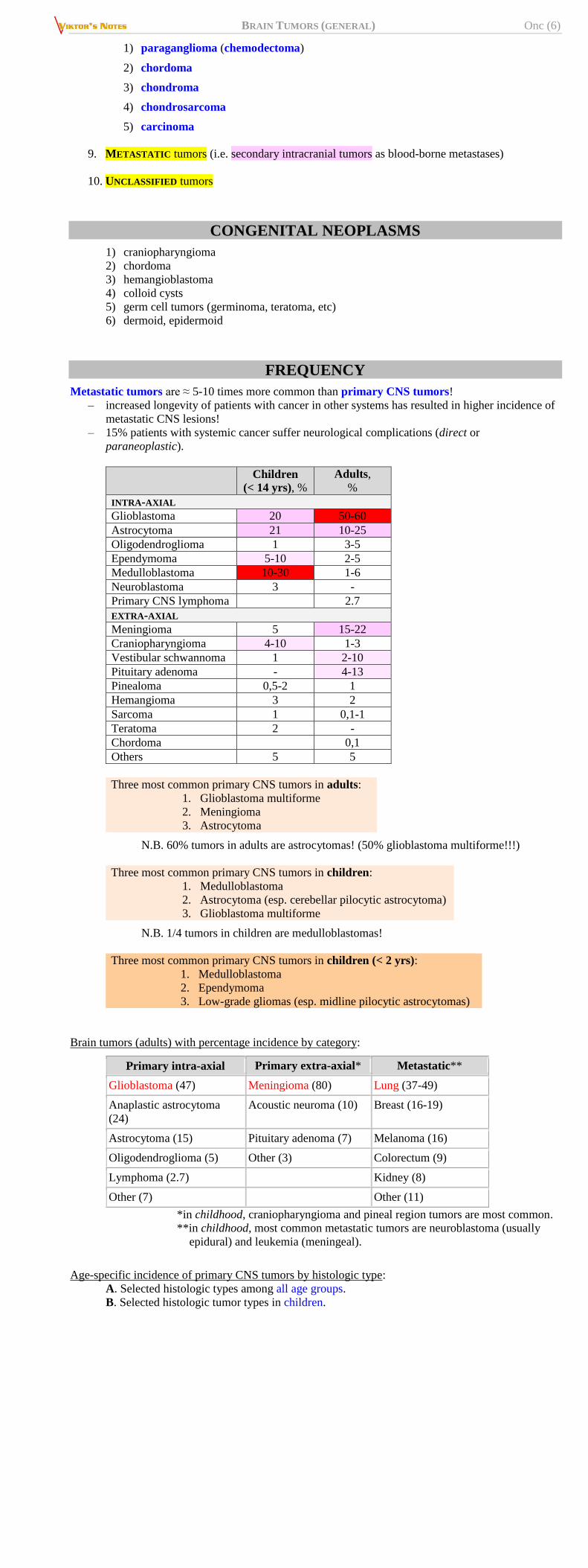
FREQUENCY
Metastatic tumors are ≈ 5-10 times more common than primary CNS tumors!
– increased longevity of patients with cancer in other systems has resulted in higher incidence of
metastatic CNS lesions!
– 15% patients with systemic cancer suffer neurological complications (direct or
paraneoplastic).
Children
(< 14 yrs), %
Adults,
%
INTRA-AXIAL
Glioblastoma 20 50-60
Astrocytoma 21 10-25
Oligodendroglioma 1 3-5
Ependymoma 5-10 2-5
Medulloblastoma 10-30 1-6
Neuroblastoma 3 -
Primary CNS lymphoma 2.7
EXTRA-AXIAL
Meningioma 5 15-22
Craniopharyngioma 4-10 1-3
Vestibular schwannoma 1 2-10
Pituitary adenoma - 4-13
Pinealoma 0,5-2 1
Hemangioma 3 2
Sarcoma 1 0,1-1
Teratoma 2 -
Chordoma 0,1
Others 5 5
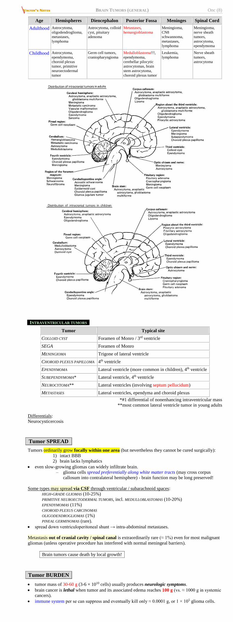
Three most common primary CNS tumors in adults:
1. Glioblastoma multiforme
2. Meningioma
3. Astrocytoma
N.B. 60% tumors in adults are astrocytomas! (50% glioblastoma multiforme!!!)
Three most common primary CNS tumors in children:
1. Medulloblastoma
2. Astrocytoma (esp. cerebellar pilocytic astrocytoma)
3. Glioblastoma multiforme
N.B. 1/4 tumors in children are medulloblastomas!
Three most common primary CNS tumors in children (< 2 yrs):
1. Medulloblastoma
2. Ependymoma
3. Low-grade gliomas (esp. midline pilocytic astrocytomas)
Brain tumors (adults) with percentage incidence by category:
Primary intra-axial Primary extra-axial* Metastatic**
Glioblastoma (47) Meningioma (80) Lung (37-49)
Anaplastic astrocytoma
(24)
Acoustic neuroma (10) Breast (16-19)
Astrocytoma (15) Pituitary adenoma (7) Melanoma (16)
Oligodendroglioma (5) Other (3) Colorectum (9)
Lymphoma (2.7) Kidney (8)
Other (7) Other (11)
*in childhood, craniopharyngioma and pineal region tumors are most common.
**in childhood, most common metastatic tumors are neuroblastoma (usually
epidural) and leukemia (meningeal).
Age-specific incidence of primary CNS tumors by histologic type:
A. Selected histologic types among all age groups.
B. Selected histologic tumor types in children.
BRAIN TUMORS (GENERAL) Onc (7)
PATHOLOGY
In most neoplasms, three zones may be identified:
1) central region of necrosis
2) densely cellular ring (area of CT/MRI contrast enhancement)
3) peripheral edema zone of lesser cellular density ("tumoral infiltration") with fingers
extending peripherally from main mass.
Rosenthal fibers are characteristic feature of:
1) JUVENILE PILOCYTIC ASTROCYTOMAS 2) CRANIOPHARYNGIOMAS
3) around EPENDYMOMAS
4) ALEXANDER DISEASE (Rosenthal fibers radiate from vessels)
Rosenthal fibers in neuropil:
BENIGN vs. MALIGNANT
≈ 1/3 brain tumors can be called BENIGN (mainly extra-axial tumors - meningiomas, acoustic
neuromas).
Concept of malignancy in CNS has different meaning from that which applies to systemic cancers;
term "malignant" has nothing to do with metastasis out of CNS, which is extraordinarily rare.
term "malignant" describes:
1) histologic features:
BENIGN - grow slowly, low cellularity, few mitoses, no necrosis, no vascular
proliferation.
MALIGNANT – 1) rapid growth (frequent mitotic figures), 2) invasiveness, 3) vascular
proliferation (endothelial hyperplasia), 4) necrosis.
2) anatomic location - can have lethal consequences irrespective of histologic classification. e.g. benign meningioma, by compressing medulla, can cause cardiorespiratory arrest
3) possibility of complete surgical removal - unless tumor can be completely excised to last
cell, all intracranial neoplasms are potentially malignant in that they may recur, and often
do. e.g. gliomas are rarely curable by surgical excision - fundamentally malignant!
Neuroectodermal tumors are never "benign"
N.B. because cranial vault allows no room for expansion, even BENIGN tumors can be serious! - not
clearly separable into BENIGN and MALIGNANT forms. e.g. histologically benign PITUITARY ADENOMAS may invade adjacent dura mater and bone and grow into
cavernous or sphenoid sinus.
e.g. malignant GLIOBLASTOMA MULTIFORME invades brain locally but seldom spreads elsewhere.
Distinction between "benign" and "malignant" is less important than for systemic cancers
Tumor LOCATION & TYPES
ADULTS - most commonly (70%) above tentorium;
most common tumors above tentorium (intra-axial tumors predominate) – gliomas and
metastases; meningiomas.
most common tumors below tentorium (extra-axial tumors predominate) – neuromas;
metastases and hemangioblastomas.
CHILDREN (2-12 yrs) - most commonly (70%) below tentorium (posterior cranial fossa, often in
midline): medulloblastomas, cerebellar astrocytomas, ependymomas, brain stem or optic nerve
gliomas, germinomas, congenital tumors.
ADOLESCENTS (> 12 yrs) and INFANTS (< 2 yrs) - equal frequencies below tentorium and above
tentorium.
distribution of parenchymal tumors is directly related to mass of lobe or region.
BRAIN TUMORS (GENERAL) Onc (8)
Age Hemispheres Diencephalon Posterior Fossa Meninges Spinal Cord
Adulthood Astrocytoma,
oligodendroglioma,
metastases,
lymphoma
Astrocytoma, colloid
cyst, pituitary
adenoma
Metastases,
hemangioblastoma
Meningioma,
CN8
schwannoma,
metastases,
lymphoma
Meningioma,
nerve sheath
tumors,
astrocytoma,
ependymoma
Childhood Astrocytoma,
ependymoma,
choroid plexus
tumor, primitive
neuroectodermal
tumor
Germ cell tumors,
craniopharyngioma
Medulloblastoma!!!,
ependymoma,
cerebellar pilocytic
astrocytomas, brain
stem astrocytoma,
choroid plexus tumor
Leukemia,
lymphoma
Nerve sheath
tumors,
astrocytoma
INTRAVENTRICULAR TUMORS
Tumor Typical site
COLLOID CYST Foramen of Monro / 3rd ventricle
SEGA Foramen of Monro
MENINGIOMA Trigone of lateral ventricle
CHOROID PLEXUS PAPILLOMA 4th ventricle
EPENDYMOMA Lateral ventricle (more common in children), 4th ventricle
SUBEPENDYMOMA* Lateral ventricle, 4th ventricle
NEUROCYTOMA** Lateral ventricles (involving septum pellucidum)
METASTASES Lateral ventricles, ependyma and choroid plexus
*#1 differential of nonenhancing intraventricular mass
**most common lateral ventricle tumor in young adults
Differentials:
Neurocysticercosis
Tumor SPREAD
Tumors ordinarily grow focally within one area (but nevertheless they cannot be cured surgically):
1) intact BBB
2) brain lacks lymphatics
even slow-growing gliomas can widely infiltrate brain.
– glioma cells spread preferentially along white matter tracts (may cross corpus
callosum into contralateral hemisphere) - brain function may be long preserved!
Some types may spread via CSF through ventricular / subarachnoid spaces:
HIGH-GRADE GLIOMAS (10-25%)
PRIMITIVE NEUROECTODERMAL TUMORS, incl. MEDULLOBLASTOMAS (10-20%)
EPENDYMOMAS (11%)
CHOROID PLEXUS CARCINOMAS
OLIGODENDROGLIOMAS (1%)
PINEAL GERMINOMAS (rare).
spread down ventriculoperitoneal shunt → intra-abdominal metastases.
Metastasis out of cranial cavity / spinal canal is extraordinarily rare (≈ 1%) even for most malignant
gliomas (unless operative procedure has interfered with normal meningeal barriers).
Brain tumors cause death by local growth!
Tumor BURDEN
tumor mass of 30-60 g (3-6 × 10¹⁰ cells) usually produces neurologic symptoms.
brain cancer is lethal when tumor and its associated edema reaches 100 g (vs. ≈ 1000 g in systemic
cancers).
immune system per se can suppress and eventually kill only ≈ 0.0001 g, or 1 × 10⁵ glioma cells.
BRAIN TUMORS (GENERAL) Onc (9)
CELLULAR HETEROGENEITY
While tumors are monoclonal in origin (i.e. they originate from single cell),
as they grow they progress through series of genomic changes that permit
evolution to more and more malignant stages.
parental cell population is genetically unstable → tumors are heterogeneous in cellular content:
a) genotypic (incl. chromosomal content [ranges from near diploid to hypo- or hypertetraploid]
and molecular aberrations).
b) phenotypic (cells that are immediately adjacent to one another may have very different
histologic appearance).
REGIONAL DIFFERENCES develop when tumor cells begin to invade surrounding normal brain -
during migration, some cells develop additional abnormalities that confer selective advantage for
growth → tumor is seeded with microfoci that are both genotypically and phenotypically different.
TUMOR MARKERS / IMMUNOHISTOCHEMISTRY, STAINS
Alcian blue – stain for mucin (e.g. myxopapillary ependymoma)
α-fetoprotein – embryonal carcinoma, endodermal sinus (yolk sac) tumor.
Anti-Leu 7 antibody – schwannomas.
N.B. uniformly negative in meningiomas
ATRX (alpha-thalassemia/mental retardation syndrome x-linked) gene
ATRX is present in every cell!
loss of ATRX = astrocytic lineage (grade II/III astrocytomas and secondary GBM).
Brachyury (protein encoded by the TBXT gene, transcription factor within the T-box family of genes)
early mutational event in chordoma evolution (discriminates chordoma from chondrosarcoma).
present in majority of hemangioblastomas (helps to differentiate from clear cell renal cell
carcinoma metastases in von Hippel-Lindau syndrome).
CD68 (protein highly expressed by cells in the monocyte lineage: microglia, histiocytes) –
differentiates histiocytosis from lymphoma.
CD3 – T-cell lymphoma.
Desmin – tumors containing muscle (rhabdomyosarcoma, teratoma, etc), primitive neuroectodermal
tumor.
EGFR (epidermal-derived growth factor receptor) – aberrantly expressed (usually amplified*) in
many gliomas.
*poor prognostic factor!
EMA (epithelial membrane antigen) – epithelia marker (ependymoma, meningioma, epithelial areas
of teratomas, rhabdoid tumor).
N.B. not present in melanoma!
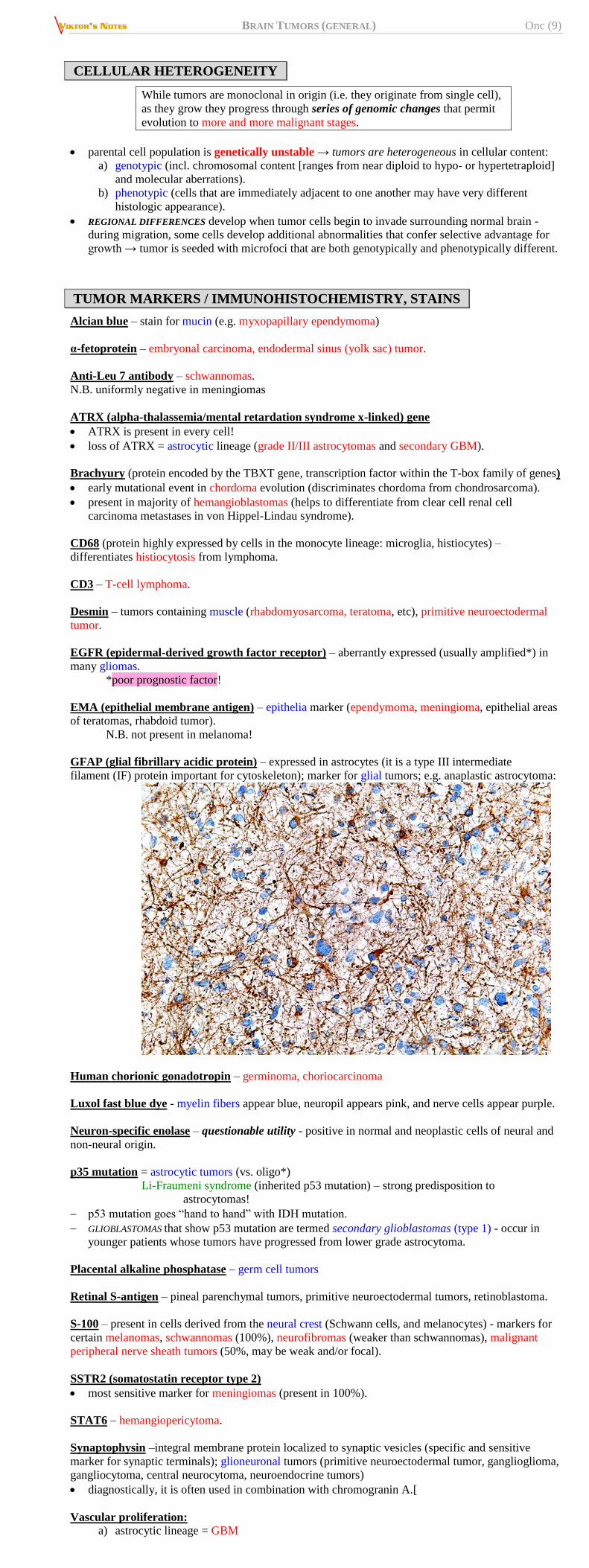
GFAP (glial fibrillary acidic protein) – expressed in astrocytes (it is a type III intermediate
filament (IF) protein important for cytoskeleton); marker for glial tumors; e.g. anaplastic astrocytoma:
Human chorionic gonadotropin – germinoma, choriocarcinoma
Luxol fast blue dye - myelin fibers appear blue, neuropil appears pink, and nerve cells appear purple.
Neuron-specific enolase – questionable utility - positive in normal and neoplastic cells of neural and
non-neural origin.
p35 mutation = astrocytic tumors (vs. oligo*)
Li-Fraumeni syndrome (inherited p53 mutation) – strong predisposition to
astrocytomas!
p53 mutation goes “hand to hand” with IDH mutation.
GLIOBLASTOMAS that show p53 mutation are termed secondary glioblastomas (type 1) - occur in
younger patients whose tumors have progressed from lower grade astrocytoma.
Placental alkaline phosphatase – germ cell tumors
Retinal S-antigen – pineal parenchymal tumors, primitive neuroectodermal tumors, retinoblastoma.
S-100 – present in cells derived from the neural crest (Schwann cells, and melanocytes) - markers for
certain melanomas, schwannomas (100%), neurofibromas (weaker than schwannomas), malignant
peripheral nerve sheath tumors (50%, may be weak and/or focal).
SSTR2 (somatostatin receptor type 2)
most sensitive marker for meningiomas (present in 100%).
STAT6 – hemangiopericytoma.
Synaptophysin –integral membrane protein localized to synaptic vesicles (specific and sensitive
marker for synaptic terminals); glioneuronal tumors (primitive neuroectodermal tumor, ganglioglioma,
gangliocytoma, central neurocytoma, neuroendocrine tumors)
diagnostically, it is often used in combination with chromogranin A.[
Vascular proliferation:
a) astrocytic lineage = GBM
BRAIN TUMORS (GENERAL) Onc (10)
b) 1p/19q co-deletion = anaplastic oligo
Vimentin – type III intermediate filament (IF) protein, major cytoskeletal component of mesenchymal
cells (significant role in supporting and anchoring the position of the organelles in the cytosol);
staining may confirm mesenchymal origin (e.g. sarcomas, meningioma, lymphoma, chordoma) but
numerous exceptions so relatively nonspecific.
ETIOLOGY, RISK FACTORS
SEIZURES
Seizures may herald development of cerebral tumors by several years!
British study (Journal of Neurology, Neurosurgery and Psychiatry, online March 28, 2011):
— risk for any cerebral tumor after first admission for epilepsy is increased 20-fold (risk for
malignant tumors is more than twice that for benign tumors).
— risk is still elevated several years after first admission for epilepsy → need for continued
surveillance of patients with new-onset seizures.
ENVIRONMENTAL EXPOSURE
Numerous epidemiologic studies* suggest statistically significant increased incidence of astrocytomas
in people exposed to petrochemicals (e.g. in rubber industry) or electromagnetic radiation.
*equally impressive studies, however, have not confirmed association.
well-documented environmental risk factor (Israeli study) - ionizing radiation (e.g. given for
treatment of tinea capitis) - increases risk for meningiomas almost 10 times and for gliomas 2.5
times.
insufficient epidemiologic evidence to support or refute claims, that hand-held cellular telephones
generate electromagnetic radiation and cause brain tumors.
both RNA and DNA viruses can induce animal brain tumors, but few viruses have been found to
account for specific human tumor (e.g. Epstein-Barr virus evidence in primary CNS lymphoma
tissue).
immunosuppression (transplant recipients, AIDS patients, Wiskott-Aldrich syndrome, ataxia-
telangiectasia) substantially increases risks for primary CNS lymphoma but not gliomas.
role of trauma is unproven.
The only proven environmental risk factor for brain tumor
is previous exposure to high-dose ionizing radiation
TUMORIGENESIS
- multistep process (probably at least 4-6 separate steps - multiple local mutations and clonal
expansion). see p. 3781-3788 >>
Most important genetic markers – see above >>
PROTO-ONCOGENES
Proto-oncogenes mutated / overexpressed in brain tumors:
1) EGFR (erb-B) - encodes epidermal-derived growth factor receptor; aberrantly expressed
(usually amplified) in many gliomas!
2) c-sis - encodes platelet-derived growth factor
3) c-myc
4) ros1
5) H-ras
6) gli
medulloblastomas, 50% glioblastomas have homogeneously staining regions and double minute
chromosomes - may contain amplified proto-oncogenes.
GROWTH FACTORS
- have potent growth stimulatory effects on glioma cells in culture:
1) platelet-derived growth factor (PDGF)
2) epidermal-derived growth factor (EGF)
3) transforming growth factor-α (TGF-α) - 50% homology with EGF; secreted by numerous
tumors (incl. high-grade malignant gliomas).
4) fibroblast growth factor (FGF)
5) insulin-like growth factor (IGF).
many glioma cells produce growth factors and express appropriate growth factor receptor on their
surface membranes - constantly stimulate own growth and division (AUTOCRINE GROWTH).
N.B. normal brain cells are kinetically quiescent (neurons are incapable of division
after birth; glial cells are minimally proliferative in reactive or reparative gliosis)
cardinal histopathologic features that define malignant glioma - cellular atypia, cellularity,
mitoses, endothelial hyperplasia, necrosis;
- all (with exception of necrosis - attributed to growth beyond capacity of blood supply)
are subject to modulation by growth factors.
> 60% gliomas have TELOMERASE activity (correlates with tumor grading, being lowest in low-
grade tumors). see p. 599-600 >>
TUMOR SUPPRESSOR GENES
Tumor suppressor genes associated with nervous system tumors:
p53 (17p13) – loss predisposes to astrocytoma and neurofibrosarcoma.
progression from low-grade astrocytoma to glioblastoma strongly correlates with loss of p53 gene.
Li-Fraumeni syndrome - familial cancer syndrome in young adults (< 45 yrs) - breast cancer, soft
tissue sarcomas, brain tumor (esp. astrocytoma), osteosarcoma, leukemia, adrenocortical
carcinoma.
– affected people inherit one mutant p53 allele.
– sporadic (nonfamilial) forms of cancers associated with Li-Fraumeni syndrome also
show p53 inactivation.
NF1 (17q11.2), NF2 (22q12) - loss predispose to neurofibromatosis.
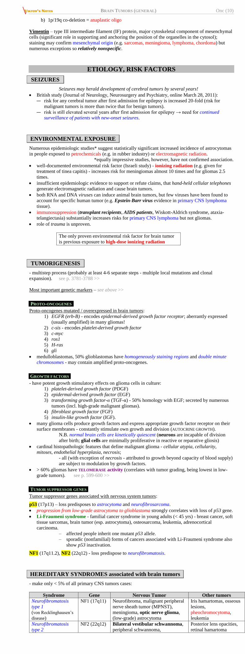
HEREDITARY SYNDROMES associated with brain tumors
- make only < 5% of all primary CNS tumors cases:
Syndrome Gene Nervous Tumor Other tumors
Neurofibromatosis
type 1
(von Recklinghausen’s
disease)
NF1 (17q11) Neurofibroma, malignant peripheral
nerve sheath tumor (MPNST),
meningioma, optic nerve glioma,
(low-grade) astrocytoma
Iris hamartomas, osseous
lesions,
pheochromocytoma,
leukemia
Neurofibromatosis
type 2
NF2 (22q12) Bilateral vestibular schwannoma,
peripheral schwannoma,
Posterior lens opacities,
retinal hamartoma
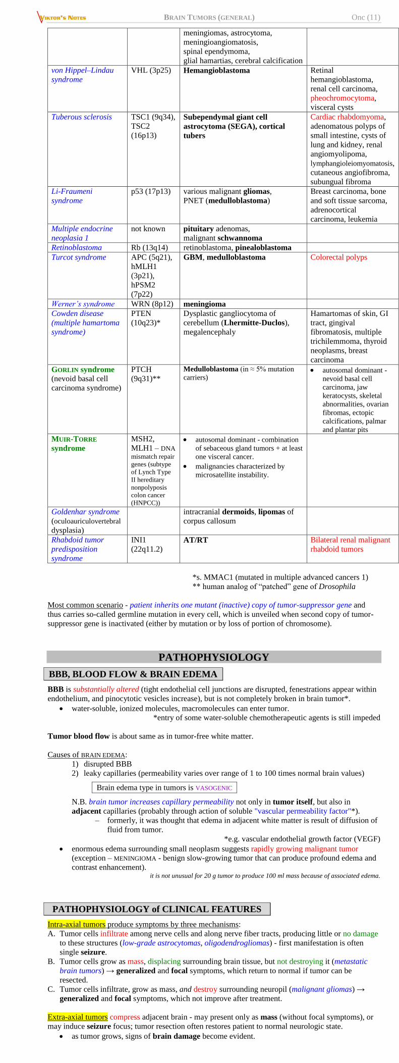
BRAIN TUMORS (GENERAL) Onc (11)
meningiomas, astrocytoma,
meningioangiomatosis,
spinal ependymoma,
glial hamartias, cerebral calcification
von Hippel–Lindau
syndrome
VHL (3p25) Hemangioblastoma Retinal
hemangioblastoma,
renal cell carcinoma,
pheochromocytoma,
visceral cysts
Tuberous sclerosis TSC1 (9q34),
TSC2
(16p13)
Subependymal giant cell
astrocytoma (SEGA), cortical
tubers
Cardiac rhabdomyoma,
adenomatous polyps of
small intestine, cysts of
lung and kidney, renal
angiomyolipoma,
lymphangioleiomyomatosis,
cutaneous angiofibroma,
subungual fibroma
Li-Fraumeni
syndrome
p53 (17p13) various malignant gliomas,
PNET (medulloblastoma)
Breast carcinoma, bone
and soft tissue sarcoma,
adrenocortical
carcinoma, leukemia
Multiple endocrine
neoplasia 1
not known pituitary adenomas,
malignant schwannoma
Retinoblastoma Rb (13q14) retinoblastoma, pinealoblastoma
Turcot syndrome APC (5q21),
hMLH1
(3p21),
hPSM2
(7p22)
GBM, medulloblastoma Colorectal polyps
Werner’s syndrome WRN (8p12) meningioma
Cowden disease
(multiple hamartoma
syndrome)
PTEN
(10q23)*
Dysplastic gangliocytoma of
cerebellum (Lhermitte-Duclos),
megalencephaly
Hamartomas of skin, GI
tract, gingival
fibromatosis, multiple
trichilemmoma, thyroid
neoplasms, breast
carcinoma
GORLIN syndrome (nevoid basal cell
carcinoma syndrome)
PTCH
(9q31)**
Medulloblastoma (in ≈ 5% mutation
carriers) autosomal dominant -
nevoid basal cell
carcinoma, jaw
keratocysts, skeletal
abnormalities, ovarian
fibromas, ectopic
calcifications, palmar
and plantar pits
MUIR-TORRE
syndrome
MSH2,
MLH1 – DNA
mismatch repair
genes (subtype
of Lynch Type
II hereditary
nonpolyposis
colon cancer
(HNPCC))
autosomal dominant - combination
of sebaceous gland tumors + at least
one visceral cancer.
malignancies characterized by
microsatellite instability.
Goldenhar syndrome
(oculoauriculovertebral
dysplasia)
intracranial dermoids, lipomas of
corpus callosum
Rhabdoid tumor
predisposition
syndrome
INI1
(22q11.2) AT/RT Bilateral renal malignant
rhabdoid tumors
*s. MMAC1 (mutated in multiple advanced cancers 1)
** human analog of “patched” gene of Drosophila
Most common scenario - patient inherits one mutant (inactive) copy of tumor-suppressor gene and
thus carries so-called germline mutation in every cell, which is unveiled when second copy of tumor-
suppressor gene is inactivated (either by mutation or by loss of portion of chromosome).
PATHOPHYSIOLOGY
BBB, BLOOD FLOW & BRAIN EDEMA
BBB is substantially altered (tight endothelial cell junctions are disrupted, fenestrations appear within
endothelium, and pinocytotic vesicles increase), but is not completely broken in brain tumor*.
water-soluble, ionized molecules, macromolecules can enter tumor.
*entry of some water-soluble chemotherapeutic agents is still impeded
Tumor blood flow is about same as in tumor-free white matter.
Causes of BRAIN EDEMA:
1) disrupted BBB
2) leaky capillaries (permeability varies over range of 1 to 100 times normal brain values)
Brain edema type in tumors is VASOGENIC
N.B. brain tumor increases capillary permeability not only in tumor itself, but also in
adjacent capillaries (probably through action of soluble "vascular permeability factor"*).
– formerly, it was thought that edema in adjacent white matter is result of diffusion of
fluid from tumor.
*e.g. vascular endothelial growth factor (VEGF)
enormous edema surrounding small neoplasm suggests rapidly growing malignant tumor
(exception – MENINGIOMA - benign slow-growing tumor that can produce profound edema and
contrast enhancement). it is not unusual for 20 g tumor to produce 100 ml mass because of associated edema.
PATHOPHYSIOLOGY of CLINICAL FEATURES
Intra-axial tumors produce symptoms by three mechanisms:
A. Tumor cells infiltrate among nerve cells and along nerve fiber tracts, producing little or no damage
to these structures (low-grade astrocytomas, oligodendrogliomas) - first manifestation is often
single seizure.
B. Tumor cells grow as mass, displacing surrounding brain tissue, but not destroying it (metastatic
brain tumors) → generalized and focal symptoms, which return to normal if tumor can be
resected.
C. Tumor cells infiltrate, grow as mass, and destroy surrounding neuropil (malignant gliomas) →
generalized and focal symptoms, which not improve after treatment.
Extra-axial tumors compress adjacent brain - may present only as mass (without focal symptoms), or
may induce seizure focus; tumor resection often restores patient to normal neurologic state.
as tumor grows, signs of brain damage become evident.
BRAIN TUMORS (GENERAL) Onc (12)
How intracranial neoplasms increase ICP:
1) tumor mass
2) cerebral edema adjacent to neoplasm
3) obstruction of CSF pathways (producing hydrocephalus):
a. intraventricular (at Monro foramen, aqueduct, 4th ventricle)
b. leukemic or carcinomatous involvement of meninges
4) obstruction of venous pathways.
75% infants < 6 months of age have tumor volumes > 1/3 of their intracranial volume - plasticity of
cranial vault allows asymptomatic growth.
CLINICAL FEATURES
Characteristic feature of all intracranial neoplasms is that they produce progressive symptoms!
Clinical presentation depends primarily on:
1. Age of patient (ability of skull bones to adjust to growing intracranial mass).
N.B. symptoms in young children and infants are nonspecific and are frequently mistaken for
non-CNS problems - diagnosis of pediatric brain tumor can be extremely difficult to make
without very high index of suspicion!
2. Primary histology – determines rate of symptom evolution.
e.g. benign tumors may achieve considerable size before producing symptoms (grow slowly,
cerebral edema occurs infrequently).
3. Tumor location
e.g. extra-axial tumors - usually well circumscribed with benign histology - clinical
presentation is directly related to CNS structures immediately adjacent to lesion.
e.g. posterior fossa tumors or tumors near foramen of Monro tend to obstruct CSF pathways
early.
Symptoms do not differ much by tumor histology but rather relate to area of brain affected
Asymptomatic cases:
1) silent areas (tumors may grow large): parietal or frontal association cortices, nondominant
temporal lobe.
2) slow growth (brain can accommodate to slowly growing mass).
Manifestations can be divided (but it may not be possible to differentiate these except in retrospect):
a) FOCAL SYMPTOMS due to tumor itself (direct compression or infiltration)
b) GENERALIZED SYMPTOMS due to secondary consequences* (mass effect causing ICP↑) –
tumor volume, peritumoral edema, hydrocephalus, shift of critical structures.
*these may cause false-localizing signs!
Systemic symptoms (malaise, weight loss, anorexia, fever) suggests metastatic rather than primary
brain tumor!
KARNOFSKY performance scale - objective measurement of functional ability (useful in assessing
and following patients with CNS neoplasm):
100 – Normal (no evidence of disease)
90 – Minor symptoms (able to carry on normal activity)
80 – Some symptoms (normal activity with effort)
70 – Unable to carry on normal activity (cares for self) - level of function justifying
aggressive therapy!
60 – Cares for most needs (requires occasional assistance)
50 – Requires considerable assistance
40 – Disabled
30 – Severely disabled
20 – Active supportive treatment needed (very sick)
10 – Moribund
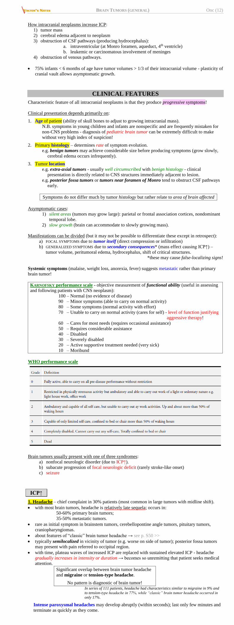
WHO performance scale
Brain tumors usually present with one of three syndromes:
a) nonfocal neurologic disorder (due to ICP↑).
b) subacute progression of focal neurologic deficit (rarely stroke-like onset)
c) seizure
ICP↑
1. Headache – chief complaint in 30% patients (most common in large tumors with midline shift).
with most brain tumors, headache is relatively late sequela; occurs in:
50-60% primary brain tumors;
35-50% metastatic tumors.
rare as initial symptom in brainstem tumors, cerebellopontine angle tumors, pituitary tumors,
craniopharyngiomas.
about features of “classic” brain tumor headache → see p. S50 >>
typically semilocalized in vicinity of tumor (e.g. worse on side of tumor); posterior fossa tumors
may present with pain referred to occipital region.
with time, plateau waves of increased ICP are replaced with sustained elevated ICP - headache
gradually increases in intensity or duration → becomes so unremitting that patient seeks medical
attention.
Significant overlap between brain tumor headache
and migraine or tension-type headache.
No pattern is diagnostic of brain tumor! In series of 111 patients, headache had characteristics similar to migraine in 9% and
to tension-type headache in 77%, while “classic” brain tumor headache occurred in
only 17%.
Intense paroxysmal headaches may develop abruptly (within seconds); last only few minutes and
terminate as quickly as they come.
BRAIN TUMORS (GENERAL) Onc (13)
– ominous sign of markedly increased ICP (ICP monitoring shows that peak pressure
coincides with plateau waves).
– during episode, patient may vomit, lose vision, consciousness↓, fall.
– possible mechanism - acute hydrocephalus (ball-valve obstruction of CSF outflow
with tumor in ventricular system).
2. Vomiting
associated with nausea and headache.
direct compression of vomiting center → projectile vomiting - highly characteristic of posterior
fossa tumors!
N.B. “projectile” is misnomer - nothing pathognomonic about forcefulness of ejection; term
“projectile” more appropriately refers to vomiting without antecedent nausea or headache
(precedes appearance of headache by weeks).
3. Deterioration in mental status (psychomotor retardation, sleep / cognitive / social disturbances,
confusion, lethargy). see p. S50 >>
- frequent clinical manifestation of intracranial tumor!
often subtle in presentation and onset and may not attract attention of friends and family
members until patient begins to behave unusually.
N.B. it is not unusual for patient to seek psychiatric help (up to 20% of all patients)
4. Cushing reflex signals life-threatening ICP↑↑↑. see p. S50 >>
5. Brain mass shifts (may manifest as false-localizing signs) - CN6 palsy, CN3 palsy, ipsilateral
hemiplegia (compression of opposite cerebral peduncle against Kernohan's notch), ipsilateral visual
field defects (compression of opposite PCA), nuchal rigidity & torticollis* (herniation of cerebellar
tonsils), etc.
*torticollis also may be due to CN4 palsy
6. Enlarging head (tense fontanelle, ‘sun set’ eyes, dilated scalp veins) in young children.
SYMPTOMS due to TUMOR ITSELF (FOCAL BRAIN DYSFUNCTION)
- may be absent in tumors growing in silent areas.
result from compression of neurons and white matter tracts by expanding tumor and
accompanying edema.
vascular compression may produce focal brain ischemia.
1. Seizures - occur in 20-71% patients (as presenting symptom in 18-50% cases);
focal or generalized.
most common with SLOWLY GROWING tumors affecting cortex (esp. meningiomas,
oligodendrogliomas, low-grade gliomas). Even small meningiomas that compress adjacent cerebral cortex may present with seizures!
Epilepsy rates range 60-100% in low-grade gliomas and 25-60% in high-grade gliomas
suggestive features: status epilepticus at onset, prolonged postictal paralysis*, resistance to medical
control, focal symptoms.
*brain tumor patients have higher incidence of postictal neurologic deficit!
2. Negative signs - hemiparesis, sensory loss, aphasia, cranial nerve palsies, visual deficits, hearing
impairment, anosmia, personality changes, etc.
multiple metastases or diffuse brain infiltration (by glioma or lymphoma) may present as
dementia or decline in level of alertness.
hand preference in child < 3-5 yrs may signify hemiparesis.
3. Hyperactive function:
pituitary / pineal tumors → hormone overproduction.
choroid plexus papilloma → CSF overproduction.
REGIONAL FEATURES
SUPRATENTORIAL TUMORS
progressive focal neurologic signs and seizures predominate:
Frontal lobe
1. Seizures - may precede other symptoms by months or years.
2. Intellectual impairment (esp. with bilateral tumors, e.g. butterfly glioma)
3. Impairment of initiative and spontaneity: abulia → akinetic mutism.
4. Personality changes: see also p. Psy5 >>
a) dorsolateral prefrontal lesions → apathetic & indifferent (pseudodepressed)
b) orbital prefrontal lesions → loss of inhibition & euphoric (pseudopsychopathic).
5. Motor disturbances – hemiparesis, precipitate urination (tumor of medial surfaces of frontal
lobe).
6. Motor aphasia.
7. Anosmia (e.g. meningioma of olfactory groove).
Temporal lobe
1. Personality change (bizarre thinking, trance-like states, mood symptoms, immature emotional
behavior; bilateral amygdaloid lesions → Klüver-Bucy syndrome).
2. Sensory aphasia, anomia.
3. Seizures - complex partial (psychomotor).
4. Contralateral hemianopia (or superior quadrantanopia).
5. Impairment of recent memory (bilateral hippocampal lesions → Korsakoff amnesia)
N.B. temporal tumors (esp. in nondominant hemisphere) are often relatively "silent"!
Parietal lobe
1. Seizures - generalized or sensory focal seizures.
2. Impaired contralateral cortical sensory modalities (position sense, two-point discrimination,
stereognosis)
3. Contralateral homonymous hemianopia (or inferior quadrantanopia).
4. Mixed expressive-receptive aphasia, anosognosia.
5. Dominant hemisphere – Gerstmann’s syndrome (agraphia, acalculia, finger agnosia).
6. Nondominant hemisphere – apraxia, contralateral hemineglect.
Occipital lobe
- contralateral quadrantanopia or hemianopia with sparing of macula; visual misperceptions &
hallucinations; bilateral lesions – cortical blindness.
Thalamus
1. Hydrocephalus.
2. Contralateral sensory abnormality, neuropathic pain, intermittent paresthesias.
3. Involvement of basal ganglia → contralateral intention tremor, hemiballistic movement.
4. Involvement of hypothalamus → eating disorders, precocious puberty.
BRAIN TUMORS (GENERAL) Onc (14)
POSTERIOR FOSSA
- more devastating than supraventricular tumors (limited space + vital brain stem nuclei)
1) early CSF flow obstruction → hydrocephalus (rapidly worsening mental status)
2) projectile vomiting
3) common symptoms – cranial nerve dysfunction (CN6, CN7), nystagmus, ataxia, long tract signs.
commonest tumor of brain stem is astrocytoma.
DIAGNOSIS
BLOOD TESTS
Primary* brain tumors typically do not produce blood abnormalities (anemia, ESR↑ or tumor-specific
antigens).
*vs. CNS metastases, depending on primary tumor, may be
associated with systemic features of malignancy.
polycythemia associated with cerebellar tumor - presumptive evidence of HEMANGIOBLASTOMA.
Tumor Markers – see above >>
With MRI ability to image tumors clearly, role of tumor markers is more limited than in other parts of
body!
URINE TESTS
Two markers in urine can be effective, noninvasive way of detecting presence / recurrence of brain
tumors:
1) matrix metalloproteinase-2 (MMP-2)
2) vascular endothelial growth factor (VEGF)
- both are secreted by tumor tissue (have role in tumor angiogenesis).
OPHTHALMOSCOPY
1. Papilledema - most reliable sign of ICP↑ (but present in only ≈ 20% patients) see p. Eye62 >>
more common with tumors that occlude CSF ways – infratentorial, pineal, thalamic, 3rd
ventricle tumors.
2. Other signs of ICP↑ see p. S50 >>
thorough ophthalmologic examination (incl. visual field testing) is important in pre- and
postoperative evaluation of tumors adjacent to visual / oculomotor pathways.
SKULL X-RAY
- only rare indications:
1) screening skull for metastatic disease
2) assessing integrity of various shunts
may show signs of raised ICP. see p. S50 >>
tumor calcification.
MENINGIOMAS: hyperostotic bone reaction, enlargement of middle meningeal artery grooves.
DERMOID CYSTS, SCHWANNOMAS: bone thinning → enlargement of middle cranial fossa or internal
auditory meatus.
PNEUMOENCEPHALOGRAPHY
- historical method for diagnosing brain tumors.
CSF
LP should not be performed if intracranial mass is suspected!!!
does not provide significant diagnostic information: raised opening pressure, protein↑, mild
lymphocytic pleocytosis.
– ASTROCYTOMAS that extend to ventricular surface, or EPIDERMOID CYST rupture, can
produce intense CSF inflammation simulating infectious meningitis.
positive CSF cytology postoperatively is common, but seeding and new growth may not occur.
Indications - diagnosing:
1) neoplastic meningitis (malignant cells in CSF) – LP indicated only if:
a) symptoms suggest meningeal involvement.
b) parenchymal tumor has propensity to seed (e.g. MEDULLOBLASTOMA,
EPENDYMOMA, CHOROID PLEXUS CARCINOMA, some EMBRYONAL PINEAL and
SUPRASELLAR TUMORS) – combine with spinal MRI (CSF is negative in ≈ 50%
MRI-positive cases!)
N.B. routine CSF examination in all patients with tumors, searching for
malignant cells, is discouraged.
2) benign intracranial hypertension (pseudotumor cerebri)
N.B. both conditions are not emergency - wait until tumor (if present) has been brought under control
by surgical decompression, corticosteroids, radiation, or chemotherapy.
e.g. LP is safe about 10-21 days after intracranial decompression.
EEG
- no role in diagnosis of brain tumors, does not assist in choice of anticonvulsant drugs.
seizure focus or slow wave focus over hemisphere tumor.
generalized slowing suggests either involvement of deep midline centers or metabolic problems.
unresponsive patient often requires EEG to rule out subclinical seizures.
OTOLOGIC EXAM
(audiometry, auditory evoked potential testing, electronystagmography) - for tumors of
cerebellopontine angle or posterior skull base.
NEUROIMAGING
- indispensable component of modern diagnosis - confirms presence, but not type, of brain tumor! One type of tumor can look like another or even resemble non-neoplastic mass lesion, such as brain
abscess, fungal infection, parasitic invasion, demyelinating disease, or stroke.
because human brain possesses remarkable capacity to make room for growing tumor, patient
usually appears better clinically than might be expected from degree of abnormality seen on
imaging!
CT WITH CONTRAST

BRAIN TUMORS (GENERAL) Onc (15)
- most common screening examination (but MRI is test of choice!)
CT without contrast enhancement is of little value in
diagnosis of brain tumors or other mass lesions! although hemorrhage, calcifications, hydrocephalus, shifts can be well seen on non-
contrast CT, underlying causative structural abnormality can be missed.
better definition (than MRI) of calcification – suggests more indolent growth;
tumors that tend to calcify: oligodendrogliomas (90%), meningiomas, craniopharyngioma,
teratoma, chordoma, choroid plexus tumors, ependymoma, central neurocytoma.
CT preferable (over MRI) for evaluating bones, intratumoral hemorrhage.
CT-guided localization (in stereotactic biopsies) is more precise than MRI (because of “MRI
distortion”).
on enhanced CT – most commonly as ring-like hyperdense region around central radiolucent area.
– enhancement is stronger with more malignant tumors.
– enhanced CT may be completely normal (± subtle mass effect).
on nonenhanced CT:
– tumors can be hypo-, iso- or hyperdense (depends on histological tumor type and
presence of calcification or necrosis) relative to surrounding structures.
– associated vasogenic edema (low attenuation in white matter).
contrast enhancement is sign of malignancy / high-grade! (exceptions exist)
Tumors that enhanced strongly: meningiomas, neuromas, pilocytic astrocytoma,
malignant tumors (high-grade gliomas, metastases, CNS lymphoma)
Pituitary adenomas always enhance less than normal pituitary gland!
Tumors that show no enhancement: low-grade gliomas (astro, oligo), epidermoids
in presence of leaky tumor vessels there is some risk of precipitating seizure by iodinated contrast
material used for CT scanning;
H: pretreatment with 10 mg IV DDIIAAZZEEPPAAMM or 4 mg LLOORRAAZZEEPPAAMM 10 min before
contrast administration.
MRI WITH CONTRAST
- most sensitive test of choice for detection of brain tumor (MRI reveals greater extent of tumor than
does CT!!!; MRI may detect additional tumors not suspected with CT), esp.:
1) posterior fossa tumors – no bony artefacts as in CT.
2) low-grade gliomas – MRI shows extensive brain infiltration when CT fails
to produce any image abnormality.
most protocols include T1, proton density, and T2 images.
Many brain tumors will not be seen unless contrast medium is used
(small lesions that lack mass effect and edema may only be
detectable on contrast-enhanced MRI)
delineates tumor in all three planes without requiring patient to change position.
important application - use of sagittal MRI image in planning radiation treatment.
MRI has supplanted CT as preferred test of choice in follow-up of patients undergoing active
therapy.
Features of tumors:
1) signal alteration – depends on MRI type. see below
irregular tumor borders suggest invasiveness (histologic malignancy).
Feature that most affects MRI appearance is increased water content
2) mass effect (volume of neoplastic tissue + surrounding vasogenic edema*)
*malignant tumors are associated with considerable edema
MRI is more accurate (than CT) in defining extent of infiltrating tumor.
features of extra-axial mass (differentiation from intra-axial mass):
– ‘buckling’ and medial displacement of grey–white matter interface;
– CSF cleft separating base of mass from adjacent brain.
3) contrast enhancement (reflects BBB breakdown in neovascular structures)
N.B. volume of enhancement represent major tumor mass, but tumor cells typically
extend beyond this boundary (important in planning therapy for MALIGNANT GLIOMAS).
contrast enhancement is sign of malignancy! (exceptions exist). see above >>
degree of enhancement homogeneity varies - more benign lesions tend to be more
homogeneous.
border between tumor and edema may not be clear (important when planning biopsy);
neoplastic infiltration frequently extends some distance into zone of edema.
corticosteroid use can significantly diminish contrast enhancement!!!
postoperative enhancement and radionecrosis may be difficult to distinguish from residual
or recurrent tumor; consider TRAM protocol – see p. Rx11 >>
4) necrotic core
how to distinguish from cystic tumor – DWI (diffusion restriction in necrosis), GRE (old
hemorrhagic cavity).
T1 - well-demarcated area of low density.
T1 with gadolinium - most precise way to image brain tumor!
patients can be followed up during and after treatment with T1 alone.
T2 - bright whiteness in more extensive region (signal of surrounding brain edema);
FLAIR - most precise way to spot brain tumor!
better contrast between normal and abnormal tissue than in T1.
T2 may miss some brain metastases!!!
tumors that are hypointense on T2:
BRAIN TUMORS (GENERAL) Onc (16)
METASTATIC MELANOMA (paramagnetic properties of melanin)
DERMOID (due to fat)
COLLOID CYSTS
INTRATUMORAL HEMORRHAGE
T2 also delineates demyelinating effects of radiation (FLAIR, variant of T2, is even better
for this).
MENINGIOMAS are usually isointense on all image sequences!!!
Tumor type T1 with gadolinium Contrast CT
GLIOBLASTOMA ring configuration
ANAPLASTIC
ASTROCYTOMAS
solidly bright or patchy or do not enhance.
LOW-GRADE
ASTROCYTOMAS
do not enhance (except pilocytic
astrocytoma)
invisible (or vague low
density)
OLIGODENDROGLIOMAS do not enhance (unless anaplastic) invisible (unless calcified)
PITUITARY ADENOMAS always enhance less than normal pituitary
gland.
CT is inferior in every way
METASTASES variable: some enhance brightly and solidly,
others are in ring configuration (central
necrosis & cavitation)
many are invisible
ACOUSTIC NEUROMAS,
MENINGIOMAS
intensely contrasted (≈ homogeneously) contrasted
PRIMARY CNS
LYMPHOMA
smoothly rounded homogeneous
enhancement; periventricular location is
common; multiple in 25% cases (easily
mistaken for metastases)
hyperdense even without
contrast (due to
hypercellularity)
N.B. for tumors with propensity for leptomeningeal spread (MEDULLOBLASTOMAS, EPENDYMOMAS,
CHOROID PLEXUS CARCINOMAS, malignant PINEAL REGION TUMORS), spinal MRI must be done!
Any child found to have posterior fossa tumor (that is not obviously benign)
→ contrast MRI of entire spinal axis; vice versa - detection of extramedullary,
intradural spinal tumor → immediate brain MRI.
Cyst + mural nodule:
1) pilocytic astrocytoma
2) pleomorphic xanthoastrocytoma (PXA)
3) hemangioblastoma
4) ganglioglioma, esp. desmoplastic infantile ganglioglioma / astrocytoma (DIG/DIA)
5) metastasis
6) neurocysticercosis
PERFUSION-WEIGHTED MRI (PW-MRI)
markedly increased rCBV - excess vascularization (growth of high-grade tumors);
increased rCBV - low-grade tumors;
decreased rCBV - vasogenic edema or radiation necrosis.
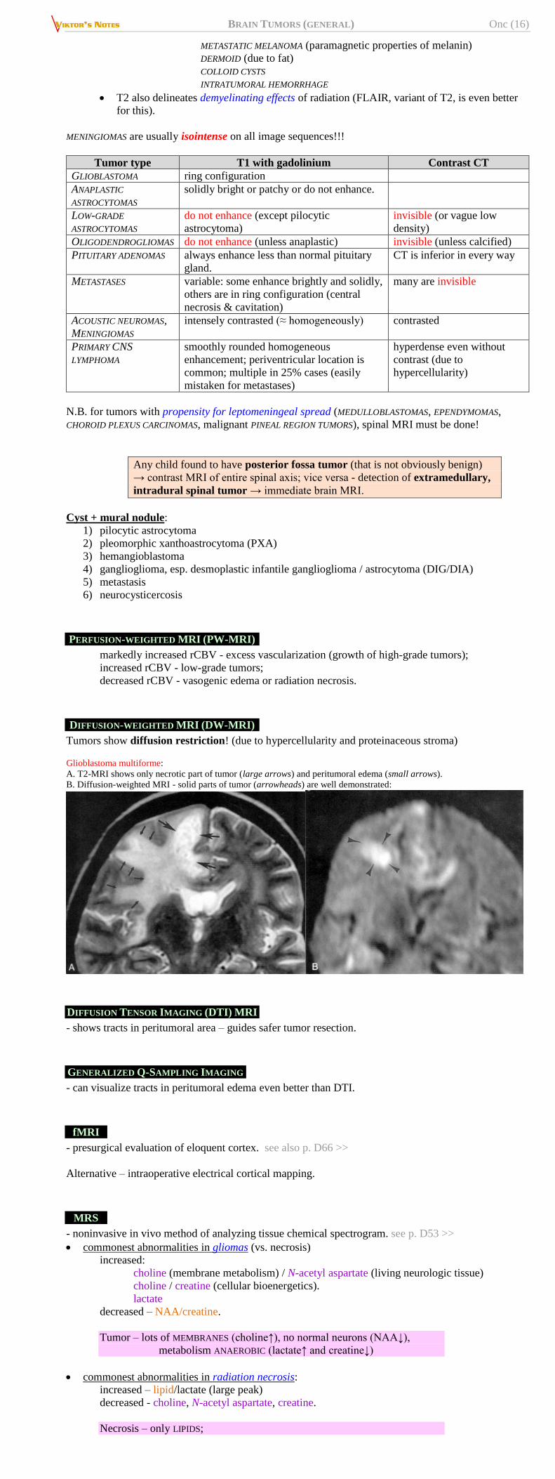
DIFFUSION-WEIGHTED MRI (DW-MRI)
Tumors show diffusion restriction! (due to hypercellularity and proteinaceous stroma)
Glioblastoma multiforme:
A. T2-MRI shows only necrotic part of tumor (large arrows) and peritumoral edema (small arrows).
B. Diffusion-weighted MRI - solid parts of tumor (arrowheads) are well demonstrated:
DIFFUSION TENSOR IMAGING (DTI) MRI
- shows tracts in peritumoral area – guides safer tumor resection.
GENERALIZED Q-SAMPLING IMAGING
- can visualize tracts in peritumoral edema even better than DTI.
fMRI
- presurgical evaluation of eloquent cortex. see also p. D66 >>
Alternative – intraoperative electrical cortical mapping.
MRS
- noninvasive in vivo method of analyzing tissue chemical spectrogram. see p. D53 >>
commonest abnormalities in gliomas (vs. necrosis)
increased:
choline (membrane metabolism) / N-acetyl aspartate (living neurologic tissue)
choline / creatine (cellular bioenergetics).
lactate
decreased – NAA/creatine.
Tumor – lots of MEMBRANES (choline↑), no normal neurons (NAA↓),
metabolism ANAEROBIC (lactate↑ and creatine↓)
commonest abnormalities in radiation necrosis:
increased – lipid/lactate (large peak)
decreased - choline, N-acetyl aspartate, creatine.
Necrosis – only LIPIDS;
BRAIN TUMORS (GENERAL) Onc (17)
no normal neurons (NAA↓), no normal membranes (choline↓),
no metabolism (creatine↓, lactate↓)
As comparison – infarction (stroke) region:
lactate↑
N-acetylaspartate (NAA)↓, creatine↓, choline↓*
*choline is only difference from tumor
Tumor – lots of membranes (choline) and anaerobic metabolism (lactate)
Stroke – everything is down except anaerobic metabolism (lactate)↑
Necrosis – everything is down except dead lipids↑
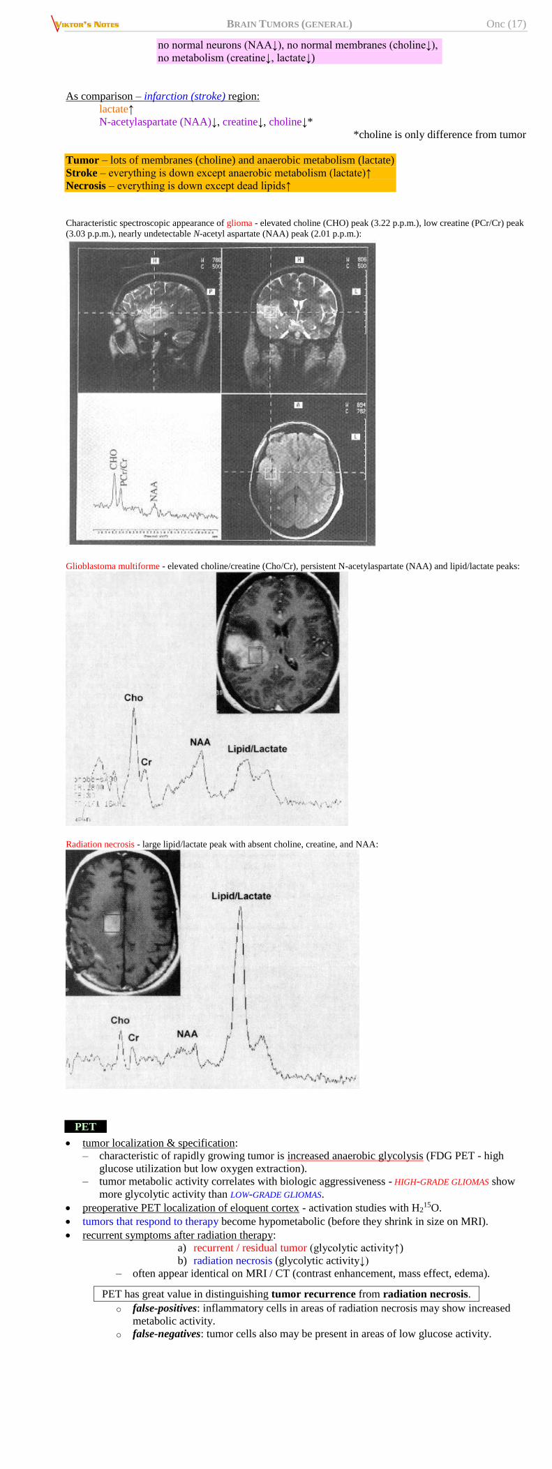
Characteristic spectroscopic appearance of glioma - elevated choline (CHO) peak (3.22 p.p.m.), low creatine (PCr/Cr) peak
(3.03 p.p.m.), nearly undetectable N-acetyl aspartate (NAA) peak (2.01 p.p.m.):
Glioblastoma multiforme - elevated choline/creatine (Cho/Cr), persistent N-acetylaspartate (NAA) and lipid/lactate peaks:
Radiation necrosis - large lipid/lactate peak with absent choline, creatine, and NAA:
PET
tumor localization & specification:
– characteristic of rapidly growing tumor is increased anaerobic glycolysis (FDG PET - high
glucose utilization but low oxygen extraction).
– tumor metabolic activity correlates with biologic aggressiveness - HIGH-GRADE GLIOMAS show
more glycolytic activity than LOW-GRADE GLIOMAS.
preoperative PET localization of eloquent cortex - activation studies with H215O.
tumors that respond to therapy become hypometabolic (before they shrink in size on MRI).
recurrent symptoms after radiation therapy:
a) recurrent / residual tumor (glycolytic activity↑)
b) radiation necrosis (glycolytic activity↓)
– often appear identical on MRI / CT (contrast enhancement, mass effect, edema).
PET has great value in distinguishing tumor recurrence from radiation necrosis.
o false-positives: inflammatory cells in areas of radiation necrosis may show increased
metabolic activity.
o false-negatives: tumor cells also may be present in areas of low glucose activity.
BRAIN TUMORS (GENERAL) Onc (18)
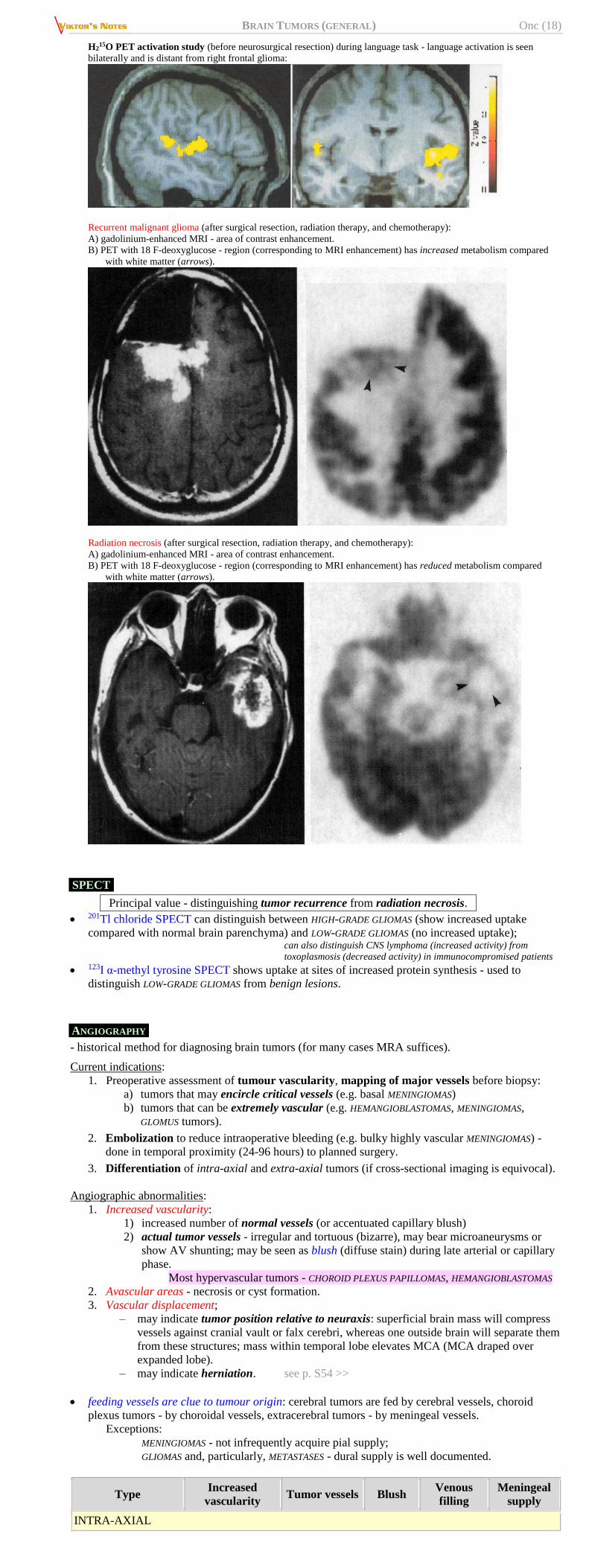
H215O PET activation study (before neurosurgical resection) during language task - language activation is seen
bilaterally and is distant from right frontal glioma:
Recurrent malignant glioma (after surgical resection, radiation therapy, and chemotherapy):
A) gadolinium-enhanced MRI - area of contrast enhancement.
B) PET with 18 F-deoxyglucose - region (corresponding to MRI enhancement) has increased metabolism compared
with white matter (arrows).
Radiation necrosis (after surgical resection, radiation therapy, and chemotherapy):
A) gadolinium-enhanced MRI - area of contrast enhancement.
B) PET with 18 F-deoxyglucose - region (corresponding to MRI enhancement) has reduced metabolism compared
with white matter (arrows).
SPECT
Principal value - distinguishing tumor recurrence from radiation necrosis.
201Tl chloride SPECT can distinguish between HIGH-GRADE GLIOMAS (show increased uptake
compared with normal brain parenchyma) and LOW-GRADE GLIOMAS (no increased uptake); can also distinguish CNS lymphoma (increased activity) from
toxoplasmosis (decreased activity) in immunocompromised patients
123I α-methyl tyrosine SPECT shows uptake at sites of increased protein synthesis - used to
distinguish LOW-GRADE GLIOMAS from benign lesions.
ANGIOGRAPHY
- historical method for diagnosing brain tumors (for many cases MRA suffices).
Current indications:
1. Preoperative assessment of tumour vascularity, mapping of major vessels before biopsy:
a) tumors that may encircle critical vessels (e.g. basal MENINGIOMAS)
b) tumors that can be extremely vascular (e.g. HEMANGIOBLASTOMAS, MENINGIOMAS,
GLOMUS tumors).
2. Embolization to reduce intraoperative bleeding (e.g. bulky highly vascular MENINGIOMAS) -
done in temporal proximity (24-96 hours) to planned surgery.
3. Differentiation of intra-axial and extra-axial tumors (if cross-sectional imaging is equivocal).
Angiographic abnormalities:
1. Increased vascularity:
1) increased number of normal vessels (or accentuated capillary blush)
2) actual tumor vessels - irregular and tortuous (bizarre), may bear microaneurysms or
show AV shunting; may be seen as blush (diffuse stain) during late arterial or capillary
phase.
Most hypervascular tumors - CHOROID PLEXUS PAPILLOMAS, HEMANGIOBLASTOMAS
2. Avascular areas - necrosis or cyst formation.
3. Vascular displacement;
– may indicate tumor position relative to neuraxis: superficial brain mass will compress
vessels against cranial vault or falx cerebri, whereas one outside brain will separate them
from these structures; mass within temporal lobe elevates MCA (MCA draped over
expanded lobe).
– may indicate herniation. see p. S54 >>
feeding vessels are clue to tumour origin: cerebral tumors are fed by cerebral vessels, choroid
plexus tumors - by choroidal vessels, extracerebral tumors - by meningeal vessels.
Exceptions:
MENINGIOMAS - not infrequently acquire pial supply;
GLIOMAS and, particularly, METASTASES - dural supply is well documented.
Type Increased
vascularity Tumor vessels Blush
Venous
filling
Meningeal
supply
INTRA-AXIAL
BRAIN TUMORS (GENERAL) Onc (19)
Type Increased
vascularity Tumor vessels Blush
Venous
filling
Meningeal
supply
Glioma (low grade) rare (+) (+) normal v. rare
Glioblastoma increased (50%) ++ + early rare
Metastases increased (50%) +++ ++ early (+)
Lymphoma normal (+) rare normal rare
Hemangioblastoma ++ ++ to +++ ++ rapid (+)
INTRA-VENTRICULAR
Choroid pl. papilloma increased + + early no
Meningioma increased + + to ++ early no
Colloid cyst no no no normal no
EXTRA-AXIAL
Meningioma increased (75%) + (angioblastic) ++ early/normal typical
Neuromas normal/increased (+) (+) can be early +
Pituitary adenoma can be increased no (+) normal from ICA
Craniopharyngioma normal no no normal no
Chemodectoma +++ +++ ++ rapid ++
Chordoma normal/increased + to ++ + to ++ early +
BIOPSY
- definitive tissue diagnosis necessary for adequate treatment planning. see p. D34 >>
most primary brain tumors are verified histologically, but 80% metastatic tumors are diagnosed
& treated empirically.
biopsy is not indicated in CHIASMAL GLIOMAS and DIFFUSE BRAIN STEM GLIOMAS* - characteristic
MRI features and uniform histology - biopsy rarely influences treatment (prognosis is dismal in
diffuse brain stem tumors regardless of biopsy results + biopsy is hazardous).
*unless brain stem glioma has exophytic component (which may be biopsied)
Open biopsies (without tumor removal) are not justifiable! - if skull and dura are to be opened, surgeon
should do gross total resection. any tumor causing mass effect or neurologic symptoms in relatively noneloquent area of brain should be
removed (so biopsy is part of surgical resection)
All brain regions may be approached by MR-guided stereotactic biopsy!
stereotactic biopsy usually provides enough tissue to make diagnosis of glioma but may not
provide enough to grade tumor (most informative specimen is one taken from area of contrast
enhancement).
Gliomas are of heterogeneous nature - areas of low-grade histology
are commonly noted in many high-grade tumors!
stereotactic biopsy is reserved for poor-surgical risk patients* (but if tumor has prominent blood
vessels or hemorrhage within tumor, open biopsy is preferable).
*open excision may result in unacceptable functional impairment
without positive influence on survival
There is no indication for craniotomy when purpose is merely to biopsy (and not resect) tumor
DIFFERENTIAL DIAGNOSIS
1. Hematomas (may be mistaken for acute bleeding into tumor)
2. Abscesses*
3. Granulomas*
4. Parasitic infections (such as cysticercosis)
5. Vascular malformations (esp. without AV shunts)
6. Solitary large MS plaque, concentric sclerosis of Balo (but T2-MRI usually reveals additional
asymptomatic lesions)
7. Progressive strokes (rare)
*usually cannot be distinguished from tumors by CT or MRI alone -
reliable management may demand biopsy
N.B. immunosuppressed patients are at risk for both primary CNS lymphomas and CNS
infections (such as toxoplasmosis or cryptococcosis) - patients treated empirically with
antibiotics should undergo prompt biopsy of lesions that are not responding to therapy.
COMPLICATIONS
HYDROCEPHALUS
A. Obstructive hydrocephalus - obstruction at ventricular atrium → foramen of Monro → aqueduct
→ 4th ventricle.
– tumor can act as valve (e.g. tumor in region of foramen of Monro) → sudden potentially
life-threatening hydrocephalus.
B. Communicating hydrocephalus a) tumor seeding to meninges
b) reaction to previous therapy
if depressed consciousness persists despite steroid administration, CSF diversion procedure should
be strongly considered.
N.B. posterior fossa tumors can cause reverse herniation after ventricular shunt
insertion (therefore, drain EVD at 15 cmH2O)
hydrocephalus requiring permanent shunt develops in 25-33% patients after posterior fossa tumor
removal.
INTRATUMOR HEMORRHAGE
tumors that most often cause hemorrhage (stroke-like onset of focal neurologic deficit):
1) oligodendrogliomas, high-grade astrocytomas
2) some metastatic tumors (melanoma!!!, renal cell carcinoma, choriocarcinoma*,
testicular carcinomas).
3) WNT among medulloblastomas
*”menses in brain”
may be provoked by iatrogenic thrombocytopenia (associated with chemotherapy).
clinically: insignificant ÷ dramatic.
treatment - osmotic agents and glucocorticoids ± surgical decompression.

BRAIN TUMORS (GENERAL) Onc (20)
BIBLIOGRAPHY for ch. “Neuro-Oncology” → follow this LINK >>
Viktor’s Notes℠ for the Neurosurgery Resident
Please visit website at www.NeurosurgeryResident.net