Embed Size (px)
Citation preview

BOVINE VIRAL DIARRHEA VIRUS: EVALUATION OF PERSISTENT
INFECTIONS, ACUTE TRANSMISSION, AND VACCINATION
PROTECTION IN ALPACAS
By
STACEY RENEE BYERS
A thesis submitted in partial fulfillment of the requirements for the degree of
MASTERS OF SCIENCE IN VETERINARY SCIENCE
WASHINGTON STATE UNIVERSITY College of Veterinary Medicine
Department of Veterinary Clinical Sciences
MAY 2009

ii
To the Faculty of Washington State University:
The members of the Committee appointed to examine the thesis of
STACEY RENEE BYERS find it satisfactory and recommend that it be accepted.
____________________________________ George M. Barrington, DVM, PhD, Chair
____________________________________ James F. Evermann, PhD
____________________________________ Steven M. Parish, DVM
____________________________________ Ahmed Tibary, DVM, PhD

iii
ACKNOWLEDGEMENT
I would like to express my sincere appreciation to my committee chair Dr. George
Barrington for his patience, assistance, encouragement, and great advice. I also want to
thank my committee members for all their assistance and support in completing this
endeavor: Dr. Evermann for his expertise, enthusiasm, and always appearing to be happy
to see me knocking on his door; Dr. Tibary for his advice on the projects, assistance with
the camels, and obtaining research animals for my projects; and Dr. Parish for his support
and encouragement and if not for his persistence and persuasion there would not have
been a residency position for me.
Finally, I can not begin to express my gratitude to my husband Thomas, who has
never stopped encouraging and supporting me. I can not imagine making this journey
without him.

iv
BOVINE VIRAL DIARRHEA VIRUS: EVALUATION OF PERSISTENT
INFECTIONS, ACUTE TRANSMISSION, AND VACCINATION
PROTECTION IN ALPACAS
Abstract
by Stacey Renee Byers, DVM, MS
Washington State University May 2009
Chair: George M. Barrington
The purpose of this project was to characterize bovine viral diarrhea virus (BVDV)
type 1b infection in alpacas to determine the effects of persistent infection (PI), acute
transient disease, and vaccine efficacy. The first specific aim was designed to follow three
PI alpacas from identification of the BVDV infection through the end of life. Antemortem
and postmortem testing identified viral shedding patterns and organs affected by virus.
Results indicated variability in antigen location between alpacas; however, viremia and
viral shedding was consistent in all three PI alpacas until death. Specific aim two
characterized BVDV transmission and acute infection in previously unexposed alpacas.
The study involved two groups of alpacas, one experimentally inoculated with BVDV,
and the second housed with PI alpacas to facilitate direct, natural transmission. Clinical
signs in the previously unexposed alpacas were mild, although alterations in blood cell
parameters indicated immunosuppression similar to that seen in bovidae infected with
BVDV. The third specific aim evaluated the efficacy of a commercial, modified-live,
BVDV vaccine in alpacas. Five previously unexposed alpacas were vaccinated and their
serologic antibody response was documented. The five vaccinated and two unvaccinated

v
controls were challenged by nasal and ocular inoculation with BVDV Type 1b obtained
from a PI alpaca. Vaccinated alpacas experienced no adverse side effects and failed to
become viremic whereas the unvaccinated control alpacas developed viremia and
experienced mild signs of acute BVDV infections. The vaccine appeared efficacious for
use in nonpregnant alpacas.

vi
TABLE OF CONTENTS
Abstract .............................................................................................................................. iv
List of Tables .................................................................................................................... vii
List of Figures .................................................................................................................. viii
Chapter
1 Introduction ...................................................................................................................... 1
2 Literature Review ............................................................................................................. 4
3 Evaluation of Persistently Infected Alpaca Crias ............................................................ 8
4 Disseminated bovine viral diarrhea virus in a persistently infected alpaca (Vicugna
pacos) cria ......................................................................................................................... 25
5 Experimental bovine viral diarrhea virus infection in male alpacas (Vicugna pacos) .. 36
6 Acute bovine viral diarrhea virus infection, transmission, and disease effects in alpacas
exposed to persistently infected alpacas ........................................................................... 48
7 Evaluation of a Bovine Viral Diarrhea Virus Commercial Vaccine in Alpacas ............ 68
8 Conclusions .................................................................................................................... 82
References ......................................................................................................................... 83
Appendix
A. Virus Culture and Isolation .......................................................................................... 90
B. Polymerase Chain Reaction ......................................................................................... 92
C. Antigen ELISA ............................................................................................................. 94
D. Serum Neutralization ................................................................................................... 96

vii
LIST OF TABLES
Chapter 3
Table 1. Results of testing to verify BVDV persistent infection status ............................ 22
Table 2. Monthly complete blood counts performed on WSU-D ..................................... 23
Table 3. Monthly complete blood counts performed on WSU-S ..................................... 23
Table 4. Monoclonal antibodies used to identify cell populations for flow cytometry. ... 24
Table 5. Flow cytometry results from BVDV persistently infected alpacas. ................... 24
Chapter 5
Table 1. Summary of diagnostic results from male alpacas experimentally infected with
BVDV ............................................................................................................................... 47
Chapter 6
Table 1. Monoclonal antibodies used to identify cell populations in whole blood .......... 65
Table 2. Diagnostic results for BVDV PI alpacas ............................................................ 65
Table 3. Diagnostic results of BVDV negative males exposed to BVDV PI alpacas ...... 66
Table 4. Physical examination findings for alpacas housed with BVDV PI alpacas ....... 67
Table 5. Flow cytometry results for BVDV transiently infected alpacas ......................... 67
Chapter 7
Table 1. Diagnostic testing results on vaccinated and unvaccinated alpacas ................... 81

viii
LIST OF FIGURES
Chapter 3
Figure 1. Phylogenetic analysis and relatedness of BVDV persistently infected alpaca
crias to other known BVDV strains. ................................................................................. 22
Chapter 4
Figure 1. Parotid salivary gland with immunohistochemical staining for BVDV antigen 34
Figure 2. Immature testis with immunohistochemical staining for BVDV antigen ......... 34
Figure 3. Panel A: Hematoxylin and eosin stained section of thymus. Panel B:
Immunohistochemical staining of the thymus .................................................................. 35
Chapter 5
Figure 1. Phylogenetic tree of alpaca isolate WSU 05-10966 in comparison with other
pestivirus isolates .............................................................................................................. 46
Figure 2. Immunohistochemical staining for BVDV antigen in parotid salivary gland ... 47
Chapter 6
Figure 1. Phylogenetic analysis and relatedness of the BVDV persistently infected alpaca
crias to other known BVDV strains .................................................................................. 66

1
CHAPTER 1
INTRODUCTION
This thesis consists of an introduction, a literature review, a summary on three
persistently infected alpacas, four manuscripts submitted for publication, a conclusion,
and appendices. This graduate student was the principle contributor and author of this
thesis and manuscripts. The manuscripts have been reviewed by the committee
members. Their comments and advice have been considered and changed made where
deemed appropriate. All final changes made were the decision of this graduate student.
Bovine viral diarrhea virus (BVDV) primarily affects cattle however it can
infect other even-toed ungulates such as alpacas. In recent years, there have been an
increasing number of reports of clinical disease and persistent infections similar to that
observed in cattle. The cause of the sudden increase in clinical BVDV in alpacas has
not been identified but is thought to be related to the mutagenic aspects of the virus.
Since clinical BVDV has only recently emerged in alpacas, there is a paucity of
information about all aspects of the disease in this species. Some information can be
extrapolated from research in the bovine species but aspects such as diagnostic
methods, transmission, and protection need to be verified in alpacas.
The purpose of this study is to characterize BVDV in alpacas to determine
persistent infection consequences, transmission modes, disease effects, and vaccine
protection. I hypothesize that the clinical disease and transmission of BVDV in
alpacas is comparable to that of cattle and a commercial bovine vaccine protects
against experimental infection.
2
Chapter 2 is a literature review of BVDV in alpacas including pertinent
research information from the bovine species.
Chapters 3 and 4 summarize the information learned from three persistently
infected (PI) alpacas. The alpacas were verified to be PI on a monthly basis until
death. Various bovine diagnostic tests including polymerase chain reaction (PCR),
virus isolation (VI), antigen capture enzyme-linked immunosorbent assay (AgELISA),
and serum neutralization (SN) were verified for use in alpacas. All three alpacas
eventually succumbed to secondary bacterial infections. Postmortem testing found
variations in tissue infectivity. The severity of clinical illness discovered on
postmortem testing corresponded to the degree of BVDV infection detected in each
alpaca, with the least severe signs and longest life span in the alpaca with minimal
antigen presence in tissues. All three alpacas had viral antigen present in saliva and
nasal secretions and urine, two had antigen present in gastrointestinal tissues, the
female had viral antigen detected in uterine tissues and feces, and one of the two males
had antigen present in testicular tissues. The results indicated all body fluids were
potential shedding routes for transmission to other alpacas. The study on the first PI
alpaca to succumb was published in the Journal of Veterinary Diagnostic
Investigations and is found in Chapter 4.
Chapters 5 and 6 summarize the results from a preliminary project performed
to evaluate the effects on alpacas experimentally inoculated with a BVDV strain from
a PI alpaca, followed by a natural transmission study using two of the PI alpacas
described above. The preliminary study found minimal signs of disease following
intravenous and intrarectal inoculation. All five inoculated alpacas became viremic

3
and virus was detected in salivary gland and saliva of two of the alpacas. The natural
transmission study was then performed with six alpacas to evaluate methods of
infection, shedding, and duration of clinical signs following exposure to PI alpacas.
All six alpacas became viremic however were not found to shed virus in nasal or oral
secretions. Clinical signs were minimal. Transmission occurred primarily by the
respiratory route since the direct contact and fence-line contact animals became
infected. The experimental inoculation study will be submitted to Veterinary
Pathology and the natural transmission study has been submitted to the Canadian
Veterinary Journal.
Chapter 7 describes the results from the BVDV vaccination trial. Five alpacas
were vaccinated with a commercial modified-live BVDV vaccine commonly used in
cattle. Two other alpacas were not vaccinated and all seven were inoculated by the
intranasal route with BVDV cultured from one of the PI alpacas discussed above. The
PI alpaca succumbed prior to the start of this phase of the project so direct challenge
could not be performed. The vaccinated alpacas did not become viremic or develop
clinical signs of BVDV infection. The two unvaccinated alpacas developed viremia
and signs of upper respiratory infections before serologic titers were detected and they
cleared the viremia and respiratory infections. This study has been submitted to
Veterinary Microbiology.

4
CHAPTER 2
LITERATURE REVIEW
Bovine viral diarrhea virus (BVDV) is one of several pestiviruses in the
Flaviviridae family. It is related to Border Disease and Classical Swine Fever. Bovine
are the primary species infected by BVDV, however research has identified it in most
even-toed ruminants with varying degrees of disease produced.1,2 Research on BVDV
has been performed since it was first identified in cattle in 1946 and since then much
has been identified about the virus and disease it produces in cattle.3
The two classification methods used to identify BVDV are genotype and
biotype. Bovine viral diarrhea virus can be divided into two genotypes: BVDV type 1
and BVDV type 2. Type 1 BVDV is found worldwide whereas type 2 is found
predominantly in North America.4 Within each genotype are numerous subgenotypes
indicating the dynamic genome and mutagenic feature of the virus. There are
approximately 11 subtypes of BVDV type 1 and 2 subtypes of BVDV type 2.5,6 The
significance of the multiple subtypes is not well understood since virulence and
disease is not only dependent on the subtype but also infectious dose, individual
animal immune response, and environmental conditions. The numerous subtypes
likely contribute to the failure of various vaccination control strategies and the
development of over 100 commercial vaccines.7
Biotype refers to the cytopathic (cp) or noncytopathic (ncp) feature of the virus
in cell cultures. Both type 1 and type 2 BVDV can have cp and ncp forms and both
can be found in natural infections. Only the ncp forms are thought to be capable of

5
causing persistent infections in the fetus however cp forms have been identified in
fetal tissues and serum.4,8-10 A BVDV persistently infected (PI) bovine co-infected
with a closely related cp subtype can develop a rare and fatal form of BVDV known as
mucosal disease.11
Bovine viral diarrhea virus has the ability to cause multiple forms of disease:
persistent infections, transient infections, and mucosal disease. Persistent infections
can develop if the fetus is exposed to the virus prior to maturation of the fetal immune
system at around 125 days gestation.12 In these cases, the immune system does not
recognize the virus as a foreign antigen and does not mount an immune response.
These animals will have detectable virus by tests such as PCR, VI, AgELISA, and
immunohistochemistry (IHC), but will be antibody negative to that strain of virus.
Transient infections occur in previously unexposed animals and are typically
acute and subclinical in 70-90% of infected cattle.13 However, severity does vary and
can include respiratory disease, infertility, abortions, congenital defects, hemorrhagic
disorders, and even death in severe cases.12,14 Mucosal disease occurs sporadically and
is typically fatal due to the severe ulcerations of all mucosal surfaces and secondary
infections.12 Bovine viral diarrhea virus is also known to induce immune suppression
leading to many secondary illnesses such as the respiratory disease complex
commonly observed in feedlot cattle.12,15
Bovine viral diarrhea virus transmission occurs by separate but interrelated
vertical and horizontal modes. Horizontal transmission of a pregnant bovine can lead
to vertical transmission to the fetus causing a persistent infection. The PI animal is the
primary source for horizontal transmission of BVDV in a herd since large amounts of

6
virus are shed in all body fluids, but transiently infected (TI) animals can also be a
major source of infection.16,17 Horizontal transmission occurs within one hour of direct
contact with a PI animal and fence line transmission from both PI and TI animals has
been reported.18,19 Inhalation of virus-laden body fluids is thought to be the primary
route of infection for acute infections.20
A variety of diagnostic tests are available and some are more suited for
distinguishing the PI animal from the TI animal. Persistent infections can be detected
using a combination of VI of the buffy coat layer or serum, IHC staining for viral
antigen in skin, AgELISA of skin or blood samples, and PCR of whole blood or serum
samples.21,22 Tests such as VI or PCR cannot differentiate persistent from transient
infections so retesting several weeks later in combination with serum neutralization is
used.
Various control and eradication strategies have evolved over time and
currently are based on testing and culling PI animals, vaccinations, and biosecurity.
Detection of the PI animal is important as this animal is the primary source of
infection within the herd. Even so, producers utilizing these control points continue to
observe problems due to transient infections and vaccination or biosecurity failures.
Prior to 2005, very little research had been published evaluating BVDV in
camelids. Only one research project evaluated BVDV inoculation in pregnant
llamas.23 Other reports indicated camelids formed antibodies to the virus but clinical
signs were either absent or mild so the prevailing thought was the virus did not readily
affect camelids.24-27 In 2005, the first reports of suspected clinical BVDV cases and PI
alpacas emerged.28,29 At that time, most bovine diagnostic tests had not been validated

7
for use in alpacas so the initial cases of suspected PI alpaca crias were difficult to
confirm. Diagnostic laboratories appeared to settle on PCR and serology as the
primary methods to test alpacas.
Since 2005, other reports have been published indicating an increasing
incidence of BVDV cases in alpacas.30-33 Current research indicates the majority of
BVDV cases in alpacas have been classified as type 1b but some type 2 cases have
been detected.28,29,31-35 Alpacas also have not exhibited the severe mucosal disease
seen in cattle. Due to the recent emergence of BVDV in alpacas, most reports have
only focused on detection, genetics, and prevalence so basic questions regarding
transmission, duration of infection, shedding routes, clinical effects, and control
mechanisms have not been well defined.
The purpose of this study was to determine the effects of BVDV persistent
infections, transient infections, transmission, and vaccination protection in alpacas.

8
CHAPTER 3
EVALUATION OF PERSISTENTLY INFECTED ALPACA CRIAS
Abstract
Bovine viral diarrhea virus (BVDV) is an emerging infectious pathogen of
concern to the alpaca industry. Three alpaca crias from a single farm were diagnosed
as persistently infected with BVDV based on repeated positive antemortem PCR and
VI assays, and negative antibody titers to BVDV. Various postmortem diagnostic tests
were performed with virus identified in multiple tissues demonstrating disseminated
BVDV type 1b infections in all three alpacas. Virus was detected in various body
fluids indicated potential routes of shedding and transmission to other alpacas.
Introduction
Three alpacas crias: a 6-month-old female (WSU-D) and two approximately 5-
month-old, intact males (WSU-S and WSU-R), presented to the Washington State
University Veterinary Teaching Hospital (Pullman, WA) with a history of decreased
weight gain, ill thrift, and anorexia. The alpacas were from a herd of 45 animals and
diagnosed as PI alpacas infected with BVDV type 1, based on repeated positive tests
by PCR and VI on EDTA blood samples obtained at monthly intervals staring at 2-3
months of age, and negative SN antibody titers on serum samples. Upon presentation,
all three crias were bright, alert, and responsive with normal vital parameters
(temperatures, pulse, and respiration), however all three were deemed to be
significantly underweight compared to age-matched uninfected crias. Because of the

9
grave prognosis due to their PI status and general poor health, as well as financial
considerations, the owners declined further antemortem diagnostics tests or treatment
and elected to donate the alpaca crias to WSU for monitoring.
WSU-R
WSU-R was five months old and weighed 13.6 kg at presentation. Besides a
small stature and poor body condition score (1.5/5), all other findings on physical
examination were within normal limits. Complete blood cell count revealed a mild left
shift (band neutrophils 309/µl; reference [ref.] range <200/µl), high normal fibrinogen
(400 mg/dl; ref. range 100–400 gm/dl), anemia (PCV 21%, ref. range 27–45%),
decreased hemoglobin (9.4 g/dl; ref. range 11.9–19.4 g/dl), and marked anisocytosis.
Serum biochemical profile was within normal limits.
Therapy with tulathromycin (5.5 mg/kg intramuscular, Draxxin®, Pfizer
Animal Health, New York, NY) and a nutritional supplement had been initiated prior
to admittance to WSU due to a suspected upper respiratory tract infection. Over the
next eight days at WSU, the cria was observed to be ambulating, eating, drinking,
urinating, and defecating normally. Florfenicol therapy (22 mg/kg, subcutaneous, q 48
h; Nuflor®, Intervet/Schering-Plough, Union, NJ) was initiated on day 4 because of a
suspected respiratory infection. On the morning of day 9, the cria was found dead in
its stall and transferred to the Washington Animal Disease Diagnostic Laboratory
(WADDL, Pullman, WA) for complete postmortem examination.

10
WSU-D
WSU-D was six months old and weighed 19.8 kg at presentation. Besides a
small stature and poor body condition score (1.5/5), all other findings on physical
examination were within normal limits. Complete blood cell count revealed a
neutrophilia (segmented neutrophils 1501/µl; ref. range 4700-14900/µl), anemia (PCV
24%, ref. range 27–45%), decreased hemoglobin (10.4 g/dl; ref. range 11.9–19.4 g/dl),
and moderate anisocytosis. Serum biochemical profile revealed an elevated SDH (32
U/L; ref. range 1-5 U/L), elevated AP (190 U/L; ref. range 27-132 U/L), mildly
elevated CK (106 U/L; ref. range 8-77 U/L), mildly decreased total protein (5.0 g/dl;
ref. range 5.1-7.8 g/dl) and mildly decreased albumin (3.0 g/dl; ref. range 3.1-5.2 g/dl).
All other values were within normal limits.
Therapy with tulathromycin (5.5 mg/kg IM) had been initiated prior to
admittance to WSU due to a suspected upper respiratory tract infection. Over the next
three months of monitoring, the cria was observed to be ambulating, eating, drinking,
urinating, and defecating normally. Florfenicol therapy (22 mg/kg, SC, q 48 h) was
initiated on day 4 because of a suspected respiratory infection and chronic pyrexia. A
recurrent fever occurred every few weeks for 4-8 days at a time and ranged from
102.8-103.8°F. Antipyretics were not administered as the cria remained bright and
appetent. The febrile episodes would typically be accompanied by nasal and or ocular
discharge consistent with an upper respiratory tract infection. Due to the persistent
respiratory tract signs, the cria remained on florfenicol for approximately three weeks
before therapy was changed to ceftiofur crystalline free acid (6.6 mg CE/kg SC, q 7 d;
Excede, Pfizer Inc., New York, NY). Monthly sampling was performed to monitor

11
BVDV PI status and shedding (Table 1) and CBC values (Table 2). Three months after
arrival, the cria was found to have a sinus abscess that eroded through the frontal bone.
Due the cria’s continued deterioration and immune suppression, it was euthanized and
transferred to WADDL for complete postmortem examination.
WSU-S
WSU-S was 4-1/2 months old and weighed 15.5 kg at presentation. Besides a
small stature, and poor body condition score (2.0/5), all other findings on physical
examination were within normal limits. Blood work was performed the day after
arrival at WSU. Complete blood cell count revealed only mild changes including a
lymphocytosis (6302/µl; ref. range 700-4800/µl), high normal fibrinogen (400 mg/dl;
ref. range 100–400 gm/dl), and marked anisocytosis with slight to moderate
polychromasia. Serum biochemical profile revealed mild increases in GGT (51 U/L;
ref. range 9-29 U/L), ALP (468 U/L; ref. range 27-132 U/L), and CK (246 U/L; ref.
range 8-77 U/L).
Over the next 11 months of monitoring, the cria was observed on a daily basis
with only mild intermittent health problems. Florfenicol therapy (22 mg/kg, SC, q 48
h) was initiated on day 4 because of suspected respiratory infections in the two other
PI crias housed with this animal and potential BVDV induced immune suppression in
conjunction with shipping and environmental stresses. The cria did not develop signs
of respiratory disease similar to the other two PI crias until just prior to death. The
alpaca was relatively healthy with minor illnesses including a corneal ulcer due to a
foreign body and a chronic hyperplasia along the oral mucocutaneous junction.

12
Monthly sampling was performed to monitor BVDV PI status and shedding (Table 1)
and CBC values (Table 3). At 15 months old, the cria developed a respiratory tract
infection with anorexia and suddenly died while undergoing medical treatment to
stabilize its condition. The cria was transferred to the WADDL for complete
postmortem examination.
Diagnostic Testing and Results
WSU-R
Gross postmortem examination revealed acute, locally extensive
bronchopneumonia of the cranial lung lobes; caudal lung lobes were mottled red to
pink and moderately firm. Several well delineated (0.5–1.5 cm in diameter) ulcers
were observed on the mucosal surface of gastric compartments 1 (C1), 2 (C2), and 3
(C3). Numerous petechiae were noted on the surface of the spleen. No other gross
lesions were identified. Tissues were collected in 10% neutral buffered formalin,
routinely processed, and embedded in paraffin blocks. From the paraffin blocks, 4-µm
sections were cut and stained with hematoxylin and eosin. Lung, liver, and a tracheal
swab submitted for aerobic culture revealed mixed bacterial growth; similar samples
were negative for Mycoplasma culture.
Histological examination of tissue from the cranial lung lobes revealed
moderate, multifocal, suppurative bronchopneumonia. Sections from the remaining
lung lobes were characterized by patchy infiltrates of minimal to low numbers of
lymphocytes, plasma cells, and rare neutrophils within the interlobular and alveolar
septa. Gastric compartments 1 and 2 had multifocal gastritis, characterized by a mild

13
to moderate infiltration of the lamina propria by variable admixtures of lymphocytes,
plasma cells, eosinophils, neutrophils, and macrophages. No fungi were identified. In
several sections of both large and small intestine, the lamina propria was mildly to
moderately expanded by neutrophils, lymphocytes, and plasma cells. The liver
exhibited moderate, multifocal, random hepatocellular necrosis consistent with
bacteremia. Submandibular, inguinal, and mesenteric lymph nodes had mildly to
markedly reduced numbers of lymphocytes within the cortex and medulla; mesenteric
lymph nodes were most severely affected. The cortex and medulla of the thymic
lobules and the white pulp of the spleen had moderately to severely reduced
lymphocyte numbers; the thymic corticomedullary junction was indistinct.
Immunohistochemical (IHC) detection of BVDV antigen was performed on
sections of formalin-fixed, paraffin-embedded tissues using monoclonal antibody
(mAb) 15c5 (IDEXX MoAB 15c-5 (anti-BVDV EO), IDEXX Laboratories,
Westbrook, ME, USA) at a 1:1000 dilution in a streptavidin-biotin-immunoperoxidase
technique with diaminobenzidine as a chromogen (LSAB™2 Kit, Dako North
America Inc., Carpinteria, CA, USA).36 Large amounts of BVDV antigen were
detected in the following tissues: parotid salivary gland, testis, prostate, esophagus,
C1, C2, C3, right kidney, bone marrow, liver, lung, spleen, thymus, and the mesenteric
and submandibular lymph nodes. In sections of parotid salivary gland, there was
strong intracytoplasmic immunoreactivity within acinar epithelial cells and occasional
ductular epithelial cells. Similarly, a section of testis revealed frequent cells within the
seminiferous tubules that had strong intracytoplasmic immunoreactivity for BVDV
antigen. Scattered groups of epithelial cells within the convoluted tubules of the

14
kidney had mild to moderate intracytoplasmic immunoreactivity for BVDV, while rare
macrophages within the interstitium had intracytoplasmic immunoreactivity for
BVDV. Within the esophagus and C1, there was BVDV immunoreactivity within the
cytoplasm of basaloid epithelial cells. Compartment 2 had immunoreactivity of both
the basaloid epithelial cells within the areas of stratified squamous epithelium and
within the glandular epithelium of the saccules. In C3, there were frequent areas of
immunoreactivity within the cytoplasm of glandular epithelial cells and within some
of the gastric ulcers. Within the lung, thymus, spleen, and lymph nodes, there were
rare to frequent cells (presumptive macrophages) that had intracytoplasmic
immunoreactivity for BVDV antigen, while scattered Kupffer cells within the liver
had similar immunoreactivity. Frequent myeloid precursor cells and presumptive
macrophages within the bone marrow had similar intracytoplasmic immunoreactivity
for BVDV antigen.
Real-time reverse transcription polymerase chain reaction (real-time RT-PCR)
to detect and subtype BVDV in tissues was performed using a duplex TaqMan real-
time PCR procedure as previously described.37 Following extraction of total RNA
with TRIzol (Invitrogen Corp., Carlsbad, CA, USA), BVDV-1 was detected in kidney,
liver, spleen, lung, and thymus. Bovine viral diarrhea virus was isolated from the same
tissues in BVDV-free bovine turbinate cells.22 Bovine viral diarrhea virus RNA was
extracted from the supernatants of virus-infected cells using a commercial RNA
extraction kit (QIAamp® Viral RNA Mini Kit, Qiagen Inc., Valencia, CA, USA) and
detected via TaqMan real-time PCR procedure as previously described.37 To obtain a
longer amplicon for sequencing, a separate RT-PCR reaction was run, using

15
recombinant Thermus thermophilus (rTth) DNA polymerase (Applied Biosystems,
Foster City, CA, USA) to amplify a highly conserved 290-bp portion of the 5’-
untranslated region (5’ UTR) of the BVDV genome as previously described.38
Polymerase chain reaction amplicons were visualized on a 1.5% agarose gel
containing ethidium bromide, excised using a sterile scalpel blade under ultraviolet
illumination, and purified using a nucleic acid purification kit (Bio-Rad Freeze ‘N
Squeeze™ Kit, Bio-Rad Laboratories, Hercules, CA, USA) according to the
manufacturer’s directions. Amplicon DNA was sequenced directly on both strands by
a local vendor (Amplicon Express Inc., Pullman, WA, USA) using a commercial
sequencing kit (Amersham DYEnamic ET Terminator Cycle Sequencing Kit,
Amersham Biosciences, Piscataway, NJ, USA) with analysis on a DNA sequencer
(Applied Biosystems, Foster City, CA). Sequencing reactions were done in duplicate,
and sequences were confirmed by sequencing of both strands. Forward and reverse
sequences were aligned using the ClustalW algorithm.39 Each sequence was compared
to the GenBank nucleotide sequence database for similarity using BLASTn
(nucleotide Basic Local Alignment Search Tool;
http://www.ncbi.nlm.nih.gov/blast/Blast.cgi). The sequence most closely matched that
of BVDV-1b (98% sequence identity to GenBank accession # AY159530 and other
type 1b accessions) when compared with sequences in GenBank.
WSU-D
Antemortem samples collected from WSU-D and tested as described above
verified constant BVDV PI status and viral shedding in oral, nasal, fecal, and urine

16
samples with PCR, VI, and AgELISA (Table 1). Skin biopsy samples collected from
ear notching were positive by the commonly used bovine AgELISA test. The alpaca
was consistently negative on serology. Postmortem samples were collected and
processed as describe above. Whole blood samples were collected at 7 and 9 months
of age for flow cytometry (see Chapter 6 for processing details) and processed with
llama specific monoclonal antibodies to determine percentages of leukocytes (Table
4). Cell population percentages were variable between both samples and were
generally within the normal expected values for juvenile alpacas (Table 5).
Gross postmortem examination revealed thymic atrophy with scant amounts of
ill defined thymic tissue in the mediastinal and distal cervical region. At
approximately midline overlying the frontal sinuses there was a 1 cm circular
ulceration of the skin with caseous to mucopurulent discharge with a draining
inflammatory tract extended into the subjacent bone. An abundant amount of ale
yellow purulent material was present within the frontal sinuses and the nasal cavity.
Diffusely within the wall of C2, there were accumulations of edema fluid that
expanded the wall up to 1 cm in thickness. Compartment 3 had a focal, 1.5 x 0.5 cm
dark red ulcer overlaid by a 0.5 cm fibrinous pseudomembrane. The mucosa of the
urinary bladder was stippled by numerous petechia. No other gross lesions were
identified. Bacterial cultures of swabs taken from the trachea and nasal exudates
revealed Bordetella bronchiseptica and Arcanobacterium pyogenes. Lung and liver
swabs revealed primary growth of Bordetella bronchiseptica. Samples were negative
for Mycoplasma culture.

17
Histological examination of tissue from sections of tonsil, thymus, spleen and
submandibular, cervical, mesenteric and inguinal lymph nodes had generalized,
moderate to severe lymphocyte depletion. Occasional lymphoid cortical follicles and
splenic white pulp were admixed with pyknotic nuclear debris (lympholysis). Lymph
node medullary cords were further severely depleted of macrophages, plasma cells and
lymphocytes. Subcapsular and medullary sinuses of the submandibular lymph node
were multifocally expanded by numerous degenerate neutrophils with fewer
macrophages interspersed with flocculent eosinophilic proteinaceous material. The
liver exhibited rare, multifocal hepatocellular necrosis. Gastric compartment 3 had
focal ulcerative gastritis, characterized by lamina propria that was replaced by a
coagulum of serocellular debris admixed with numerous bacterial colonies and
degenerate neutrophils. The underlying submucosa was expanded by a mixture of
neutrophils, lymphocytes, macrophages, and eosinophils interspersed with fibrin and
edema residue. Replicate sections stained with Brown & Hopps tissue gram stain
revealed large numbers of long slender gram negative rods along the advancing edge
of the ulcerated mucosa. The uterus had suppurative, mild, diffuse, acute endometritis
with scattered neutrophils, lymphocytes, and decreased plasma cells. Several large
rafts of neutrophils are free within the uterine lumen. Sections of the lung lobes
showed moderate, multifocal, peracute pulmonary congestion and atelectasis. The
brain had a single vessel within the cerebral cortex bordered by mildly increased
numbers of lymphocytes. The pulmonary changes detected grossly were attributed to
multifocal congestion and atelectasis with agonal aspiration of mucocellular debris
from the upper respiratory tract. The hepatic changes were speculated to be secondary

18
to septicemia or endotoxemia from the ulcerative gastritis. Suppurative endometritis
was regarded as an incidental finding likely secondary to immunosuppression.
Immunohistochemical detection of BVDV antigen was performed as described
above. Numerous skin and pancreatic acinar epithelial cells had strongly positive
cytoplasmic immunoreactivity. Parotid salivary gland acinar and ductal epithelial cells
comprised approximately 5% of the section, and were stippled with strongly positive
cytoplasmic immunoreactivity. Polymerase chain reaction testing was positive for
BVDV on lung, tracheal and nasal exudate swabs, whole blood and serum, feces,
kidney, spleen, and thymus. Virus isolation was positive for BVDV on lung, kidney,
spleen, thymus, and liver. Antigen ELISA was positive for BVDV on serum. The
alpaca was serology negative for BVDV. The fecal sample also indicated a large
population of giardia.
WSU-S
Antemortem samples collected from WSU-S and tested as described above
verified constant BVDV PI status and viral shedding in oral, nasal, and urine samples
with PCR, VI, and AgELISA (Table 1). Skin biopsy samples collected from ear
notching were positive by the commonly used bovine AgELISA test. The alpaca was
consistently negative on serology. Whole blood samples were obtained at 5, 7, and 11
months of age for flow cytometric analysis. Values were variable between the three
samples and were generally within the normal reference values (Table 5). Postmortem
samples were collected and processed as describe above.

19
Gross postmortem examination revealed severe consolidated and hyperemia of
the right cranioventral lung lobes which affected approximately 20% of the tissue. On
cut section, medium sized airways exuded green feed material. The left caudoventral
lobes had irregular zones of consolidation affecting approximately 5% of the left lung.
Sections of affected lung lobes did not float in 10% formalin. Tracheobronchial lymph
nodes were moderately to severely enlarged. Lung and tissue pool samples submitted
for aerobic culture revealed a mixed bacterial growth.
Histologic examination of lung tissue had broad regions with dense aggregates
of degenerate neutrophils, cellular debris, colonies of mixed bacteria, and fragments of
plant material filling bronchi and bronchioles. Adjacent alveoli were frequently filled
with neutrophils and fewer macrophages admixed with large numbers of mixed
bacteria. The trachea had a mild infiltration of lymphocytes and plasma cells within
the subepithelial stroma. Many portal areas of the liver were surrounded by
disorganized, proliferative bile ducts and biliary epithelial cells that did not form
recognizable ductules. The adrenal gland contained scattered cortical foci expanded by
mild hemorrhage. Several lymph node sections contained moderate numbers of
neutrophils within the medullary sinuses and moderate numbers of macrophages that
contain phagocytosed erythrocytes. The spleen had mild depletion of lymphocytes
within the white pulp and numerous macrophages in the red pulp contained
phagocytosed erythrocytes. The small intestinal villi in several segments were
moderately to severely blunted and fused. The lamina propria was infiltrated with
minimal numbers of lymphocytes and plasma cells.

20
Immunohistochemical detection of BVDV antigen was performed as described
above. Only skin epithelial cells showed signs of positive cytoplasmic
immunoreactivity.
Discussion
The results of the diagnostic testing indicated disseminated BVDV infections
in all three alpacas. Interestingly, there was significant variation between the three
animals as to which tissues were affected. Alpaca WSU-S had the least amount of
detectable virus in the tissue samples and this alpaca also had the fewest clinical signs
and survived the longest. The variation in presence of virus is may be due in part to
different gestational infection time frames and is similar to that reported in other PI
alpacas.28,30-32,34 The exact time frame for development of persistent infections in
alpacas has not yet been identified but is suspected to be in the first trimester as is
reported in cattle. The dams of these three alpacas were all exposed to an acute or
persistently infected alpaca at the breeding farm prior to being shipped home at 4-5
months pregnant.
Diagnosis of the PI status was verified consistently with positive PCR, VI,
AgELISA, and negative serology. Viral antigen was detected in nasal and oral swabs,
urine or urinary bladder, testicles, and gastrointestinal tract tissue or feces, which
indicates potential routes of transmission similar to that observed in cattle. Flow
cytometry results from WSU-S and WSU-D were generally within the normal
expected range for healthy juvenile alpacas. Similar findings have been reported in PI
cattle with lymphocytes typically within normal ranges.40 All the PI alpacas in this

21
report were stunted and unable to gain weight, had poor fiber quality, and were
predisposed to secondary infections to which they all eventually succumbed.
.
22
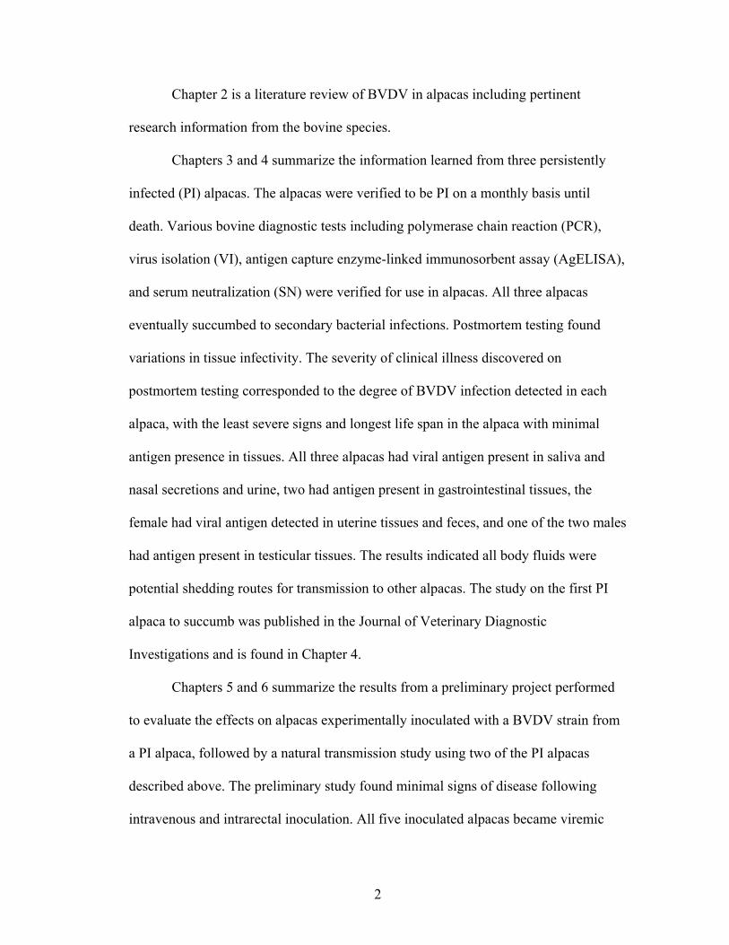
Figure 1. Phylogenetic analysis and relatedness of the BVDV persistently infected alpaca crias to other known BVDV strains. Persistently infected alpacas are identified as WSU alpaca S, WSU alpaca R, and WSU alpaca D.
Table 1. Monthly testing to verify BVDV persistent infection status on WSU-R, WSU-D, and WSU-S. Time 0 for month tested indicates initial testing on arrival at WSU. Tests with spaces not containing ‘+’ or ‘-‘ indicates test not performed. Oral and nasal samples were obtained by swabbing buccal and nasal mucosal surfaces.
Month Tested
WSU Alpaca Sample PCR VI AgELISA Serology
0 R, D, S blood + + + -
1 D, S blood + + + -
oral & nasal + +
2 D, S blood + -
3 D, S blood + -
ear notch +
4 D feces, urine + +
D, S blood + -
5 S blood + + -
6 S blood + -
oral & nasal + +
7 S blood + -
8 S blood + -
9 S blood + -
oral & nasal - -
10 S blood + + -
urine, oral, nasal + +
WSU alpaca S
WSU alpaca E
WSU alpaca D
WSU alpaca R
BVDV1b-NY-1
BVDV1a-NADL
BVDV1c-Trangie
BVDV2a-890
BVDV2b-VS-63
BD-31
100.0%100.0%
100.0%
95.7%
87.7%
75.2%
80.6%
56.6%
53.2%
WSU alpaca S
WSU alpaca E
WSU alpaca D
WSU alpaca R
BVDV1b-NY-1
BVDV1a-NADL
BVDV1c-Trangie
BVDV2a-890
BVDV2b-VS-63
BD-31
100.0%100.0%
100.0%
95.7%
87.7%
75.2%
80.6%
56.6%
53.2%

23
Table 2. Monthly complete blood counts performed on WSU-D. Months represent age of the alpaca cria. NR: Not Reported
Ref. Range 6 Mos 7 Mos 8 Mos 9 Mos 10 Mos
WBC (/µL) 8000-12000 7900 11400 3688 6603 4000
Band Neutrophils (/µL) <200 37 66 400
Segmented Neutrophils (/µL) 4700-14900 1501 1140 443 3037 960
Lymphocytes (/µL) 700-4800 2765 4788 2102 2509 2080
Monocytes (/µL) <1100 237 798 1033 726 560
Eosinophils (/µL) 700-4900 3318 4496 73 264 NR
Hemoglobin (g/dL) 11.9-19.4 10.4 10.6 10.1 10.3 8.1
PCV (%) 27-45 24 22 23 24 18
Plasma Protein (g/dL) 5.1-7.8 5.7 6.0 6.8 5.7 3.4
Fibrinogen (mg/dL) 100-400 300 500 NR 400 200
Table 3. Monthly complete blood counts performed on WSU-S. Months represent age of the alpaca cria.
Cells (Ref Range) 4 Mos 5 Mos 6 Mos 7 Mos 12 Mos 15 Mos
WBC 8000-12000/µL 13694 6500 8200 7300 10000 5344
Band Neutrophils <200/µL 742
Segmented Neutrophils 4700-14900/µL 6439 1820 3116 2117 6600 2491
Lymphocytes 700-4800/µL 6302 3380 4182 3796 2700 1590
Monocytes <1100/µL 959 1170 492 1022 500 424
Eosinophils 700-4900/µL 410 219 100
Hemoglobin 11.9-19.4 g/dL 12 11.9 12.2 12.3 10.8 8.9
PCV 27-45% 29 26 27 27 25 21
Plasma Protein 5.1-7.8 g/dL 6.5 6.4 6.5 6.1 4.5 6.0
Fibrinogen 100-400 mg/dL 400 400 300 700

24
Table 4. Monoclonal antibodies (mAb) used to identify cell populations in whole blood collected from BVDV PI alpacas
mAb Isotype Specificity DH59B1A IgG1 CD172a
GB45A IgG1 WC1 (expressed on subset of and γδ T cells)
GC50A1 IgM CD4 LH41A IgG2a B cells
LT3A1 IgG1 CD5 (predicted)
LT5A IgG2a CD8
LT10A IgG2a CD6 (predicted)
LT97A IgG2b αβ and γδ T cells (predicted)
Table 5. Flow cytometry results from BVDV persistently infected alpacas WSU-D and WSU-S. Months indicate age of the alpaca.
Cell Phenotype Control WSU-Alpaca D WSU Alpaca S Normal Values
Mean 7 Mos 9 Mos 5 Mos 7 Mos 11 Mos Mean Range αβ T lymphocytes 26 64 31 25 51 29 27 11-47
CD4+ 16 39 17 22 21 21 21 11-49
CD8+ 11 14 12 4 25 9 10 4-28 CD4/CD8 ratio
1.5 2.8 1.4 5.5 0.8 2 1-5
CD4/CD8 dbl pos
0 11 0 2 1 0
γδ T lymphocytes
17 6 9 7 1 9 1 4-26
B lymphocytes
53 19 42 54 21 32 4 26-60
Monocytes 4 11 16 8 31 13 19 6-39

25
CHAPTER 4
DISSEMINATED BOVINE VIRAL DIARRHEA VIRUS IN A PERSISTENTLY
INFECTED ALPACA (VICUGNA PACOS) CRIA
S.R. Byers, K.R. Snekvik, D.J. Righter, J.F. Evermann, D.S. Bradway,
S.M. Parish, and G.M. Barrington
From the Departments of Veterinary Clinical Sciences (Byers, Snekvik, Righter,
Evermann, Parish, Barrington) and Veterinary Microbiology and Pathology (Snekvik,
Righter), and the Washington Animal Disease Diagnostic Laboratory (Snekvik,
Righter, Evermann, Bradway), College of Veterinary Medicine, Washington State
University, Pullman, WA.
The American Association of Veterinary Laboratory Diagnosticians holds the
copyright to this material.
Abstract
Bovine viral diarrhea virus (BVDV) is an emerging infectious pathogen of
concern to the alpaca industry. A 4-month-old, intact, male alpaca cria was diagnosed
as persistently infected with BVDV based on repeated positive antemortem
polymerase chain reaction (PCR) and virus isolation (VI) assays, and negative
serologic titers to BVDV. Immunohistochemistry, real time reverse transcription PCR,

26
and VI performed on tissues collected at necropsy demonstrated disseminated BVDV-
1b infection. Virus was detected in multiple tissues, including parotid salivary gland,
testes, prostate, kidneys, skin, and gastrointestinal tract. Demonstration of BVDV in
previously unreported tissues suggests additional potential routes of BVDV
transmission in alpacas.
Introduction
A 4-month-old, intact, male alpaca (Vicugna pacos) cria presented to the
Washington State University Veterinary Teaching Hospital (Pullman, WA) with a
history of decreased weight gain, ill thrift, and anorexia. The cria was from a herd of
45 animals and was diagnosed as persistently infected (PI) with bovine viral diarrhea
virus type 1 (BVDV-1), based on three repeated positive tests by polymerase chain
reaction (PCR) and virus isolation (VI) on EDTA blood samples obtained at 3, 4, and
5 months of age, and negative serologic titers to BVDV on serum samples. Upon
presentation, the cria was bright, alert, and responsive with normal temperature, pulse,
and respiration. The cria was deemed to be significantly underweight (13.6 kg)
compared to his herd mates. All other findings on physical examination were within
normal limits. Complete blood cell count revealed a mild left shift (band neutrophils
309/µl; reference [ref.] range <200/µl), high normal fibrinogen (400 mg/dl; ref. range
100–400 gm/dl), anemia (PCV 21%, ref. range 27–45%), decreased hemoglobin (9.4
g/dl; ref. range 11.9–19.4 g/dl), and marked anisocytosis. Serum biochemical profile
was within normal limits.

27
Therapy with tulathromycin (5.5 mg/kg IM) and a nutritional supplement had
been initiated approximately one week earlier due to a suspected upper respiratory
tract infection. Because of the grave prognosis due to PI status and general poor health
of the cria, as well as financial considerations, the owners declined further antemortem
diagnostics tests or treatment and elected to have the cria hospitalized for temporary
monitoring. Over the next eight days, the cria was observed to be ambulating, eating,
drinking, urinating, and defecating normally. Florfenicol therapy (22 mg/kg,
subcutaneous, q 48 h) was initiated on day four because of a suspected respiratory
infection. On the morning of day nine, the cria was found dead in its stall and
transferred to the Washington Animal Disease Diagnostic Laboratory (Pullman, WA)
for complete postmortem examination.
Diagnostic Testing and Results
Gross postmortem examination revealed acute, locally extensive
bronchopneumonia of the cranial lung lobes; caudal lung lobes were mottled red to
pink and moderately firm. Several well delineated (0.5–1.5 cm in diameter) ulcers
were observed on the mucosal surface of gastric compartments 1 (C1), 2 (C2), and 3
(C3). Numerous petechiae were noted on the surface of the spleen. No other gross
lesions were identified. Tissues were collected in 10% neutral buffered formalin,
routinely processed, and embedded in paraffin blocks. From the paraffin blocks, 4-µm
sections were cut and stained with hematoxylin and eosin. Lung, liver, and a tracheal
swab submitted for aerobic culture revealed mixed bacterial growth; similar samples
were negative for Mycoplasma culture.

28
Histologic examination of tissue from the cranial lung lobes revealed
moderate, multifocal, suppurative bronchopneumonia. Sections from the remaining
lung lobes were characterized by patchy infiltrates of minimal to low numbers of
lymphocytes, plasma cells, and rare neutrophils within the interlobular and alveolar
septa. Gastric compartments 1 and 2 had multifocal gastritis, characterized by a mild
to moderate infiltration of the lamina propria by variable admixtures of lymphocytes,
plasma cells, eosinophils, neutrophils, and macrophages. No fungi were identified. In
several sections of both large and small intestine, the lamina propria was mildly to
moderately expanded by neutrophils, lymphocytes, and plasma cells. The liver
exhibited moderate, multifocal, random hepatocellular necrosis consistent with
bacteremia. Submandibular, inguinal, and mesenteric lymph nodes had mildly to
markedly reduced numbers of lymphocytes within the cortex and medulla; mesenteric
lymph nodes were most severely affected. The cortex and medulla of the thymic
lobules and the white pulp of the spleen had moderately to severely reduced
lymphocyte numbers; the thymic corticomedullary junction was indistinct.
Immunohistochemical (IHC) detection of BVDV antigen was performed on
sections of formalin-fixed, paraffin-embedded tissues using monoclonal antibody
(mAb) 15c5 (IDEXX MoAB 15c-5 (anti-BVDV EO), IDEXX Laboratories,
Westbrook, ME, USA) at a 1:1000 dilution in a streptavidin-biotin-immunoperoxidase
technique with diaminobenzidine as a chromogen (LSAB™2 Kit, Dako North
America Inc., Carpinteria, CA, USA).36 Large amounts of BVDV antigen were
detected in the following tissues: parotid salivary gland, testis, prostate, esophagus,
C1, C2, C3, right kidney, bone marrow, liver, lung, spleen, thymus, and the mesenteric

29
and submandibular lymph nodes. In sections of parotid salivary gland, there was
strong intracytoplasmic immunoreactivity within acinar epithelial cells and occasional
ductular epithelial cells (Fig. 1). Similarly, a section of testis revealed frequent cells
within the seminiferous tubules that had strong intracytoplasmic immunoreactivity for
BVDV antigen (Fig. 2). Scattered groups of epithelial cells within the convoluted
tubules of the kidney had mild to moderate intracytoplasmic immunoreactivity for
BVDV, while rare macrophages within the interstitium had intracytoplasmic
immunoreactivity for BVDV. Within the esophagus and C1, there was BVDV
immunoreactivity within the cytoplasm of basaloid epithelial cells. Compartment 2
had immunoreactivity of both the basaloid epithelial cells within the areas of stratified
squamous epithelium and within the glandular epithelium of the saccules. In C3, there
were frequent areas of immunoreactivity within the cytoplasm of glandular epithelial
cells and within some of the gastric ulcers. Within the lung, thymus (Fig. 3), spleen,
and lymph nodes, there were rare to frequent cells (presumptive macrophages) that
had intracytoplasmic immunoreactivity for BVDV antigen, while scattered Kupffer
cells within the liver had similar immunoreactivity. Frequent myeloid precursor cells
and presumptive macrophages within the bone marrow had similar intracytoplasmic
immunoreactivity for BVDV antigen.
Real-time reverse transcription polymerase chain reaction (real-time RT-PCR)
to detect and subtype BVDV in tissues was performed using a duplex TaqMan real-
time PCR procedure as previously described.37 Following extraction of total RNA
with TRIzol (Invitrogen Corp., Carlsbad, CA, USA), BVDV-1 was detected in kidney,
liver, spleen, lung, and thymus. Bovine viral diarrhea virus was isolated from the same

30
tissues in BVDV-free bovine turbinate cells.22 Bovine viral diarrhea virus RNA was
extracted from the supernatants of virus-infected cells using a commercial RNA
extraction kit (QIAamp® Viral RNA Mini Kit, Qiagen Inc., Valencia, CA, USA) and
detected via TaqMan real-time PCR procedure as previously described.37 To obtain a
longer amplicon for sequencing, a separate RT-PCR reaction was run, using
recombinant Thermus thermophilus (rTth) DNA polymerase (Applied Biosystems,
Foster City, CA, USA) to amplify a highly conserved 290-bp portion of the 5’-
untranslated region (5’ UTR) of the BVDV genome as previously described.38
Polymerase chain reaction amplicons were visualized on a 1.5% agarose gel
containing ethidium bromide, excised using a sterile scalpel blade under ultraviolet
illumination, and purified using a nucleic acid purification kit (Bio-Rad Freeze ‘N
Squeeze™ Kit, Bio-Rad Laboratories, Hercules, CA, USA) according to the
manufacturer’s directions. Amplicon DNA was sequenced directly on both strands by
a local vendor (Amplicon Express Inc., Pullman, WA, USA) using a commercial
sequencing kit (Amersham DYEnamic ET Terminator Cycle Sequencing Kit,
Amersham Biosciences, Piscataway, NJ, USA) with analysis on a DNA sequencer
(Applied Biosystems, Foster City, CA). Sequencing reactions were done in duplicate,
and sequences were confirmed by sequencing of both strands. Forward and reverse
sequences were aligned using the ClustalW algorithm.39 Each sequence was compared
to the GenBank nucleotide sequence database for similarity using BLASTn
(nucleotide Basic Local Alignment Search Tool;
http://www.ncbi.nlm.nih.gov/blast/Blast.cgi). The sequence most closely matched that

31
of BVDV-1b (98% sequence identity to GenBank accession # AY159530 and other
type 1b accessions) when compared with sequences in GenBank.
Discussion
Bovine viral diarrhea virus is considered to be primarily an infectious pathogen
in cattle, but other even-toed ungulates including camelids have been reported to be
susceptible.41 Early studies in camelids suggested either resistance or decreased ability
to transmit BVDV due to low seroprevalence, minimal clinical disease, and lack of
pathology after experimental infections.24,27 However, current evidence suggests that
BVDV may be an emerging and significant disease of alpacas.23,28,29,32,34 The recent
increase in cases within alpacas is likely multifactoral and may involve the emergence
of a novel viral strain, differences in animal management practices, or increased
awareness of BVDV by alpaca owners. The BVDV genome is known to be dynamic,
with replication of any isolate giving rise to a viral swarm until a predominant strain
emerges, and one of these may have an increased predilection for alpacas.34,42
Preliminary research indicates the recent BVDV infections in alpacas have been
primarily noncytopathic BVDV-1b strain. 23,28,29,32
Research on PI bovine fetuses and calves has identified virus in all tissues and
shedding occurring from multiple sites.43,44 Previous reports on four confirmed PI
alpacas identified virus in various organs but did not mention reproductive tissues or
salivary gland.28,32,34 Unlike those cases, the cria in the present report had
disseminated BVDV infections which included parotid salivary gland, esophagus, C1–
3, testes, prostate, and kidneys. Identification of virus in these organs indicates

32
potential routes for transmission through communicative and reproductive behaviors.
Alpacas spit as a communication tool within the herd, and breeders frequently use the
behavior to evaluate sexual receptivity of the female. Breeding involves cervical
penetration by the penis and deposition of semen deep inside both uterine horns. The
uterine trauma occurring during the breeding process may increase the risk of BVDV
transmission.45
Bovine viral diarrhea virus infection in calves is also associated with “weak
calf syndrome” and “shipping fever,” in part due to viral immunosuppressive effects.
Clinically, poor growth and recurrent infection in the animal in the present study
mimicked that seen with many PI calves. Similar signs have also been reported in
other PI alpacas.28,32,34 The similarities between the disease in cattle and alpacas
suggest that the viral pathogenesis may be comparable.
The fetal outcome of BVDV infection in naive, pregnant cattle is variable and
dependent on the stage of gestation at which infection occurs. It is believed that cattle
become persistently infected when fetal infection occurs prior to immunologic
development, and viral proteins are recognized as “self antigens” and therefore
tolerated.46 The gestational exposure time for BVDV immunotolerance in alpacas has
not been fully identified, but appears to occur in the first trimester. The dam of the PI
cria in the present study was exposed to a suspected nonclinical PI or transiently
infected alpaca while located at the breeding facility during the first 90 days of her
pregnancy. It was unknown if the female was exposed prior to pregnancy or
continually during this time period. The animal was not exposed to cattle during her
pregnancy.

33
Research in the cattle industry has identified fetal infection rates of over 10%,
even in vaccinated herds, with the rate of PI calves approximately 0.5%.47 Fetal
infection rates are suspected to be lower within the alpaca industry due to the lower
population and the recent emergence of the virus within the species; however, because
potential PI crias are typically euthanized to prevent herd exposure and limit farm
reputation damage, this cannot be confirmed.
The alpaca cria in the current case had a disseminated BVDV infection, which
included the reproductive, salivary, and upper gastrointestinal tract tissues. These
findings suggest a significant risk for BVDV transmission due to alpaca behavior and
management practices. Subclinical to acute infections have been estimated to cost the
cattle industry $50 to $400 per head, for all animals in the herd, not just those
suffering disease.48 With the current value of many female alpacas beginning at
approximately $10,000 per animal, the potential economic losses for alpaca producers
due to BVDV are significant therefore continued testing and biosecurity efforts are
warranted.

34
Figure 1. Parotid salivary gland with immunohistochemical staining for Bovine viral diarrhea virus (BVDV) antigen. Bovine viral diarrhea virus immunoreactivity within the cytoplasm of acinar epithelial cells and rare ductular epithelial cells.
Figure 2. Immature testis with immunohistochemical staining for BVDV antigen. Bovine viral diarrhea virus immunoreactivity within the cytoplasm of cells of the seminiferous tubules.

35
Figure 3. Hematoxylin and eosin stained section of thymus (Panel A) showing severe thymic atrophy with lymphoid depletion. Immunohistochemical staining of the thymus shows BVDV antigen within the cytoplasm of presumptive macrophages (Panel B).
Panel A Panel B

36
CHAPTER 5
EXPERIMENTAL BOVINE VIRAL DIARRHEA VIRUS INFECTION IN MALE
ALPACAS (VICUGNA PACOS)
S.R. Byers, J.F. Evermann, G. Haldorson, S.M. Parish, D.S. Bradway, F.R. Rurangirwa, C.L. Fite, J.R. Ridpath, A.Tibary
Department of Veterinary Clinical Sciences (Byers, Evermann, Parish, Fite, Tibary);
Department of Veterinary Microbiology and Pathology (Haldorson, Rurangirwa);
Washington Animal Disease Diagnostic Laboratory (Evermann, Haldorson, Bradway).
College of Veterinary Medicine, Washington State University, Pullman, WA; National
Animal Disease Center (Ridpath), ARS/USDA, Ames, IA.
Abstract
Bovine viral diarrhea virus (BVDV) has been reported to infect multiple
animal species and determining its pathogenesis in alpacas has been of importance to
the camelid industry. This research involved the experimental inoculation of five male
alpacas with an alpaca isolate of BVDV (WSU 05-10966). This virus genotype (1b),
was representative of other alpaca BVDV isolates. Alpacas were studied over the
course of 14 days, and sampled to determine antemortem viral shedding and the
location of BVDV antigen postmortem by immunohistochemistry. Clinical symptoms
were mild over the 14 day observation period. There was a transient leukopenia noted

37
in three animals. By day 7 post inoculation, virus was obtained from the saliva of two
alpacas and from the blood of all five alpacas. At necropsy, BVDV antigen was
detected in the parotid salivary gland of one animal and saliva of two animals, but not
in the testicular tissues or accessory sex glands. This is the first report to describe the
presences of BVDV in saliva and salivary glands of alpacas suggesting an important
potential route of transmission between animals.
Introduction
The pathogenesis of bovine viral diarrhea virus (BVDV) in camelids, primarily
alpacas and llamas, is not completely understood. Earlier studies concentrated on
serological evaluation which detected variable levels of BVDV seropositive
populations.24,27 Experimental studies have been limited due to the lack of availability
of alpaca or llama field isolates of BVDV. One experimental study using a bovine
strain of BVDV in pregnant llamas indicated that while the animals seroconverted,
there were no ill effects noted upon fetal development and subsequent birth of crias.27
Natural studies have expanded our knowledge regarding BVDV infection in alpacas to
include the occurrence of a persistently infected (PI) state similar to what has been
reported in cattle.28,31,32,34 This study was designed to determine what clinical effects
an alpaca-derived isolate of BVDV would have on naïve, healthy, male alpacas, and to
determine if BVDV could be detected in selected body tissues and secretions which
would account for the spread of BVDV amongst reproductively active animals.

38
Materials and methods
Test subjects
Five intact male alpacas (#1-5) ranging in age from 2.5 months to 48 months were
selected for experimental inoculation. One male alpaca (#6), aged 48 months, was not
inoculated and served as a contact control. Serum samples for BVDV serology and
whole blood samples for BVDV polymerase chain reaction (PCR) were collected six
days prior to experimental inoculation and every four days thereafter. All six animals
were seronegative (BVDV serum neutralization antibody titer) and BVDV PCR
negative at the beginning of the experiment.
Challenge virus inoculum
A pestivirus (WSU 05-10966) was initially isolated from a 12 month old
alpaca that was anorexic. The alpaca was tentatively identified as being persistently
infected with BVDV based upon its negative BVDV serologic status coincidental with
successful isolation of BVDV from whole blood. The virus was cultured on bovine
turbinate (BT) cells (BVDV free) in minimum essential media, supplemented with
10% fetal bovine serum (BVDV and BVDV antibody free), penicillin, streptomycin,
gentamicin and Fungizone (JR Scientific, Inc., Woodland, CA, USA). The virus was
typed as BVDV 1b by nested PCR (Figure 1). The low passage (p5) virus inoculum
used to challenge the experimental animals was amplified in BT cells, and frozen at -
80°C until animals were inoculated. The virus titer was determined to be
106.5TCID50/ml.

39
Experimental Design
Five male alpacas were inoculated by the intravenous and intrarectal routes,
utilizing 10ml of BVDV inoculum per route. The intrarectal route was chosen as a
convenient route for mucous membrane experimental infection. Alpacas were
randomly assigned to be euthanized on days 7 and 14 post inoculation. The single in-
contact control alpaca was euthanized on day 15. This study was conducted with
approval, and in accordance with the Washington State University Institutional
Animal Care and Use Committee. A sample size of five alpacas was chosen to limit
animal use and because the effects of transient BVDV infection in camelids was
unknown.
Sample collection and analysis
All six animals received initial physical examinations, and blood was obtained for a
CBC, serology, virus isolation, and PCR. Saliva was collected antemortem by placing
a 2 inch cotton/gauze roll in the animal’s mouth for 5 minutes. The saliva was
extracted using a 60cc syringe and expressed into a sterile red top tube. At necropsy, a
complete set of tissues were collected and divided between fresh-frozen and fixed in
10% buffered formalin. At necropsy, seminal fluid was collected for virus isolation
and PCR.
Antibodies to BVDV were determined by the serum neutralization (SN) assay,
using the Singer strain of BVDV as the challenge virus. Animals were considered
positive if antibody levels were equal to or greater than 1:8 dilution of the serum.
Virus isolation was performed in 24-well trays pre-seeded 48 hours earlier with BT

40
cells. Detection of BVD by RT-PCR was conducted according to Bhudevi and
Weinstock.37 Briefly, RNA was extracted from tissues, EDTA blood, and body fluids
using the QIA amp viral RNA extraction kit (Qiagen Inc., Valencia, CA, USA). Real
time Taqman PCR was performed using a Cepheid Smart Cycler (Cepheid, Sunnyvale,
CA, USA) to detect and type the BVDV from the experimental animals. The sequence
matched that of BVDV 1b (99% sequence identity) when compared with sequences in
Genbank.
Genotyping of BVDV alpaca isolate
Genotyping of the alpaca BVDV isolate was based on phylogenic analysis of
highly conserved sequences from the 5’ untranslated region (UTR). Total RNA was
extracted from cells infected with each isolate and a 268 nucleotide sequence from the
5’ UTR was amplified and sequenced as described previously. Determination of
BVDV1 subgenotypes was based on comparison of type strains for BVDV1
subgenotype identified by Vilcek et al.6 The mean sequence homology was
determined using an unpaired geometric mean analysis (UPGMA) (Higgins-Sharp
algorithm/CLUSTAL4, MacDNASIS software package, Hitachi Software Engineering
Co., Yokohama, Japan).
Histopathology and Immunohistochemistry
A complete compliment of tissues was collected at necropsy for
histopathologic examination. Briefly, tissue samples were collected into 10% buffered
formalin, processed overnight in an automatic processor (Tissue-Tek VIP, Sakura

41
Finetek Japan Co., LTD, Toyko, Japan), embedded in paraffin, and 4 µm thick
sections were placed onto glass slides. The sections were de-parafinized with xylene
and stained with hematoxylin and eosin.
Sections of fixed parotid salivary gland and reproductive tissues including
testes, vas deferens, epididymis, ampulla, prostate, and bulbourethral gland were
evaluated by immunohistochemistry (IHC) for evidence of BVDV antigens using the
avidin-biotin-peroxidase method. Briefly, sections were deparafinized and treated with
3% H2O2 in methanol for 10 minutes. The samples were treated with 0.06% Pronase in
Tris buffered saline (Dako North America, Inc., Carpinteria, CA, USA) for 10 minutes
for antigen retrieval, and blocked with 5% normal goat serum for 10 minutes.
Monoclonal antibody 15c-5 (Syracuse Bioanalytical, Inc., Syracuse, NY, USA) was
diluted 1:5000 in blocker of 5% normal goat serum and incubated for 30 minutes at
room temperature for antigen detection. Replicate sections were incubated with
isotype-matched, irrelevant monoclonal antibody as a negative control. Primary
antibody binding was detected using biotinylated goat anti-mouse IgG (Signet
Laboratories, Inc., Bedham, MA, USA) for 30 minutes and labeled with Ultra
Streptavidin (Signet Laboratories, Inc., Bedham, MA, USA) for 30 minutes. Dako
K3464 AEC (Dak North America, Inc., Carpinteria, CA, USA) was used as the
chromagen. Positive control tissue included skin from a BVDV infected bovine calf.
Negative control tissues were incubated with an irrelevant isotype matched
monoclonal antibody.

42
Results
Clinical profiles on the experimentally infected alpacas revealed that three
alpacas (#2, 3, and 4) developed pyrexia (maximum of 104.8, 103, and 102.6°F
respectively) at 6-7 days post inoculation (dpi). Three alpacas (#1, 4, and 5) developed
neutropenia beginning 3 dpi, with alpaca #1 remaining neutropenic until euthanasia on
14 dpi. The other two animals returned to normal white blood cell counts by the next
blood sampling. Alpaca #3 was lymphopenic at 7 dpi at which time it was euthanized.
None of the animals developed anemia, however four alpacas (#1, 2, 3, 5) had
decreasing pack cell volumes beginning at 7 dpi until euthanasia. Alpaca #5 developed
transient diarrhea during days 6-9 pi. The in-contact control animal (#6) remained
clinically normal during the course of the experiment
The serological and virological results indicated that three of the five
experimentally infected animals seroconverted by 14 dpi (Table 1). However, the two
animals that did not seroconvert were euthanized at 7 dpi and may not have had
sufficient time to develop detectable BVDV antibodies. All five experimental animals
were viremic by 5 dpi. Two of the five animals were positive for BVDV antigen in
saliva samples at 7 dpi. The in-contact control animal remained seronegative to BVDV
and no virus was detected from any salivary samples collected. Gross examination at
necropsy did not revealed any abnormalities in the alpacas. Similarly, there were no
histologic lesions observed, however the parotid gland on one animal was BVDV
antigen positive by immunohistochemistry (IHC) (Figure 2). No virus was identified
in seminal fluid collected at necropsy nor was virus detected by IHC in the

43
reproductive tissues. No gross or histologic lesions were observed in the control
animal, and none of this alpaca’s tissues were reactive to BVDV antigen by IHC.
Discussion
The results of this study indicated that following experimental inoculation of
male alpacas with an alpaca-derived isolate of BVDV, there was a short-term viremia
coincident with seroconversion to BVDV. There were effects noted upon blood
profiles with animals displaying neutropenia, lymphopenia, and decreasing hematocrit
values. Several of the alpacas developed transient fevers and one had diarrhea for
three days. Virus isolation from salivary samples was positive in two of the five
alpacas yet no virus was identified in seminal fluid samples.
There were no gross or histopathologic lesions in animals necropsied at 7 and
14 days post inoculation. Staining of parotid salivary gland and reproductive tissues by
BVDV-specific IHC revealed that the salivary gland was positive in one of the
experimentally infected animals.
The one in-contact control animal did not seroconvert during the 14 day
observation period indicating that either the experimentally infected alpacas were not
shedding communicable levels of BVDV, or that it took longer than the 14 day
observation period to detect an immune response by the BVDV SN assay.
Bovine viral diarrhea virus was not detected in testicular tissues and seminal
fluid in this group of alpacas which was in contrast to what has been reported with an
experimental inoculation of BVDV into three 2-year-old bulls.49 Although no clinical
abnormalities were noted other than a spike in temperature (104-105.8°F), there was

44
evidence of sustained viral infection in the testes out to 7 months post infection in two
of three of the bulls. Testicular biopsies were not obtained prior to this time to
determine when the infection was first detectable; however semen from two bulls was
found to be PCR positive at days 8 and 10 post inoculation. The differences between
BVDV infection in alpaca males and young bovine bulls is of importance in
understanding the epidemiology of the virus in non-bovine species. Although our
study concluded at 14 days post infection, there was no evidence that the virus
replicated in, or established infection in the sexual glands of the male alpacas. The
lack of occurrence of BVDV in the seminal fluid and other reproductive tissues,
suggests that sexual transmission of BVDV in alpacas may not be as common as other
routes. It has been documented that uterine trauma occurs during the breeding process
potentially exposing the female to infectious agents.45 However, the apparent lack of
BVD virus in seminal fluids could suggest a lower risk for females to acquire this
disease during breeding.
The occurrence of BVDV in the salivary secretions of two of the male alpacas
suggests however, that saliva may be an important route of disease transmission in
alpacas. Immunohistochemistry positive BVDV staining in the parotid salivary gland
indicates that viral replication was occurring, and that virus may have been amplified
prior to the onset of an immune response. Alpacas commonly use “spitting” as a
communication tool between animals. This trait and the presence of BVDV in saliva
and salivary gland should be further evaluated to determine ease of viral transmission
between animals.

45
Finally, conclusions drawn from a single in-contact control animal must not be
overstated. The failure of this animal to seroconvert or produce detectable virus might
suggest that transmission requires longer contact time or that it is not as efficient in
other species. Clearly, studies involving larger populations of animals are necessary to
more definitively determine the frequency and efficiency of various routes of
transmission within this species.

46
Figure 1. Phylogenetic tree of alpaca isolate WSU 05-10966 in comparison with other pestivirus isolates. Note that the virus is placed in the BVDV 1b subgenotype together with two prototype bovine strains, TGAC and NY-1, and another alpaca isolate 023909 (see arrow).

47
Figure 2. Immunohistochemical staining for BVDV antigen in parotid salivary gland of animal # 3 counterstained with hematoxylin (×400).
Table 1. Summary of diagnostic results from male alpacas experimentally infected with alpaca BVDV isolate (WSU 05-10966).
Animal number Maximum BVDV antibodya titer post infection
BVDV isolationb
Blood Saliva Semen 1 1:16 (14 dpi) + + -
2 <1:8 (7 dpi) + + -
3 <1:8 (7 dpi) + - -
4 1:32 (14 dpi) + - -
5 1:64 (14 dpi) + - -
6 (in-contact control)
<1:8 (15 dpe)c - - -
a Serum neutralization antibodies b Virus isolation in bovine turbinate cells, confirmed by PCR c Days post exposure

48
CHAPTER 6
ACUTE BOVINE VIRAL DIARRHEA VIRUS INFECTION, TRANSMISSION,
AND DISEASE EFFECTS IN ALPACAS EXPOSED TO PERSISTENTLY
INFECTED ALPACAS
S.R. Byers, J.F. Evermann, D.S. Bradway, A.L. Grimm, J. Ridpath,
W. Davis, S.M. Parish, A. Tibary, and G.M. Barrington
From the Departments of Veterinary Clinical Sciences (Byers, Evermann, Parish,
Tibary, Barrington) and Veterinary Microbiology and Pathology (Grimm), the
Washington Animal Disease Diagnostic Laboratory (Evermann, Bradway), College of
Veterinary Medicine, Washington State University, Pullman, WA, and the National
Animal Disease Center (Ridpath), ARS/USDA, Ames, IA.
Abstract
Bovine viral diarrhea virus (BVDV) reports in alpacas have been increasing;
however, much is still unknown about the mechanisms of disease in this species. This
report describes research performed to evaluate transmission of BVDV from

49
persistently infected (PI) alpacas to naïve alpacas, document shedding patterns, and
determine the effects on the immunological status. Two PI alpacas were determined
to shed BVDV Type 1b virus in most body fluids, and commonly available diagnostic
tests were used to verify their status. The alpacas exhibited signs of ill thrift and
increased susceptibility to secondary infections. Transient infections with the BVDV
Type 1b strain produced only mild signs of disease in BVDV naïve alpacas. Viral
shedding during the acute phase was not detected and antibody titers appeared to be
protective upon re-exposure to the virus. Immunosuppressive effects were observed,
which could lead to secondary infections similar to that seen in cattle.
Introduction
Bovine viral diarrhea viruses (BVDV-1 and BVDV-2) are members of the
pestivirus genus that includes genetically similar viruses such as Border Disease virus
of sheep and Classical Swine Fever (Hog Cholera virus) in swine.50,51 Bovine viral
diarrhea virus was first recognized as a pathogen of cattle in 1946, however BVDV
does infect other even-toed ungulates with varying degrees of disease severity.2,3
Recent reports indicate an increased number of BVDV-1b infections occurring in
alpacas and describe clinical disease in species such as goats, bison, yaks, mountain
goats, and white tailed deer.2,23,28,29,31,32,34,52-56
The recent increase in reported cases of BVDV in alpacas and other species is
likely multifactoral and may involve variations in virus strain, animal management
practices, as well as increased awareness and testing by producers. As with other
RNA viruses, the BVDV genome is dynamic resulting from genetic mutation and gene

50
recombination.57-59 It is suspected that certain BVDV strains may adapt to new hosts
resulting in increased infectivity in non-bovine species such as alpacas.1,34 Despite the
increasing number of alpaca BVDV cases, much is still unknown regarding
transmission, fetal infections, and clinical disease.
Research has shown that transiently (TI) and persistently infected (PI) cattle
can transmit BVDV in various body fluids, with the primary portal of infection via the
oronasal mucosa.12,17,60 Transiently infected cattle typically shed BVDV for only a few
days whereas PI cattle are believed to either continuously shed large quantities of virus
or in some cases, inconsistently shed as evidenced by periods of intermittent viremia.
20,61-63 Both TI and PI cattle are sources of infection to other cattle. Similar shedding
and transmission patterns are suspected in PI and TI alpacas, but have not been
reported. Based on identification of the virus in tissues such as salivary glands,
kidneys, gastrointestinal tract, lungs, testicles, and prostate, it is likely alpacas shed
virus in a range of body fluids similar to cattle.28,31,32,34
Cattle become persistently infected with BVDV if the fetus is exposed to the
virus before the immune system is fully developed, resulting in immune tolerance
whereby the fetus does not recognize virus as foreign.12 Fetal infection can occur if the
dam develops a transient infection or is a PI herself.64 Persistently infected calves
typically show signs of ill-thrift and chronic illnesses; however, they can appear
clinically normal.14
Research in BVDV PI alpacas is lacking in part due to the relative infrequency
of the condition, the long gestation period of alpacas (11+ months), and the likelihood
that most crias suspected of being PI’s are euthanized and not reported. The present

51
study was designed to evaluate the serologic and viral status of alpacas suspected of
being persistently infected with BVDV; to evaluate transmission of BVDV from PI
alpacas to naïve alpacas; to document shedding patterns in transiently infected
animals; and to determine the effects of BVDV on the immunological status of PI and
TI alpacas.
Materials and Methods
Animals
Two alpaca crias from a single farm (a five-month-old intact male (WSU
alpaca S) and a six-month-old female (WSU alpaca D) were donated to Washington
State University Veterinary Teaching Hospital. The two crias were verified as
persistently infected with BVDV based on positive BVD PCR and virus isolation (VI),
and negative BVDV serology on monthly checks starting at 2-3 months of age.22,37
Six intact male alpacas (5 to 6-month old) were used as BVDV-negative,
infection controls. The crias were verified as BVDV negative based on PCR and
antibody by SN.22,37 The cutoff for negative SN titers was 1:8. All six animals were
clinically healthy.
The studies were conducted in accordance with the Washington State
University Institutional Animal Care and Use Committee. The limited number of
experimental animals was chosen and justified based on humanitarian grounds since
BVDV is known to induce immunosuppression and increase the risk of infection with
secondary pathogens in other species.

52
Sampling
Blood, nasal and oral swabs, and fecal samples were obtained every three days
during weeks 1-3. Blood samples were then obtained weekly during the next month
(weeks 4-8), then every two weeks for the next three months (weeks 9-20). Twelve
milliliters of blood were aseptically obtained from the jugular vein and divided
between tubes containing ethylenediaminetetraacetic acid (EDTA), acid citrate
dextrose (ACD), and serum blood collection tubes (BD Vacutainer Blood Collection
Tubes; Becton, Dickinson and Company, Franklin Lakes, NJ, USA). Individual nasal
and oral swabs were collected from the lateral wall of the nasopharynx and buccal
surface of the oral cavity. Swabs were placed in individual viral collection vials (BD
Universal Viral Transport Kit (220222), Becton, Dickinson and Company, Franklin Lakes,
NJ, USA). Fecal samples were obtained by digital collection from the rectum and
placed in sterile plastic specimen bags. When possible, free-catch urine samples were
collected in sterile specimen cups. Samples not immediately processed were stored at
-20°C until analysis.
Four months following the initial exposure period, the same six male alpacas
were re-introduced to the remaining PI alpaca (WSU-S) in the same facility and pen
configuration as described above. Blood, nasal and oral swabs were collected daily
for one week, every third day for one week, then at one month and three months post
exposure.

53
Virus Isolation and PCR
Whole blood and oral and nasal swabs from the PI alpacas and BVDV exposed
alpacas were analyzed using virus isolation in bovine turbinate cells free of BVDV.22
Bovine viral diarrhea virus RNA was extracted from the supernatants of virus-infected
cells or from EDTA blood using the Magmax-96 Blood RNA Isolation Kit (Ambion,
Inc. Austin, TX, USA). Real Time Taqman PCR to detect BVDV was performed
using an ABI 7500 Fast Real-Time PCR System (Applied Biosystems, Foster City,
CA, USA) and using the ABI AgPath-ID™ BVDV Reagent Kit. The sequences were
compared to the GenBank nucleotide sequence database for similarity using BLASTn
(nucleotide Basic Local Alignment Search Tool;
http://www.ncbi.nlm.nih.gov/blast/Blast.cgi).
Antigen ELISA
Serum samples were tested for BVD antigen by the IDEXX HerdChek Bovine
Virus Diarrhea Antigen Test Kit (IDEXX Laboratories, Westbrook, ME, USA).
Briefly, 100 µL of serum samples was placed in pre-wetted wells, along with positive
and negative standards, and in-house reference sera. Testing was done according to
IDEXX protocols. Samples with standard to positive (S/P) ratios of <0.20 were
classified as “BVDV-Negative”. Samples with S/P ratios of 0.20 – 0.39 were
classified as “Suspect”, and samples with an S/P ratio of >0.39 were classified as
“BVDV-Positive”. Suspect and positive reactors were assayed twice, first using the
standard working detector reagent, and then using a “modified” working detector
reagent to check for non-specific reactivity. Optical densities were measured using a

54
Bio-Tek EL808 Ultra Microplate Reader (Bio-Tek Instruments Inc., Winooski, VT,
USA), and calculations were made with KC-4 Bio-Tek software.
Serum Neutralization
Serum was analyzed for BVDV-specific antibodies using the SN assay as
described by Evermann, et al.22 Alpaca serum was diluted in two-fold serial dilutions
using minimal essential media (MEM) plus antibiotics [2.5 µg/ml Fungizone (JR
Scientific, Inc., Woodland, CA), 100 International Units (IU)/ml penicillin G, 100
IU/ml streptomycin sulfate, and 50 µg/ml gentamicin] beginning at a screening
dilution of 1:4. Briefly, 0.050 ml of MEM plus was added to wells of a flat-bottom
96-well microtiter plate and 0.050 ml alpaca serum and known positive control serum
were added to the appropriate wells. Serial dilutions were performed with an end
volume of 0.050 ml in each well. The plate was incubated at room temperature for one
hour after which 0.050 ml of BT cells (BVDV-free) were added to each well as the
indicator cell type followed by incubation in a 10% humidified, 5% CO2 incubator at
37°C. At 72 hours, the plate was read for serum neutralization of virus or cytopathic
effect. The Singer strain of cytopathic BVDV was used as the challenge virus at 100-
1000 TCID50. Serum antibody titers were expressed as the reciprocal of the highest
dilution of serum providing 100 percent cell protection.
Flow Cytometry
Whole blood was collected in ACD, and peripheral blood mononuclear cells
(PBMC) separated by density gradient centrifugation using Accu-Paque (density,

55
1.086 g/ml; Accurate Chemical & Scientific Corps., Westbury, NY, USA). The buffy
coat layer was collected from the interface and washed three times in phosphate
buffered saline (PBS) containing 20% ACD to remove excess platelets. The cells were
resuspended in PBS/ACD. Concentration of the cell suspension was determined using
a hemocytometer.
One million cells were added to wells of a 96 well v-bottom microtiter plate
containing two mAbs (50 µl each, 15 µg/ml) in 100 µl of dilution buffer [PBS
containing 0.5% horse serum (Invitrogen/GIBCO, Gaithersburg, MD, USA), 20%
ACD and 0.02% azide]. Cells were incubated for 15 min (4°C) and then washed three
times in dilution buffer. Following washing, cells were incubated for 15 min (4°C)
with 50 µl of isotype specific fluorescein or phycoerythrin conjugated goat anti-mouse
immunoglobulin (isotype specific) antibodies (Invitrogen/Caltag, Burlingame, CA,
USA). The cells were again washed two times with dilution buffer without horse
serum, resuspended in 2% formaldehyde in PBS (200 µl/well) and stored refrigerated
in the dark until analyzed.
An anti-pan T lymphocyte mAb was used to identify T and B lymphocytes
(Table 1). T lymphocyte subsets were further differentiated using mAbs specific for
CD4+, CD8+, and γδ T cells (Table 1). A FACS Calibur flow cytometer with Cell
Quest software (Becton Dickinson Immunocytometry Systems, San Jose, CA, USA)
was used to collect data. The FCS Express software (De Novo Software, Thornton,
Ontario, Canada) was used to analyze data.

56
Experimental Exposure
Three BVDV negative alpacas (1A-1C) and the two PI alpacas (WSU-S,
WSU-D) were placed together in an 8 foot by 10 foot pen (pen 1) and the other three
alpacas (2D-2F) were placed in a similar sized adjacent pen (pen 2) separated by a six
foot high chain link fence. Both groups were provided free choice grass and alfalfa
hay, and a commercial pelleted alpaca ration with trace minerals. Alpacas in pen 1
were also provided a commercial calf starter ration due to the low body condition
score of the PIs (BCS 1.5/5). Both groups had separate water sources. Pens were
cleaned on a daily basis. The alpacas were housed for four weeks (weeks 1-4) after
which the six non-PI alpacas were removed to a separate facility and maintained as
separate groups in order to monitor serum titers. Vital parameters (temperature, pulse,
and respiration rates), and compartment 1 (C1) contraction rates were obtained twice
daily day on all animals during weeks 1-3 of the experiment. During the second
exposure period (weeks 16 - 20) vital parameters and C1 contraction rates were
obtained daily during week 16.
Results
Verification of PI status:
Throughout the duration of the project, PI status was verified on a monthly
basis by PCR and serology (Table 2). Both alpacas (WSU-D and WSU-S) retained
their PI status until euthanasia. Antigen ELISA was performed intermittently on both
serum and ear notch skin samples and were found to be positive in both animals. Viral

57
shedding was verified by PCR and VI testing of periodic oral and nasal swabs. Urine
samples obtained prior to euthanasia were PCR and VI positive from both PI alpaca
and on feces from WSU-D.
Viral genomic sequence patterns from both PI animals most closely matched
that of BVDV-1b (98% sequence identity to GenBank accession # AY159530 and
other type 1b accessions) (Figure 1).
PCR and Serology
Two alpacas (1A and 1C) housed in direct contact and one alpaca (2F) housed
in fence-line contact with the PI alpacas became PCR positive from EDTA blood
samples on day 5; all others became PCR positive by day 8 of exposure (Table 3). All
six animals remained PCR positive through day 18. Two alpacas (1A, 1C)
seroconverted by day 18, three alpacas (2D, 2E, 2F) by day 21, and the final alpaca
(1B) by day 27. Titers continued to increase after separation from the PI alpacas.
Titers remained elevated then declined slightly around three months post exposure.
Oral and nasal swabs collected from all six alpacas during the viremic stage were
BVDV negative by VI and PCR testing.
During week 7, one of the PI alpacas (WSU-D) was euthanized for humane
reasons. Upon re-exposure to the remaining PI alpaca (WSU-S) beginning at week 16,
none of the six previously exposed alpacas produced detectable virus by PCR
performed on blood samples and no significant changes occurred in serum antibody
titer levels (data not shown). WSU-S was verified to be shedding BVDV prior to and
throughout the second exposure period.

58
Complete Blood Cell Counts
Complete blood cell counts in the six non-PI alpacas revealed only mild
changes. All six alpacas developed transient low-normal neutrophil counts 2-5 days
after becoming viremic. Neutrophil counts remained decreased for approximately
one week before returning to normal levels. None of the alpacas demonstrated
increased numbers of immature neutrophils. Three to five days after becoming
viremic, three alpacas (1A, 1B, 2E) developed low-normal lymphocyte counts which
persisted for less than a week. Four alpacas (1B, 1C, 2D, 2E) developed mildly
decreased hematocrit values (23-26%; reference range 27-45%) 3-7 days after
becoming PCR positive. Mild anemia persisted for 1-3 weeks. No other
abnormalities were detected.
Physical Examination
Mild changes in physical examination findings were noted (Table 4). Elevated
rectal temperatures (39.3-39.6°C; reference range 37.2-39.2°C) were detected in three
alpacas (1B, 1C, 2D) for 1-3 days beginning on day 11 of exposure. Alpacas in both
pens developed soft stools for up to 11 days beginning on day 8 of exposure. Two
alpacas (1A, 1C) housed in pen 1 developed diarrhea on days 15 and 17, respectively.
Nasal discharge was present in two alpacas (2E, 2F) days 5 and 6 of exposure, though
no coughing or sneezing occurred and respiratory rates remained within normal limits.
Two alpacas (1B, 2D) developed transient lameness for several days, however the

59
cause was not identified and the lameness resolved without treatment. No
abnormalities were noted during the second exposure period.
Flow cytometry
Blood from the six transiently infected alpacas, obtained four weeks prior to,
and four weeks and three months after initial exposure to the PI alpacas, was analyzed
by flow cytometry (Table 5). Pre-exposure lymphocyte values were within the normal
range for camelids; however monocyte levels were increased in five alpacas. Pre and
postexposure values were compared using a paired Student’s t-Test (significance set at
p < 0.5). Pre-exposure and week 4 values were significantly different for αβ T
lymphocytes, B lymphocyte, monocytes, CD4+ and CD8+ T lymphocytes subsets; but
not for the γδ T lymphocytes. The average percentage of T lymphocytes increased
where as the percentage of B lymphocytes and monocytes were decreased four weeks
following exposure to the PI alpacas. The average percentage of CD4+ and CD8+ T
lymphocytes were increased four weeks post exposure as well. Samples obtained
three months post exposure revealed cell distributions and percentages that were
similar to the four week post exposure samples.
Discussion
Research on PI bovine calves has identified BVD virus in most tissues and
viral shedding occurring from multiple sites.43,44 Previous reports on five confirmed PI
alpacas identified virus in various organs including gastrointestinal tract, testes,
prostate, kidney, and parotid salivary gland.28,31,32,34 Identification of virus in these

60
organs suggests potential routes for viral transmission. In the present study, viral
shedding was documented from urine, nasal and oral swabs obtained from both PI
alpacas and from the feces of one PI alpaca (WSU-D).
Both pens had naïve alpacas that became viremic within five days of exposure
to the PI crias. The remainder became viremic before the next blood sampling three
days later. Transmission of virus was suspected to be via aerosolized bodily
secretions with infection likely through the oronasal mucus membrane contact since
the indirect fence-line contact alpacas readily developed transient infections. Other
potential exposure routes that could not be ruled out included nose to nose contact
through the fence, direct contact with bodily fluids, and spitting. However, during the
experiment period, manure and urination areas were not located adjacent to the shared
fence line and no evidence of spitting was observed. However, during the experiment
period, manure piles were not located adjacent to the shared fence line and no
evidence of spitting was observed. Alpacas in pen 1 shared feed, water buckets, and a
manure pile suggesting that both indirect and direct transmission was probable. Both
housing situations mimicked that commonly seen on alpaca farms where either direct
physical contact or indirect (adjacent, fence line) contact can occur.
After a presumed incubation period of 5-8 days, BVDV antigen was detectable
by PCR for 10-13 days in previously BVDV-naïve alpacas. This finding is similar to
that reported in cattle where acute BVDV infections have an incubation period of 5-7
days and viremia persists for up to 15 days.12,14 Interestingly viral shedding was not
detected in the oral or nasal secretions of the transiently infected alpacas whereas
transiently infected cattle shed virus in these secretions.65 The lack of shedding by

61
transiently infected alpacas may contribute to the relatively low prevalence of BVDV
disease and persistent infections in alpacas.
Following the initial exposure of four weeks to the PI alpacas, the six exposed
alpacas were transferred to a remote area where their BVDVantibody titers were tested
every two weeks and were found to peak between weeks 9-13. At approximately
week 16, the six male alpacas were re-exposed to the remaining PI alpaca (WSU-S). ).
Upon this secondary or re-exposure, the antibody titers did not significantly change,
and in fact, appeared to be protective, as all six remained PCR negative. Again, our
findings were similar to that described with acute BVDV infections in cattle wherein
detectable serum antibodies were present 2-3 weeks post exposure and peaked at 2-3
months, unless exposure to PI cattle was maintained.66,67
Acute BVDV infections within endemic cattle herds are usually described as
inapparent or subclinical. Previous reports and the results from this project, suggest a
similar, mild clinical disease process in alpacas. Several animals from both pens
developed mildly elevated temperatures and soft stools, and a smaller proportion
developed mild diarrhea and nasal discharge. Such mild clinical signs may not be
appreciated in production units unless close, daily monitoring of animals is practiced.
The origin of the transient lameness that resolved in two alpacas was not discovered.
However, the smooth nature of the flooring in the pens was considered a potential
cause of minor trauma.
Bovine viral diarrhea virus has been demonstrated to have an
immunosuppressive effect in both TI and PI cattle. Immunosuppression increases the
likelihood of clinically significant secondary infections, especially when animals are

62
introduced to new or stressful environments such as feedlots or sale yards.68 In cattle,
BVDV affects both production and function of lymphocytes, monocytes, and
neutrophils and alters immune-related cytokines involved in the immune response.14
Transiently infected cattle typically experience lymphoid tissue depletion, leukopenia,
and lymphopenia.69,70 Some of the alpacas in this study also revealed temporary
decreases in circulating neutrophil, lymphocyte, and monocyte populations.
Lymphoid biopsies were not obtained, therefore the existence of lymphoid depletion
could not be determined. Mild anemia was observed in the majority of transiently
infected alpacas, and the condition resolved in all alpacas over time without treatment.
The transient neutropenia and anemia may have resulted from temporary, viral
induced, suppression of bone marrow activity.
Flow cytometric analysis of peripheral leukocyte populations revealed
consistent changes in the TI alpacas after four weeks of exposure to the PI animals.
Changes included increased average proportions of αβ T lymphocytes, CD4+ and
CD8+ T lymphocytes, and decreased average proportions of B lymphocytes and
monocytes. Decreases in B lymphocyte and monocyte populations persisted through
the time of the follow up testing, three months after initial exposure to the PI animals.
In contrast, acutely infected cattle have been found to have decreased proportions of
CD8+ and CD4+ T lymphocytes, variable changes in B lymphocytes, and no
significant variation in monocytes or γδ T lymphocytes.70,71 In light of the sampling
periods, it is possible that transient changes were not identified and the values
observed may represent a rebound effect following depression of lymphocyte
populations. However, the persistent depression in B lymphocyte and monocytes

63
populations may indicate differences in the alpaca immunological response to the
virus versus that in cattle.
The poor growth and recurrent infections in the PI alpacas in the present study
mimicked that seen with many PI calves and the few reports of PI alpacas.28,31,32,34
Throughout the project, WSU-D suffered from recurrent upper respiratory tract
infections which were responsive to treatment with hydrochloride (Excenel®, Pfizer
Animal Health, New York, NY, USA) or florfenicol (Nuflor®, Intervet/Schering-
Plough Animal Health, Summit, NJ, USA) antibiotics. Physical examinations
revealed recurrent bouts of fever up to 39.8°C with signs of upper respiratory tract
disease, but no anorexia, depression, or other clinical signs during these episodes.
Blood work indicated a chronic process with consistently low normal leukocyte
counts, neutropenia and anemia, with normal fibrinogen and globulin concentrations.
Due to WSU-D’s deteriorating condition, failure to respond to therapy, the alpaca was
humanely euthanized at 8 months of age. Post mortem examination revealed an upper
and lower respiratory tract infection with Bordetella bronchiseptica and
Arcanobacterium pyogenes, together with evidence of disseminated BVDV infection.
The second PI alpaca (WSU-S), was also stunted but suffered from less
frequent recurrent infections. Physical examination findings remained within normal
limits and blood work revealed intermittent mild leukopenia and neutropenia similar to
WSU-D. The last month prior to euthanasia, WSU-S began showing signs of anorexia
with weight loss and depression, and blood work indicated an inflammatory process.
Despite treatment with florfenicol (Nuflor®), the alpaca continued to decline and was
humanely euthanized at 15 months of age. Post mortem examination found aspiration

64
pneumonia, biliary duct hyperplasia, and diffuse small intestinal villous atrophy.
Bovine viral diarrhea virus antigen was only detected in the skin and urinary bladder
by immunohistochemistry.
Results of the present study suggest that a likely route of BVDV infection in
alpacas is via the oronasal mucosa, presumably after inhalation of viral particles
present in the respiratory secretions of infected animals. The mild clinical signs
observed during acute, transient infections in alpacas suggest that infections may be
inapparent to owners and producers. However, the potential for immunosuppression
and associated secondary infections may result in increased risks of disease for alpacas
either traveling or co-mingling at various events. While the animals evaluated in this
project appeared to resist subsequent infection upon secondary re-exposure to the
viral-shedding, PI animals, the duration of this potentially protective effect was not
determined.
Acknowledgements
The authors would like to thank Dr. Martha Delaney, Dr. Bill Davis, Ms. Sallie Bayly,
Mr. John VanderSchalie, Ms. Carol Petersen, and Ms. Lorraine Tanaka for their
assistance in this project.

65
Table 1. Monoclonal antibodies (mAb) used to identify cell populations in whole blood collected from male alpacas exposed to BVDV PI alpacas.
mAb Isotype Specificity DH59B1A IgG1 CD172a
GB45A IgG1 WC1 (expressed on subset of and γδ T cells)
GC50A1 IgM CD4 LH41A IgG2a B cells
LT3A1 IgG1 CD5 (predicted)
LT5A IgG2a CD8
LT10A IgG2a CD6 (predicted)
LT97A IgG2b αβ and γδ T cells (predicted)
Table 2. Diagnostic results for BVDV PI alpacas [WSU-S (S) and WSU-D (D)]. The exposure project started during month 1. Diagnostic testing techniques with a (+) or (–) indicates samples were BVDV positive or negative; tests without a (+) or (-) were not run on the samples. PCR and virus isolation testing of blood samples were performed on whole blood and serum and AgELISA testing was performed on serum or ear notch skin samples. Oral and nasal samples were secretions obtained by swabbing the mucus membranes.
Month Tested
WSU Alpaca Sample PCR VI AgELISA Serology
0 D, S blood + + + -
1 D, S blood + + + -
oral & nasal + +
2 D, S blood + -
3 D, S blood + -
ear notch +
4 D feces, urine + +
D, S blood + -
5 S blood + + -
6 S blood + -
oral & nasal + +
7 S blood + -
8 S blood + -
9 S blood + -
oral & nasal - -
10 S blood + + -
urine, oral, nasal + +

66
Figure 1. Phylogenetic analysis and relatedness of the BVDV persistently infected alpaca crias to other known BVDV strains. Persistently infected alpacas utilized in this project are identified as WSU alpaca S (WSU-S in text) and WSU alpaca D (WSU-D).
Table 3. Diagnostic results of BVDV negative males exposed to BVDV PI alpacas. Pen 1 alpacas were housed directly with the PI animals and Pen 2 provided fence line contact with the animals in Pen 1. Serology titer results represent the duration the alpacas were in contact with the PI alpacas (Days 0-28) and duration after contact was terminated (Days 63-112). Day 0 represents the start of the exposure project.
Pen Animal PCR Positive
Dates Serology Dates
0 7 18 27 63 95 112 (Housed with PI's) (Not housed with PI's)
1
A Days 5-18 <4 <4 4 16 128 256 256 B Days 8-18 <4 <4 <4 4 256 128 64 C Days 5-18 <4 <4 4 32 256 512 256
2
D Days 8-18 <4 <4 <4 64 >512 512 >512 E Days 8-18 <4 <4 <4 16 256 256 256 F Days 5-18 <4 <4 <4 8 >512 512 512
WSU alpaca S
WSU alpaca E
WSU alpaca D
WSU alpaca R
BVDV1b-NY-1
BVDV1a-NADL
BVDV1c-Trangie
BVDV2a-890
BVDV2b-VS-63
BD-31
100.0%100.0%
100.0%
95.7%
87.7%
75.2%
80.6%
56.6%
53.2%
WSU alpaca S
WSU alpaca E
WSU alpaca D
WSU alpaca R
BVDV1b-NY-1
BVDV1a-NADL
BVDV1c-Trangie
BVDV2a-890
BVDV2b-VS-63
BD-31
100.0%100.0%
100.0%
95.7%
87.7%
75.2%
80.6%
56.6%
53.2%

67
Table 4. Abnormal physical examination findings for alpacas housed with BVDV PI alpacas.
Pen Animal Physical Examination
Elevated Rectal Temp °C (°F) Other Findings
1
A WNL Diarrhea (days 15-17) Soft stool in
pen (days 11-19)
B Day 13: 39.5 (103.1) Hind limb lameness (day 11)
C Day 11: 39.5 (103.1), Day 13: 39.3 (102.8) Diarrhea (day 17)
2
D Days 11-12: 39.6 (103.3) Left hind lameness (day 13) Soft stool in
pen (days 8-19)
E WNL Nasal discharge (day 6)
F WNL Nasal discharge (day 5)
Table 5. Flow cytometry results for BVDV transiently infected alpacas. Pre-exposure sampling occurred prior to housing with the PI alpacas. Post-exposure sampling occurred during housing with the PI alpacas. Results are percent of labeled lymphocytes.
Cell Phenotype Normal Range
Pre-exposure Post-exposure Mean +/- SD Range Mean +/- SD Range
αβ T Lymphocytes 11-47 17 + 8 5-27 52 + 5 46-58
CD4+ T Lymphocytes 11-49 17 + 7 7-25 33 + 5 26-38
CD8+ T Lymphocytes 4-28 5 + 3 2-11 20 + 4 14-25
B Lymphocytes 26-60 38 + 10 28-55 22 + 4 17-28
Monocytes 6-39 34 + 19 7-56 6 + 3 4-12

68
CHAPTER 7
EVALUATION OF A BOVINE VIRAL DIARRHEA VIRUS COMMERCIAL
VACCINE IN ALPACAS
S.R. Byers, J.F. Evermann, D.S. Bradway, S.M. Parish, A. Tibary, and G.M.
Barrington
Departments of Veterinary Clinical Sciences (Byers, Evermann, Parish, Tibary,
Barrington) and the Washington Animal Disease Diagnostic Laboratory (Evermann,
Bradway); College of Veterinary Medicine, Washington State University, Pullman,
WA.
Abstract
Bovine viral diarrhea virus (BVDV) is an emerging pathogen in alpacas.
Eradication efforts have focused on testing and culling persistently infected alpaca
crias. Vaccination as part of a control strategy was previously discouraged due to
concurrent utilization of diagnostic tests for detection of infection. However, some
veterinarians were administering commercial BVDV vaccinations in various alpaca
herds in an attempt to reduce the economic losses from reproductive failures and
persistent infections. The purpose of this research was to evaluate a commercial
BVDV vaccine for safety and efficacy in alpacas. Five nonpregnant alpacas were
vaccinated with a modified-live vaccine and challenged by nasal and ocular
inoculation with a BVDV type 1b strain isolated from a confirmed persistently

69
infected alpaca. Two other nonpregnant alpacas, serving as non-vaccinate controls,
were similarly challenged with the same strain. Results indicated that BVDV virus
could not be detected from the vaccinated alpacas; however, unvaccinated alpacas
developed signs of acute BVDV infection and virus was detected by BVDV-specific
PCR. Results suggest that administration of modified-live BVDV vaccine protected
the alpacas in this study from experimental challenge and no adverse effects from the
vaccine were observed.
Introduction
Bovine viral diarrhea virus (BVDV) is a pestivirus that primarily causes
disease in the bovine species, but it is known to infect even-toed ungulates including
alpacas, small ruminants, and wild ungulates.2,52-54,56,72 Earlier reports suggest BVDV
is likely a minor pathogen that does not cause disease or persistent infections in
alpacas.24,27 However recent reports indicate an increased number of BVDV infections
and persistent infections occurring within alpacas.28,31,32,34,35,54,73 The cause for the
sudden emergence of pathogenic BVDV in alpacas has not been identified, however
the virus is known to be dynamic and prone to genetic modification so a strain may
have emerged to which alpacas have increased susceptibility1,34,35
Bovine viral diarrhea virus transmission in alpacas appears to occur through
separate, but interrelated vertical and horizontal modes similar to that observed in
cattle. Both modes are interrelated since horizontal transmission to a pregnant alpaca
or bovine can lead to vertical transmission to the fetus. Based on research in cattle,
control programs including test and cull, biosecurity, and vaccination, should be

70
evaluated for use in alpacas to reduce or eliminate both horizontal and vertical
transmission.
There are multiple testing modalities for use in cattle including various
polymerase chain reaction (PCR) techniques, virus isolation (VI), antigen capture
enzyme-linked immunosorbent assay (AgELISA), immunohistochemistry (IHC), and
serum neutralization (SN). Sensitivity and specificity vary amongst the tests, and
some are more appropriate for herd versus individual animals, and for distinguishing
persistent versus transient infections. Validation for using the tests in alpacas has
occurred over the past several years and the currently recommended diagnostic testing
methods are PCR, VI, and SN. However PCR cannot distinguish PI from TI animals,
therefore PCR positive alpacas are retested to verify PI status before recommending
culling.
Biosecurity protocols can be used to prevent exposure of a herd to infected
animals.74 Research in the cattle industry has shown that factors such as transportation,
change in diet, and immediate comingling increases the morbidity risk due to stress
induced immune suppression and exposure to novel infectious agents.19 A lack of
biosecurity was a significant factor in the spread of BVDV through the alpaca industry
in the United States.33 Management practices included shipping juvenile and
pregnant alpacas to shows and breeding facilities across the country. Herds were
typically not “closed” and alpacas were brought onto farms and immediately housed
with other alpacas without quarantining or testing for diseases which increased the
potential for disease transmission.

71
Vaccination programs are the third major BVDV control mechanism in cattle,
and when properly used, can significantly reduce morbidity, prevent fetal infections,
and reduce reproductive losses.7 However, during the initial outbreaks of BVDV in
alpaca herds, the use of bovine-labeled, BVDV vaccines was discouraged for several
reasons including the lack of safety and efficacy data, the potential for confounding
diagnostic test results, and the need to validate testing strategies in alpacas.
Nonetheless, the use of commercial bovine-labeled, killed BVDV vaccines occurred in
an attempt by veterinarians and producers to reduce persistent infections and
reproductive losses (Byers, unpublished data). Fortunately, no adverse side effects
were reported from these various vaccination programs. Conversely, no beneficial
effects of the vaccines were documented.
Since the cause of pathogenic BVDV emerging in alpacas has not been
identified there is the risk another strain may emerge. Therefore control and potential
elimination of BVDV in the alpaca species will also likely require the same multiple
control modes utilized in cattle. Testing and culling PI alpacas will remain important
to reduce exposure to other alpacas. Even with biosecurity methods, management
factors and breeding methods maintain a higher level of exposure risk than in cattle
therefore vaccination should not be discounted.
Currently, no vaccines are labeled for use in alpacas, so usage is considered
“off label”, and the manufacturer does not guarantee efficacy or safety. Various
bovine and equine vaccines are however used in alpacas and most have only anecdotal
reports of protection and side effects. The purpose of this study was to evaluate the

72
effectiveness of a commercial modified-live (MLV) bovine BVDV vaccine to provide
protection against infection in alpacas.
Materials and Methods
Animals:
Seven clinically normal, 3-12 year-old, female alpacas were used in the study.
The alpacas were test negative for BVDV by PCR on whole blood and for BVDV
antibody by SN.22,37 Five alpacas were randomly selected for vaccination and two
served as non-vaccinate controls.
The study was conducted in accordance with the Washington State University
Institutional Animal Care and Use Committee. The size of treatment groups was
justified based on the possibility of viral-induced immunosuppression and the
potential for secondary infections.
Blood and tissue sampling
Six milliliters of blood was obtained from the jugular vein and divided
between ethylenediaminetetraacetic acid (EDTA) and serum blood collection tubes
(BD Vacutainer Blood Collection Tubes; Becton, Dickinson and Company, Franklin
Lakes, NJ, USA). Samples not immediately processed were stored at -20°C until
analyzed.

73
Virus Isolation and PCR:
The EDTA blood samples were inoculated on to confluent cultures of bovine
turbinate (BT) cells free of BVDV.22 Bovine viral diarrhea virus RNA was extracted
from the supernatants of virus-infected cells or from EDTA blood using the Magmax-
96 Blood RNA Isolation Kit (Ambion, Inc. Austin, TX, USA). Real Time Taqman
PCR to detect BVDV was performed using an ABI 7500 Fast Real-Time PCR System
(Applied Biosystems, Foster City, CA, USA) and using the ABI AgPath-ID™ BVDV
Reagent Kit.37
Serum Neutralization
Serum was analyzed for BVDV-specific antibodies using the SN assay.22
Alpaca serum was diluted in two-fold serial dilutions using minimal essential media
(MEM) plus antibiotics [2.5 µg/ml Fungizone (JR Scientific, Inc., Woodland, CA),
100 International Units (IU)/ml penicillin G, 100 IU/ml streptomycin sulfate, and 50
µg/ml gentamicin] beginning at a screening dilution of 1:4. Briefly, 0.050 ml of MEM
plus antibiotics was added to wells of a flat-bottom 96-well microtiter plate and 0.050
ml alpaca serum or positive control serum was added to the appropriate wells. Serial
dilutions were performed with an end volume of 0.050 ml in each well. The plate was
incubated at room temperature for one hour after which 0.050 ml of BT cells (BVDV-
free) were added to each well as the indicator cell type followed by incubation in a
10% humidified, 5% CO2 incubator at 37°C. At 72 hours, the plate was read for
serum neutralization of virus or cytopathic effect. The Singer strain of cytopathic
BVDV was used as the challenge virus at 100-1000 TCID50. Serum antibody titers

74
were expressed as the reciprocal of the highest dilution of serum providing 100
percent cell protection.
Vaccination and Inoculation
Virus for inoculation was obtained from an alpaca previously determined to be
persistently infected with BVDV (WSU 2007-12591). Virus was cultured on BT cells
(BVDV-free) in MEM plus antibiotics with 10% fetal bovine serum (BVDV and
BVDV antibody free). The virus was typed as a BVDV 1b using nested PCR and
comparing to sequences to the GenBank nucleotide sequence database for similarity
using BLASTn (nucleotide Basic Local Alignment Search Tool;
http://www.ncbi.nlm.nih.gov/blast/Blast.cgi). A low passage (p5) virus inoculum was
amplified in BT cells and stored at -80°C until its use in the challenge study. The viral
titer of the inoculum was determined to be 106.5TCID50/ml.
Approximately four weeks prior to experimental challenge, five alpacas were
transported to an isolation facility. Blood samples were obtained to confirm negative
SN antibody titers, and they were vaccinated subcutaneously with 2 ml of a
commercial MLV BVDV vaccine [Vista 3 SC Vaccine (Modified-live Bovine
Rhinotracheitis, BVD Type 1 and Type 2), Intervet/Schering-Plough Animal Health,
Millsboro, DE, USA] according to product directions. The alpacas were provided
free-choice grass hay, water, and a trace mineral supplement. Three weeks after
vaccination, blood samples were obtained to document the immunologic response by
serum neutralization.

75
Immediately prior to inoculation, two unvaccinated control alpacas were
transferred to the same facility and all seven alpacas were inoculated with 13 ml of
BVDV. Administration involved applying drip wise approximately 6 ml of inoculum
into each nostril over a 3-5 minute period, and instilling 0.5 ml of inoculum to the
conjunctival membrane of each eye. None of the alpacas displayed spitting behavior
or adverse respiratory signs (sneezing or snorting), though most were observed to
swallow during the intranasal inoculation. It was concluded that some of the inoculum
was likely administered via the pharyngeal mucous membranes or gastrointestinal
tract.
Blood samples were aseptically collected from the jugular vein every 2-3 days
post inoculation (pi) for four weeks, then monthly thereafter for three months.
Alpacas were observed daily for clinical symptoms (lethargy, inappetence, respiratory
discharge, changes in fecal consistency) for four weeks post inoculation and weekly
thereafter. Vital parameters (temperature, pulse, and respiration) were only measured
if clinical symptoms were detected.
Results
PCR and Serology
The vaccinated alpacas (A – E) remained BVDV PCR negative for the
duration of the study while the two unvaccinated animals (F and G) were BVDV PCR
positive days 6-17 pi. Serologic titers were measured prior to vaccination of alpacas
A-E, and immediately prior to challenge inoculation and periodically thereafter on all
seven alpacas. All vaccinated animals demonstrated negative antibody titers below

76
1:4 prior to vaccination and variable titers at the time of viral challenge with values
ranging from 1:8 to 1:512 (Table 1). Antibody titers continued to increase after
inoculation and peaked in four of five animals at >1:512. Unvaccinated alpacas G and
F were antibody negative prior to challenge inoculation and seroconverted on days 23
and 50 pi, respectively. Antibody titers continued to increase until completion of
sampling on day 85.
Physical Examinations
No abnormal clinical signs were observed in the five vaccinates (A-E)
following vaccination or after challenge inoculation. The unvaccinated, challenged
alpacas (F and G) developed serous nasal discharge and increased respiratory effort on
day 15 pi. Respiratory signs and nasal discharge resolved within two weeks and no
treatments were required. Vital parameters (temperature, pulse rate, respiratory rate)
of alpacas F and G were monitored daily during the first week of clinical signs and
remained within normal limits.
Complete Blood Cell Counts
Due to the persistent respiratory signs in the unvaccinated alpacas (F and G), a
complete blood count was performed on both animals on day 23 pi. Leukocyte
profiles were within normal limits, however hematocrit values were in the low normal
range (28-29%; reference range 27-45%).

77
Discussion
The five alpacas (A-E) vaccinated with a commercial MLV BVDV vaccine
appeared to be protected from challenge with a BVDV Type 1b strain when compared
to unvaccinated controls. Virus was not detected by PCR in any of the vaccinated
alpacas for the duration of the study. Serum neutralization titers were variable prior to
inoculation and steadily increased post challenge inoculation in all five alpacas.
Bovine viral diarrhea virus was detected by PCR in the unvaccinated alpacas
on days 6-17 post inoculation. This time frame is comparable to research in cattle and
alpacas in which the incubation period is approximately 5-9 days with viremia
persisting up to 15 days depending on virulence.12,14 Serological titers were not
detectable until day 23 and day 50 for each unvaccinated alpaca. Research in cattle
indicates that an antibody response is detectable 2-3 weeks post infection and
continues to rise and plateau 10-12 weeks post-infection.66 It is possible that the
delayed seroconversion of alpaca F until day 50 was a result of the
immunosuppressive effects of the BVDV infection.
Acute, transient BVDV infections in cattle are typically asymptomatic or mild
in nature.13 In general, infections are manifested as a mild fever, leukopenia, anorexia,
ocular or nasal discharge, oral lesions, and diarrhea, with short term viremia and
shedding. Signs of increased severity can include pneumonia, secondary bacterial
infections, and death.12,14 In this study, clinical signs of disease were not observed in
the vaccinated alpacas and were mild in the unvaccinated alpacas. Unvaccinated
alpacas demonstrated prolonged serous nasal discharge and increased respiratory
effort for up to two weeks beginning on day 15 post inoculation. During this time, the

78
two unvaccinated alpacas remained viremic, yet produced no detectable antibody
titers. Neither anorexia nor changes in fecal consistency were observed, further
indicating the mild nature of acute infection in these alpacas. Respiratory signs were
attributed to either a primary effect of BVDV or BVDV-induced immunosuppression
facilitating the effects of other respiratory pathogens similar to that described in the
respiratory disease complex of cattle.68
Results of complete blood counts in the unvaccinated alpacas were
unremarkable with the exception of mild decreases in hematocrit when compared to
the vaccinated herdmates. One of the unvaccinated animals apparently had a true
depression in hematocrit as values returned to the middle of the normal reference
range by the end of the evaluation. The second unvaccinated alpaca continued to have
a persistently low normal hematocrit suggesting this was the normal range for the
alpaca. Since neither was clinically affected, further diagnostics to elucidate the cause
of the low hematocrit, were not pursued.
A large number of commercial BVDV vaccines, both killed and MLV, are
currently available for use in cattle. Newer vaccines may aid in protection against
fetal infection, reproductive losses, and persistent infections. Modified live virus
vaccines typically offer several benefits including the use of smaller amounts of virus,
single dosing, increased immunity due to antigen replication, and increased duration of
immunity.75 Potential side effects include vaccine contamination with an unattenuated
BVDV strain, vaccine induced fetal infections, short term viremia and shedding, and
immunosuppression.75,76 Since commercially available bovine respiratory vaccines
contain multiple viruses in addition to BVDV, the vaccine selected for this project was

79
a MLV vaccine that only contained BVDV types 1 and 2 and bovine herpesvirus 1
(BHV-1, infectious bovine rhinotracheitis virus) in order to reduce the potential for
confounding results. No adverse effects were seen in the vaccinated alpacas.
The single inoculation protocol used in this study was similar to that used in other
BVDV vaccine studies, although a continuous exposure challenge may have been
more representative of natural infections.76-78 A similar study was planned, however
the persistently infected alpaca planned for the exposure challenge succumbed to a
secondary bacterial infection prior to the start of the project.
Research in cattle has identified the oronasal mucus membranes as the primary
entrance for BVDV infection therefore most studies use either direct inoculation or
nebulization techniques.60 Previous work in alpacas has identified the oronasal mucus
membranes as the probable route of natural infection in alpacas as well (Byers,
unpublished). In this project, both nasal and ocular mucus membranes were
inoculated to ensure adequate exposure to virus.
This is the first report of a challenge study evaluating the use of a commercial
BVDV vaccine in alpacas. The single dose of MLV BVDV vaccine produced variable
serologic titers ranging from 1:8 to >1:512. Despite this variation in titers, none of the
five vaccinated alpacas developed clinical signs or produced detectable BVDV virus
after challenge with live BVDV type 1. This was in contrast to the two unvaccinated
alpacas that did developed minor clinical signs as well as viremia.
Results of the present study support the hypothesis that administration of a
MLV BVDV vaccine containing BVD type 1 is protective against experimental
challenge with BVD type 1. Because the study was conducted in non-pregnant female

80
alpacas, it is unknown if a MLV BVDV vaccine may be safe for use in pregnant
alpacas or protective against fetal infections and reproductive losses. Furthermore the
exact timing for the onset of protection after immunization has not been determined.
Nonetheless, use of a MLV BVDV vaccine may be a viable option for assisting in the
control of BVDV infections in alpacas. In addition to vaccination, measures to control
BVDV in alpaca herds should include strategic testing, adoption of appropriate
biosecurity measures, and the incorporation of specific management practices directed
at minimizing risk factor associated disease.
Acknowledgement
The authors would like to thank Dr. Martha Delaney, Ms. Sallie Bayly, Mr.
John VanderSchalie, Ms. Carol Petersen, and Ms. Lorraine Tanaka for their assistance
in this project.

81
Table 1. Diagnostic testing results on alpacas vaccinated with a commercial bovine vaccine and then inoculated with BVDV Type 1b cultured from a confirmed persistently infected alpaca.
Serum Neutralization Antibody Titers by Animal Statusa Day (Post Challenge Inoculation)
-25b 0c 8 15 23 35 50 56 85 A Vaccinated <4 >512 >512 >512 >512 >512 NTd 512 >512
B Vaccinated <4 64 512 >512 >512 >512 NT >512 >512
C Vaccinated <4 64 128 256 256 128 NT 256 256
D Vaccinated <4 32 64 64 512 512 NT >512 >512
E Vaccinated <4 8 32 256 256 256 NT >512 >512
F Unvaccinated <4 <4 <4 <4 <4 <4 8 16 128
G Unvaccinated <4 <4 <4 <4 32 64 NT 128 256
a Alpacas A-E were vaccinated on Day -25 with Vista 3 SC Vaccine (Modified-live Bovine Rhinotracheitis, BVD Type 1 and Type 2), Intervet/Schering-Plough Animal Health, Millsboro, DE, USA. b Initial BVDV antibody titers. Antibody titers were measured on all 7 alpacas prior to vaccination of alpacas A-E. c Values for alpacas A-E represent post vaccination antibody titer response obtained immediately prior to BVDV challenge. d NT = Not tested; indicates first time alpaca F had a detectable BVDV antibody titer.

82
CHAPTER 8
CONCLUSIONS
Bovine viral diarrhea virus is an emerging pathogen in alpacas. The virus is
capable of causing persistent and transient infections in alpacas similar to that
observed in cattle. Persistently infected alpacas appear to be capable of shedding virus
in all body fluids. The PI alpaca is likely exposed to BVDV in utero during the first
trimester with varying degrees of tissue infectivity and clinical signs. Generally,
physical manifestations appear to be poor growth, ill-thrift, and fiber abnormalities,
and the PI alpaca eventually succumbs to secondary illnesses.
Transiently infected alpacas generally experience acute, mild infections
manifested as respiratory or gastrointestinal disease due to immune compromise and
secondary illnesses. Transmission by TI alpacas appears to be limited.
Control of BVDV in alpacas will involve a multifaceted program which should
include test and cull, biosecurity, and vaccination. The commonly used BVDV
antemortem diagnostic tests: PCR, VI, SN, and AgELISA, have been validated for use
in alpacas. Biosecurity will assist in limiting the spread of BVDV but management
and breeding strategies used in the alpaca industry maintain the risk of BVDV
transmission. Finally, the use of a commercial modified-live BVDV vaccine appears
to be protective against acute BVDV infection of nonpregnant alpacas. However
protection provided against fetal infections and reproductive losses needs to be
determined.

83
REFERENCES
1. Evermann JF. Pestiviral infection of llamas and alpacas. Small Rumin Res 2006;61:201-206. 2. Loken T. Ruminant pestivirus infections in animals other than cattle and sheep. Vet Clin North Am Food Anim Pract 1995;11:597-614. 3. Olafson P, McCallum A, Fox F. An apparently new transmissible disease of cattle. Cornell Vet 1946;36:205-213. 4. Ridpath JF. Classification and molecular biology In: Goyal SM, Ridpath JF, eds. Bovine viral diarrhea virus: diagnosis, management, and control. Ames, IA: Blackwell Publishing, 2005;65-80. 5. Flores EF, Ridpath JF, Weiblen R, et al. Phylogenetic analysis of Brazilian bovine viral diarrhea virus type 2 (BVDV-2) isolates: evidence for a subgenotype within BVDV-2. Virus Res 2002;87:51-60. 6. Vilcek S, Paton DJ, Durkovic B, et al. Bovine viral diarrhea virus genotype 1 can be separated into at least eleven genetic groups. Arch Virol 2001;146:99-115. 7. Fulton RW. Vaccines In: Goyal SM, Ridpath JF, eds. Bovine Viral Diarrhea Virus: Diagnosis, Management, and Control. Ames, IA: Blackwell Publishing, 2005;209-222. 8. Brownlie J, Clarke MC, Howard CJ. Experimental infection of cattle in early pregnancy with a cytopathic strain of bovine viral diarrhoea virus. Res Vet Sci 1989;46:307-311. 9. Bolin SR, Matthews PJ, Ridpath JF. Methods for detection and frequency of contamination of fetal calf serum with bovine viral diarrhea virus and antibodies against bovine viral diarrhea virus. J Vet Diagn Invest 1991;3:199-203. 10. Bolin SR, Ridpath JF. Prevalence of bovine viral diarrhea virus genotypes and antibody against those viral genotypes in fetal bovine serum. J Vet Diagn Invest 1998;10:135-139. 11. Brownlie J, Clarke MC, Howard CJ. Experimental production of fatal mucosal disease in cattle. Vet Rec 1984;114:535-536. 12. Evermann JF, Barrington GM. Clinical Features In: Goyal SM, Ridpath JF, eds. Bovine Viral Diarrhea Virus: Diagnosis, Management, and Control. Ames, IA: Blackwell Publishing, 2005;105-119.

84
13. Ames TR. The causative agent of BVD: Its epidemiology and pathogenesis. Vet Med 1986;81:848-869. 14. Grooms DL, Baker JC, Ames TR. Diseases caused by bovine viral diarrhea virus In: Smith BP, ed. Large Animal Internal Medicine. St. Louis: Mosby, 2002;707-714. 15. Chase CC, Elmowalid G, Yousif AA. The immune response to bovine viral diarrhea virus: A constantly changing picture. Vet Clin North Am Food Anim Pract 2004;20:95-114. 16. Mills J, Luginbuhl R. Distribution and persistence of mucosal disease virus in experimentally exposed calves. Am J Vet Res 1968;29:1367-1375. 17. Houe H. Epidemiology of bovine viral diarrhea virus. Vet Clin North Am Food Anim Pract 1995;11:521-547. 18. Traven M, Alenius S, Fossum C, et al. Primary bovine viral diarrhoea virus infection in calves following direct contact with a persistently viraemic calf. J Vet Med B 1991;38:453-462. 19. Larson RL. Management Systems and Control Programs In: Goyal SM, Ridpath JF, eds. Bovine viral diarrhea virus: diagnosis, management, and control. Ames, IA: Blackwell Publishing, 2005;223-238. 20. Duffell SJ, Harkness JW. Bovine viral diarrhea-mucosal disease infection in cattle. Vet Rec 1985;117:240-245. 21. Dubovi EJ. Laboratory diagnosis of bovine viral diarrhea virus infections. Vet Med 1996;91:867-872. 22. Evermann JF, Berry ES, Baszler TV, et al. Diagnostic approaches for the detection of bovine viral diarrhea (BVD) virus and related pestiviruses. J Vet Diagn Invest 1993;5:265-269. 23. Belknap EB, Collins JK, Larsen S, et al. Bovine viral diarrhea virus in New World camelids. J Vet Diagn Invest 2000;12:568-570. 24. Mattson DE. Viral Diseases. Vet Clin North Am Food Anim Pract 1994;10:345-351. 25. Motha MXA, Tham K. Pestivirus infection in a llama (Lama glama). N Z Vet J 1992;40:126. 26. Thedford TR, Johnson LW. Infectious Diseases of New-World Camelids. Vet Clin North Am Food Anim Pract 1989;5:145-157.

85
27. Wentz PA, Belknap EB, Brock KV, et al. Evaluation of bovine viral diarrhea virus in New World camelids. J Am Vet Med Assoc 2003;223:223-228. 28. Carman S, Carr N, DeLay J, et al. Bovine viral diarrhea virus in alpaca: abortion and persistent infection. J Vet Diagn Invest 2005;17:589-593. 29. Goyal SM, Bouljihad M, Haugerud S, et al. Isolation of bovine viral diarrhea virus from an alpaca. J Vet Diagn Invest 2002;14:523-525. 30. Barnett J, Twomey DF, Millar MF, et al. BVDV in British alpacas. Vet Rec 2008;162:795. 31. Byers SR, Snekvik KR, Righter DJ, et al. Disseminated bovine viral diarrhea virus in a persistently infected alpaca (Vicugna pacos) cria. J Vet Diagn Invest 2009;21:145-148. 32. Foster AP, Houlihan MG, Holmes JP, et al. Bovine viral diarrhoea virus infection of alpacas (Vicugna pacos) in the UK. Vet Rec 2007;161:94-99. 33. Topliff CL, Smith DR, Clowser SL, et al. Prevalence of bovine viral diarrhea virus infections in alpacas in the United States. J Am Vet Med Assoc 2009;234:519-529. 34. Mattson DE, Baker RJ, Catania JE, et al. Persistent infection with bovine viral diarrhea virus in an alpaca. J Am Vet Med Assoc 2006;228:1762-1765. 35. Kim SG, Anderson RR, Yu JZ, et al. Genotyping and phylogenetic analysis of bovine viral diarrhea virus isolates from BVDV infected alpacas in North America. Vet Microbiol 2008;In Press, Corrected Proof. 36. Haines D, Clark E, Dubovi E. Monoclonal antibody-based immunohistochemical detection of bovine viral diarrhea virus in formalin-fixed, paraffin-embedded tissues. Vet Pathol 1992;29:27-32. 37. Bhudevi B, Weinstock D. Fluorogenic RT-PCR assay (TaqMan) for detection and classification of bovine viral diarrhea virus. Vet Microbiol 2001;83:1-10. 38. Weinstock D, Bhudevi B, Castro A. Single-tube single-enzyme reverse transcriptase PCR assay for detection of bovine viral diarrhea virus in pooled bovine serum. J Clin Microbiol 2001;39:343-346. 39. Thompson J, Higgins D, Gibson T. CLUSTAL W: improving the sensitivity of progressive multiple sequence alignment through sequence weighting, position-specific gap penalties and weight matrix choice. Nucleic Acids Res 1994;22:4673-4680.
86
40. Kapil S, Walz P, Wilkerson M, et al. Immunity and Immunosuppression In: Goyal SM, Ridpath JF, eds. Bovine Viral Diarrhea Virus: Diagnosis, Management and Control. Ames, IA: Blackwell Publishing, 2005;157-170. 41. Ames TR. Hosts In: Goyal SM, Ridpath JF, eds. Bovine viral diarrhea virus: diagnosis, management, and control. Ames, IA: Blackwell Publishing, 2005;171-175. 42. Dubovi EJ. Genetics diversity and BVD virus. Comp Immunol Microbiol Infect Dis 1992;15:155-162. 43. Bielefeldt-Ohmann H. BVD virus antigens in tissues of persistently viremic, clinically normal cattle: Implications for the pathogenesis of clinically fatal disease. Acta Vet Scand 1988;29:77-84. 44. Fredrickson B, Press C, Loken T, et al. Distribution of viral antigen in uterus, placenta, and foetus of cattle persistently infected with bovine viral diarrhoea virus. Vet Microbiol 1999;36:109-122. 45. Tibary A, Vaughan J. Reproductive physiology and infertility in male South American camelids: a review and clinical observations. Small Rumin Res 2006;61:283-298. 46. Liebler-Tenorio EM. Pathogenesis In: Goyal SM, Ridpath JF, eds. Bovine Viral Diarrhea Virus: Diagnosis, Management, and Control. Ames, IA: Blackwell Publishing, 2005;121-143. 47. Munoz-Zanzi CA, Hietala SK, Thurmond MC, et al. Quantification, risk factors, and health impact of natural congenital infection with bovine viral diarrhea virus in dairy calves. Am J Vet Res 2003;64:358-365. 48. Houe H. Risk Assessment In: Goyal SM, Ridpath JF, eds. Bovine Viral Diarrhea Virus: Diagnosis, Management, and Control. Ames: Blackwell Publishing, 2005;35-64. 49. Givens MD, Heath AM, Brock KV, et al. Detection of bovine viral diarrhea virus in semen obtained after inoculation of seronegative postpubertal bulls. Am J Vet Res 2003;64:428-434. 50. Darbyshire J. Agar gel diffusion studies with mucosal disease of cattle. II. A serologic relationship between a mucosal disease and swine fever. Res Vet Sci 1962;3:123-128. 51. Plant D, Littlejohns I, Gardiner A, et al. Immunological relationship between border disease, mucosal disease, and swine fever. Vet Rec 1973;92:455.

87
52. Broaddus CC, Holyoak GR, Dawson L, et al. Transmission of bovine viral diarrhea virus to adult goats from persistently infected cattle. J Vet Diagn Invest 2007;19:545-548. 53. Deregt D, Tessaro SV, Baxi MK, et al. Isolation of bovine viral diarrhoea viruses from bison. Vet Rec 2005;157:448-450. 54. Mishra N, Vilcek S, Rajukumar K, et al. Identification of bovine viral diarrhea virus type 1 in yaks (Bos poephagus grunniens) in the Himalayan region. Res Vet Sci 2008;84:507-510. 55. Ridpath JF, Mark CS, Chase CCL, et al. Febrile response and decrease in circulating lymphocytes following acute infection of white-tailed deer fawns with either a BVDV1 or a BVDV2 strain. J Wildl Dis 2007;43:653-659. 56. Nelson DD, Dark MJ, Bradway DS, et al. Evidence for persistent bovine viral diarrhea virus infection in a captive mountain goat (Oreamnos americanus). J Vet Diagn Invest 2008;20:752-759. 57. Evermann JF, Ridpath JF. Clinical and epidemiologic observations of bovine viral diarrhea virus in the northwestern United States. Vet Microbiol 2002;89:129-139. 58. Ridpath JF, Bolin SR, Dubovi EJ. Segregation of bovine viral diarrhea virus into genotypes. Virology 1994;205:66-74. 59. Ridpath JF, Neill JD, Frey M, et al. Phylogenetic, antigenic and clinical characterization of type 2 BVDV from North America. Vet Microbiol 2000;77:145-155. 60. Niskanen R, Lindberg A. Transmission of bovine viral diarrhoea virus by unhygienic vaccination procedures, ambient air, and from contaminated pens. Vet J 2003;165:125-130. 61. Brock KV, Grooms DL, Ridpath JF, et al. Changes in levels of viremia in cattle persistently infected with bovine viral diarrhea virus. J Vet Diagn Invest 1998;10:22-26. 62. Kelling CL, Steffen DJ, Topliff CL, et al. Comparative virulence of isolates of bovine viral diarrhea virus type II in experimentally inoculated six- to nine-month-old calves. Am J Vet Res 2002;63:1379-1384. 63. Niskanen R, Lindberg A, Larsson B, et al. Lack of virus transmission from bovine viral diarrhoea virus infected calves to susceptible peers. Acta Vet Scand 2000;41:93-99.

88
64. Brownlie J. The pathways for bovine viral diarrhea virus biotypes in the pathogenesis of disease. Arch Virol 1991;3:79-96. 65. Thurmond MC. Virus Transmission In: Goyal SM, Ridpath JF, eds. Bovine viral diarrhea virus: diagnosis, management, and control. Ames, IA: Blackwell Publishing, 2005;99-104. 66. Howard CJ, Clarke MC, Sopp P, et al. Immunity to bovine viral diarrhea virus in calves: The role of different T-cell subpopulations analyzed by specific depletion in vivo with monoclonal antibodies. Vet Immunol Immunopathol 1992;32:303-314. 67. Brackenbury LS, Carr BV, Charleston B. Aspects of the innate and adaptive immune responses to acute infections with BVDV. Vet Microbiol 2003;96:337-344. 68. Callan R, Garry F. Biosecurity and bovine respiratory disease. Vet Clin North Am Food Anim Pract 2002;18:57-77. 69. Bolin SR, McClurkin AW, Coria MF. Effects of bovine viral diarrhea virus on the percentages and absolute numbers of circulating B and T lymphocytes in cattle. Am J Vet Res 1985;46:884-886. 70. Ellis JA, Davis W, EL B, et al. Flow cytofluorimetric analysis of lymphocyte subset alterations in cattle infected with bovine viral diarrhea virus. Vet Pathol 1988;25:231-236. 71. Ganheim C, Johannisson A, Ohagen P, et al. Changes in Peripheral Blood Leucocyte Counts and Subpopulations after Experimental Infection with BVDV and/or Mannheimia haemolytica. J Vet Med B 2005;52:380-385. 72. Nettleton P. Pestivirus infections in ruminants other than cattle. Rev Sci Tech 1990;9:131-150. 73. Foster AP, Houlihan M, Higgins RJ, et al. BVD virus in a British alpaca. Vet Rec 2005;156:718-719. 74. Barrington GM, Allen AJ, Parish SM, et al. Biosecurity and biocontainment in alpaca operations. Small Rumin Res 2006;61:217-225. 75. Kelling CL. Evolution of bovine viral diarrhea virus vaccines. Vet Clin North Am Food Anim Pract 2004;20:115-129. 76. Fulton R, Briggs R, Ridpath J, et al. Transmission of bovine viral diarrhea virus 1b to susceptible and vaccinated calves by exposure to persistently infected calves. Can J Vet Res 2005;69:161-169.

89
77. Fulton R, Johnson B, Briggs R, et al. Challenge with bovine viral diarrhea virus by exposure to persistently infected calves: protection by vaccination and negative results of antigen testing in nonvaccinated acutely infected calves. Can J Vet Res 2006;70:121-127. 78. Kelling CL, Hunsaker BD, Steffen DJ, et al. Characterization of protection against systemic infection and disease from experimental bovine viral diarrhea virus type 2 infection by use of a modified-live noncytopathic type 1 vaccine in calves. Am J Vet Res 2007;68:788-796.

90
APPENDIX
A. VIRUS CULTURE AND ISOLATION
Primary Cultivation
1. Place the tissue(s) in a sterile Petri® dish, and snip off representative samples
(size of a 25 cent coin) from each tissue using sterile forceps and scissors. Mince
each sample into small pieces.
2. Add approximately 7 ml of Hood Media to a labeled stomacher bag and then add
sample to be homogenized.
3. Place secured bag into the stomacher, and stomach until sufficient
homogenization is achieved (120 seconds).
4. Transfer a portion (2 to 3 mls) of the homogenate into a prelabeled 5 ml sterile
snap cap tube using a wide mouth disposable bulb-type pipette, and place in 4ºC
refrigerator until ready to process.
5. Prepare growth media using Minimal Essential Media (MEM-Hepes), with 10%
fetal bovine serum (BVDV free and BVDV-antibody free), 2.5 µg/ml Fungizone
(JR Scientific, Inc., Woodland, CA), 100 IU/ml penicillin G, 100 IU/ml
streptomycin sulfate, and 50 µg/ml gentamicin.
6. Seed 24-well plate with BTurb cell line and growth media. Incubate for 24-48
hours. Examine to determine if monolayer covers 80-90% of well.
7. Prepare ice bath and keep samples chilled throughout inoculation procedure.
Prechill centrifuge and buckets.
8. Centrifuge sample homogenates in snap cap tubes for 15 minutes at 2500 x g at
4ºC. Transfer to ice bath immediately.

91
9. Using a 10 ml pipette to remove growth media from wells as needed to keep cell
cultures moist. Rotate plate to keep wells moist.
10. Inoculate 0.25 ml of the centrifuged sample supernate into each of two wells of
all appropriate cell lines. Inoculate the two cell control wells with 0.25 ml
Hood’s media.
11. Place inoculated plates in 37ºC incubator for 60 minutes (±15 minutes).
12. After incubation, pipette 1.0 ml of Hood’s media into each well of all cell lines.
Change pipettes between wells.
13. Return plates to 37ºC incubator.
14. Examine daily for cytotoxicity (CTE), bacterial contamination, or cytopathic
effect (CPE) for up to 5 days. If CTE or bacterial contamination occurs and
progresses to involve 50% of the monolayer in either or both wells, collect the
cells and culture fluid, freeze at -20ºC, thaw at 37ºC, centrifuge at 2500 x g at
4ºC for 15 minutes, and re-passage supernate. [Passage 2 (P2)]. Where bacterial
contamination is suspected, supernate is to be filtered through a 0.45 µl filter
prior to inoculation of P2. If bacterial contamination persists, filter through a
0.22 µl filter.
15. If CPE / CTE is observed, monitor the process daily for 5 days. If not observed,
see Subcultivation Procedure.
Subcultivation Procedures (Passage 2)
1. Primary cell cultures not expressing CPE at 5 days post inoculation are
subpassaged to appropriate cells to ensure that no viral agents are present.

92
2. Collect cells and culture fluid from both wells of each sample, using a disposable
bulb pipette, and transfer to sterile labeled snap cap tubes in an ice bath.
3. Freeze at -60ºC, quickly thaw in 37ºC water bath, and centrifuge at 2500 x g at
4ºC for 15 minutes. Return tubes to ice bath.
4. Inoculate 0.25 ml of sample supernate onto appropriate cell cultures and
continue as for primary cultivation.
5. Samples are negative if no CPE is observed at the end of 5 days. Samples can be
evaluated by PCR.
B. POLYMERASE CHAIN REACTION
Preparation
1. Oligonucleotide primers and probes used in the TaqMan assay (Positions based
on the NADL genotype I strain BVD) Designation Description Sequence (5´–
3´).
• BVD Forward PCR Primer TAG CCA TGC CCT TAG TAG GAC (103-123
of the 5’ UTR)
• BVD Reverse PCR Prime GAC GAC TAC CCT GTA CTC AGG (176-196 of
the 5’ UTR)
• BVD I Probe 6FAM- AAC AGT GGT GAG TTC GTT GGA TGG CTT -
BHQ1 (145-171 of the 5’ UTR)
• BVD II Probe 6TET- TAG CAG TGA GTC CAT TGG ATG GCC GA -
BHQ1 (147-172 of the 5’ UTR)
93
2. Primers are ordered from Invitrogen and diluted to 40 uM in Tris EDTA (pH
8.0) buffer for storage. Working dilutions of primers are diluted to 5uM (5
pmole/µl) in PCR water (Nuclease free DEPC-treated filtered autoclaved
distilled water).
3. Probes are ordered from Biosearch Technologies and diluted to 40 uM in Tris
EDTA (pH 8.0) buffer for storage, and diluted to 5 uM (5 pmole/µl) in PCR
water for working stocks.
RNA Extractions (Qiagen RNA extraction kits)
1. For EDTA blood, serum, tissue culture samples, extract RNA using QIAamp
Viral RNA Mini-Kit.
Real-Time RT-PCR
1. Place “clean” pipettors, racks, tips, etc. into the “clean” Airclean hood, and
expose to UV germicidal light for 5 to 10 minutes. Place the sample equipment
into the “sample” Airclean hood, and expose to UV germicidal light for 5 to 10
minutes.
2. Prepare Master Mix for PCR, and sample addition.
a. Remove needed reagents from freezer to thaw.
b. In the “clean” Airclean hood, add reagents to a 2.0 ml sterile microfuge tube
to make the master mix, adding PCR water first, then adding reagents in the
order on the worksheet.
c. RNase inhibitor and the enzyme mix should be kept cool, and the light
sensitive probe must be kept in the dark at all times. Vortex to mix the
reagents.

94
d. Set up Smart Cycler tubes into cooling racks which have been kept at -20ºC
and pipet 17 µl of master mix into each tube.
e. Using the “sample” Airclean hood, dispense 8 µl of each extracted RNA
sample into each Smart Cycler tube prepared above.
f. Fasten Smart Cycler tubes securely and briefly centrifuge for 5 seconds in
the Smart Cycler microcentrifuge.
3. Run the assay:
a. Stage 1: 1 repeat @48C for 1800 sec for RT step; optics off; 95C for 600 sec
to deactivate RT; optics off.
b. Stage 2: 40 repeats of 95C for 15 sec (optics off) to denature; 60C for 60 sec
for annealing and extension (optics on); Cycle threshold set at 30.
C. ANTIGEN ELISA
IDEXX HerdChek Bovine Virus Diarrhea Antigen Test Kit (IDEXX Laboratories,
Westbrook, ME, USA).
Sample Preparation
1. Ear notch biopsy samples.
a. Ear notches samples should be kept refrigerated except for when processing.
b. Transfer 1 square cm tissue portion into 13 ml snap-cap polypropylene tube.
c. Add 2 ml of phosphate buffered saline (IDEXX formula with ProClin
biostat: 11.93g sodium phosphate dibasic, 2.21g sodium phosphate
monobasic, 9.0g sodium chloride, 15uL ProClin per liter deionized water, pH
to 7.4) to tube, and mix by vortexing 7-10 seconds.

95
d. Allow the vortexed ear notch to soak for 10 minutes at room temperature,
then vortex again as before, and pour approximately 1 ml of the liquid into a
ScreenMates tube for testing.
2. Warm microtiter tubes to room temperature.
3. 10X wash concentrate solution from ELISA kit should be brought to room
temperature and mixed to dissolve any particles. The concentrate must be diluted
with diH2O.
Test Procedure:
1. All reagents must be at room temperature and swirled to mix. Break away only
as many strip wells as are needed for the samples.
2. Pre-wet wells with 200 µl of wash buffer before addition of samples. Aspirate
and slap out all pre-wetting buffer before addition of samples, but do not let the
wells dry out.
3. Dispense 100µL of positive and negative controls and samples into appropriate
wells.
4. Cover the wells with self-adhesive film and incubate for 60 minutes at room
temperature.
5. Prepare working Detector Reagent: mix 8 parts Reagent Diluent Buffer with 1
part NSB Reagent (10x concentrate) and 1 part Detector Reagent (10x
concentrate).
6. Wash each well three times with about 300µL of wash buffer. Following the
final aspiration, firmly tap residual wash fluid from each plate onto absorbent

96
material. Avoid drying between washes by leaving the plate inverted on the
damp paper towel until the next reagent is dispensed into the wells.
7. Dispense 100uL of the Detector Reagent into each well.
8. Incubate for 60 minutes at room temperature.
9. Prepare working Enzyme Conjugate Reagent near the end of the incubation
period (one part 10x enzyme conjugate, one part 10x NSB reagent, and eight
parts reagent diluent buffer).
10. Wash each well three times with 300µL of wash buffer. Tap residual moisture
out as above and avoid drying between washes.
11. Dispense 100uL of the enzyme conjugate into each well.
12. Incubate for 60 minutes at room temperature.
13. Wash each well three times with about 300µL of wash buffer as above.
14. Dispense 100µL of TMB substrate into each well.
15. Incubate for 10 minutes at room temperature in the dark for color development
(blue) and then add 100µL of stop solution to each well which will change the
blue color to yellow.
16. Measure and record absorbance at 450 nm through the KC-4 program.
D. SERUM NEUTRALIZATION
1. All the sera need to be heat inactivated for 30 minutes at 57°C.
2. Label one sterile bottle or tube. Keep tubes cold in an ice bath.
3. Dilute virus to working concentration based on the current virus stock and make
dilutions for the back titration.
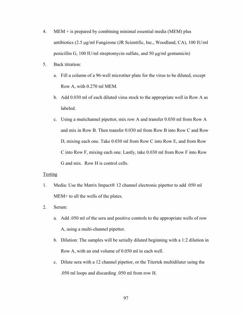
97
4. MEM + is prepared by combining minimal essential media (MEM) plus
antibiotics (2.5 µg/ml Fungizone (JR Scientific, Inc., Woodland, CA), 100 IU/ml
penicillin G, 100 IU/ml streptomycin sulfate, and 50 µg/ml gentamicin)
5. Back titration:
a. Fill a column of a 96-well microtiter plate for the virus to be diluted, except
Row A, with 0.270 ml MEM.
b. Add 0.030 ml of each diluted virus stock to the appropriate well in Row A as
labeled.
c. Using a mutichannel pipettor, mix row A and transfer 0.030 ml from Row A
and mix in Row B. Then transfer 0.030 ml from Row B into Row C and Row
D, mixing each one. Take 0.030 ml from Row C into Row E, and from Row
C into Row F, mixing each one. Lastly, take 0.030 ml from Row F into Row
G and mix. Row H is control cells.
Testing
1. Media: Use the Matrix Impact® 12 channel electronic pipettor to add .050 ml
MEM+ to all the wells of the plates.
2. Serum:
a. Add .050 ml of the sera and positive controls to the appropriate wells of row
A, using a multi-channel pipettor.
b. Dilution: The samples will be serially diluted beginning with a 1:2 dilution in
Row A, with an end volume of 0.050 ml in each well.
c. Dilute sera with a 12 channel pipettor, or the Titertek multidiluter using the
.050 ml loops and discarding .050 ml from row H.

98
3. Virus:
a. Dispense the back titration first, with a Costar pipettor, into the four identical
columns as marked.
b. Pour the working virus into a sterile reservoir and drop .050 ml of the
appropriate virus into each of the remaining wells of the plates with a Matrix
pipettor set on the fastest speed setting, taking care not to touch the tips to
any well.
c. Tap gently to mix. Incubate at room temperature for 1 hour in the hood.
Prepare bovine turbinate cells during this incubation.
4. For Bovine Turbinate (BT) cells:
a. Cells should be between 6 and 13 days old.
b. Make up the appropriate amount of MEM+ and set in ice water bath.
c. Pour the growth media off the flask of BT cells.
d. Wash with room temperature, sterile 1X CMF at least 3 times to rinse off all
the media.
e. Add 2 mls ATV (trypsin) per jumbo flask, tilt to cover the growth surface
completely.
f. Incubate at room temperature until the cells come off the bottom of the flask,
about 2-5 minutes. The flasks can be tapped vigorously during this time to
help shake the cells loose.
g. Add about 10 ml MEM+ per flask to triturate the cells and wash the sides of
the flask.

99
h. Remove all cells and MEM+ to the iced bottle and keep in an ice bath or
refrigerated so they don't attach to the flask or glass bottle.
5. After 1 hour incubation of the virus, drop .050 ml of BVDV free bovine
turbinate cells into each well of the plates using the Matrix pipettor. The Singer
strain of cytopathic BVDV is used as the challenge virus at 100-1000 TCID50.
6. Incubation in a 10% humidified, 5% CO2 incubator at 37°C.
7. Check plates at 72 hours for serum neutralization of virus or cytopathic effect.
Serum antibody titers are expressed as the reciprocal of the highest dilution of
serum providing 100 percent cell protection. Dilutions are 1:4 through 1:512.