Embed Size (px)
Citation preview
Page 1 of 31 Medical Coverage Policy: 0118
Medical Coverage Policy
Effective Date ............................................. 2/15/2020 Next Review Date ....................................... 2/15/2021 Coverage Policy Number .................................. 0118
Bone, Cartilage and Ligament Graft Substitutes
Table of Contents Overview .............................................................. 1 Coverage Policy ................................................... 1 General Background ............................................ 3 Coding/Billing Information .................................. 18 References ........................................................ 21
Related Coverage Resources Autologous Platelet-Derived Growth Factors (Platelet-
Rich Plasma [PRP]) Bone Growth Stimulators: Electrical (Invasive,
Noninvasive), Ultrasound Intervertebral Disc (IVD) Prostheses Lumbar Fusion for Spinal Instability and Degenerative
Disc Conditions, Including Sacroiliac Fusion Minimally Invasive Spine Surgery Procedures and
Trigger Point Injections Percutaneous Vertebroplasty, Kyphoplasty and
Sacroplasty Stem Cell Therapy for Orthopaedic Applications Tissue-Engineered Skin Substitutes
INSTRUCTIONS FOR USE The following Coverage Policy applies to health benefit plans administered by Cigna Companies. Certain Cigna Companies and/or lines of business only provide utilization review services to clients and do not make coverage determinations. References to standard benefit plan language and coverage determinations do not apply to those clients. Coverage Policies are intended to provide guidance in interpreting certain standard benefit plans administered by Cigna Companies. Please note, the terms of a customer’s particular benefit plan document [Group Service Agreement, Evidence of Coverage, Certificate of Coverage, Summary Plan Description (SPD) or similar plan document] may differ significantly from the standard benefit plans upon which these Coverage Policies are based. For example, a customer’s benefit plan document may contain a specific exclusion related to a topic addressed in a Coverage Policy. In the event of a conflict, a customer’s benefit plan document always supersedes the information in the Coverage Policies. In the absence of a controlling federal or state coverage mandate, benefits are ultimately determined by the terms of the applicable benefit plan document. Coverage determinations in each specific instance require consideration of 1) the terms of the applicable benefit plan document in effect on the date of service; 2) any applicable laws/regulations; 3) any relevant collateral source materials including Coverage Policies and; 4) the specific facts of the particular situation. Coverage Policies relate exclusively to the administration of health benefit plans. Coverage Policies are not recommendations for treatment and should never be used as treatment guidelines. In certain markets, delegated vendor guidelines may be used to support medical necessity and other coverage determinations.
Overview This Coverage Policy addresses bone graft substitute and other orthobiologic-type materials. The materials in scope of this Medical Coverage Policy include bone graft substitutes, bone void fillers, articular cartilage materials, recombinant morphogenetic proteins, and ligament and meniscus reconstruction materials.
Coverage Policy The effectiveness of some bone graft materials, including those that undergo advanced processing methods, has not been demonstrated as clinically superior to those which do not undergo advanced processing methods. Bone graft substitutes that undergo advanced processing methods are often more costly. Many health plans administered by Cigna contain definitions of medical necessity which include a cost comparison component. When medical necessity criteria have been met, coverage of a bone graft
Page 2 of 31 Medical Coverage Policy: 0118
substitute may be limited to the lowest cost, clinically equivalent alternative. Please refer to the applicable plan document to determine benefit availability and the terms, conditions and limitations of coverage. Dental implants are specifically excluded under many benefit plans. When coverage for dental implants is excluded, the use of bone graft materials in conjunction with a dental implant, including sinus and/or alveolar ridge augmentation, is similarly not covered. Bone Graft Materials/Substitutes The following bone graft materials and/or substitutes, used alone or in combination, are each considered medically necessary for enhancement of bone healing:
• autografts • allograft-based, including demineralized bone matrix (DBM) • ceramic or polymer-based synthetic bone graft substitutes • bone graft substitutes containing anorganic bone material (e.g., bovine, coral) when used alone or
combined with another covered bone graft substitute The following bone graft materials and/or substitutes are each considered experimental, investigational or unproven for the enhancement of bone healing:
• human amniotic membrane bone graft substitute materials, including amniotic fluid stem cell substitutes • cell-based substitutes (e.g., mesenchymal stem cells used alone, added to other biomaterials for
grafting, or seeded onto scaffolds) • human growth factor substitutes (e.g., fibroblast growth factor, insulin-like growth factor) • platelet rich plasma (e.g., autologous platelet derived growth factor) • bone marrow aspirate processed to concentrate growth factors (e.g., concentrated bone marrow
aspirate, centrifuged bone marrow aspirate), used alone or in combination with other bone graft materials (e.g., allograft)
• bone graft substitutes containing anorganic bone material (e.g., bovine, coral) when combined with any non-covered bone graft substitute
• allograft bone graft substitutes used exclusively as stand-alone stabilization devices for fusion (e.g., TruFuse® for isolated facet fusion, NuFix™ for isolated facet fusion, BacFast® HD for isolated facet fusion)
• bone graft substitutes used to reduce donor site morbidity (e.g., iliac crest donor site reconstruction) Recombinant Bone Morphogenetic Protein (rhBMP) rhBMP-2 (i.e., INFUSE® Bone Graft) is considered medically necessary when criteria is met for EITHER of the following conditions:
in combination with a fusion device for a single-level anterior interbody lumbar or lumbosacral
fusion* surgery when ALL of the following criteria have been met: degenerative disc disease at one level from L2–S1 no more than Grade I spondylolisthesis at the involved level
in surgical repair of an acute, open tibial shaft fractures when BOTH of the following criteria are met:
fracture is stabilized with intramedullary (IM) nail fixation rhBMP–2 is applied within 14 days of the fracture
*Note: For specific medical necessity criteria for lumbar fusion please reference the Cigna Lumbar Fusion for Spinal Instability and Degenerative Disc Conditions Coverage Policy.
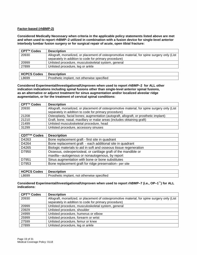
rhBMP–2 is considered experimental, investigational or unproven for ALL other indications, including the following:
Page 3 of 31 Medical Coverage Policy: 0118
• rhBMP–2 (i.e., INFUSE® Bone Graft) when used for spinal fusion procedures other than single-level anterior spinal fusion (e.g., posterior lumbar fusion, transforaminal lumbar fusion, more than single-level fusion)
• rhBMP–2 (i.e., INFUSE® Bone Graft) as an alternative or adjunct treatment for sinus augmentation and/or localized alveolar ridge augmentation
• rhBMP–2 (i.e., INFUSE® Bone Graft) for the treatment of cervical spine conditions rhBMP-7 (i.e., OP–1™ ) is considered experimental, investigational or unproven for ALL indications. Ligament/Meniscus Reconstruction The following are each considered experimental, investigational or unproven when used alone or as part of a ligament or meniscus reconstruction, regeneration, or transplantation:
• autologous platelet-derived growth factors (e.g., platelet rich plasma) • bioactive scaffolds (e.g., collagen meniscal implants) • bioresorbable porous polyurethane • meniscal prosthesis • tissue-engineered menisci • xenografts
Bone Filler / Articular Cartilage The following bone filler and/or articular cartilage materials are each considered experimental, investigational, or unproven:
• synthetic resorbable polymers (e.g., PolyGraft™ BGS, TruFit® [cylindrical plug], TruGraft™ [granules]) • juvenile cartilage allograft tissue implantation, including minced cartilage (e.g., DeNovo® NT Natural
Tissue Graft, DeNovo® ET™ Engineered Tissue Graft [ISTO Technologies, Inc., St. Louis, MO / Zimmer, Inc., Warsaw IN]; BioCartilage® [Arthrex, Naples, Florida])
• decellularized osteochondral allograft implant (e.g., Chondrofix® Osteochondral Allograft [Zimmer Biomet, Warsaw, IN])
General Background Bone grafts can be harvested from the patient (autograft), a cadaver (allograft), or they can be synthetic. The composition of allograft and synthetic bone graft substitutes and their mechanism of action can vary widely. Bone graft materials are often combined to extend graft availability and enhance healing. Used alone or in combination, bone graft substitutes may be utilized for many orthopaedic applications including spinal fusion, for enhancement of fracture healing, for filling cavities and defects, bridging joints, establishing the continuity of long bone and providing bone blocks. More recently, bone graft materials such as calcium phosphate and bioglass combined with antibiotics are being investigated as a method of delivering antimicrobials to the site of infection, such as for treatment of osteomyelitis, although additional studies are needed to support safety and efficacy. For most of the indications noted above, there is sufficient evidence to support safety and effectiveness, although for some indications clinical studies are limited, for others there is no evidence, and for some types of materials, clinical studies are not required. Many of the bone graft substitute products are regulated by the United States Food and Drug Administration (FDA). For example, nonstructural allograft and cellular allograft materials are considered human cells, tissues and cellular tissue-based products and as such do not require preclinical or clinical data by the FDA. Synthetic bone grafts and demineralized bone matrices (DBM) are considered Class II materials and fall under the FDA 510(k) regulatory process and upon approval are considered “substantially equivalent” to another marketed device/material used for the same purpose. Other materials, such as those that are considered drug-device combinations require premarket approval (PMA); ta PMA approval requires an investigational device exemption clinical trial prior to the PMA application (Abjornson, et al., 2018).
Page 4 of 31 Medical Coverage Policy: 0118
Bone Graft Materials Autografts Autografts are considered the established standard graft material and are typically retrieved from the patient’s tibia, fibula, ileum or iliac crest, by way of a surgical procedure and are then placed at the injury site. The advantage of autograft is the high probability of success—autograft possesses all of the necessary characteristics such as osteoconductivity, osteogenicity, and osteoinductivity. The disadvantages associated with autografts are that the amount of autogenous bone available for grafting is limited; autografts are associated with increased morbidity; increased anesthesia time and blood loss; and post-operative donor site complications. The iliac crest is the most common site for autograft harvesting. Once the actual bone graft is obtained the site is allowed to heal independently without backfilling. However, there is often post-operative harvest site pain associated with this procedure. The use of various bone graft substitutes are being investigated for backfilling of iliac crest harvest sites, particularly when used for spinal surgery, as a method of reducing pain and for improving cosmesis. Despite this proposed use there is insufficient data in the peer-reviewed published scientific literature supporting the effectiveness of iliac reconstruction with any type of graft material. Most of the published studies involve small sample populations with inconsistent clinical outcomes for reducing donor site morbidity. The use of bone graft substitutes for this indication is not recommended at this time due to lack of data supporting safety, efficacy and improved clinical outcomes. Autologous bone marrow aspirate obtained from the iliac crest is also commonly used during orthopedic procedures as an adjunct to other graft materials to enhance bone healing. Freshly harvested bone marrow aspirate contains osteogenic precursor cells (mesenchymal stem cells, growth factors) and once aspirated may be injected directly into defects or mixed with other grafting materials. In theory, combining bone marrow aspirate with an osteoconductive and osteoinductive bone graft material will avoid associated disadvantages of iliac crest graft harvest and improve healing. Although injecting aspirate directly into defects or mixing with other allograft materials is commonly performed, it is integral to the surgical procedure. Another proposed use of bone marrow is aspirate involves various cell retention and processing methods which are now being utilized to increase cell concentration. It has been suggested that stem cell concentration is directly related to overall effectiveness and as a result, in order to increase the concentration of osteoprogenitor cells various cell retention processing methods (e.g., centrifugation) may be employed. Although the amount of aspirate required and proposed indications vary, the process has also been referred to as bone marrow nucleated cell concentrate (BMAC) or autologous bone marrow mononuclear cells (BMMC). The techniques for concentrating bone marrow aspirate vary, as well as the resulting cell concentration and cell viability. Comparative data in the medical literature is insufficient to support clinical effectiveness of concentrated bone marrow aspirate and strong evidence based conclusions cannot be made at this time. Allografts One alternative to autograft is the use of allografts. Allograft offers the advantage of avoiding additional surgery and potential complications associated with harvesting host bone during the primary procedure. Allograft materials are frequently used during various orthopedic procedures, and may also be used alone or in combination with other materials. Allografts are readily available from bone banks and provide both osteoinductive and osteoconductive (e.g., structural support) properties, however they lack osteogenic properties. Allografts may give less consistent clinical results, and there is an increased risk of disease transmission and immunogenic response. When allografts are intensively processed to decrease these risks, the osteoinductive potential is lessened, and the processing removes osteogenic cells and reduces mechanical strength. AlloGro® Demineralized Bone Matrix (Wright Medical, Arlington, TN); Dynagraft-D™ (Citagenix, Laval, Quebec, Canada); Opteform® (Exactech, Inc., Gainesville, FL); Grafton® (Osteotech, Eatontown, NJ); OrthoBlast (IsoTis Orthobiologics, Irvine, CA); TruFuse® (minSURG™ Corp., Clearwater, FL) ; and NuFix™ (Nutech Medical, Birmingham, AL) are examples of allograft-based bone graft substitutes. More recently, processing methods used for preparation of some allografts have been refined and products are now available that manufacturers claim retain higher concentrations of naturally occurring growth factors and/or stem cells. Human growth factors such as fibroblast growth factor, insulin-like growth factor, platelet-derived growth factor, transforming growth factor-beta, and microglobulin-B, are examples of osteogenic growth factors that are naturally found within the matrix of bone. Despite availability and current use, clinical superiority has not
Page 5 of 31 Medical Coverage Policy: 0118
been demonstrated in the medical literature. How these allograft bone graft materials, processed to retain higher concentrations of inherent growth factors and/or stem cells, improve the rate and quality of bone formation compared to other available allograft bone graft substitutes, has not yet been firmly established. Demineralized bone matrix (DBM) is also a type of allograft. It is produced through a process that involves the decalcification of cortical bone; substantially decreasing the structural strength. However, it is more osteoinductive than ordinary allograft. Although the reason for this is not completely understood, it has been speculated that the osteoinductive growth factors contained in the extracellular bone matrix are more easily accessed once the mineral phase of the bone has been removed. Allograft DBM preparations available for use include Osteotech’s Grafton®, Regeneration Technology’s Osteofil® and Medtronic’s Magnifuse to name a few. These preparations differ in shape and size of DBM particles, the amount of inherent growth factors, the amount of residual minerals, and the type of carrier materials. Anorganic Bone Graft Materials Anorganic bone graft material is a type of xenograft bone graft substitute made from other than human material, such as cow (i.e., bovine) or coral, and is typically used in combination with other types of bone graft materials, for example with collagen or a calcified matrix. The animal bone is processed to remove any organic components (i.e., anorganic bone material) reducing concerns of disease transmission or immunogenic reactions. Some of the anorganic type xenograft materials (e.g., Bio-Oss) may be used as stand-alone graft material to enhance healing, such as when used for dental implants. When used according to U.S. Food and Drug Administration (FDA) approved indications, either alone or combined with other bone graft materials proven effective, anorganic bone graft materials are considered safe and effective for promoting bone formation. Bone Graft Substitutes Due to the limitations of autogenous bone and allograft material, and the number of surgeries that require grafting, investigators have developed grafting alternatives, some of which are available for current use and others which are still in developmental stages. Bone graft substitutes have overlapping properties and are often made of a variety of materials such as polymers (degradable and nondegradable), ceramics and composites (calcium phosphate, calcium sulfate, and bioactive glass), factor-based materials (recombinant growth factors) and cell-based materials (mesenchymal stem cells). Some authors classify bone graft substitutes according to these materials. However, these substitutes can also be classified based on their characteristics, such as osteoconduction (e.g., calcium sulfate, ceramics, calcium phosphate, cements, collagen), osteoinduction (e.g., DMB, rhBMPs, growth factors), osteogenesis (e.g., bone marrow aspirate), or combined (composites). Nonetheless, the ideal bone graft substitute must provide scaffolding for osteoconduction, growth factors for osteoinduction and progenitor cells for osteogenesis. In addition, the bone graft substitute must be able to integrate with the host. The role of bone graft substitutes is to provide a medium for osteoconduction rather than osteoinduction and can provide variable levels of structural support. These materials appear to be safe when used according to FDA indications; however each type of product is under varying degrees of regulation and in some cases safety and efficacy of these products remain unproven through human trials. For the intent of this coverage policy, bone graft substitutes are described as those that are cell-based, ceramic-based, polymer-based and factor-based. Synthetic substitutes generally consist of ceramic and polymer based materials. Cell-based: Bone graft substitutes that are cell-based use cells to generate new tissue either alone, with other biomaterials, or seeded onto a support matrix (e.g., in combination with allograft material). Support matrix materials may include xenograft (i.e., bovine) or human type I collagen. Examples of cell-based substitutes include but are not limited to:
• Mesenchymal stem cells (MSCs): MSCs are multipotent stem cells that express a variety of different cell surface proteins and can differentiate into a variety of cell types, such as osteoblasts, chondrocytes, myocytes, adipocytes, and neuronal cells. Although processing techniques vary, and the optimal number of MSCs to be transplanted/seeded has not been established, following autologous bone marrow collection MSCs are either concentrated for direct injection, or cultured and incubated. Once cultured the MSCs can be mixed with biomaterials, such as gels or pastes; the biomaterials hold the cells in suspension and provide a matrix for filling defects. MSCs can also be seeded on scaffolds, and have been investigated when used with a support matrix for implantation (e.g., tissue engineered). In theory,
Page 6 of 31 Medical Coverage Policy: 0118
MSCs are responsive to osteogenic growth factors and aid in the healing of bone. Nevertheless evidence in the published peer-reviewed scientific literature evaluating the use of MSCs to enhance bone healing consists mainly of animal trials and a paucity of human trials. At present the evidence is insufficient to support improved clinical outcomes, when used alone, added to other biomaterials, or as cultured/seeded on a support matrix.
• Trinity® Evolution™ (Orthofix®, Inc., Lewisville, TX) is a viable cell allograft that according to the manufacturer contains active adult mesenchymal stem cells. Trinity Evolution contains osteoprogenitor cells, mesenchymal stem cells, and demineralized cortical bone to promote osteoconduction, osteogenisis, and osteoinduction for successful bone grafting.
• Osteocel® Plus (NuVasive, Inc, San Diego, CA) is an allograft cellular matrix that contains viable stem cells. The manufacturer suggests this material promotes fusion in cervical, thoracic, and lumbar procedures. According to the manufacturer, Osteocel Plus mimics the biologic properties of autograft and contains mesenchymal cells, cancellous bone, and demineralized bone matrix. Osteocel Plus also provides a scaffold for new bone to grow on, theoretically avoiding limitations of other more traditional bone graft alternatives.
• Amniotic membrane, the innermost layer of the fetal membrane, is considered a source of collagen that acts as a scaffold for the attachment of cells. Recently, amniotic membrane allografts have been investigated for various uses including use as bone void fillers during spinal and other orthopedic surgeries to enhance bone healing. Classifications of these products differ depending on the specific product but are generally classified as human tissue and/or cell-based products. Some are claimed to be non-immunogenic by the manufacturer. Amniotic membrane allografts currently under investigation include NuCel® (Nutech Medical, Birmingham, AL, USA), Ovation® cellular repair matrix (Osiris Therapeutics, Inc., Columbia, MD, USA) derived from placental mesenchyme to name a few. In addition, amniotic fluid stem cells, derived from amniotic fluid, are being investigated to form tissue-engineered bone from scaffolds in animal trials; human trials are ongoing and limited.
The use of mesenchymal and other cell-based bone graft substitutes has been and continues to be investigated for various procedures, including spinal fusion, intervertebral disc regeneration and other orthopedic procedures. Although currently under investigation, data published in the medical literature evaluating cell-based substitutes is in preliminary stages and mainly in the form of nonhuman trials or case reports; data supporting safety and efficacy for these indications are lacking. Ceramic-based: Ceramic-based bone graft substitutes include materials such as calcium phosphate, calcium sulfate and bioactive glass, used alone or in combination with other grafts. Some ceramic-based products (e.g., calcium phosphate-collagen composites, beta-tricalcium phosphate) are combined with collagen to augment healing; collagen composites may include bovine material similar to that used with cell-based products. Several types of calcium phosphates, including tricalcium phosphate, synthetic hydroxyapatite, and coralline hydroxyapatite are available in pastes, putties, solid matrices, and granules. When used, calcium sulfate is less desirable for weight bearing applications due to loss of mechanical properties during degradation. When implanted into living tissue, bioactive glass forms a bond with pre-existing bone, however there are only a few products commercially available and use is primarily in dental applications. Synthetic hydroxyapatite (e.g., ProOsteon® Implant 500 [Interpore Cross, Int., Irvine, CA]) is brittle, has little tensile strength and is typically used for bone defects with internal fixation. A pure beta-tricalcium phosphate scaffold, Vitoss® Synthetic Cancellous Bone Filler (Orthovita, Inc., Malvern, PA) is intended for use in small defects in the extremities, pelvis, and spine. Other ceramic-based materials include but are not limited to:
• Osteograf® (Ceramed, Lakewood, CO) • Norian SRS (Skeletal Repair System) (Synthes, Inc., West Chester, PA) • Osteoset® (Wright Medical, Arlington, TN) • Actifuse™ (ApaTech Limited, Elstree, Hertfordshire, UK) • Integra MOZAIK™ Osteoconductive Scaffold (Integra LifeSciences, Plainsboro, NJ) • PRO-DENSE® Bone Graft Substitute paste(Wright Medical Technology, Inc., Arlington, TN)
Subchondral injection of calcium phosphate bone substitute, into the area of subchondral bone edema, as part of treatment for osteochondritis dissecans of the knee, has been reported in the literature (Cohen, Sharkey, 2016; Abrams, et al., 2013). Conservative treatment of osteoarthritis-related bone marrow lesions generally includes
Page 7 of 31 Medical Coverage Policy: 0118
pain control, reduction in weight bearing, activity modification, and appropriate nutrition including additional calcium and vitamin D during treatment if appropriate. One procedure aimed at treating such defects, the Subchondroplasty® (SCP®) procedure (Zimmer Holdings, Inc.; Warsaw, IN), is a minimally invasive surgery designed to access and treat bone defects associated with chronic bone marrow lesions by filling them with a biomimetic bone substitute material. Evidence in the peer reviewed scientific literature evaluating injection of a calcium phosphate bone substitute into the area of subchondral bone edema, or of the Subchondroplasty® procedure, in the treatment of chronic bone marrow lesions / bone marrow edema is lacking. As a result, evidence based conclusions regarding safety, efficacy, and impact on health outcomes cannot be firmly established. Polymer-based: Polymer-based substitutes are polymers that are either degradable or nondegradable and may be used alone or in combination with other materials. Degradable polymers are resorbed by the body allowing it to heal itself without foreign bodies remaining. Types of polymer-based substitutes include but are not limited to:
• Cortoss® (Orthovita, Inc., Malvern, PA) • OPLA (TMH Biomedical, Inc., Duluth, MN) • Immix (Osteobiologics, Smith and Nephew, Memphis, TN).
Factor-based: Factor-based bone graft substitutes consist of human growth factors and recombinant growth factors used alone or in combination with other materials. Factor-based osteogenic bone graft substitutes include but are not limited to:
• human growth factors (e.g., fibroblast growth factor, insulin-like growth factor, transforming growth factor-beta), used alone or in combination with other materials
• recombinant bone morphogenetic proteins (rhBMP), used as an adjunct to autografts Human Growth Factors Fibroblast growth factor (FGF), insulin-like growth factor (ILGF), transforming growth factor-beta (TGF-beta) and bone morphogenetic protein (BMP) are human growth factors found in the matrix of bone. Some of these factors have been isolated in research settings for use alone or in combination with other materials; however evidence in the published, peer-reviewed scientific literature is insufficient to support safety and efficacy at this time. Recombinant Bone Morphogenetic Proteins (rhBMP) RhBMP is a unique subgroup of graft substitutes and many published trials support safety and efficacy. The function of BMP is to promote differentiation of mesenchymal cells into chondrocytes and osteoblasts, to promote differentiation of osteoprogenitors into osteoblasts, and to influence skeletal pattern formation. Recombinant human bone morphogenetic proteins act as an adjunct to autogenous bone grafts, and are used commonly with spinal instrumentation devices (i.e., cages) during lumbar fusion and for fracture repair. According to the FDA, (FDA, 2008) safety and effectiveness of rhBMP for the treatment of cervical spine conditions has not been demonstrated. RhBMP–2 appear to be safe when used appropriately, placed accurately, not allowed to come into contact with decompressed areas (i.e., rhBMP carrier must be protected from compression to avoid the forcing out of implant into surrounding tissues) and contained in the region of surgical fusion. BMPs must be used with caution in the presence of defects in the dura. In spinal fusion surgery, BMP cannot resist compression or shear forces within a vertebral motion segment; thus, they cannot be used as stand-alone devices. RhBMP–2 must be used with a cage or some type of supportive structure within the vertebral interspace. The benefits of rhBMP versus autogenous iliac crest bone graft (AICBG) are the decreases in operating room time, blood loss, and morbidity due to the avoidance of an additional procedure to harvest AICBG. None of the early studies utilizing rhBMP–2 or rhBMP–7 documented any adverse systemic effects occurring as a result of their use. A small percentage (≤10%) of patients develops antibodies to rhBMPs, although there has been no documented evidence of harm resulting from this. Carreon et al. (2008) studied wound related and anaphylactic related adverse events in a case series of patients (n=90) who were re-exposed to rhBMP-2 and reported that multiple exposures from either a secondary primary surgery or revision (through the same approach or a different approach) does not increase the risk of those adverse events.
Page 8 of 31 Medical Coverage Policy: 0118
Although early evidence supports safety and efficacy when used according to FDA indications, adverse events have been reported which include ectopic bone formation, bone resorption or remodeling at the graft site, hematoma, neck swelling, and painful seroma (Benglis, et al., 2008). Dural tears, bowel/bladder and sexual dysfunction, failure to fuse and paralysis have also been reported (Epstein, 2011), as well as carcinogenicity and teratogenic effects. Recently there has been concern more specifically safety and efficacy of rhBMP–2 used in spinal fusion surgeries. According to Carragee, et al. (2011), who in a systematic review compared conclusions regarding safety and efficacy published in the original rhBMP–2 industry-sponsored trials when used for spinal fusion, to data published following the FDA approval, the risk of adverse events associated with rhBMP–2 for spinal fusion was found to be “10 to 50 times the original estimates calculated from the industry-sponsored peer-reviewed publications.” As a result of reported complications and the recent concern regarding safety and efficacy, use of RhBMP–2 product should be limited to the FDA-approved labeling indications. Evidence in the peer-reviewed scientific literature is insufficient and does not provide strong support to safety and efficacy of rhBMP–7 to enhance bone healing. RhBMP–2/ INFUSE® Bone Graft RhBMP–2 is marketed in the U.S. as INFUSE® Bone Graft in conjunction with specific spinal and non-specific tibial fusion devices, as an alternative to autogenous bone graft for sinus augmentation, and for localized alveolar ridge augmentation for defects associated with extraction sockets. Anterior Lumbar Spinal Fusion: The FDA gave new device approval to the InFuse™ Bone Graft/LT-CAGE™ (Medtronic Sofamor Danek, Memphis, TN) in July 2002. The InFuse Bone Graft /LT Cage is intended to be implanted via an anterior open or an anterior laparoscopic approach. According to the FDA approval, safety and effectiveness for approaches other than anterior have not been established. In December 2003 the approval was broadened to include additional fusion cages, specifically the INTER FIX™ Threaded Fusion Device and the INTER FIX™ RP Threaded Fusion Device (FDA, 2004). The device is to be used by surgeons experienced in spinal fusion surgery and adequately trained in the use of the device. The use of these devices in conjunction with surgical spinal fusion has been approved by the FDA, with numerous supplements to the original PMA, for patients who meet all of the following criteria:
• skeletally mature • degenerative disc disease at one level from L2–S1 • no more than Grade I spondylolisthesis at the involved level • failure of at least six months of nonoperative therapy
The device is contraindicated in patients with the following conditions:
• hypersensitivity to rhBMP–2, bovine Type I collagen or to other components of the formulation • resected or extant tumor at the operative site • active infection at the operative site • allergy to titanium or titanium alloy • possible or confirmed pregnancy
Evidence in the published peer-reviewed scientific literature consists of case series, randomized control trials, and literature reviews, many of which demonstrate safety and efficacy for the use of rhBMP–2 during spinal fusion (Burkus, et al., 2005; Glassman, et al., 2005; Baskin, et al., 2003; Burkus, et al., 2002;Kleeman, et al., 2002). In general, sample populations vary in size from 11 subjects to as many as 279 subjects and follow-up periods range from six to 24 months. Most of the patients have single-level lumbar disc disease. The approach to lumbar fusion and the use of instrumentation varies among clinical trials. Most of the control groups had standard lumbar fusion using autograft bone from the iliac crest. The results of early clinical trials (Boden, et al., 2000; Boden, et al, 2002; Burkus, et al., 2005) demonstrate that patients who received rhBMP–2 during spinal fusion surgery had shorter operating room times, shorter length of stay, and less blood loss compared to patients who received autogenous bone graft. Several studies published since that time has demonstrated similar results.
Page 9 of 31 Medical Coverage Policy: 0118
Posterior Spinal Fusion: RhBMP-2 has been used as part of posterior fusion techniques for treating both single and multi-level spinal deformity, either alone or combined with other osteoconductive scaffolds, and some authors have reported improved clinical outcomes (e.g., earlier and higher fusion rates) compared to iliac crest bone graft (Dawson, et al., 2009; Dimar, et al., 2009; Mulconrey, et al., 2008; Luhmann, et al., 2005). However, this represents an off-label use as it is not FDA-approved for posterolateral, posterior lumbar interbody fusion or multilevel fusion. Cervical Spinal Fusion: Although it is an off-label use, rhBMP–2 has been evaluated for use in cervical spinal fusion. In 2006, Smucker et al. conducted a retrospective case-control study comparing the incidence of perioperative cervical swelling complications after anterior cervical fusion with (=69) or without the use of rhBMP–2 (n=165). The authors found that rhBMP–2 was significantly associated with increased cervical swelling and reported that 27.5% (19 of 69) of the rhBMP–2 group had a clinically significant swelling event compared to 3.6% in the non-rhBMP–2 group. The results were statistically significant (p<0.001). Boakye et al. (2005) retrospectively reviewed outcomes in 24 consecutive patients who had undergone anterior cervical discectomy and fusion of one to three levels using polyetheretherketone (PEEK) spacers filled with rhBMP–2. Fusion was documented by radiograph in all cases, and clinical outcomes were rated as good to excellent in 95% of cases. Complications included transient recurrent laryngeal nerve injury in one case, transient C–5 paresis in one, cerebrospinal fluid leakage in one, and transient dysphagia in two. Further clinical trials are required to support safety and efficacy for treatment for cervical spine conditions. Fracture Repair: RhBMP–2 has also been studied in patients with tibial fractures; this application received premarket approval from the FDA in April 2004. It is marketed as the INFUSE® Bone Graft device (Wyeth Pharmaceuticals, Inc., Philadelphia, PA) and consists of rhBMP–2 in an absorbable collagen sponge. The FDA approval for this device is for the treatment of patients with acute, open tibial shaft fractures when all of the following criteria are met:
• The fracture must be stabilized with intramedullary (IM) nail fixation after appropriate wound management.
• The rhBMP–2 must be applied within 14 days after the initial fracture. • The prospective patient should be skeletally mature.
The FDA notes the following contraindications to use of the product:
• possible or confirmed pregnancy • sensitivity to titanium, titanium alloy, cow (bovine) Type I collagen, or rhBMP–2 • infection near the area of the surgical incision • previous or current tumor at the site of use • high risk of amputation of the affected leg • compartment syndrome of the affected leg
Published clinical studies evaluating the use of rhBMP–2 in patients with tibial fractures support safety and efficacy (Swiontkowski, et al., 2006; Jones, et al., 2006; Govender, yet al., 2002). Sinus Augmentation/Alveolar Ridge Augmentation: In March 2007 the INFUSE® Bone Graft (Medtronic Sofamor Danek, Memphis, TN) was granted premarket approval from the FDA for use in oral surgical procedures, (i.e., sinus augmentation and localized alveolar ridge augmentation for defects associated with extraction sockets), as an alternative to autograft. According to the FDA, INFUSE Bone Graft is used to fill space where bone is needed in order to place endosseous dental implants. Dental implants should be placed if there is sufficient bone to stabilize them. When the sinus wall is thin, there is not enough bone to place dental implants. In a procedure known as sinus augmentation, a sinus graft is inserted into the floor of the sinus (i.e., the roof of the upper jaw). Dental implants can then be inserted and stabilized in the new sinus bone. The alveolar ridge of the jaw is the bone that surrounds the roots of the teeth. When a tooth is extracted, a socket remains which later
Page 10 of 31 Medical Coverage Policy: 0118
heals; however, typically, previous height and width are not restored. Alveolar ridge augmentation is a procedure performed to increase bone volume, making treatment with dental implants possible. The FDA notes the following contraindications to use for oral surgical procedures:
• in patients with an active infection at the operative site • in patients who are pregnant • in patients who are hypersensitive to rhBMP–2 or bovine type I collagen • in an area where there was a tumor
Evidence in the published scientific literature evaluating rhBMP–2 for oral maxillofacial surgery consists of few published clinical trials (Esposito, et al., 2007; Boyne, et al., 2005; Fiorellini, et al., 2005; Jung, et l., 2003). Although the study results suggest that this technique may be a promising treatment option, the evidence in the published, peer-reviewed, scientific literature is insufficient to allow strong conclusions regarding the long-term effectiveness of rhBMP–2 for sinus augmentation and alveolar ridge augmentation. Published studies have been small in sample size, and data on long-term outcomes are lacking. Patient selection criteria are not well-defined. Some studies have indicated that rhBMP–2 is safe and enhances bone maturation. However, additional well-designed clinical trials assessing long-term health outcomes are needed to validate these results. RhBMP–7/ OP–1™ Putty A second type of human bone morphogenetic protein is rhBMP–7, marketed in the United States as OP–1™ Implant for use in healing fractures of the long bones, and OP–1™ Putty for use in spinal fusion. The FDA approved the OP–1 Implant and the OP–1 Putty for use in specifically-defined patients under a humanitarian device exemption (HDE). Posterolateral Lumbar Spinal Fusion: The FDA granted HDE approval in April 2004 for the use of OP–1™ Putty (Stryker Biotech, Hopkinton, MA) for use as an alternative to autograft in compromised patients requiring revision posterolateral spinal fusion of the lower back. OP–1 Putty is made from a manufactured human protein powder and bovine collagen that is mixed with a saline solution and a thickening agent to form a putty-like material. During surgery, the putty is placed on each side of the spinal levels to be fused. The FDA approval specifies patient selection criteria as those who meet both of the following:
• failed previous spinal fusion surgery • not candidates for autograft because of a condition such as osteoporosis, diabetes, or smoking
The use of the product is contraindicated in patients with the following conditions:
• allergy to OP–1 or collagen • existing tumor, tumor removed at or near the fracture, or history of malignancy • previous history of cancer • skeletal immaturity • pregnancy
The data presented to the FDA for consideration of approval is contained in the FDA Summary of Safety and Probable Benefit (FDA, 2004). Forty-eight patients with single-level degenerative lumbar spondylolisthesis and spinal stenosis received rhBMP–7 alone, rhBMP–7 combined with autograft, or autograft alone. The patients receiving rhBMP–7 alone demonstrated superior success, as evidenced by radiograph and clinical improvement, over those receiving autograft alone. The FDA decision is based on the clinical data, as well as the following rationale provided in the approval summary: “When revision of a failed fusion is required, most patients are limited to either living with pain and altered function or repeating the original procedure with additional autologous bone, which may result in depletion of the bone stock and further risk to the patient. Allograft bone and bone graft substitutes are not considered feasible alternatives to autograft in revision surgery due to their lack of osteogenic potential. For certain patients, e.g., those with implanted leads, bone growth stimulators would not be considered as feasible
Page 11 of 31 Medical Coverage Policy: 0118
options. OP–1 Putty has the potential to eliminate the risk and complications associated with these treatment alternatives while providing a feasible and beneficial alternative treatment.” Some of the outcomes reported for osteoinductive properties and fusion rates are comparable to those of autograft, and the use of rhBMP–7 does avoid the need for autogenous bone graft harvest. Overall, a paucity of studies have assessed rhBMP–7 in spine fusion surgery (Delawi, et al., 2010; Vaccaro, et al., 2008; Kanayama, et al., 2006; Vaccaro, et al., 2004; Vaccaro, et al., 2003; Johnsson, et al., 2002). Limitations to these available studies such as use of combined materials, small sample size and short-term follow-up does not lead to formation of evidence-based conclusions regarding safety, efficacy and improved clinical outcomes. Additionally these trials evaluated primary fusion surgery and not revision surgery as mentioned in the HDE approval, and are therefore not in agreement with FDA limitations. Studies evaluating safety and efficacy of on-label use in the peer-reviewed published scientific literature are lacking. Other variables precluding generalizations include inconsistency with regard to instrumentation; some procedures were instrumented while others were not. Additional studies are needed to assess clinical outcomes. Fracture Repair: The FDA gave HDE approval for the use of rhBMP–7 to treat nonunion of long bones. It is a powder that is mixed with normal saline to form a paste which is applied during surgery. The substance is marketed in the U.S. as OP–1™ Implant (Stryker Biotech, Hopkinton, MA). The FDA approval indicates that the substance is appropriate for use in the surgical repair of long bone nonunion when both of the following patient selection criteria are met:
• autograft is not feasible • alternative treatments have failed
The use of the product is contraindicated in patients with the following conditions:
• allergy to OP–1 or collagen • existing tumor or tumor removed at or near the fracture or history of malignancy • previous history of cancer • skeletal immaturity • pregnancy
Similar to posterolateral lumbar fusion surgery, studies evaluating the use of rhBMP–7 for nonunion of long bones are limited by small sample size and short term follow-up. Although there is some evidence of successful clinical outcomes resulting from the use of rhBMP-7 for the treatment of nonunion in the published scientific literature (Ronga, et al., 2006; Maniscalco, et al., 2002; Friedlaender, et al., 2001; Geesink, et al., 1999) evidence is insufficient to draw strong conclusions regarding safety and efficacy. Platelet Rich Plasma (PRP) for Bone Healing Platelet rich plasma (plasma having a platelet concentration above baseline) is an approach being investigated for the treatment of bone healing. PRP is also referred to as autologous platelet derived growth factor, platelet enriched plasma, platelet-rich concentrate, and autogenous platelet gel or platelet releasate. When activated in the body, platelets release growth factors which accelerate healing, including platelet-derived growth factor, transforming growth factor beta (TGF-β) and insulin-like growth factor to name a few. It is hypothesized that a concentrated preparation of platelets, which contain higher concentrations of growth factors, may promote more rapid healing. Platelet concentrates are not osteoinductive since they do not include BMPs (Marx, 2004), although in theory they promote osteoblast proliferation and differentiation (Veillette, McKee, 2007). During the procedure, a small amount of the patient’s blood is drawn and centrifuged to separate red blood cells from the platelet rich plasma. The platelet rich plasma is then mixed with the patient’s bone graft material. Theoretically, the growth factors signal the local mesenchymal and epithelial cells to migrate, divide, and increase collagen and matrix synthesis increasing bone regeneration. Overexposure of cells to PRP yields many cells but limited differentiation of those cells into appropriate cell lines. It has been suggested that the inability to control differentiation is a reason to not use PRP for healing of tissue (Mehta, Watson, 2008).
Page 12 of 31 Medical Coverage Policy: 0118
Platelet concentration devices are approved by the FDA as part of the 510(k) approval process for preparation of platelet rich plasma. Examples of devices that are FDA approved include PCCS(TM) Platelet Concentrate Separation Kit (3i [Implant Innovations Inc.] and Magellan(TM) Autologous Platelet Separator System (Medtronic Perfusion Systems). Evidence in the published scientific literature is inconsistent and does not lend strong support to the clinical utility of using PRP to augment bone grafting. There are few randomized controlled trials with comparison groups available to support routine use of PRP. Some authors have reported PRP successfully promotes graft incorporation in oral and maxillofacial surgery (Marx, 2004; Kassoulis, Reynolds, 2005; Camargo, et al., 2005) and there is a growing body of evidence evaluating the safety and efficacy of PRP as a treatment for knee osteoarthritis and other degenerative conditions (Lai, et al., 2015; Campbell, et al., 2015; Chang, et al., 2014; Koshbin, et al, 2013; Kon, et al., 2010; Filardo, et al., 2010; Vidriero, et al., 2010; Sanchez, et al., 2009). However, the outcomes of these reports are variable and the utility of PRP for use in bone repair is debatable. For other applications, such as spinal fusion the studies are encouraging but limited in quantity and quality, therefore no conclusions can be drawn. Additionally, there is a lack of controlled clinical trials to support safety and efficacy of platelet rich plasma for the treatment of long bone nonunion or delayed union. Lenza et al. (2013) conducted a systematic review to evaluate the effectiveness of PRP as an adjunctive therapy for the union of long bones and concluded there was no significant difference with the use of PRP. In a prospective case series published by Mariconda and colleagues (2008) the authors failed to demonstrate the clinical utility of platelet gel supplementation in the treatment of long bone nonunions. Calorie et al. (2008) conducted a prospective randomized clinical study comparing efficacy of rhBMP–7 and PRP in the treatment of fracture nonunions and reported that rhBMP–7 resulted in superior clinical and radiological efficacy when compared to PRP. In a review published by Viellette and McKee (2008) the authors noted that there was a lack of evidence evaluating the efficacy of PRP in management of acute fractures, to assess the effect of PRP on new bone formation for long bone defects and nonunions, and that there is no clinical evidence to support the use of PRP to augment spinal fusion. In 2009 Miyazaki et al. published a systematic review regarding bone graft substitutes for spinal fusion and reported that the evidence for autologous platelet concentrates is weak and the clarity of risk/benefit is unclear. In 2016 the Washington State Health Care Authority (WSHCA) conducted a technology assessment to evaluate the safety and efficacy of the use of PRP and/or autologous blood injection (ABI), for the treatment of various musculoskeletal and orthopedic conditions, including but not limited to tendinopathies, rotator tendinosis, rotator tears, as well as plantar fasciitis, acute injuries and OA (e.g., osteochondral lesions of the talus, OA of the hip, knee). As part of the technology assessment a total of 54 randomized controlled trials and eight cohort studies were included and reviewed. Limitations of the studies noted by the Committee generally included small sample populations, short-term follow-up, inconsistency of measured outcomes, potential for risk bias, and lack of high quality evidence. The authors concluded there was insufficient evidence to draw strong conclusions regarding safety and efficacy. Moreover, the Committee reported despite its current use, standardization of PRP preparation is lacking, and although the technology to obtain PRP is FDA-approved, PRP is currently not indicated for direct injection (WSHCA, 2016). A formal position statement supporting platelet rich plasma for enhancement of bone healing from the American Academy of Orthopaedics (AAOS) could not be found. The AAOS does provide general information regarding PRP and suggests that further studies are needed to define the role of PRP and to determine for which settings PRP may be valuable. Furthermore, within the AAOS evidence-based guidelines on osteoarthritis of the knee (2013), the AAOS states they are unable to recommend for or against growth factor injections and/or platelet-rich plasma for the treatment of symptomatic osteoarthritis of the knee; there is a lack of compelling evidence and the balance of clinical benefit and harm has not been established. Currently, whether or not platelet rich plasma facilitates osteoinduction and improves clinical outcomes remains unproven in the published scientific literature. (Please refer to CIGNA Coverage Policy: Autologous Platelet-Derived Growth Factors [Platelet Rich Plasma] for further detail regarding autologous platelet derived growth factor). U.S Food and Drug Administration (FDA)
Page 13 of 31 Medical Coverage Policy: 0118
The FDA classifies most orthobiologicals (e.g., rhBMP–2, Osteoset, Grafton) as Class II devices. Many of the bone graft substitutes are approved through the 510(k) process and are based on a predicate device clearance although some require premarket approval (i.e., Class III devices). In addition, some of the bone graft materials are regulated as human tissue and do not require clinical trials or FDA approval for marketing, such as some of the DBM products and cell-based materials. Humanitarian Device Exemption (HDE) was granted by the FDA for the OP-1 Implant and the OP-1 Putty, also referred to as rhBMP–7, for use in specifically defined patients (FDA, H010002, H020008). According to the FDA, a humanitarian use device (HUD) is a device that is intended to benefit patients by treating or diagnosing a disease or condition that affects fewer than 4,000 individuals in the United States per year. An HDE application is not required to contain the results of scientifically valid clinical investigations demonstrating that the device is effective for its intended purpose. Ligament/Meniscus Reconstruction The use of adjunctive treatments such as autologous platelet-derived growth factors (e.g., centrifuged platelet aggregates) have been utilized to assist in healing of tissues, however, there is insufficient evidence in the medical literature at this time, in particular with ACL/PCL reconstruction using allograft tissue or meniscal transplant, to support any improvement in health outcomes with the use of these adjunctive treatments. Other options under investigation for meniscal regeneration and/or transplantation include tissue-engineered menisci, bioactive scaffolds (collagen meniscal implants, bioresorbable porous polyurethane), and synthetic devices (e.g., hydrogel) (Packer and Rodeo, 2009). Collagen meniscal implants have been proposed by some authors for filling defects of partial meniscectomy with functional repair tissue. Authors hypothesize the collagen meniscal implant may help prevent or delay the progression of osteoarthritis, protecting from degenerative joint disease. In addition, xenografts and meniscal prostheses are under investigation for use as an alternative approach to meniscal allograft transplantation (Verdonk, et al., 2007). Autologous Platelet-derived Growth Factors (e.g., platelet rich plasma [PRP]): Platelet-rich plasma, derived from autologous blood, is being investigated for cartilage regeneration and for anterior ligament reconstruction. Theoretically, delivering high concentrations of growth factors, found in the platelet rich plasma, to focal areas may enhance healing, improve mechanical properties during the remodeling phase, and improve the rate of graft incorporation and maturation. One purported use involves autologous platelet-derived growth factors (e.g., centrifuged platelet aggregates, platelet rich plasma) with tendon and ligament repair. Nevertheless, the applicability for this use is controversial. U.S. Food and Drug Administration (FDA): The systems used for preparing autologous platelet-derived growth factors are FDA approved under the 510(k) notification process. In general, the systems are approved to be used at the patient’s point of care and/or in a clinical laboratory to prepare autologous platelet-rich plasma (PRP)/platelet concentrate from the patient’s own blood. Literature Review: The use of platelet rich plasma to enhance soft tissue healing such as with tendon and ligament repair continues to be investigated, however controlled clinical trials that support clinical improvement are lacking (Hall, et al., 2013). Regarding anterior cruciate ligament reconstruction in particular, Nin et al.(2009) reported the results of a randomized trial (n=100) evaluating the effect of platelet-enriched gel on the inflammatory process following primary ACL reconstruction with BPTB allograft ay two year follow-up. The authors acknowledged the therapeutic role of platelet derived growth factor currently remains unclear. The results of a more recent randomized controlled clinical trial (n=150) comparing two different platelet rich plasma preparations to a non-gel control group as part of ACL reconstruction continues to support further clinical trials are needed. The results of this study demonstrate there were no statistically significant differences in functional outcome, graft healing on MRI, or complications; the authors reported there was no clinical or pain improvement compared with the control group (Valenti Azcarate, et al., 2014). In addition, multiple formulations with varying content of platelet rich plasma, platelets and white cells are available, resulting in inconsistent clinical outcomes among studies. There is insufficient evidence in the medical literature at this time, in particular with ACL/PCL reconstruction to support any improvement in health outcomes with the use of these adjunctive platelet-derived orthobiologic materials.
Page 14 of 31 Medical Coverage Policy: 0118
In 2013 the American Academy of Orthopaedic Surgeons published recommendations for treatment of osteoarthritis of the knee. Within these guidelines the Academy notes that they are unable to recommend for or against growth factor injections and/or platelet rich plasma for patients with symptomatic OA of the knee. The inconclusive recommendation is based on lack of compelling evidence that has resulted in an unclear balance between benefits and potential harm (AAOS, 2013). NICE issued guidance (2014) for platelet rich plasma injections as treatment of osteoarthritis of the knees and concluded that the procedure should only be used with special arrangements for clinical governance, consent, and audit or research. Meniscus Regeneration/Transplantation: The meniscus is a crescent-shaped wedge of fibrocartilage located in the knee joint between the femoral condyle and tibial plateau. Small meniscal tears can be sutured, however, management of more severe meniscal injury involves arthroscopic or open surgery, often with meniscal allograft transplant. Other options under investigation for meniscal regeneration and/or transplantation include tissue-engineered menisci, bioactive scaffolds (collagen meniscal implants, bioresorbable porous polyurethane), and synthetic devices (e.g., hydrogel) (Packer and Rodeo, 2009). Collagen meniscal implants have been proposed by some authors for filling defects of partial meniscectomy with functional repair tissue. Authors hypothesize the collagen meniscal implant may help prevent or delay the progression of osteoarthritis, protecting from degenerative joint disease. In addition, xenografts and meniscal prostheses are under investigation for use as an alternative approach to meniscal allograft transplantation (Verdonk, et al., 2007). U.S. Food and Drug Administration (FDA): Menaflex™ (ReGen Biologics, Inc., Hackensack, NJ), was granted a 510(k) approval from the FDA in December 2008. Menaflex is a resorbable collagen matrix regulated by the FDA as a Class II device. The collagen scaffold is used to reinforce weakened soft tissue and provides a resorbable scaffold that is replaced by the patient’s own tissue. According to the FDA, the scaffold was approved for the reinforcement and repair of soft tissue injuries of the medial meniscus (FDA, 2008); the device was not cleared for use in lateral meniscal injuries. However, in 2010 the FDA announced that the Menaflex device should not have been cleared for marketing in the U.S. and implemented a rescission. A rescission is an action by the FDA to revoke a marketing clearance later determined to be erroneous. The FDA concluded that the Menaflex device is intended to be used for different purposes and is technologically different from predicate devices (i.e., devices already on the market); these differences can affect the safety and effectiveness of the device. Literature review: Evidence evaluating the safety and efficacy of collagen meniscal implants generally involve small patient populations. Some of the preliminary results are encouraging, suggesting meniscus regeneration occurs with an associated reduction in patient symptoms (Zaffagnini, et al., 2007). One prospective randomized trial (n=311) conducted by Rodkey et al. (2008) demonstrated the use of a collagen meniscus implant appeared safe, supported new tissue ingrowth and improved clinical outcomes (e.g., pain scores, Lysholm scores and patient assessment scores) in patients with chronic meniscal injury at an average follow-up of 59 months. The authors noted that patients who received the implant regained significantly more of their lost activity when compared to a group of patients who underwent repeat partial meniscectomy. A technology assessment conducted by the California Technology Assessment Forum (2010) concluded that the collagen meniscal implant for irreparable medial meniscus injury did not meet CTAF technology assessment criterion. The published evidence did not support improvement in health outcomes or that clinical improvement was attainable outside of the investigational setting. Although promising, long-term data supporting safety, efficacy and improved clinical outcomes, including prevention of osteoarthritis, are not yet available to support widespread use of this bioactive scaffold for meniscal regeneration. There is a paucity of evidence in the peer-reviewed published scientific literature evaluating meniscal scaffolds and implants (Zaffagnini, et al., 2007; Rodkey et al., 2008; California Technology Assessment Forum, 2010). For other emerging technologies, much of the evidence is in the form of animal, cadaveric or short-term clinical trials and does not support safety and efficacy. Additionally there is no consensus opinion with regard to their widespread clinical application. Use Outside of the US: Meniscal implant devices are available for use in countries outside the U.S. For example, Actifit™ (Orteq® LTD, United Kingdom), a biodegradable polyurethane scaffold designed to help repair
Page 15 of 31 Medical Coverage Policy: 0118
meniscal tears, is currently approved for use in Europe. According to the manufacturer, NUsurface® Meniscus Implant (Active Implants® LLC, Memphis TN),a free-floating non-degradable polycarbonate-urethane device intended for total meniscal replacement, is also undergoing clinical trials in Europe, Israel, France, Germany, Belgium, the Netherlands, United Kingdom and Sweden. Bone and/or Articular Cartilage Filler Materials Synthetic Resorbable Polymers: Synthetic bone void fillers can be categorized into ceramics, polymers and composites. Ceramics are osteoconductive and are composed of calcium; total degradation time depends on the composition. Composite grafts combine osteoconductive matrix with bioactive agents that provide osteoinductive and osteogenic properties. Polymers are osteoconductive and when used with marrow could provide a biodegradable osteoinductive implant for repairing large defects. Synthetic bone void fillers have been proposed by some researchers an alternative to allografting. U.S. Food and Drug Administration (FDA): PolyGraft BGS (bone graft substitute), a resorbable bone void filler, was granted 510(k) marketing approval by the FDA in 2003 because it was considered to be substantially equivalent to another device already on the market (i.e., Wright Plaster of Paris Pellets [K963562] and ProOsteon 500R [K980817]). The device is a Class II device intended for filling bony voids or gaps caused by trauma or surgery that are not intrinsic to the stability of bony structure. Literature review: Although synthetic resorbable polymers, such as PolyGraft, are available and have been proposed as bone graft substitute materials (Gardiner and Weitzel, 2007; Niederauer, et al., 2006), the available evidence for their use consists mainly of studies performed on animals. Human studies in the published scientific literature are limited and consist mainly of a few case reports and case series. Although some clinical outcomes are encouraging, poor clinical outcomes such as persistent pain, functional deficits and failure of graft incorporation have been reported and lend support to problems with biocompatibility when using synthetic implants for some individuals. Consequently, evidence in the medical literature is insufficient to support the potential value of synthetic resorbable polymers as an alternative to allograft or autograft for the repair of osteochondral defects. Minced Juvenile Cartilage Allograft (DeNovo® NT Natural Tissue Graft (, DeNovo® ET™ Engineered Tissue Graft [ISTO Technologies, Inc., St. Louis, MO, Zimmer, Inc., Warsaw IN]): Filling defects with minced articular cartilage (autologous or allogeneic), is a single-stage procedure that is being investigated for cartilage repair. DeNovo® NT Natural Tissue Graft is a juvenile cartilage allograft tissue intended for the repair of articular cartilage defects (e.g., knee, ankle, hip, shoulder, elbow, great toe). The DeNovo NT Graft consists of particulated natural articular cartilage with living cells. Tissues are recovered from juvenile donor joints. The cartilage is manually minced to help with cell migration from the extracellular matrix and facilitate fixation. During implantation, the minced cartilage is mixed in a fibrin glue adhesive. According to the National Institutes of Health Clinical trials.gov studies are being conducted to evaluate long-term outcomes, including pain relief and improvement of function, for both knee and ankle cartilage repair. DeNovo® ET™ Engineered Tissue Graft (i.e., RevaFlex™) is a scaffold free tissue-engineered juvenile cartilage graft proposed for the treatment of articular cartilage lesions. DeNovo ET uses juvenile articular cartilage cells applied to defects of the joint surface using a protein-based adhesive. Other cartilage matrices under investigation include the Cartilage Autograft Implantation System (CAIS, Johnson and Johnson, Phase III trial) that purportedly harvests cartilage and disperses chondrocytes on a scaffold in a single-stage treatment and BioCartilage® (Arthrex, Naples, Florida) which consists of a micronized allogeneic cartilage matrix that is intended to provide a scaffold for microfracture. U.S. Food and Drug Administration (FDA): DeNovo NT is classified as minimally manipulated allograft tissue and is therefore not subject to the FDA premarket approval process. The FDA requires that the manufacturers of human allograft products be registered. Currently DeNovo NT is registered on the FDA’s Human Cell and Tissue-Based Products (HCT/P) list. No listing could be found for DeNovo ET. Literature review: Evidence in the peer-reviewed published scientific literature is insufficient to support the safety and efficacy of DeNovo ET or DeNovo NT.
Page 16 of 31 Medical Coverage Policy: 0118
Professional Societies/Organizations In 2013 the American Orthopaedic Foot and Ankle Society (AOFAS, 2013) published a position statement regarding osteochondral transplantation for the treatment of osteochondral lesions of the talus. According to this position statement the AOFAS does not consider the procedure, using either autograft or allograft, experimental when the individual has failed non-operative management, particularly for large diameter lesions (>15 mm in diameter) and cystic lesions (i.e., cyst in subchondral bone). The Washington State Health Care Authority technology assessment program published a technology assessment evaluating Osteochondral Allograft/Autograft Transplantation (2011). Regarding the evidence for the effectiveness of autograft OAT/mosaicplasty in the knee and ankle, there were substantial differences in patient populations, comparators, and outcome measures across all studies and given the high potential for bias, it was noted no firm conclusions could be drawn. The American College of Rheumatology (ACR) Recommendations for the Medical Management of Osteoarthritis of the Hip and Knee (ACR, 2000) has noted that significant advances such as autologous chondrocyte transplantation, cartilage repair using mesenchymal stem cells, and autologous osteochondral plugs are being investigated; however, they do not recommend those procedures for the treatment of patients with osteoarthritis. There has been no update to the recommendations since the initial publication in 2000. Technology Assessments Technology assessments evaluating the safety and efficacy of bone graft substitutes in general were not found in the medical literature. Although the American Academy of Orthopaedic Surgeons does not have a formal position statement, the Orthopaedic Device Forum initially published a document addressing the use of bone graft substitutes in 2001(Greenwald, et al., 2001). The forum noted at that time, and again in 2006 and 2008, that the currently marketed products vary in their composition and their claimed mechanisms of action—not all substitutes perform the same. Selection should be based on reasoned burdens of proof which include the examination of the product claims and whether or not they are supported by preclinical and human studies in site specific locations, where they are to be utilized in surgery. The AAOS noted it is imperative to appreciate the level of evidence claimed in the latter studies. In 2010 the Agency for Healthcare Research and Quality (AHRQ) published a technology assessment evaluating on and off-label use of bone morphogenetic protein (AHRQ, 2010). A total of 41 articles, which included RCTs, nonrandomized comparative studies, and other observational studies, met inclusion criteria and were reviewed as part of the assessment. For on-label use, the evidence gave moderate support to clinical benefit from the use of rhBMP–2 for fusion of the lumbar-sacral spine, rhBMP–2 improved outcomes when used for the treatment of acute open tibial fractures, rhBMP–2 did not provide an advantage in prosthesis implantation and functional loading when used for sinus augmentation. The evidence was insufficient to evaluate the use rhBMP–7 for fusion of the spine. There was low strength evidence to support rhBMP–7 improved outcomes for recalcitrant long bone nonunion. Regarding off-label use, the strength of evidence that rhBMP–2 improved radiographic fusion success was moderate, however no conclusion could be drawn regarding the potential impact of off-label components on fusion success. The evidence was insufficient to draw conclusions regarding off-label use of rhBMP–7 in fusion of the lumbar spine. There was moderate support that off-label use of rhBMP–2 in anterior cervical spinal fusion increased cervical swelling and related complications. There was insufficient evidence to draw conclusions about radiographic fusion success or associated changes in neck disability scores. Although there are no recent updates, several other technology assessments specifically evaluating rhBMP are available in the medical literature. A health technology assessment was conducted by Garrison et al. (2007) to assess the clinical- and cost- effectiveness of BMP for the treatment of spinal fusions and the healing of fractures compared to current standards of care under the National Health Service, England. All studies that reported on BMP for these indications were included; however, the authors focused primarily on evidence from RCTs because of the poor quality of data from the case series. The authors reviewed eight RCTs evaluating BMP for tibial fractures, one for scaphoid fractures, and 12 for spinal fusion. The authors noted several methodological weaknesses such as unreported randomization, incomparable baseline characteristics between groups, failure to perform intention-to-treat analysis, to use independent blinded assessors and failure to report reasons for dropouts. In some studies, secondary outcomes were not measured and/or reported. Data indicated that BMP
Page 17 of 31 Medical Coverage Policy: 0118
increased fracture union for patients with acute tibial fractures and found that high-dose BMP is more effective than a lower dose for open tibial fractures. The evidence supporting safety of BMP for scaphoid nonunion was limited. Evidence indicated that BMP-2 is more effective than autogenous bone graft for radiographic fusion in patients with single-level degenerative disc disease, and that BMP was associated with a shorter hospital stay and reduced operating time compared to autograft. The California Technology Assessment Forum (CTAF) (Feldman, et al., 2005) reviewed literature to assess safety and effectiveness of rhBMP for spinal surgery and tibial fractures. The focus of the review was for rhBMP–2 for the promotion of bone fusion and to accelerate healing. The CTAF concluded that rhBMP–2 carried on a type-I collagen sponge meets CTAF criteria when used in conjunction with FDA- approved devices for the treatment of patients undergoing single-level spinal anterior lumbar interbody fusion for symptomatic single-level degenerative disc disease at L4–S1 of at least six months’ duration that has not responded to nonoperative treatment. In 2005, the Ontario Health Technology Advisory Committee published a report evaluating osteogenic protein–1 (OP–1) for long bone fracture nonunion. Based on level one evidence, the recommendation of the Committee stated that OP-1 was a reasonable alternative to autologous bone grafting in the treatment of long bone nonunions, and the decision to use it should be left to the discretion of treating physicians. In 2004, the Ontario Health Technology Advisory published a report evaluating the safety and effectiveness of rhBMP (i.e., INFUSE) for spinal surgery as an alternative to autologous bone grafting. Their recommendations indicate that the Infuse device is safe. For spinal fusion, radiologic fusion occurs at a consistently faster rate among recipients of the BMP device than among recipients of autologous bone grafts. However, no differences in clinical outcomes were noted. Regardless of technique, improvements in pain and disability are reported by similar proportions of participants in all the arms of all the trials. At the time of the report, BMP devices for cervical fusion were not approved in Canada. In 2003, the Washington Department of Labor and Industries conducted a technology assessment evaluating BMP for treatment of long bone fractures and for use in spinal fusion procedures. The authors reviewed several RCTs comparing BMP to autograft for the treatment of long bone fractures and concluded that patients with closed tibial fractures and patients with tibial nonunions had similar outcomes, regardless of treatment regimen. Patients who received BMP had shorter operative times and shorter hospital stays, although these results were not always statistically significant. Patients with BMP implants did not experience donor-site pain when compared to the autograft patients. Two case series that evaluated BMP for femoral nonunion had positive outcomes; however, the study did not have comparison groups. Additionally, RCTs of patients who underwent spinal fusion using BMP, when compared to autograft, had similar outcomes, regardless of treatment. Centers for Medicare & Medicaid Services (CMS)
• National Coverage Determination (NCD): NCD not found. • Local Coverage Determination (LCD): LCD not found.
_________________________________________________________________________________________ Appendix 1 – Procedure to Coding Crosswalk
Musculoskeletal Procedure/Orthobiologic
Intended Use (this list may not be all
inclusive)
Application CPT/HCPCS
Codes
Product HCPCS Codes
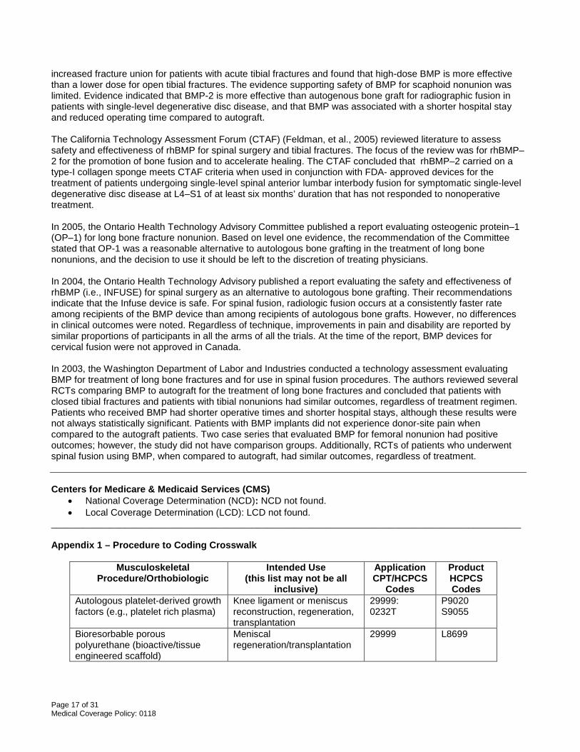
Autologous platelet-derived growth factors (e.g., platelet rich plasma)
Knee ligament or meniscus reconstruction, regeneration, transplantation
29999: 0232T
P9020 S9055
Bioresorbable porous polyurethane (bioactive/tissue engineered scaffold)
Meniscal regeneration/transplantation
29999 L8699
Page 18 of 31 Medical Coverage Policy: 0118
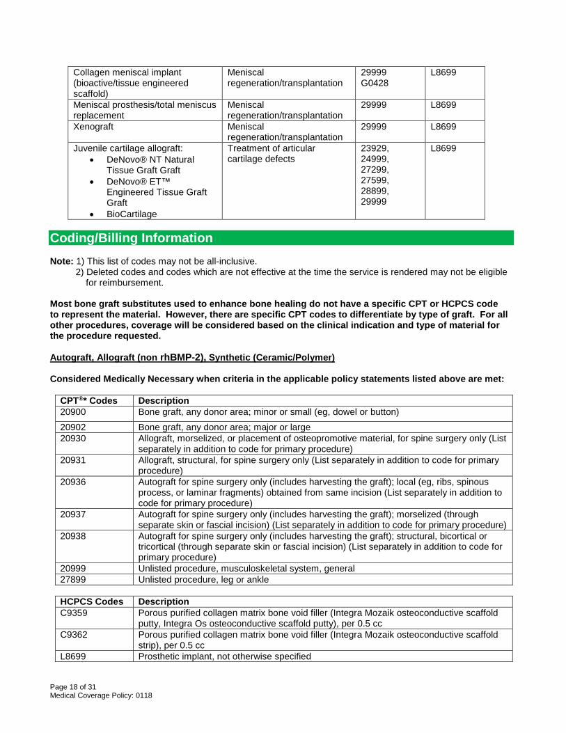
Collagen meniscal implant (bioactive/tissue engineered scaffold)
Meniscal regeneration/transplantation
29999 G0428
L8699
Meniscal prosthesis/total meniscus replacement
Meniscal regeneration/transplantation
29999 L8699
Xenograft Meniscal regeneration/transplantation
29999 L8699
Juvenile cartilage allograft: • DeNovo® NT Natural
Tissue Graft Graft • DeNovo® ET™
Engineered Tissue Graft Graft
• BioCartilage
Treatment of articular cartilage defects
23929, 24999, 27299, 27599, 28899, 29999
L8699
Coding/Billing Information Note: 1) This list of codes may not be all-inclusive. 2) Deleted codes and codes which are not effective at the time the service is rendered may not be eligible for reimbursement. Most bone graft substitutes used to enhance bone healing do not have a specific CPT or HCPCS code to represent the material. However, there are specific CPT codes to differentiate by type of graft. For all other procedures, coverage will be considered based on the clinical indication and type of material for the procedure requested. Autograft, Allograft (non rhBMP-2), Synthetic (Ceramic/Polymer) Considered Medically Necessary when criteria in the applicable policy statements listed above are met:
CPT®* Codes Description 20900 Bone graft, any donor area; minor or small (eg, dowel or button) 20902 Bone graft, any donor area; major or large 20930 Allograft, morselized, or placement of osteopromotive material, for spine surgery only (List
separately in addition to code for primary procedure) 20931 Allograft, structural, for spine surgery only (List separately in addition to code for primary
procedure) 20936 Autograft for spine surgery only (includes harvesting the graft); local (eg, ribs, spinous
process, or laminar fragments) obtained from same incision (List separately in addition to code for primary procedure)
20937 Autograft for spine surgery only (includes harvesting the graft); morselized (through separate skin or fascial incision) (List separately in addition to code for primary procedure)
20938 Autograft for spine surgery only (includes harvesting the graft); structural, bicortical or tricortical (through separate skin or fascial incision) (List separately in addition to code for primary procedure)
20999 Unlisted procedure, musculoskeletal system, general 27899 Unlisted procedure, leg or ankle
HCPCS Codes Description C9359 Porous purified collagen matrix bone void filler (Integra Mozaik osteoconductive scaffold
putty, Integra Os osteoconductive scaffold putty), per 0.5 cc C9362 Porous purified collagen matrix bone void filler (Integra Mozaik osteoconductive scaffold
strip), per 0.5 cc L8699 Prosthetic implant, not otherwise specified
Page 19 of 31 Medical Coverage Policy: 0118
Factor-based (rhBMP-2) Considered Medically Necessary when criteria in the applicable policy statements listed above are met and when used to report rhBMP–2 utilized in combination with a fusion device for single-level anterior interbody lumbar fusion surgery or for surgical repair of acute, open tibial fracture:
CPT®* Codes Description 20930 Allograft, morselized, or placement of osteopromotive material, for spine surgery only (List
separately in addition to code for primary procedure) 20999 Unlisted procedure, musculoskeletal system, general 27899 Unlisted procedure, leg or ankle
HCPCS Codes Description L8699 Prosthetic implant, not otherwise specified
Considered Experimental/Investigational/Unproven when used to report rhBMP–2 for ALL other indication indications including spinal fusions other than single-level anterior spinal fusions, as an alternative or adjunct treatment for sinus augmentation and/or localized alveolar ridge augmentation, or for the treatment of cervical spinal conditions:
CPT®* Codes Description 20930 Allograft, morselized, or placement of osteopromotive material, for spine surgery only (List
separately in addition to code for primary procedure) 21208 Osteoplasty, facial bones; augmentation (autograft, allograft, or prosthetic implant) 21210 Graft, bone; nasal, maxillary or malar areas (includes obtaining graft) 21499 Unlisted musculoskeletal procedure, head 31299 Unlisted procedure, accessory sinuses
CDT®** Codes Description D4263 Bone replacement graft - first site in quadrant D4264 Bone replacement graft - each additional site in quadrant D4265 Biologic materials to aid in soft and osseous tissue regeneration D7950 Osseous, osteoperiosteal, or cartilage graft of the mandible or
maxilla—autogenous or nonautogenous, by report D7951 Sinus augmentation with bone or bone substitutes D7953 Bone replacement graft for ridge preservation– per site
HCPCS Codes Description L8699 Prosthetic implant, not otherwise specified
Considered Experimental/Investigational/Unproven when used to report rhBMP–7 (i.e., OP–1™) for ALL indications:
CPT®* Codes Description 20930 Allograft, morselized, or placement of osteopromotive material, for spine surgery only (List
separately in addition to code for primary procedure) 20999 Unlisted procedure, musculoskeletal system, general 23929 Unlisted procedure, shoulder 24999 Unlisted procedure, humerus or elbow 25999 Unlisted procedure, forearm or wrist 27599 Unlisted procedure, femur or knee 27899 Unlisted procedure, leg or ankle
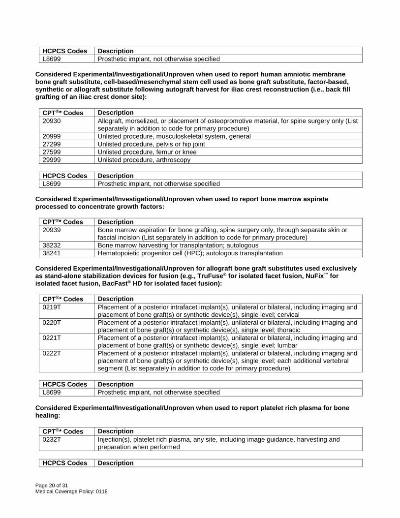
Page 20 of 31 Medical Coverage Policy: 0118
HCPCS Codes Description L8699 Prosthetic implant, not otherwise specified
Considered Experimental/Investigational/Unproven when used to report human amniotic membrane bone graft substitute, cell-based/mesenchymal stem cell used as bone graft substitute, factor-based, synthetic or allograft substitute following autograft harvest for iliac crest reconstruction (i.e., back fill grafting of an iliac crest donor site):
CPT®* Codes Description 20930 Allograft, morselized, or placement of osteopromotive material, for spine surgery only (List
separately in addition to code for primary procedure) 20999 Unlisted procedure, musculoskeletal system, general 27299 Unlisted procedure, pelvis or hip joint 27599 Unlisted procedure, femur or knee 29999 Unlisted procedure, arthroscopy
HCPCS Codes Description L8699 Prosthetic implant, not otherwise specified
Considered Experimental/Investigational/Unproven when used to report bone marrow aspirate processed to concentrate growth factors:
CPT®* Codes Description 20939 Bone marrow aspiration for bone grafting, spine surgery only, through separate skin or
fascial incision (List separately in addition to code for primary procedure) 38232 Bone marrow harvesting for transplantation; autologous 38241 Hematopoietic progenitor cell (HPC); autologous transplantation
Considered Experimental/Investigational/Unproven for allograft bone graft substitutes used exclusively as stand-alone stabilization devices for fusion (e.g., TruFuse® for isolated facet fusion, NuFix™ for isolated facet fusion, BacFast® HD for isolated facet fusion):
CPT®* Codes Description 0219T Placement of a posterior intrafacet implant(s), unilateral or bilateral, including imaging and
placement of bone graft(s) or synthetic device(s), single level; cervical 0220T Placement of a posterior intrafacet implant(s), unilateral or bilateral, including imaging and
placement of bone graft(s) or synthetic device(s), single level; thoracic 0221T Placement of a posterior intrafacet implant(s), unilateral or bilateral, including imaging and
placement of bone graft(s) or synthetic device(s), single level; lumbar 0222T Placement of a posterior intrafacet implant(s), unilateral or bilateral, including imaging and
placement of bone graft(s) or synthetic device(s), single level; each additional vertebral segment (List separately in addition to code for primary procedure)
HCPCS Codes Description L8699 Prosthetic implant, not otherwise specified
Considered Experimental/Investigational/Unproven when used to report platelet rich plasma for bone healing:
CPT®* Codes Description 0232T Injection(s), platelet rich plasma, any site, including image guidance, harvesting and
preparation when performed
HCPCS Codes Description
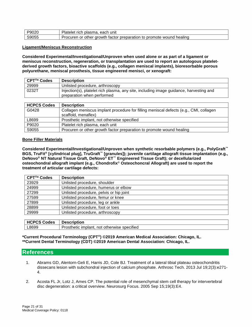
Page 21 of 31 Medical Coverage Policy: 0118
P9020 Platelet rich plasma, each unit S9055 Procuren or other growth factor preparation to promote wound healing
Ligament/Meniscus Reconstruction Considered Experimental/Investigational/Unproven when used alone or as part of a ligament or meniscus reconstruction, regeneration, or transplantation are used to report an autologous platelet-derived growth factors, bioactive scaffolds (e.g., collagen meniscal implants), bioresorbable porous polyurethane, meniscal prosthesis, tissue engineered menisci, or xenograft:
CPT®* Codes Description 29999 Unlisted procedure, arthroscopy 0232T Injection(s), platelet rich plasma, any site, including image guidance, harvesting and
preparation when performed
HCPCS Codes Description G0428 Collagen meniscus implant procedure for filling meniscal defects (e.g., CMI, collagen
scaffold, menaflex) L8699 Prosthetic implant, not otherwise specified P9020 Platelet rich plasma, each unit S9055 Procuren or other growth factor preparation to promote wound healing
Bone Filler Materials Considered Experimental/Investigational/Unproven when synthetic resorbable polymers (e.g., PolyGraft™ BGS, TruFit® [cylindrical plug], TruGraft™ [granules]); juvenile cartilage allograft tissue implantation (e.g., DeNovo® NT Natural Tissue Graft, DeNovo® ET™ Engineered Tissue Graft); or decellularized osteochondral allograft implant (e.g., Chondrofix® Osteochoncral Allograft) are used to report the treatment of articular cartilage defects:
CPT®* Codes Description 23929 Unlisted procedure, shoulder 24999 Unlisted procedure, humerus or elbow 27299 Unlisted procedure, pelvis or hip joint 27599 Unlisted procedure, femur or knee 27899 Unlisted procedure, leg or ankle 28899 Unlisted procedure, foot or toes 29999 Unlisted procedure, arthroscopy
HCPCS Codes Description L8699 Prosthetic implant, not otherwise specified
*Current Procedural Terminology (CPT®) ©2019 American Medical Association: Chicago, IL. **Current Dental Terminology (CDT) ©2019 American Dental Association: Chicago, IL. References
1. Abrams GD, Alentorn-Geli E, Harris JD, Cole BJ. Treatment of a lateral tibial plateau osteochondritis dissecans lesion with subchondral injection of calcium phosphate. Arthrosc Tech. 2013 Jul 19;2(3):e271-4.
2. Acosta FL Jr, Lotz J, Ames CP. The potential role of mesenchymal stem cell therapy for intervertebral
disc degeneration: a critical overview. Neurosurg Focus. 2005 Sep 15;19(3):E4.
Page 22 of 31 Medical Coverage Policy: 0118
3. Abjornson C, Brecevich A, Callanan T, Dowe C, Cammisa FP, Lorio MP. ISASS Recommendations and Coverage Criteria for Bone Graft Substitutes used in Spinal Surgery. Int J Spine Surg. 2018 Dec 21;12(6):757-771.
4. Agarwal R, Williams K, Umscheid CA, Welch WC. Osteoinductive bone graft substitutes for lumbar
fusion: a systematic review. J Neurosurg Spine. 2009 Dec;11(6):729-40. 5. Agency for Healthcare Research and Quality (AHRQ). Technology assessment: the role of bone growth
stimulating devices and orthobiologics in healing nonunion fractures. Updated 2005 Sep 21. Accessed January 8, 2020. Available at URL address: http://archive.ahrq.gov/clinic/techarch.htm
6. Agency for Healthcare Research and Quality (AHRQ). Bone Morphogenetic Protein: The State of
Evidence for On-Label and Off-Label Use. August 6, 2010. Accessed January 8, 2020. Available at URL address: http://archive.ahrq.gov/clinic/techarch.htm
7. American Academy of Orthopaedic Surgeons. Nonunions. Updated 2007 September, reviewed March
2014. Copyright © 1995-2019. Accessed January 8, 2020. Available at URL address: http://orthoinfo.aaos.org/topic.cfm?topic=A00374
8. American Academy of Orthopaedic Surgeons. Treatment of Osteoarthritis of the knee. Evidence-Based
Guidelines, 2nd edition, May 18, 2013. Accessed January 8, 2020. Available at URL address: http://www.aaos.org/CustomTemplates/Content.aspx?id=6396
9. American Academy of Orthopaedic Surgeons. Spinal fusion. Updated 2010 June. Accessed January 7,
2014. Available at URL address: http://orthoinfo.aaos.org/fact/thr_report.cfm?Thread_ID=156 10. Arnold PM, Sasso RC, Janssen ME, et al. Efficacy of i-Factor™ Bone Graft versus Autograft in Anterior
Cervical Discectomy and Fusion: Results of the Prospective Randomized Single-blinded Food and Drug Administration Investigational Device Exemption Study. Spine (Phila Pa 1976). 2016.
11. Arnold PM, Sasso RC, Janssen ME, Fehlings MG, Heary RF, Vaccaro AR, Kopjar B. i-Factor™ Bone
Graft vs Autograft in Anterior Cervical Discectomy and Fusion: 2-Year Follow-up of the Randomized Single-Blinded Food and Drug Administration Investigational Device Exemption Study. Neurosurgery. 2018 Sep 1;83(3):377-384.
12. Arora NS, Ramanayake T, Ren YF, Romanos GE. Platelet-rich plasma in sinus augmentation
procedures: a systematic literature review: Part II. Implant Dent. 2010 Apr;19(2):145-57. 13. Bapat MR, Chaudhary K, Garg H, Laheri V. Reconstruction of large iliac crest defects after graft harvest
using autogenous rib graft: a prospective controlled study. Spine (Phila Pa 1976). 2008 Nov 1;33(23):2570-5.
14. Baskin DS, Ryan P, Sonntag V, Westmark R, Widmayer MA. A prospective, randomized, controlled
cervical fusion study using recombinant human bone morphogenetic protein-2 with the CORNERSTONE-SR allograft ring and the ATLANTIS anterior cervical plate. Spine. 2003;28(12):1219-25.
15. Beaman FD; Bancroft LW; Peterson JJ; Kransdorf MJ. Bone graft materials and synthetic substitutes.
Radiol Clin North Am. 2006 May;44(3):451-61. 16. Benglis D, Wang MY, Levi AD. A comprehensive review of the safety profile of bone morphogenetic
protein in spine surgery. Neurosurgery. 2008 May;62(5 Suppl 2):ONS423-31; discussion ONS431. 17. Block MS, Achong R. Bone morphogenetic protein for sinus augmentation. Atlas Oral Maxillofac Surg
Clin North Am. 2006 Mar;14(1):99-105.
Page 23 of 31 Medical Coverage Policy: 0118
18. Boakye M, Mummaneni PV, Garrett M, Rodts G, Haid R. Anterior cervical discectomy and fusion involving a polyetherethereketone spacer and bone morphogenetic protein. J. Neurosurg: Spine. 2005 May;2:521-5.
19. Boden SD, Zdeblick TA, Sandhu HS, Heim SE. The use of rhBMP-2 in interbody fusion cages. Definitive
evidence of osteoinduction in humans: a preliminary report. Spine. 2000;25(3):376-81. 20. Boden SD, Kang J, Sandhu H, Heller JG. Use of recombinant human bone morphogenetic protein-2 to
achieve posterolateral lumbar spine fusion in humans: a prospective, randomized clinical pilot trial: 2002 Volvo Award in clinical studies. Spine. 2002;27(23):2662-73.
21. Bohner M. Design of ceramic-based cements and putties for bone graft substitution. Eur Cell Mater.
2010 Jul 1;20:1-12. 22. Bornes TD, Adesida AB, Jomha NM. Mesenchymal stem cells in the treatment of traumatic articular
cartilage defects: a comprehensive review. Arthritis Res Ther. 2014;16(5):432. 23. Boyne PJ, Lilly LC, Marx RE, Moy PK, Nevins M, Spagnoli DB, Triplett RG. De novo bone induction by
recombinant human bone morphogenetic protein-2 (rhBMP-2) in maxillary sinus floor augmentation. J Oral Maxillofac Surg. 2005 Dec;63(12):1693-707.
24. Burkus JK, Gornet MF, Dickman CA, Zdeblick TA. Anterior lumbar interbody fusion using rhBMP-2 with
tapered interbody cages. J Spinal Disord Tech. 2002;15(5):337-49. 25. Burkus JK, Sandhu HS, Gornet MF, Longley MC. Use of rhBMP-2 in combination with structural cortical
allografts: clinical and radiographic outcomes in anterior lumbar spinal surgery. J Bone Joint Surg Am. 2005 Jun;87-A(6):1205-12.
26. Calcei JG, Rodeo SA. Orthobiologics for Bone Healing. in Sports Med. 2019 Jan;38(1):79-95. 27. Calori GM, Tagliabue L, Gala L, d'Imporzano M, Peretti G, Albisetti W. Application of rhBMP-7 and
platelet-rich plasma in the treatment of long bone non-unions: A prospective randomised clinical study on 120 patients. Injury. 2008 Dec;39(12):1391-402..
28. Camargo PM, Lekovic V, Weinlaender M, Vasilic N, Madzarevic M, Kenney EB. A reentry study on the
use of bovine porous bone mineral, GTR, and platelet-rich plasma in the regenerative treatment of intrabony defects in humans. Int J Periodontics Restorative Dent. 2005 Feb;25(1):49-59.
29. Campbell KA, Saltzman BM, Mascarenhas R, Khair MM, Verma NN, Bach BR Jr, Cole BJ. Does intra-
articular platelet-rich plasma injection provide clinically superior outcomes compared with other therapies in the treatment of knee osteoarthritis? A systematic review of overlapping meta-analyses. Arthroscopy. 2015 Nov;31(11):2213-21.
30. Carragee EJ, Hurwitz EL, Weiner BK. A critical review of recombinant human bone morphogenetic
protein-2 trials in spinal surgery: emerging safety concerns and lessons learned. Spine J. 2011 Jun;11(6):471-91.
31. Carragee EJ, Mitsunaga KA, Hurwitz EL, Scuderi GJ. Retrograde ejaculation after anterior lumbar
interbody fusion using rhBMP-2: a cohort controlled study. Spine J. 2011 Jun;11(6):511-6. 32. Carreon LY, Glassman SD, Anekstein Y, Puno RM. Platelet gel (AGF) fails to increase fusion rates in
instrumented posterolateral fusions. Spine. 2005 May 1;30(9):E243-6; discussion E247. 33. Carreon LY, Glassman SD, Brock DC, Dimar JR, Puno RM, Campbell MJ. Adverse events in patients re-
exposed to bone morphogenetic protein for spine surgery. Spine. 2008 Feb 15;33(4):391-3.
Page 24 of 31 Medical Coverage Policy: 0118
34. Carlisle E, Fischgrund JS. Bone morphogenetic proteins for spinal fusion. Spine. 2005;5:240S-9S. 35. Chang KV, Hung CY, Aliwarga F, Wang TG, Han DS, Chen WS. Comparative effectiveness of platelet-
rich plasma injections for treating knee joint cartilage degenerative pathology: a systematic review and meta-analysis. Arch Phys Med Rehabil. 2014 Mar;95(3):562-75. doi: 10.1016/j.apmr.2013.11.006.
36. Chaua AMT, Mobbs RJ. Bone graft substitutes in anterior cervical discectomy and fusion. Eur Spine J.
2009 Apr;18(4):449-64. 37. Cohen SB, Sharkey PF. Subchondroplasty for Treating Bone Marrow Lesions. J Knee Surg. 2016
Oct;29(7):555-563. 38. Dawson E, Bae HW, Burkus JK, Stambough JL, Glassman SD. Recombinant human bone
morphogenetic protein-2 on an absorbable collagen sponge with an osteoconductive bulking agent in posterolateral arthrodesis with instrumentation. A prospective randomized trial. J Bone Joint Surg Am. 2009 Jul;91(7):1604-13.
39. Delawi D, Dhert WJ, Rillardon L, Gay E, Prestamburgo D, Garcia-Fernandez C. A prospective,
randomized, controlled, multicenter study of osteogenic protein-1 in instrumented posterolateral fusions: report on safety and feasibility. Spine (Phila Pa 1976). 2010 May 20;35(12):1185-91.
40. Dimar JR 2nd, Glassman SD, Burkus JK, Pryor PW, Hardacker JW, Carreon LY. Clinical and
radiographic analysis of an optimized rhBMP-2 formulation as an autograft replacement in posterolateral lumbar spine arthrodesis. J Bone Joint Surg Am. 2009 Jun;91(6):1377-86.
41. Dimar JR, Glassman SD, Burkus KJ, Carreon LY. Clinical outcomes and fusion success at 2 years of
single-level instrumented posterolateral fusions with recombinant human bone morphogenetic protein-2/compression resistant matrix versus iliac crest bone graft. Spine. 2006 Oct 15;31(22):2534-9; discussion 2540.
42. Dmitriev AE, Lehman RA Jr, Symes AJ. Bone morphogenetic protein-2 and spinal arthrodesis: the basic
science perspective on protein interaction with the nervous system. Spine J. 2011 Jun;11(6):500-5. 43. D'Souza M, Macdonald NA, Gendreau JL, Duddleston PJ, Feng AY, Ho AL. Graft Materials and
Biologics for Spinal Interbody Fusion. Biomedicines. 2019 Sep 26;7(4). 44. Dziadosz M, Basch RS, Young BK. Human amniotic fluid: a source of stem cells for possible therapeutic
use. Am J Obstet Gynecol. 2016 Jan 6. 45. Einhorn TA. Clinical applications of recombinant human BMPs: early experience and future
development. J Bone Joint Surg Am. 2003;85-A(Suppl 3):82-8. 46. Epstein NE. Pros, cons, and costs of INFUSE in spinal surgery. Surg Neurol Int. 2011 Jan 24;2:10. 47. Epstein NE, Hollingsworth R. Does donor site reconstruction following anterior cervical surgery diminish
postoperative pain? J Spinal Disord Tech. 2003 Feb;16(1):20-6. 48. Esposito M; Grusovin MG; Coulthard P; Worthington HV. Interventions for replacing missing teeth: bone
augmentation techniques for dental implant treatment. The Cochrane Database of Systematic Reviews 2007 Issue 4, Copyright © 2007 The Cochrane Collaboration.
49. Feldman MD. Recombinant human bone morphogenetic protein-2 for spinal surgery and treatment of
open tibial fractures. February 16, 2005. Accessed January 11, 2016. Available at URL address: http://ctaf.org/assessments?field_condition_tid=All&field_specialty_tid=38&field_met_ctaf_criteria__tid=All&items_per_page=10&page=1
Page 25 of 31 Medical Coverage Policy: 0118
50. Filardo G, Kon E, Buda R, Timoncini A, Di Martino A, Cenacchi A, Fornasari PM, Giannini S, Marcacci M. Platelet-rich plasma intra-articular knee injections for the treatment of degenerative cartilage lesions and osteoarthritis. Knee Surg Sports Traumatol Arthrosc. 2010 Aug 26.
51. Fiorellini JP, Howell TH, Cochran D, Malmquist J, Lilly LC, Spagnoli D, Toljanic J, Jones A, Nevins M.
Randomized study evaluating recombinant human bone morphogenetic protein-2 for extraction socket augmentation. J Periodontol. 2005 Apr;76(4):605-13.
52. Friedlaender GE, Perry CR, Cole JD, Cook SD, Cierny G, Muschler GF, et al. Osteogenic protein-1
(bone morphogenetic protein-7) in the treatment of tibial nonunions: a prospective, randomized clinical trial comparing rhOP-1 with fresh bone autograft. J Bone Joint Surg Am. 2001;83-A(Suppl 1 Pt 2):S151-8.
53. Freymiller EG, Aghaloo TL. Platelet-rich plasma: ready or not? J Oral Maxillofac Surg. 2004
Apr;62(4):484-8. 54. Garrison KR, Donell S, Ryder J, Shemilt I, Mugford M, Harvey I, Song F. Clinical effectiveness and cost-
effectiveness of bone morphogenetic proteins in the non-healing of fractures and spinal fusion: a systematic review. Health Technol Assess. 2007 Aug;11(30):1-150, iii-iv.
55. Gautschi OP, Frey SP, Zellweger R. Bone morphogenetic proteins in clinical applications. ANZ J Surg.
2007 Aug;77(8):626-31. 56. Geesink RG, Hoefnagels NH, Bulstra SK. Osteogenic activity of OP-1 bone morphogenetic protein
(BMP-7) in a human fibular defect. J Bone Joint Surg Br. 1999;81-B(4):710-8. 57. Glassman SD, Dimar JR 3rd, Burkus K, Hardacker JW, Pryor PW, Boden SD, Carreon LY. The efficacy
of rhBMP-2 for posterolateral lumbar fusion in smokers. Spine. 2007 Jul 1;32(15):1693-8. 58. Glassman SD, Dimar JR, Carreon LY, Campbell MJ, Puno RM, Johnson JR. Initial fusion rates with
recombinant human bone morphogenetic protein-2/compression resistant matrix and a hydroxyapatite and tricalcium phosphate/collagen carrier in posterolateral spinal fusion. Spine. 2005;30(15):1694-8.
59. Govender S, Csimma C, Genant HK, Valentin-Opran A, Amit Y, Arbel R, et al; BMP-2 Evaluation in
Surgery for Tibial Trauma (BESTT) Study Group. Recombinant human bone morphogenetic protein-2 for treatment of open tibial fractures: a prospective, controlled, randomized study of four hundred and fifty patients. J Bone Joint Surg Am. 2002;84-A(12):2123-34.
60. Granjeiro JM, Oliveira RC, Bustos-Valenzuela JC, Sogayar MC, Taga R. Bone morphogenetic proteins:
from structure to clinical use. Braz J Med Biol Res. 2005 Oct;38(10):1463-73. 61. Grawe B, Burge A, Nguyen J, Strickland S, Warren R, Rodeo S, Shubin Stein B. Cartilage Regeneration
in Full-Thickness Patellar Chondral Defects Treated with Particulated Juvenile Articular Allograft Cartilage: An MRI Analysis. Cartilage. 2017 Oct;8(4):374-383.
62. Greenwald AS, Boden SD, Goldberg VM, Khan Y, Laurencin CT, Rosier RN, American Academy of
Orthopaedics. The Committee on Biological Implants. Bone-graft substitutes: facts, fictions, and applications. J Bone Joint Surg Am. 2001;83-A Suppl 2 Pt 2:98-103. (Presented at the 2006 73rd Annual Meeting, and 2008 75th Annual Meeting, in slightly different form).
63. Hayes, Clinical Research Response. i-Factor Bone Graft (Cerapedics, Inc) for Spinal Fusion. Lansdale,
PA: Hayes, Inc.; Aug 2017. 64. Hayes, Clinical Research Response. i-Factor (Cerapedics, Inc) Versus Trinity Elite (Orthofix) for Spinal
Procedures. Lansdale, PA: Hayes, Inc.; May 2018.
Page 26 of 31 Medical Coverage Policy: 0118
65. Hayes, Inc. Search and Summary. Accufill Bone Substitute Material (Zimmer) for Ankle Subchondroplasty. Lansdale, PA: Hayes, Inc.; December 10, 2015.
66. Helgeson MD, Lehman RA Jr, Patzkowski JC, Dmitriev AE, Rosner MK, Mack AW. Adjacent vertebral
body osteolysis with bone morphogenetic protein use in transforaminal lumbar interbody fusion. Spine J. 2011 Jun;11(6):507-10.
67. Helm GA, Gazit Z. Future uses of mesenchymal stem cells in spine surgery. Neurosurg Focus. 2005
Dec 15;19(6):E13. 68. Hess DE, Werner BC, Deal DN. Use of Particulated Juvenile Articular Cartilage Allograft for
Osteochondral Lesions of the Wrist. Hand (N Y). 2017 Sep;12(5):NP62-NP67. 69. Hing KA, Annaz B, Saeed S, Revell PA, Buckland T. Microporosity enhances bioactivity of synthetic
bone graft substitutes. J Mater Sci Mater Med. 2005 May;16(5):467-75. (Abstract only). 70. Ho-Shui-Ling A, Bolander J, Rustom LE, Johnson AW, Luyten FP, Picart C. Bone regeneration
strategies: Engineered scaffolds, bioactive molecules and stem cells current stage and future perspectives. Biomaterials. 2018 Oct;180:143-162.
71. Jahangir A, Nunley RM, Menta S, Sharan A, Washingotn Health Policy Fellows. Bone graft substitutes in
orthopaedic surgery. American Academy of Orthopaedic Surgeons. January 2008, AAOS Now. 72. Jones AL, Bucholz RW, Bosse MJ, Mirza SK, Lyon TR, Webb LX, Pollak AN, Golden JD, Valentin-Opran
A; BMP-2 Evaluation in Surgery for Tibial Trauma-Allgraft (BESTT-ALL) Study Group. Recombinant human BMP-2 and allograft compared with autogenous bone graft for reconstruction of diaphyseal tibial fractures with cortical defects. A randomized, controlled trial. J Bone Joint Surg Am. 2006 Jul;88(7):1431-41.
73. Johnsson R, Stromqvist B, Aspenberg P. Randomized radiostereometric study comparing osteogenic
protein-1 (BMP-7) and autograft bone in human noninstrumented posterolateral lumbar fusion: 2002 Volvo Award in clinical studies. Spine. 2002;27(23):2654-61.
74. Jung RE, Glauser R, Schärer P, Hämmerle CH, Sailer HF, Weber FE. Effect of rhBMP-2 on guided bone
regeneration in humans. Clin Oral Implants Res. 2003 Oct;14(5):556-68. 75. Kanayama M, Hashimoto T, Shigenobu K, Yamane S, Bauer TW, Togawa D. A prospective randomized
study of posterolateral lumbar fusion using osteogenic protein-1 (OP-1) versus local autograft with ceramic bone substitute: emphasis of surgical exploration and histologic assessment. Spine. 2006 May 1;31(10):1067-74.
76. Kassolis JD, Reynolds MA. Evaluation of the adjunctive benefits of platelet-rich plasma in subantral
sinus augmentation. J Craniofac Surg. 2005 Mar;16(2):280-7. 77. Khan SN, Sandhu HS, Lane JM, Cammisa FP Jr, Girardi FP. Bone morphogenetic proteins: relevance in
spine surgery. Orthop Clin North Am. 2002;33(2):447-63. 78. Kleeman TJ, Ahn UM, Talbot-Kleeman A. Laparoscopic anterior lumbar interbody fusion with rhBMP-2: a
prospective study of clinical and radiographic outcomes. Spine. 2001;26(24):2751-6. 79. Kon E, Buda R, Filardo G, Di Martino A, Timoncini A, Cenacchi A, Fornasari PM, Giannini S, Marcacci
M. Platelet-rich plasma: intra-articular knee injections produced favorable results on degenerative cartilage lesions. Knee Surg Sports Traumatol Arthrosc. 2010 Apr;18(4):472-9.
Page 27 of 31 Medical Coverage Policy: 0118
80. Khoshbin A, Leroux T, Wasserstein D, Marks P, Theodoropoulos J, Ogilvie-Harris D, Gandhi R, Takhar K, Lum G, Chahal J. The efficacy of platelet-rich plasma in the treatment of symptomatic knee osteoarthritis: a systematic review with quantitative synthesis. Arthroscopy. 2013 Dec;29(12):2037-48.
81. Lai LP, Stitik TP, Foye PM, Georgy JS, Patibanda V, Chen B. Use of platelet-rich plasma in intra-
articular knee injections for osteoarthritis: A Systematic Review. PM R. 2015 Jun;7(6):637-48. 82. Laurencin CT, Calhoun JH, Magge A, Khan Y. Bone graft substitute materials. Medscape. Updated Dec
4, 2013. Accessed January 11, 2016. Available at URL address: http://emedicine.medscape.com/article/1230616-overview#a17
83. Lauweryns P. Raskin Y. Prospective Analysis of a New Bone Graft in Lumbar Interbody Fusion: Results
of a 2- Year Prospective Clinical and Radiological Study. Int J Spine Surg. 2015 Feb 3;9. 84. Lenza M, Ferraz Sde B, Viola DC, Santos OF, Cendoroglo Neto M, Ferretti M. Platelet-rich plasma for
long bone healing. Einstein (Sao Paulo). 2013 Jan-Mar;11(1):122-7. 85. Leung VY, Chan D, Cheung KM. Regeneration of intervertebral disc by mesenchymal stem cells:
potentials, limitations, and future direction. Eur Spine J. 2006 Aug;15 Suppl 3:S406-13. Epub 2006 Jul 15.
86. Lieberman JR, Daluiski A, Einhorn TA. The role of growth factors in the repair of bone. Biology and
clinical applications. J Bone Joint Surg Am. 2002;84-A(6):1032-44. 87. Luhmann SJ, Bridwell KH, Cheng I, Imamura T, Lenke LG, Schootman M. Use of bone morphogenetic
protein-2 for adult spinal deformity. Spine (Phila Pa 1976). 2005 Sep 1;30(17 Suppl):S110-7. 88. Maniscalco P, Gambera D, Bertone C, Rivera F, Crainz E, Urgelli S. Healing of fresh tibial fractures with
OP-1. A preliminary report. Acta Biomed Ateneo Parmense. 2002;73(1-2):27-33. 89. Mariconda M, Cozzolino F, Cozzolino A, D'Agostino E, Bove A, Milano C. Platelet gel supplementation in
long bone nonunions treated by external fixation. J Orthop Trauma. 2008 May-Jun;22(5):342-5. 90. Marmotti A, de Girolamo L, Bonasia DE, Bruzzone M, Mattia S, Rossi R, Montaruli A, Dettoni F, Castoldi
F, Peretti G. Bone marrow derived stem cells in joint and bone diseases: a concise review. Int Orthop. 2014 Sep;38(9):1787-801.
91. Marx RE. Platelet-rich plasma: evidence to support its use. J Oral Maxillofac Surg. 2004 Apr;62(4):489-
96. 92. Marx RE, Carlson ER, Eichstaedt RM, Schimmele SR, Strauss JE, Georgeff KR. Platelet-rich plasma:
Growth factor enhancement for bone grafts. Oral Surg Oral Med Oral Pathol Oral Radiol Endod. 1998 Jun;85(6):638-46.
93. McKay WF, Peckham SM, Badura JM. A comprehensive clinical review of recombinant human bone
morphogenetic protein-2 (INFUSE((R)) Bone Graft). Int Orthop. 2007 Dec;31(6):729-734. 94. Medtronic. Infuse Bone Graft. Oral-maxillofacial. Updated May 6, 2010. Accessed January 7, 2014.
Available at URL address: http://www.infusebonegraft.com/omf_about.html 95. Mehta S, Watson JT. Platelet rich concentrate: basic science and current clinical applications. J Orthop
Trauma. 2008 Jul;22(6):432-8. 96. Michielsen J, Sys J, Rigaux A, Bertrand C. The effect of recombinant human bone morphogenetic
protein-2 in single-level posterior lumbar interbody arthrodesis. J Bone Joint Surg Am. 2013 May 15;95(10):873-80.
Page 28 of 31 Medical Coverage Policy: 0118
97. Minamide A, Yoshida M, Kawakami M, Okada M, Enyo Y, Hashizume H, Boden SD. The effects of bone
morphogenetic protein and basic fibroblast growth factor on cultured mesenchymal stem cells for spine fusion. Spine. 2007 May 1;32(10):1067-71.
98. Mobbs RJ, Maharaj M, Rao PJ. Clinical outcomes and fusion rates following anterior lumbar interbody
fusion with bone graft substitute i-FACTOR, an anorganic bone matrix/P-15 composite. J Neurosurg Spine. 2014 Dec;21(6):867-76.
99. Mulconrey DS, Bridwell KH, Flynn J, Cronen GA, Rose PS. Bone morphogenetic protein (RhBMP-2) as
a substitute for iliac crest bone graft in multilevel adult spinal deformity surgery: minimum two-year evaluation of fusion. Spine (Phila Pa 1976). 2008 Sep 15;33(20):2153-9.
100. Mussano F, Ciccone G, Ceccarelli M, Baldi I, Bassi F. Bone morphogenetic proteins and bone defects: a
systematic review. Spine. 2007 Apr 1;32(7):824-30. 101. Nandi SK, Roy S, Mukherjee P, Kundu B, De DK, Basu D. Orthopaedic applications of bone graft & graft
substitutes: a review. Indian J Med Res. 2010 Jul;132:15-30. 102. Ng A, Bernhard K. The Use of Particulated Juvenile Allograft Cartilage in Foot and Ankle Surgery. Clin
Podiatr Med Surg. 2018 Jan;35(1):11-18. 103. NuVasive, Inc. Osteocel Plus. Accessed January 8, 2020. Available at URL address:
https://www.nuvasive.com/search/osteocel+plus/ 104. Ontario Ministry of Health and Long-Term Care, Medical Advisory Secretariat. Osteogenic protein-1 for
long bone nonunion. Health Technology Assessment Scientific Literature Review. Toronto, ON: Ontario Ministry of Health and Long-Term Care; April 2005. Accessed January 7, 2014. Available at URL address: http://www.health.gov.on.ca/english/providers/program/mas/tech/reviews/sum_osteo_070105.html
105. Ontario Ministry of Health and Long-Term Care, Medical Advisory Secretariat. Bone morphogenetic
proteins and spinal surgery for degenerative disc disease. Health Technology Scientific Literature Review. Toronto, ON: Ontario Ministry of Health and Long-Term Care; March 2004. Accessed January 11, 2016. Available at URL address: http://find.gov.on.ca/?searchType=simple&owner=moh&url=health.gov.on.ca&collection=&offset=0&lang=en&type=ANY&q=bone+morphogenetic&search.x=0&search.y=0
106. Papakostidis C, Kontakis G, Bhandari M, Giannoudis PV. Efficacy of autologous iliac crest bone graft
and bone morphogenetic proteins for posterolateral fusion of lumbar spine: a meta-analysis of the results. Spine. 2008 Sep 1;33(19):E680-92.
107. Pradhan BB, Bae HW, Patel VV, Delamarter RB. Graft resorption with the use of bone morphogenetic
protein: lessons from anterior lumbar interbody fusion using femoral ring allografts and recombinant human bone morphogenetic protein-2. Spine. 2006 May 1;31(10):E277-84.
108. Rengachary SS. Bone morphogenetic proteins: basic concepts. Neurosurg Focus [serial online].
2002;13(6):1-6. Neurosurg Focus 13(6), 2002. © 2002 American Association of Neurological Surgeons. 109. Resnick DK. Reconstruction of anterior iliac crest after bone graft harvest decreases pain: a randomized,
controlled clinical trial. Neurosurgery. 2005 Sep;57(3):526-9; discussion 526-9. 110. Ronga M, Baldo F, Zappala G, Cherubino P; BMP-7 Italian Observational Study (BIOS) Group.
Recombinant human bone morphogenetic protein-7 for treatment of long bone non-union: An observational, retrospective, non-randomized study of 105 patients. Injury. 2006 Sep;37(9 Suppl):S51-6.
Page 29 of 31 Medical Coverage Policy: 0118
111. Rush SM. Trinity Evolution. Foot Ankle Spec. 2010 Jun;3(3):144-7. 112. Russell TA, Leighton RK; Alpha-BSM Tibial Plateau Fracture Study Group. Comparison of autogenous
bone graft and endothermic calcium phosphate cement for defect augmentation in tibial plateau fractures. A multicenter, prospective, randomized study. J Bone Joint Surg Am. 2008 Oct;90(10):2057-61.
113. Saltzman BM, Lin J, Lee S. Particulated Juvenile Articular Cartilage Allograft Transplantation for
Osteochondral Talar Lesions. Cartilage. 2017 Jan;8(1):61-72. 114. Sánchez M, Anitua E, Orive G, Mujika I, Andia I. Platelet-rich therapies in the treatment of orthopaedic
sport injuries. Sports Med. 2009;39(5):345-54. 115. Simmonds MC, Brown JV, Heirs MK, Higgins JP, Mannion RJ, Rodgers MA, Stewart LA. Safety and
effectiveness of recombinant human bone morphogenetic protein-2 for spinal fusion: a meta-analysis of individual-participant data. Ann Intern Med. 2013 Jun 18;158(12):877-89.
116. Singh K, Smucker JD, Boden SD. Use of recombinant human bone morphogenetic protein-2 as an
adjunct in posterolateral lumbar spine fusion: a prospective CT-scan analysis at one and two years. J Spinal Disord Tech. 2006 Aug;19(6):416-23.
117. Smucker JD, Rhee JM, Singh K, Yoon ST, Heller JG. Increased swelling complications associated with
off-label usage of rhBMP-2 in the anterior cervical spine. Spine. 2006 Nov 15;31(24):2813-9. 118. Swiontkowski MF, Aro HT, Donell S, Esterhai JL, Goulet J, Jones A, et al. Recombinant human bone
morphogenetic protein-2 in open tibial fractures. A subgroup analysis of data combined from two prospective randomized studies. Bone Joint Surg Am. 2006 Jun;88(6):1258-65.
119. Szpalski M, Gunzburg R. Applications of calcium phosphate-based cancellous bone void fillers in trauma
surgery. Orthopedics. 2002;25(5 Suppl):S601-9. 120. Tarnow DP, Wallace SS, Testori T, Froum SJ, Motroni A, Prasad HS. Maxillary sinus augmentation
using recombinant bone morphogenetic protein-2/acellular collagen sponge in combination with a mineralized bone replacement graft: a report of three cases. Int J Periodontics Restorative Dent. 2010 Apr;30(2):139-49.
121. Termaat MF, Den Boer FC, Bakker FC, Patka P, Haarman HJ. Bone morphogenetic proteins.
Development and clinical efficacy in the treatment of fractures and bone defects. J Bone Joint Surg Am. 2005 Jun;87-A(6):1367-78.
122. Urist MR. Bone: formation by autoinduction. Science. 1965;150(698):893-9. 123. U.S. Food and Drug Administration. INTER FIX Threaded Fusion Device: important medical information.
Accessed January 3, 2018. Available at URL address: http://google2.fda.gov/search?q=INTER+FIX+Threaded+Fusion+&client=FDAgov&site=FDAgov&lr=&proxystylesheet=FDAgov&output=xml_no_dtd&getfields=*
124. U.S. Food and Drug Administration. New device approval: INFUSE® bone graft-P000054. Updated May
17, 2004. Accessed January 8, 2020. Available at URL address: http://google2.fda.gov/search?q=P000054&client=FDAgov&proxystylesheet=FDAgov&output=xml_no_dtd&site=FDAgov&requiredfields=&sort=date:D:L:d1&filter=1
125. U.S. Food and Drug Administration. New device approval: InFUSE™ bone graft/LT-CAGE™ lumbar
tapered fusion device-P000058. Updated 2002 Sep 6. Accessed January 8, 2019. Available at URL address:
Page 30 of 31 Medical Coverage Policy: 0118
https://google2.fda.gov/search?q=P000058&client=FDAgov&proxystylesheet=FDAgov&output=xml_no_dtd&site=FDAgov&requiredfields=&sort=date:D:L:d1&filter=1
126. U.S. Food and Drug Administration. Supplement S002. July 2004. InFUSE™ bone graft/LT-CAGE™
lumbar tapered fusion device-P000058. Accessed January 8, 2020. http://google2.fda.gov/search?q=P000058&client=FDAgov&site=FDAgov&lr=&proxystylesheet=FDAgov&requiredfields=-archive%3AYes&output=xml_no_dtd&getfields=*
127. U.S. Food and Drug Administration. Supplement S059. 12/2015. InFUSE™ bone graft/LT-CAGE™
lumbar tapered fusion device-P000058. Accessed January 8, 2020. Available at URL address: http://www.accessdata.fda.gov/scripts/cdrh/cfdocs/cfpma/pma.cfm?id=P000058S059
128. U.S. Food and Drug Administration. New humanitarian device approval: OP-1™ - H010002. Updated
2001 Nov 30. Accessed January 8, 2020. Available at URL address: http://google2.fda.gov/search?q=H010002&client=FDAgov&proxystylesheet=FDAgov&output=xml_no_dtd&site=FDAgov&requiredfields=&sort=date:D:L:d1&filter=1
129. U.S. Food and Drug Administration. New humanitarian device approval: OP-1 Putty- H020008. Updated
2004 Apr 27. Accessed January 8, 2020. Available at URL address: http://www.accessdata.fda.gov/scripts/cdrh/cfdocs/cftopic/pma/pma.cfm?num=H020008
130. U.S. Food and Drug Administration. InFUSE™ Bone Graft/LT-CAGE™ Lumbar Tapered Fusion Device.
PMA final decisions rendered for December 2003. Updated 2004 Jul 16. Accessed January 5, 2012. Available at URL address: http://www.fda.gov/MedicalDevices/ProductsandMedicalProcedures/DeviceApprovalsandClearances/PMAApprovals/ucm111338.htm
131. U.S. Food and Drug Administration. Osteofil Allograft Paste. 510(k) summary K043420. Accessed
January 8, 2020. Available at URL address: http://www.accessdata.fda.gov/scripts/cdrh/cfdocs/cfpmn/pmn.cfm?id=k043420
132. Vaccaro AR, Patel T, Fischgrund J, Anderson DG, Truumees E, Herkowitz HN, et al. A pilot study
evaluating the safety and efficacy of OP-1 Putty (rhBMP-7) as a replacement for iliac crest autograft in posterolateral lumbar arthrodesis for degenerative spondylolisthesis. Spine (Phila Pa 1976). 2004 Sep 1;29(17):1885-92.
133. Vaccaro AR, Patel T, Fischgrund J, Anderson DG, Truumees E, Herkowitz H, et al. A pilot safety and
efficacy study of OP-1 putty (rhBMP-7) as an adjunct to iliac crest autograft in posterolateral lumbar fusions. Eur Spine J. 2003;12(5):495-500.
134. Vaccaro AR, Whang PG, Patel T, Phillips FM, Anderson DG, Albert TJ, Hilibrand AS, Brower RS, Kurd
MF, Appannagari A, Patel M, Fischgrund JS. The safety and efficacy of OP-1 (rhBMP-7) as a replacement for iliac crest autograft for posterolateral lumbar arthrodesis: minimum 4-year follow-up of a pilot study. Spine J. 2008 May-Jun;8(3):457-65. Epub 2007 May 25.
135. van Vugt TA, Geurts J, Arts JJ. Clinical Application of Antimicrobial Bone Graft Substitute in
Osteomyelitis Treatment: A Systematic Review of Different Bone Graft Substitutes Available in Clinical Treatment of Osteomyelitis. Biomed Res Int. 2016;2016:6984656.
136. Veillette CJ, McKee MD. Growth factors--BMPs, DBMs, and buffy coat products: are there any proven
differences amongst them? Injury. 2007 Mar;38 Suppl 1:S38-48. 137. Vulcano E, Murena L, Falvo DA, Baj A, Toniolo A, Cherubino P. Bone marrow aspirate and bone
allograft to treat acetabular bone defects in revision total hip arthroplasty: preliminary report. Eur Rev Med Pharmacol Sci. 2013 Aug;17(16):2240-9.
Page 31 of 31 Medical Coverage Policy: 0118
138. Lopez-Vidriero E, Goulding KA, Simon DA, Sanchez M, Johnson DH. The use of platelet-rich plasma in arthroscopy and sports medicine: optimizing the healing environment. Arthroscopy. 2010 Feb;26(2):269-78.
139. Washington Department of Labor and Industry. Office of the Medical Director. Bone morphogenic protein
for the treatment of long bone fractures and for use in spinal fusion procedures. Olympia, WA: Washington State Department of Labor and Industries; September 29, 2003. Accessed December 22, 2010. Available at URL address: http://www.lni.wa.gov/ClaimsIns/Providers/Treatment/CovMedDev/TechAssess/default.asp
140. Washington State Health Care Authority. Health Technology Assessment. On- and off- label uses of
rhBMP-2 or rhBMP-7 for spinal fusion. February 14, 2012. Accessed January 14, 2013. Available at URL address: http://www.hta.hca.wa.gov/documents/bmp_final_report.pdf
141. Washington State Health Care Authority. Health Technology Assessment. Autologous Blood or Platelet
Rich Plasma Injections. April 15, 2016. Accessed January 3, 2020. Available at URL address: http://www.hca.wa.gov/about-hca/health-technology-assessment/autologous-blood-or-platelet-rich-plasma-injections
142. Wood GW. General principles of fracture treatment. Fracture Healing. In: Canale & Beaty: Campbell’s
Operative Orthopaedics, 11th ed. CH 50. Copyright © 2007 Mosby. 143. Yoon ST, Boden SD. Spine fusion by gene therapy. Gene Ther. 2004;11(4):360-7. 144. Yu NY, Schindeler A, Little DG, Ruys AJ. Biodegradable poly(alpha-hydroxy acid) polymer scaffolds for
bone tissue engineering. J Biomed Mater Res B Appl Biomater. 2010 Apr;93(1):285-95. 145. Zhang N, Wu YP, Qian SJ, Teng C, Chen S, Li H. Research progress in the mechanism of effect of PRP in
bone deficiency healing. Scientific World Journal. 2013 Apr 4;2013:134582.
“Cigna Companies” refers to operating subsidiaries of Cigna Corporation. All products and services are provided exclusively by or through such operating subsidiaries, including Cigna Health and Life Insurance Company, Connecticut General Life Insurance Company, Cigna Behavioral Health, Inc., Cigna Health Management, Inc., QualCare, Inc., and HMO or service company subsidiaries of Cigna Health Corporation. The Cigna name, logo, and other Cigna marks are owned by Cigna Intellectual Property, Inc. © 2020 Cigna.