Embed Size (px)
Citation preview
http://jdr.sagepub.com/Journal of Dental Research
http://jdr.sagepub.com/content/75/9/1706The online version of this article can be found at:
DOI: 10.1177/00220345960750091401
1996 75: 1706J DENT RESN. Nakabayashi and Y. SaimiBonding to Intact Dentin
Published by:
http://www.sagepublications.com
On behalf of:
International and American Associations for Dental Research
can be found at:Journal of Dental ResearchAdditional services and information for
http://jdr.sagepub.com/cgi/alertsEmail Alerts:
http://jdr.sagepub.com/subscriptionsSubscriptions:
http://www.sagepub.com/journalsReprints.navReprints:
http://www.sagepub.com/journalsPermissions.navPermissions:
http://jdr.sagepub.com/content/75/9/1706.refs.htmlCitations:
What is This?
- Sep 1, 1996Version of Record >>
at PENNSYLVANIA STATE UNIV on August 11, 2014 For personal use only. No other uses without permission.jdr.sagepub.comDownloaded from at PENNSYLVANIA STATE UNIV on August 11, 2014 For personal use only. No other uses without permission.jdr.sagepub.comDownloaded from
J Dent Res 75(9): 1706-1715, September, 1996
Bonding to Intact DentinN. Nakabayashi and Y. Saimi
Division of Organic Materials, Institute for Medical and Dental Engineering, Tokyo Medical and Dental University, 2-3-10 Kanda-Surugadai,Chiyoda-ku, Tokyo 101, Japan
Abstract. It has been reported that the presence of a smearlayer on dentinal substrates can compromise bonding.Typically, smear layers are removed by acidic agents thatselectively extract calcium salts from dentin surfaces toleave a collagen-rich substrate. Acid-conditioned dentin (i.e.,demineralized) is then primed and an adhesive agentapplied. In the present study, we removed smear layers by"polishing" dentin specimens with a hydroxyapatite pasteand ultrasonication. Bonding procedures were carried outby means of an aqueous solution of 20% 2-methacryloyloxyethyl phenyl phosphoric acid (phenyl-P)and 30% 2-hydroxyethyl methacrylate, referred to as 20P-30H, a "self-etching primer". The 20P-30H solution wasapplied to "intact" dentin (i.e., non-demineralized) for either30 or 60 s. Control samples received no application (0 s) ofthe self-etching primer. Mean tensile bond strengths (10MPa) were similar in both the 30-second- and 60-second-primed groups. The widths of formed hybrid layers variedfrom 0.3 ± 0.2 pm at 0 s application (control) to 2.1 + 0.3 pmfor the 30-second group and 4.1 ± 0.2 pm for the 60-secondgroup. SEM and TEM observations revealed that the 20P-30H self-etching primer created diffusion channels into"intact", calcium-rich dentin which permitted monomer toinfiltrate dentin substrates. Hybrid layers identified undermicroscopic examination demonstrated resistance to bothHCl and NaOCl treatments, suggesting that the hybrid layerwas not defective, and that bonding was stable.
Key words: bonding, dentin, collagen, smear layer, hybrid layer,phenyl-P.
Received January 3, 1995; Accepted April 29, 1996
Introduction
Bonding of 4-methacryloyloxyethyl trimellitate anhydridein methyl methacrylate initiated by tri-n-butyl borane in thepresence of poly(methyl methacrylate) (4-META/MMA-TBB resin) to dentin via hybridization of impregnatedmonomers that polymerize in situ was first described byNakabayashi et al. in 1982. The mechanism of bonding byhybridization currently applies to several bonding systems(Jacobsen and Finger, 1993). Hybrid layer formation on thesurface and within the subsurfaces of dentin is a function ofboth the permeability of dentin and the diffusibility ofapplied monomers (Nakabayashi and Takarada, 1992;Pashley et al., 1993). Nakabayashi (1993) proposed that a
methacrylate with both hydrophilic and hydrophobicgroups improved the diffusibility of a monomer mixtureand enhanced its impregnation into appropriatelyconditioned dentin substrates.
The presence of a smear layer created during toothpreparation has been demonstrated to have an adverseeffect on dentin bonding (Prati et al., 1990; Yu et al., 1991). Itadheres weakly to dentin (Pashley, 1991), and its removalby an acidic demineralizing agent prior to application of abonding system has been reported to result in strongerbonds. However, some investigators report that treatmentof dentin with acids can cause collapse of exposed collagenfibers due to removal of the supporting hydroxyapatiteand/or denaturation of collagen (Nakabayashi, 1992;Perinka et al., 1992; Inokoshi et al., 1993; Pashley et al., 1993;Gwinnett, 1994). The ensuing matted collagen surfacebecomes more difficult to impregnate with adhesivemonomers. To overcome this problem, investigators haveused priming agents to restore the permeability of acid-treated dentin (Watanabe et al., 1991; Nakabayashi et al.,1992a; Morra, 1993; Chigira et al., 1994; Titley et al., 1994;Eick et al., 1995), thereby facilitating the penetration ofapplied adhesive monomers, and to match the surfacetension of the applied solution to the surface energy of thewet collagen so that it can wet and penetrate the collagen-rich surface (Nakabayashi and Takarada, 1992; Inokoshi etal., 1993; Nishiyama et al., 1995). Efficient diffusion ofprimers and saturation of spaces around collagen fibers areessential to good dentin bonding (Munksgaard andAsmussen, 1984; Suzuki et al., 1990; Manabe et al., 1991). It
1706 at PENNSYLVANIA STATE UNIV on August 11, 2014 For personal use only. No other uses without permission.jdr.sagepub.comDownloaded from
Bonding to Intact Dentin
has also been proposed that removal of exposed,unsupported collagen fibers has a beneficial effect on bondstrength (Ciucchi et al., 1994; Cobb et al., 1995; Gwinnett etal., 1995). This suggests that unsupported collagen may be aweak link in the adhesive assembly.
In the current investigation of a self-etching primer, anaqueous mixture of 20 wt% 2-methacryloyloxyethyl phenylphosphoric acid (phenyl-P) and 30 wt% 2-hydroxyethylmethacrylate (HEMA) (20P-30H), we removed the weaklyadhering smear layer by polishing with a paste containingground hydroxyapatite powder. This provides a hard, clean,calcium-rich dentin surface for bonding.
The depth of demineralization and completeness ofmonomer diffusion/impregnation into this calcium-depleted zone are two factors believed to affect the qualityof dentin bonds. When the former (demineralization depth)exceeds the latter (diffusion/impregnation), a zone ofhydroxyapatite-depleted collagen fibers is left exposed, i.e.,not resin-infiltrated and non-hybridized. This zone ofexposed collagen may be unstable and subject to hydrolysisi.n zitro (Sasazaki, 1985; Kiyomura, 1987; Takarada et al.,1990; Burrow et al., 1993). The microleakage within thehyxbrid layer reported by Sano et al. (1994) might be theresult of incomplete monomer infiltration. Studies havereported that the durability of dentin bonds is excellentwhen resin-encapsulated hydroxyapatite crystals remain atthe base of hybrid layers (Takarada, 1990; Burrow et al.,1993). Vital human dentin pre-conditioned with an aqueousmixture of 1000 citric acid and 3%o ferric chloride (10-3) for10 s to expose collagen fibers becomes fully impregnatedwith adhesive monomers that polymerize in sitll to create ahybrid layer which includes incorporation of resin-enicapsulated hydroxyapatite crystals at its base(Nakabavashi et al., 1992b, 1995a).
Dentin bonding systems which do not require smearlayer removal by acidic conditioners should be developedbecause there is no discrepancy between the depth ofdemineralization and the depth of resin infiltration, sinceboth processes occur together. Watanabe developed the 20P-30H bonding system in 1992. Due to its intrinsic acidity, thesN stem has the ability to penetrate dentinal smear layers andproceed further to impregnate underlying calcium-richdentin to create three-dimensional, reticulate, diffusionclhann-els which surround dentin collagen fibers.Simultan-eous with this penetration is the deposition ofadhesixve monomers that polymerize with overlying resinsto form a unified, continuous structure with tooth substance.TEM examination of specimens bonded with thisphotocuurable material demonstrates a seamless transitionfrom the underlying dentin, through the hybrid layer, andonto the overlying adhesive resin, with no discernibleinterfaces that might be expected to permit microleakageand hydrolysis (Watanabe et al., 1994a,b).
Earlier studies (Watanabe et al., 1994a,b) of the effect of thesmear layer on dentin bonds mediated by the 20P-30H self-etching primer led to the conclusion that it is better to removethe smear layer prior to using the 20P-30H primer, sincecoarse smear layers are very weak, even when reinforced bydiffused and polymerized resins (Toida et al., 1995).
The purpose of the present study was to explore thefeasibility of bonding to smear-layer-free non-demineralizeddentin using the self-etching primer, 20P-30H, and toexamine the adhesive-dentin interfaces by SEM and TEM todetermine the optimum priming time for the developmentof hybrid layers in calcium-rich, "intact" dentin.
Materials and methods
Preparation of "polished" dentin
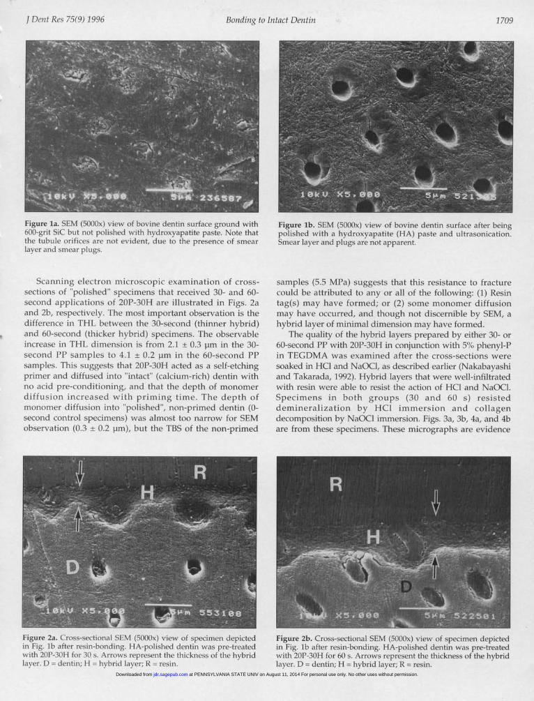
Freshly extracted bovine teeth were sequentially ground (180-to 600-grit SiC abrasive paper) under a stream of water so thatdentin would be exposed. Prepared dentin surfaces (Fig. la)were then "polished" for 30 s with 0.1 g of a hydroxyapatitepaste on a bristle brush (Kuroda Brush, Tokyo, Japan) rotatingat 1000 rpm, as previously reported (Nakabayashi, 1995;Nakabayashi et al., 1995b). The polishing paste was composedof 30 wt% pulverized dense hydroxyapatite particles (purity,99.9%0; Asahi Kogaku, Tokyo, Japan) with an average diameterof 0.1 to 0.2 mm, 49 wt% propylene glycol (Tokyo Kasei, Tokyo,Japan), and 21 wt% polyvinyl pyrrolidone (MW, 360,000; TokyoKasei, Tokyo, Japan) (Saito et al., 1993). The surfaces were thencarefully washed with water and placed in 10°C de-ionizedwater in an ultrasonic cleaner (Model 100, Kaneda Scientific Co.Ltd., Tokyo, Japan; output power, 100 W; frequency, 28 kHz) for30 min for removal of sticky residual surface contaminants andsmear plugs. Several samples of prepared dentinal surfaceswere examined by scanning electron microscopy (JSM-5400,JEOL, Tokyo, Japan) (Fig. lb).
Bonding of PMMA rods to "polished" bovine dentin
Double-sided tape (3M, St. Paul, MN, USA) having a 5.4-mm-diameter perforation was affixed to the "polished" dentinsurface. Ten pL of self-etching primer, an aqueous solution of20% 2-methacryloyloxyethyl phenyl phosphoric acid (phenyl-P;Yamauchi, 1986) and 300o 2-hydroxyethvl methacrylate(Mitsubishi Rayon, Tokyo, Japan) described as 20P-30H byWatanabe in 1992, was applied to prime the exposed dentinarea. Priming times were 0, 30, and 60 s. The 0-second controlspecimens received only photocure bonding agent; no self-etching primer (20P-30H) was applied. Thirty-second and 60-second specimens received 20P-30H application followed byphotocure bonding agent. Primed surfaces of the 30-second and60-second primer-treated samples were dried with a gentlestream of air, rather than being vigorously blown dry. A 6-mm-diameter poly(methyl methacrylate) ring was then affixed to thetape. The primed surfaces were painted with 20 pL of a
photocure bonding agent composed of 0.5 wt%camphorquinone (Tokyo Kasei, Tokyo, Japan), 0.5 wt0o N-phenylglycine (Sigma Chemical, St. Louis, MO, USA), and 5wt% phenyl-P in triethylene glycol dimethacrylate (TEGDMA,Shin-Nakamura Kagaku, Wakayama, Japan). This bondingagent was left undisturbed for 60 s, then light-cured with a Day-Light Lamp (Shofu, Kyoto, Japan) for 60 s. A resin composite(Clearfil Photo Bright, Kuraray, Osaka, Japan) was placed on
top of the bonding agent and irradiated for 60 s. Finally, a 6-mm-diameter acrylic rod (Mitsubishi Rayon, Tokyo, Japan) was
1707j Dent Res 75(9) 1996
at PENNSYLVANIA STATE UNIV on August 11, 2014 For personal use only. No other uses without permission.jdr.sagepub.comDownloaded from
1708 Nakabayashi & Saimi
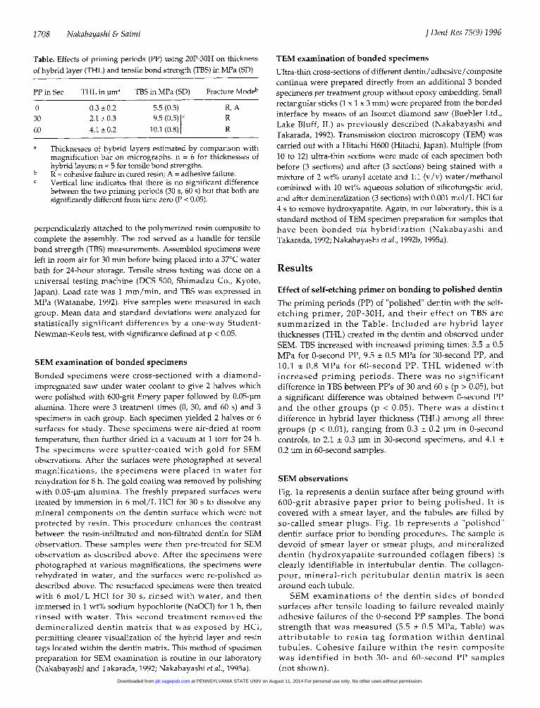
Table. Effects of priming periods (PP) using 20P-30H on thicknessof hybrid layer (THL) and tensile bond strength (TBS) in MPa (SD)
PP in Sec THL in plma TBS in MPa (SD) Fracture Modeb
0 0.3 ± 0.2 5.5 (0.5) R, A30 2.1 + 0.3 9.5 (0.5) c R60 4.1 ± 0.2 10.1 (0.8) R
a Thicknesses of hybrid layers estimated by comparison withmagnification bar on micrographs. n = 6 for thicknesses ofhybrid layers; n = 5 for tensile bond strengths.
b R = cohesive failure in cured resin; A = adhesive failure.c Vertical line indicates that there is no significant difference
between the two priming periods (30 s, 60 s) but that both aresignificantly different from time zero (P < 0.05).
perpendicularly attached to the polymerized resin composite tocomplete the assembly. The rod served as a handle for tensilebond strength (TBS) measurements. Assembled specimens wereleft in room air for 30 min before being placed into a 37°C waterbath for 24-hour storage. Tensile stress testing was done on auniversal testing machine (DCS 500, Shimadzu Co., Kyoto,Japan). Load rate was 1 mm/min, and TBS was expressed inMPa (Watanabe, 1992). Five samples were measured in eachgroup. Mean data and standard deviations were analyzed forstatistically significant differences by a one-way Student-Newman-Keuls test, with significance defined at p < 0.05.
SEM examination of bonded specimensBonded specimens were cross-sectioned with a diamond-impregnated saw under water coolant to give 2 halves whichwere polished with 600-grit Emery paper followed by 0.05-pmalumina. There were 3 treatment times (0, 30, and 60 s) and 3specimens in each group. Each specimen yielded 2 halves or 6surfaces for study. These specimens were air-dried at roomtemperature, then further dried in a vacuum at 1 torr for 24 h.The specimens were sputter-coated with gold for SEMobservations. After the surfaces were photographed at severalmagnifications, the specimens were placed in water forrehydration for 8 h. The gold coating was removed by polishingwith 0.05-pm alumina. The freshly prepared surfaces weretreated by immersion in 6 mol/L HCI for 30 s to dissolve anymineral components on the dentin surface which were notprotected by resin. This procedure enhances the contrastbetween the resin-infiltrated and non-filtrated dentin for SEMobservation. These samples were then pre-treated for SEMobservation as described above. After the specimens werephotographed at various magnifications, the specimens wererehydrated in water, and the surfaces were re-polished asdescribed above. The resurfaced specimens were then treatedwith 6 mol/L HCI for 30 s, rinsed with water, and thenimmersed in 1 wt% sodium hypochlorite (NaOCl) for 1 h, thenrinsed with water. This second treatment removed thedemineralized dentin matrix that was exposed by HCI,permitting clearer visualization of the hybrid layer and resintags located within the dentin matrix. This method of specimenpreparation for SEM examination is routine in our laboratory(Nakabayashi and Takarada, 1992; Nakabayashi et al., 1995a).
TEM examination of bonded specimens
Ultra-thin cross-sections of different dentin/adhesive/compositecontinua were prepared directly from an additional 3 bondedspecimens per treatment group without epoxy embedding. Smallrectangular sticks (1 x 1 x 3 mm) were prepared from the bondedinterface by means of an Isomet diamond saw (Buehler Ltd.,Lake Bluff, IL) as previously described (Nakabayashi andTakarada, 1992). Transmission electron microscopy (TEM) wascarried out with a Hitachi H600 (Hitachi, Japan). Multiple (from10 to 12) ultra-thin sections were made of each specimen bothbefore (3 sections) and after (3 sections) being stained with amixture of 2 wt% uranyl acetate and 1:1 (v/v) water/methanolcombined with 10 wt% aqueous solution of silicotungstic acid,and after demineralization (3 sections) with 0.001 mol/L HCI for4 s to remove hydroxyapatite. Again, in our laboratory, this is astandard method of TEM specimen preparation for samples thathave been bonded via hybridization (Nakabayashi andTakarada, 1992; Nakabayashi et al., 1992b, 1995a).
Results
Effect of self-etching primer on bonding to polished dentinThe priming periods (PP) of "polished" dentin with the self-etching primer, 20P-30H, and their effect on TBS aresummarized in the Table. Included are hybrid layerthicknesses (THL) created in the dentin and observed underSEM. TBS increased with increased priming times: 5.5 ± 0.5MPa for 0-second PP, 9.5 ± 0.5 MPa for 30-second PP, and10.1 ± 0.8 MPa for 60-second PP. THL widened withincreased priming periods. There was no significantdifference in TBS between PP's of 30 and 60 s (p > 0.05), buta significant difference was obtained between 0-second PPand the other groups (p < 0.05). There was a distinctdifference in hybrid layer thickness (THL) among all threegroups (p < 0.01), ranging from 0.3 ± 0.2 pm in 0-secondcontrols, to 2.1 ± 0.3 pm in 30-second specimens, and 4.1 +0.2 pm in 60-second samples.
SEM observationsFig. la represents a dentin surface after being ground with600-grit abrasive paper prior to being polished. It iscovered with a smear layer, and the tubules are filled byso-called smear plugs. Fig. lb represents a "polished"dentin surface prior to bonding procedures. The sample isdevoid of smear layer or smear plugs, and mineralizeddentin (hydroxyapatite-surrounded collagen fibers) isclearly identifiable in intertubular dentin. The collagen-poor, mineral-rich peritubular dentin matrix is seenaround each tubule.SEM examinations of the dentin sides of bonded
surfaces after tensile loading to failure revealed mainlyadhesive failures of the 0-second PP samples. The bondstrength that was measured (5.5 ± 0.5 MPa, Table) wasattributable to resin tag formation within dentinaltubules. Cohesive failure within the resin compositewas identified in both 30- and 60-second PP samples(not shown).
j Dent Res 75(9) 1996
at PENNSYLVANIA STATE UNIV on August 11, 2014 For personal use only. No other uses without permission.jdr.sagepub.comDownloaded from
Bondiitg to Inttact Dentin
Figure la. SEM (5000x) view of bovine dentin surface ground with600-grit SiC bUt not polished with hydroxyapatite paste. Note thatthe tubule orifices are not evident, due to the presence of smearlayer and smear plugs.
Scanning electron microscopic examination of cross-sections of "polished" specimens that received 30- and 60-second applications of 20P-30H are illustrated in Figs. 2aand 2b, respectively. The most important observation is thedifference in THL between the 30-second (thinner hybrid)and 60-second (thicker hybrid) specimens. The observableincrease in THL dimension is from 2.1 + 0.3 pm in the 30-second Pl' samples to 4.1 + 0.2 pm in the 60-second PPsamples. This suggests that 20P-30H acted as a self-etchingprimer and diffused into "intact" (calcium-rich) dentin withno acid pre-conditioning, and that the depth of monomerdiffusion increased with priming time. The depth ofmonomer diffusioll into "polished", non-primed dentin ((-second control specimens) was almost too narrow for SEMobservation ((1.3 ± 0.2 pm), but the TBS of the non-primed
-I - -Figure 2a. Cross-sectional SEM (5000x) view of specimen depictedin Fig. lb after resin-bonding. HA-polished dentin was pre-treatedwith 201'-30H foi 30 s. Arrows represent the thickniess of the hybridlaver. D = dentin; 11 hybrid layer; 1< resin.
Figure lb. SEM (5000x) view of bovine dentin surface after beingpolished with a hydroxyapatite (HA) paste and ultrasonication.Smear layer and plugs are not apparent.
samples (5.5 MPa) suggests that this resistance to fractLrecould be attributed to any or all of the following: (1) Resintag(s) may have formed; or (2) some monomer diffusionmay have occurred, and though not discernible by SEM, ahybrid layer of minimal dimension may have formed.
The quality of the hybrid layers prepared by either 30- or60-second PP with 2(P-30H in conjunction with 5"Yo phenyl-Pin TEGDMA was examined after the cross-sections weresoaked in HCI and NaOCI, as described earlier (Nakabayashiand Takarada, 1992). Hybrid layers that were well-infiltratedwith resin were able to resist the action of HCI and NaOCI.Specimens in both groups (30 and 60 s) resisteddemineralization by HCI immersion and collagendecomposition by NaOCI immersion. Figs. 3a, 3b, 4a, and 4bare from these specimens. These micrographs are evidence
Figure 2b. Cross-sectional SEM (50)(0x) view of specimen depictedin Fig. lb after resin-bonding. HA-polished dentin was pre-treatedwith 20P-30H for 60 s. Arrows represent the thickness of the hybridlayer. D dentin; H = hybrid layer; R resin.
j Dcizt Res 75(9) 1996 1 709
at PENNSYLVANIA STATE UNIV on August 11, 2014 For personal use only. No other uses without permission.jdr.sagepub.comDownloaded from

1710 Nakabayashi &SainiD
Figure 3a. Cross-sectional SEM (5000x) view of HA-polished, 20P-30Hpre-treated (30) s), resin-bonded specimen after HCI demineralization.DD = demineralized dentin; H = hybrid layer; R = resin.
that the formed hybrid layers were stable and of high quality.There was no evidence of the removal of any non-envelopedexposed collagen fibers, supporting previously publishedwork where the same techniques were used (Nakabayashiand Takarada, 1992; Nakabayashi et al., 1992a, 1995a). Ratherthan a sharp junction or interface between cured primer anddentin, one observes a continuum of surfaces.
TEM examination of bonded specimens
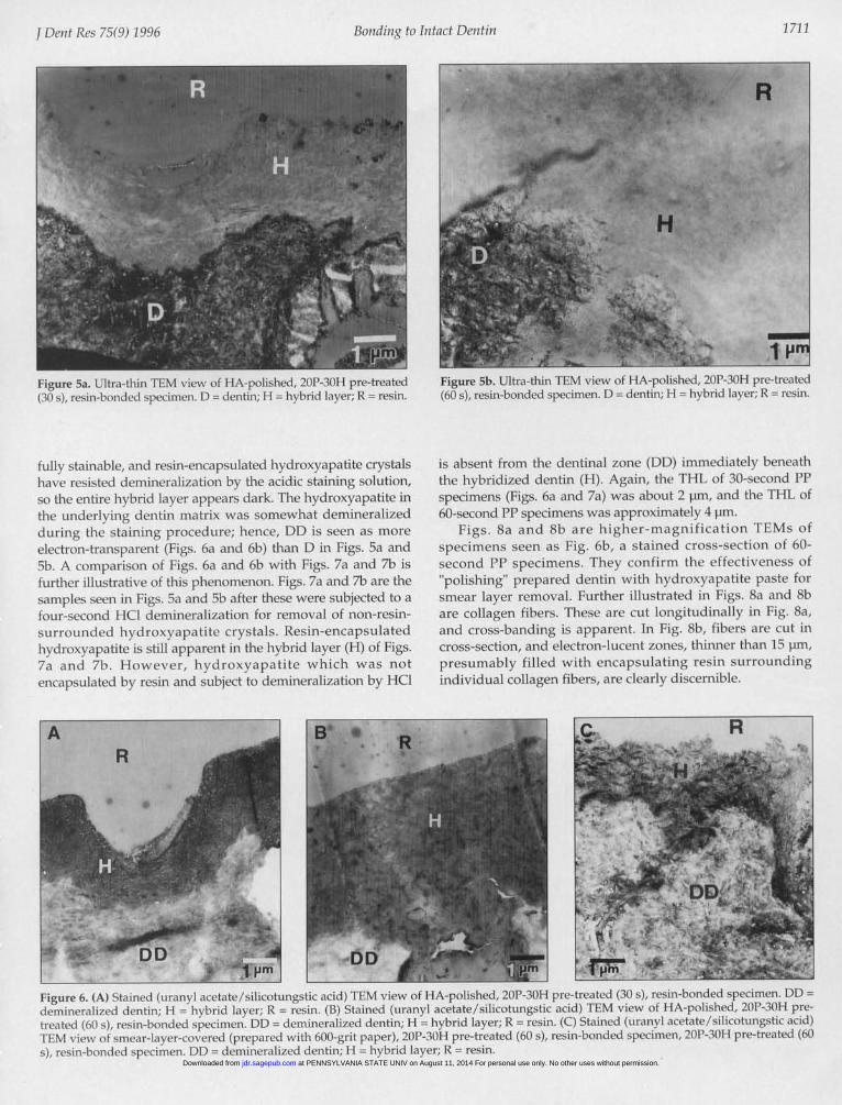
Figs. 5a and 5b show ultra-thin cross-sections of "polished"samples that have received priming with 20P-30H for 30 sand 60 s, respectively. They illustrate that the smear layer wasremoved by the hydroxyapatite-paste polishing procedure(compare Figs. 6a, 6b, and 6c). Resin (R) unites with hybrid
L: 0
t
Figure 4a. Cross-sectional SEM (5000x) view of HA-polished, 20P-30Hpre-treated (30 s), resin-bonded specimen after HCI demineralizationand NaOCI immersion to eliminate exposed organic material. DD =
demineralized dentin; H = hybrid layer; R = resin.
Figure 3b. Cross-sectional SEM (5000x) view of HA-polished, 20P-30Hpre-treated (60 s), resin-bonded specimen after HCI demineralization.DD = demineralized dentin; H = hybrid layer; R = resin.
(H) that is a continuation of calcium-rich dentin (D), with noapparent interfaces throughout the continuum. THL of the30-second PP sample (Fig. 5a) is about 2 pm, while that of the60-second PP specimen (Fig. 5b) is approximately 4 pm.
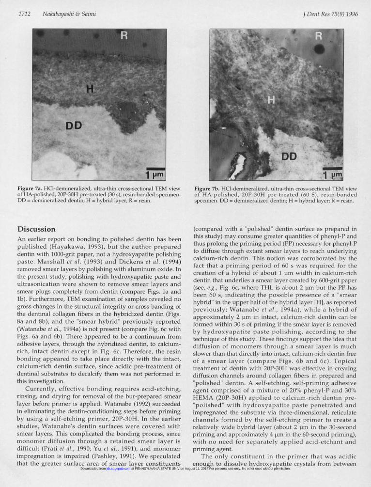
When the unstained specimens shown in Figs. 5a and 5bwere stained with heavy metals, they yielded the micrographsseen in Figs. 6a and 6b. Fig. 6c represents the same TEMpicture, but the smear layer (prepared by 600-grit paper) wasnot removed, and the priming period of 20P-30H was 60 s.The rough interface between the resin (R) and the hybrid (H)in Fig. 6c shows that it is due to the smear layer. However,once the smear layer was removed, the interfaces in Figs. 6aand 6b became smoother. These micrographs demonstrate anextremely intimate interface between the resin (R) and thehybrid layer (H). Collagen embedded in the copolymer is not
Figure 4b. Cross-sectional SEM (5000x) view of HA-polished, 20P-30H pre-treated (60 s), resin-bonded specimen after HCIdemineralization and NaOCI immersion to eliminate exposed organicmaterial. DD = demineralized dentin; H = hybrid layer; R = resin.
j Deiit Res 75(9) 1996
at PENNSYLVANIA STATE UNIV on August 11, 2014 For personal use only. No other uses without permission.jdr.sagepub.comDownloaded from
Bonding to Intact Dentin
Figure 5a. Ultra-thin TEM view of HA-polished, 20P-30H pre-treated(30 s), resin-bonded specimen. D dentin; H hybrid layer; R = resin.
fully stainable, and resin-encapsulated hydroxyapatite crystalshave resisted demineralization by the acidic staining solution,so the entire hybrid layer appears dark. The hydroxyapatite inthe underlying dentin matrix was somewhat demineralizedduring the staining procedure; hence, DD is seen as moreelectron-tranisparent (Figs. 6a and 6b) than D in Figs. 5a and5b. A comparison of Figs. 6a and 6b with Figs. 7a and 7b isfurther illustrative of this phenomenon. Figs. 7a and 7b are thesamples seen in Figs. 5a and 5b after these were subjected to afour-second HCI demineralization for removal of non-resin-surrounded hydroxyapatite crystals. Resin-encapsulatedhydroxyapatite is still apparent in the hybrid layer (H) of Figs.7a and 7b. However, hydroxyapatite which was notencapsulated by resin and subject to demineralization by HCl
Figure 5b. Ultra-thin TEM view of HA-polished, 20P-30H pre-treated(60 s), resin-bonded specimen. D = dentin; H = hybrid layer; R = resin.
is absent from the dentinal zone (DD) immediately beneaththe hybridized dentin (H). Again, the THL of 30-second PPspecimens (Figs. 6a and 7a) was about 2 pm, and the THL of60-second PP specimens was approximately 4 pm.
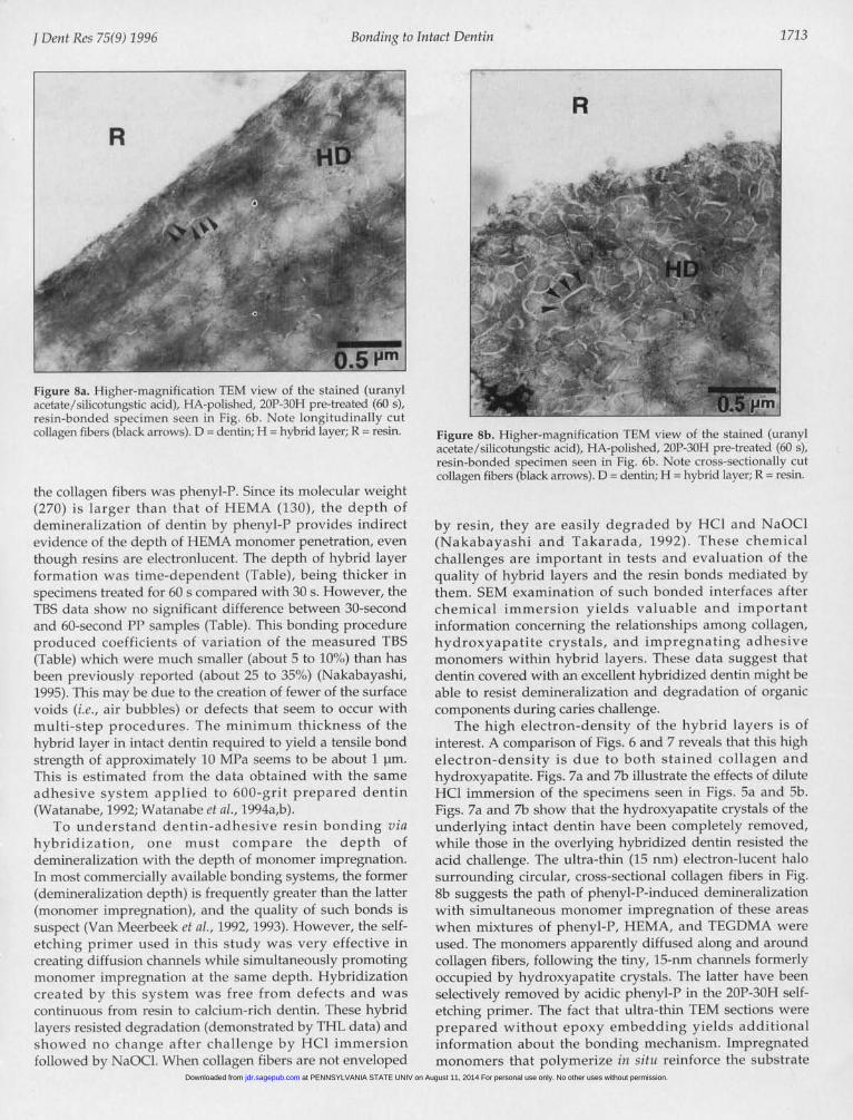
Figs. Sa and 8b are higher-magnification TEMs ofspecimens seen as Fig. 6b, a stained cross-section of 60-second PP specimens. They confirm the effectiveness of"polishing" prepared dentin with hydroxyapatite paste forsmear layer removal. Further illustrated in Figs. 8a and 8bare collagen fibers. These are cut longitudinally in Fig. 8a,and cross-banding is apparent. In Fig. 8b, fibers are cut incross-section, and electron-lucent zones, thinner than 15 ,pm,presumably filled with encapsulating resin surroundingindividual collagen fibers, are clearly discernible.
Figure 6. (A) Stained (uranyl acetate/silicotungstic acid) TEM view of HA-polished, 20P-30H pre-treated (30 s), resin-bonded specimen. DDdemineralized dentin; H = hybrid layer; R = resin. (B) Stained (uranyl acetate/silicotungstic acid) TEM view of HA-polished, 20P-30H pre-treated (60 s), resin-bonded specimen. DD = demineralized dentin; H = hybrid layer; R = resin. (C) Stained (uranyl acetate/silicotungstic acid)TEM view of smear-layer-covered (prepared with 600-grit paper), 20P-30H pre-treated (60 s), resin-bonded specimen, 20P-30H pre-treated (60s), resin-bonded specimen DD = demineralized dentin; H = hybrid layer; R resin.
j Dent Res 75(9) 1996
at PENNSYLVANIA STATE UNIV on August 11, 2014 For personal use only. No other uses without permission.jdr.sagepub.comDownloaded from
1712 Nakabayashi & Saimi
Figure 7a. HCI-demineralized, ultra-thin cross-sectional TEM viewof HA-polished, 20P-30H pre-treated (30 s), resin-bonded specimen.DD = demineralized dentin; H = hybrid layer; R = resin.
DiscussionAn earlier report on bonding to polished dentin has beenpublished (Hayakawa, 1993), but the author prepareddentin with 1000-grit paper, not a hydroxyapatite polishingpaste. Marshall et al. (1993) and Dickens et al. (1994)removed smear layers by polishing with aluminum oxide. Inthe present study, polishing with hydroxyapatite paste andultrasonication were shown to remove smear layers andsmear plugs completely from dentin (compare Figs. la andlb). Furthermore, TEM examination of samples revealed nogross changes in the structural integrity or cross-banding ofthe dentinal collagen fibers in the hybridized dentin (Figs.8a and 8b), and the "smear hybrid" previously reported(Watanabe et al., 1994a) is not present (compare Fig. 6c withFigs. 6a and 6b). There appeared to be a continuum fromadhesive layers, through the hybridized dentin, to calcium-rich, intact dentin except in Fig. 6c. Therefore, the resinbonding appeared to take place directly with the intact,calcium-rich dentin surface, since acidic pre-treatment ofdentinal substrates to decalcify them was not performed inthis investigation.
Currently, effective bonding requires acid-etching,rinsing, and drying for removal of the bur-prepared smearlayer before primer is applied. Watanabe (1992) succeededin eliminating the dentin-conditioning steps before primingby using a self-etching primer, 20P-30H. In the earlierstudies, Watanabe's dentin surfaces were covered withsmear layers. This complicated the bonding process, sincemonomer diffusion through a retained smear layer isdifficult (Prati et al., 1990; Yu et al., 1991), and monomerimpregnation is impaired (Pashley, 1991). We speculatedthat the greater surface area of smear layer constituents
rr- IrIIFigure 7b. HCI-demineralized, ultra-thin cross-sectional TEM viewof HA-polished, 20P-30H pre-treated (60 S), resin-bondedspecimen. DD = demineralized dentin; H = hybrid layer; R = resin.
(compared with a "polished" dentin surface as prepared inthis study) may consume greater quantities of phenyl-P andthus prolong the priming period (PP) necessary for phenyl-l'to diffuse through extant smear layers to reach underlyingcalcium-rich dentin. This notion was corroborated by thefact that a priming period of 60 s was required for thecreation of a hybrid of about 1 pm width in calcium-richdentin that underlies a smear layer created by 600-grit paper(see, e.g., Fig. 6c, where THL is about 2 pm but the PP hasbeen 60 s, indicating the possible presence of a "smearhybrid" in the upper half of the hybrid layer [HI, as reportedpreviously; Watanabe et al., 1994a), while a hybrid ofapproximately 2 pm in intact, calcium-rich denitini can beformed within 30 s of priming if the smear layer is removedby hydroxyapatite paste polishing, according to thetechnique of this study. These findings support the idea thatdiffusion of monomers through a smear layer is muchslower than that directly into intact, calcium-riclh dentin freeof a smear layer (compare Figs. 6b and 6c). Topicaltreatment of dentin with 20P-30H was effective in creatingdiffusion channels around collagen fibers in prepared and"polished" dentin. A self-etching, self-priming adhesiveagent comprised of a mixture of 20%, phenyl-P and 30'X)HEMA (20P-30H) applied to calcium-rich dentin pre-"polished" with hvdroxyapatite paste penetrated andimpregnated the substrate via three-dimensionial, reticulatechannels formed by the self-etching primer to create arelatively wide hybrid layer (about 2 pm in the 30-secondpriming and approximately 4 pm in the 60-second priming),with no need for separately applied acid-etchant andpriming agent.
The only constituent in the primer that was acidicenough to dissolve hydroxyapatite crystals from between
I Dent Res 75(9) 1996
at PENNSYLVANIA STATE UNIV on August 11, 2014 For personal use only. No other uses without permission.jdr.sagepub.comDownloaded from
Bonding to Intact Dentin
Figure 8a. Higher-magnification TEM view of the stained (uranylacetate/silicotungstic acid), HA-polished, 20P-30H pre-treated (60 s),resin-bonded specimen seen in Fig. 6b. Note longitudinally cutcollagen fibers (black arrows). D dentin; H = hybrid layer; R = resin.
the collagen fibers was phenyl-P. Since its molecular weight(270) is larger than that of HEMA (130), the depth ofdemineralization of dentin by phenyl-P provides indirectevidence of the depth of HEMA monomer penetration, eventhough resins are electronlucent. The depth of hybrid layerformation was time-dependent (Table), being thicker inspecimens treated for 60 s compared with 30 s. However, theTBS data show no significant difference between 30-secondand 60-second PP samples (Table). This bonding procedureproduced coefficients of variation of the measured TBS(Table) which were much smaller (about 5 to 10'S) than hasbeen previously reported (about 25 to 35/,,) (Nakabayashi,1995). This may be due to the creation of fewer of the surfacevoids (i.c., air bubbles) or defects that seem to occur withmulti-step procedures. The minimum thickness of thehybrid layer in intact dentin required to yield a tensile bondstrength of approximately 10 MPa seems to be about 1 pm.This is estimated from the data obtained with the sameadhesive system applied to 600-grit prepared dentin(Watanabe, 1992; Watanabe et al., 1994a,b).
To understand dentin-adhesive resin bonding viahybridization, one must compare the depth ofdemineralization with the depth of monomer impregnation.In most commercially available bonding systems, the former(demineralization depth) is frequently greater than the latter(monomer impregnation), and the quality of such bonds issuspect (Van Meerbeek et al., 1992, 1993). However, the self-etching primer used in this study was very effective increating diffusion channels while simultaneously promotingmonomer impregnation at the same depth. Hybridizationcreated by this system was free from defects and wascontinuous from resin to calcium-rich dentin. These hybridlayers resisted degradation (demonstrated by THL data) andshowed no change after challenge by HCI immersionfollowed by NaOCI. When collagen fibers are not enveloped
Figure 8b. Higher-magnification TEM view of the stained (uranylacetate/silicotungstic acid), HA-polished, 20P-30H pre-treated (60 s),resin-bonded specimen seen in Fig. 6b. Note cross-sectionally cutcollagen fibers (black arrows). D = dentin; H = hybrid layer; R resin.
by resin, they are easily degraded by HCI and NaOCI(Nakabayashi and Takarada, 1992). These chemicalchallenges are important in tests and evaluation of thequality of hybrid layers and the resin bonds mediated bythem. SEM examination of such bonded interfaces afterchemical immersion yields valuable and importantinformation concerning the relationships among collagen,hydroxyapatite crystals, and impregnating adhesivemonomers within hybrid layers. These data suggest thatdentin covered with an excellent hybridized dentin might beable to resist demineralization and degradation of organiccomponents during caries challenge.
The high electron-density of the hybrid layers is ofinterest. A comparison of Figs. 6 and 7 reveals that this highelectron-density is due to both stained collagen andhydroxyapatite. Figs. 7a and 7b illustrate the effects of diluteHCI immersion of the specimens seen in Figs. 5a and 5b.Figs. 7a and 7b show that the hydroxyapatite crystals of theunderlying intact dentin have been completely removed,while those in the overlying hybridized dentin resisted theacid challenge. The ultra-thin (15 nm) electron-lucent halosurrounding circular, cross-sectional collagen fibers in Fig.8b suggests the path of phenyl-P-induced demineralizationwith simultaneous monomer impregnation of these areaswhen mixtures of phenyl-P, HEMA, and TEGDMA wereused. The monomers apparently diffused along and aroundcollagen fibers, following the tiny, 15-nm channels formerlyoccupied by hydroxyapatite crystals. The latter have beenselectively removed by acidic phenyl-P in the 20P-30H self-etching primer. The fact that ultra-thin TEM sections wereprepared without epoxy embedding yields additionalinformation about the bonding mechanism. Impregnatedmonomers that polymerize in sittu reinforce the substrate
1713j Detit Rcs 75(9) 1996
at PENNSYLVANIA STATE UNIV on August 11, 2014 For personal use only. No other uses without permission.jdr.sagepub.comDownloaded from
1714 Nakabayashi & Saimi
and make direct preparation of these TEM sections possiblewithout embedding procedures. Without such monomerinfiltration and in situ polymerization, it was not possible toprepare TEM sections directly (Nakabayashi and Watanabe,1983), since the specimens lacked physical support and weretorn apart by the diamond knife. This reinforcingphenomenon suggests that hybrid layers achieve efficientand reliable bonds, and that the acid-resistance of hybridlayers might similarly resist recurrent caries and post-operative sensitivity in the clinical situation.
Adhesion of resins to dentin is carried out bypolymerization of liquid monomers after their penetrationin the matrix. In this study, the photo-cured bonding agentwas the same in all cases. However, the priming periods(PP) of 0 s, 30 s, and 60 s for 20P-30H differed. The dataindicate that the monomer mixture used in this workdiffused at least 2 pm into the substrate beforepolymerization to achieve good adhesion.
While it is theoretically possible to have bonding due tochemical reaction of phenyl-P with hydroxyapatite, since thephenyl-P contains a reactive phosphoric acid group (Kramerand McLean, 1952; Buonocore and Quigley, 1958), Figs. 5aand 5b clearly indicate that the phenyl-P in the self-etchingprimer, 20P-30H, removed the hydroxyapatite crystals.Collagen fibers and electron-lucent resin copolymers (Figs.8a and 8b) might be likened to intertwining rope braids thatbind resin and dentin together. This is the most likelymechanism of bonding at work, as demonstrated by thisinvestigation, rather than a chemical union betweencopolymers and hydroxyapatite. If chemical reaction at theinterface was essential for adhesion, higher TBS at 0-secondPP should have occurred, since there is more than enoughhydroxyapatite at the "polished" dentin surface to react withphenyl-P, and it is directly accessible to the bonding system,since no intervening smear layer was present to compromisethe presumed chemical bond (Misra, 1989; Eliades et al.,1990; Eick et al., 1991; Amory and Yvon, 1994).
AcknowledgmentsPart of this study was supported by a Grant-in-Aid forScientific Research from the Ministry of Education, Science,Sports and Culture, Japan (05453213). The authors wouldlike to express their appreciation for this support.
ReferencesAmory C, Yvon J (1994). Shear bond strength of a light-cured
composite vs. dentin characteristics. Dent Mater 10:203-209.Buonocore NG, Quigley N (1958). Bonding of a synthetic resin
material to human dentin: preliminary histological study ofthe bond area. JAm Dent Assoc 57:807-811.
Burrow NF, Tagami J, Hosoda H (1993). The long termdurability of bond strengths to dentin. Bull Tokyo Med DentUniv 40:173-191.
Chigira H, Yukitani W, Hasegawa T, Manabe A, Itoh K,Hayakawa T, et al. (1994). Self-etching dentin primerscontaining phenyl-P. J Dent Res 73:1088-1095.
Ciucchi B, Sano H, Pashley DH (1994). Bonding to sodium
hypochlorite treated dentin (abstract). J Dent Res 73(SpecIss):296.
Cobb DS, Vargas MA, Armstrong SR (1995). Shear bondstrengths between acid-etched and deproteinized dentinsurfaces (abstract). J Dent Res 74(Spec Iss):35.
Dickens S, Stangel I, Poulin S, Sacher E, Bowen RL (1994). Bondstrength and surface analyses of unaltered and chemically-and non-chemically-altered dentin (abstract). J Dent Res73(Spec Iss):277.
Eick JD, Cobb CM, Chappell RP, Spencer P, Robinson SJ (1991).The dentinal surface: Its influence on adhesion. Part 1.Quintessence Int 22:967-977.
Eick JD, Robinson SJ, Byerley TJ, Chappell RP, Spencer P,Chappelow CC (1995). Scanning transmission electronmicroscopy/energy dispersive spectroscopy analysis of thedentin adhesive interface using a labeled 2-hydroxyethylmethacrylate analogue. J Dent Res 74:1246-1252.
Eliades G, Palaghias G, Vougiouklakis G (1990). Surfacereactions of adhesives on dentin. Dent Mater 6:208-216.
Gwinnett AJ (1994). Chemically conditioned dentin. Acomparison of conventional and environmental scanningelectron microscope findings. Dent Mater 10:150-155.
Gwinnett AJ, Tay FR, Pang KM, Wei SHY (1995). Quantitativecontribution of the collagen network in dentinhybridization (abstract). J Dent Res 74(Spec Iss):403.
Hayakawa T (1993). Adhesion between the resin composite andpolished dentin without pretreatment. SJ Jpn Dent Mater12:455-465.
Inokoshi S, Hosoda H, Harnirattisai C, Shimada Y (1993).Interfacial structure between dentin and seven dentinbonding systems revealed using argon ion beam etching.Oper Dent 18:8-16.
Jacobsen T, Finger WJ (1993). Morphology of coupling sitesbetween bonding agents and dentine in vivo and in vitro. JDent 21:150-157.
Kiyomura M (1987). Bonding strength to bovine dentin with 4-META/MMA-TBB resin-long-term stability and influenceof water. Si Jpn Dent Mater 6:860-872.
Kramer IRH, McLean JW (1952). Alterations in the stainingreaction of dentine resulting from a constituent of a newself-polymerising resin. Br Dent 1 93:150-153.
Manabe A, Katsuno K, Itoh K, Wakumoto S, Miyasaka T (1991).Bonding efficacy of erythritol methacrylate solutions asdentin primers. J Dent Res 70:1294-1298.
Marshall GW Jr, Balooch N, Tench RJ, Kinney JH, Marshall SJ(1993). Atomic force microscopy of acid effects on dentin.Dent Mater 9:265-268.
Misra DN (1989). Adsorption of 4-methacryloyloxyethyltrimellitate anhydride (4-META) on hydroxyapatite and itsrole in composite bonding. J Dent Res 68:42-47.
Morra M (1993). Acid-base properties of adhesive dentalpolymers. Dent Mater 9:375-378.
Munksgaard EC, Asmussen E (1984). Bonding strength betweendentin and restorative resins mediated by mixtures ofHEMA and glutaraldehyde. J Dent Res 63:1087-1089.
Nakabayashi N (1992). The hybrid layer: A resin-dentincomposite. Proc Finn Dent Soc 88:321-329.
Nakabayashi N (1993). Dental biomaterials. In: Biomedicalapplication of polymeric materials. Tsuruta T, Hayashi T,
j Dent Res 75(9) 1996
at PENNSYLVANIA STATE UNIV on August 11, 2014 For personal use only. No other uses without permission.jdr.sagepub.comDownloaded from

Bonding to Intact Dentin
Kataoka K, Ishihara K, Kimura Y, editors. Boca Raton, FL:CRC Press, pp. 220-255.
Nakabayashi N (1995). Bonding to dentin with minimal tensilebond strength deviation (abstract). J Dent Res 74(Spec Iss):413.
Nakabayashi N, Watanabe A (1983). SEM and TEM observationof dentin surface treated for adhesion. Rep Inst Med DentEntg 17:45-55.
Nakabayashi N, Takarada K (1992). Effect of HEMA on bondingto dentin. Dent Mater 8:125-130.
Nakabayashi N, Kojima K, Masuhara E (1982). The promotionof adhesion by the infiltration of monomers into toothsubstrates. J Biomed Mater Res 16:265-273.
Nakabavashi N, Watanabe A, Gendusa NJ (1992a). Dentinadhesion of "modified" 4-META/MMA-TBB resin: functionof HEMA. Dent Mater 8:259-264.
Nakabayashi N, Ashizawa M, Nakamura M (1992b).Identification of a resin-dentin hybrid layer in vital humandentin created in vivo: Durable bonding to vital dentin.Qlulintessenice Int 23:135-141.
Nakabayashi N, Watanabe A, Ikeda W (1995a). Intraoralbonding of 4-META/MMA-TBB resin to vital humandentin. Am J Dent 8:37-42.
Nakabayashi N, Fujii B, Horiuchi H, Ishikawa I, Suda H,Yamamoto T, et al. (1995b). Occlusion of dentinal tubulesvwith reactive polymer emulsion-In vitro evaluation withSEM observation of treated surfaces. jpn J Conserv Dent38:1538-1548.
Nishiyama N, Asakura T, Suzuki K, Horie K, Nemoto K (1995).Effects of structural change in collagen upon binding toconditioned dentin studied by 13C NMR. J Biomed Mater Res29:107-111.
Pashlev DH (1991). Dentin bonding: overview of the substratewith respect to adhesive material. J Esthet Dent 3:46-50.
Pashley DH, Ciucchi B, Sano H, Horner JA (1993). Permeabilityof dentin to adhesive agents. Quintessence Int 24:618-631.
Perinka L, Sano H, Hosoda H (1992). Dentin thickness,hardness, and Ca-concentration vs. bond strength of dentinadhesives. Dent Mater 8:229-233.
Prati C, Biagini G, Rizzoli C, Nucci C, Zucchini C, Montanari G(1990). Shear bond strength and SEM evaluation ofdentinal bonding systems. Am i Dent 3:283-288.
Saito Y, Hotta K, Soma R, Nakabayashi N (1993). Effect ofphosphoric acid concentration on adhesion to polishedhuman enamel surface. Shika Zairyo 12:418-423.
Sano H, Shono T, Takatsu T, Hosoda H (1994). Microporous dentinzone beneath resin impregnated layer. Oper Dent 19:59-64.
Sasazaki H (1985). Adaptation of adhesive composite resin todentinal wall. 1. Bond strength and appearance of gap. JpnJ Conserv Dent 28:452-478.
Suzuki K, Takahashi M, Nakai H (1990). Treatment of dentin byaqueous solution of amino acid derivative-HEMA. AdhesDent 8:43-51.
Takarada K, Kojima M, Ishihara K, Nakabayashi N (1990).Durability of bonding between 4-META/MMA-TBB resinto dentin pretreated with 10-3. The effect of 10-3pretreating period and subsequent glutaraldehydetreatment. J Jpn Dent Mater 9:831-840.
Takarada K (1990). Stable adhesion to dentin-Combination ofEDTA 3-2 (NH4/Fe) pretreatment and 2% 4-META/MMA-TBB resin. J Jpn Dent Mater 9:841-849.
Titley K, Chernecky R, Maric B, Valiquette N, Smith D (1994).The morphology of the demineralized layer in primeddentin. Am J Dent 7:22-26.
Toida T, Watanabe A, Nakabayashi N (1995). Effect of smearlayer on bonding to dentin prepared with bur. J Jpni DenitMater 14:109-116.
Van Meerbeek B, Inokoshi S, Braem M, Lambrechts P, VanHerle G (1992). Morphological aspects of the resin-dentininterdiffusion zone with different dentin adhesive systems.J Dent Res 71:1530-1540.
Van Meerbeek B, Dhem A, Goret-Nicaise N, Braem M,Lambrechts P, Van Herle G (1993). Comparative SEM andTEM examination of the ultrastructure of the resin-dentininterdiffusion zone. J Dent Res 72:495-501.
Watanabe 1 (1992). Photocure bonding agents to ground dentin.J Jpn Dent Mater 11:955-973.
Watanabe I, Takarada K, Nakabayashi N (1991). Adhesion of 4-META/MMA-TBB resin to dentin treated with phosphoricacid. J Jpn Dent Mater 10:671-677.
Watanabe I, Nakabayashi N, Pashley DH (1994a). Bonding toground dentin by a phenyl-P self-etching primer. J Denit Res73:1212-1220.
Watanabe I, Saimi Y, Nakabayashi N (1994b). Effect of smearlayer on bonding to ground dentin Relationship betweengrinding condition and tensile bonding strength. J Jpni DenltMater 13:101-108.
Yamauchi J (1986). Study of dental adhesive resin containingphosphoric acid methacrylate monomer. J Jpni Denit Mater5:144-154.
Yu XY, Davis EL, Joynt RB, Wieczkowski G (1991). Bondstrength evaluation of a class V composite resin restoration.Quintessence Int 22:391-396.
I Dent Res 75(9) 1996 1715
at PENNSYLVANIA STATE UNIV on August 11, 2014 For personal use only. No other uses without permission.jdr.sagepub.comDownloaded from