Embed Size (px)
Citation preview

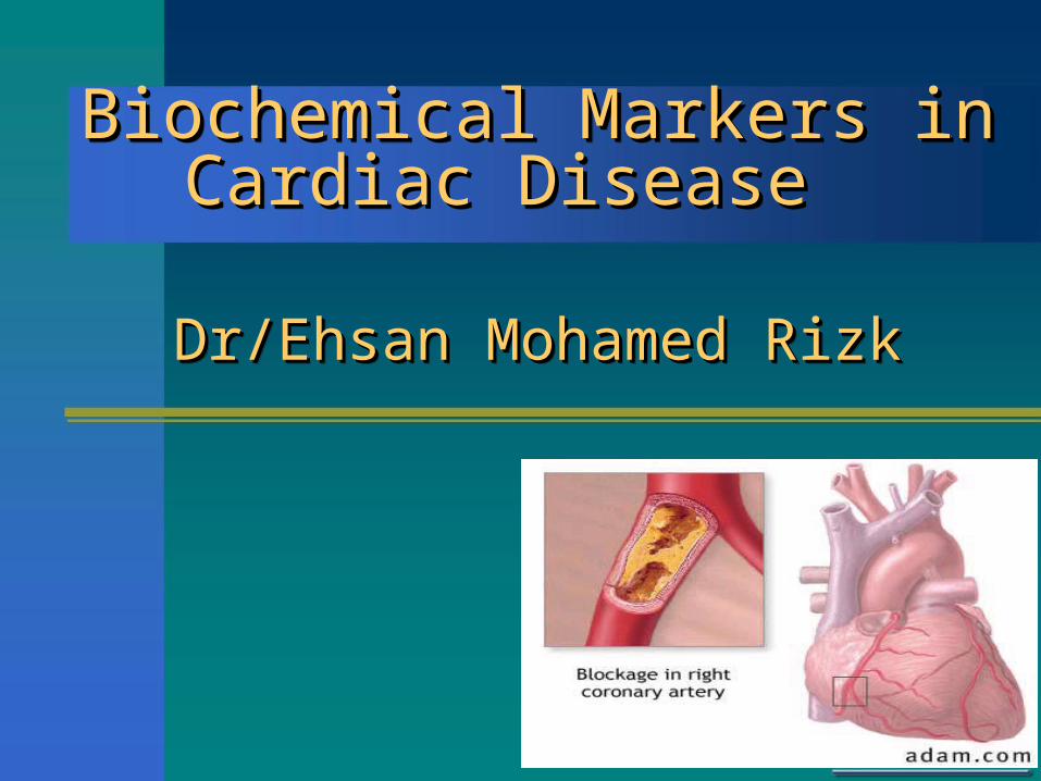
Biochemical Markers in Biochemical Markers in Cardiac DiseaseCardiac Disease
Biochemical Markers in Biochemical Markers in Cardiac DiseaseCardiac Disease
Dr/Ehsan Mohamed RizkDr/Ehsan Mohamed Rizk Dr/Ehsan Mohamed RizkDr/Ehsan Mohamed Rizk
ACUTE CORONARY SYNDROME ACUTE CORONARY SYNDROME (ACS) (ACS)
ACUTE CORONARY SYNDROME ACUTE CORONARY SYNDROME (ACS) (ACS)
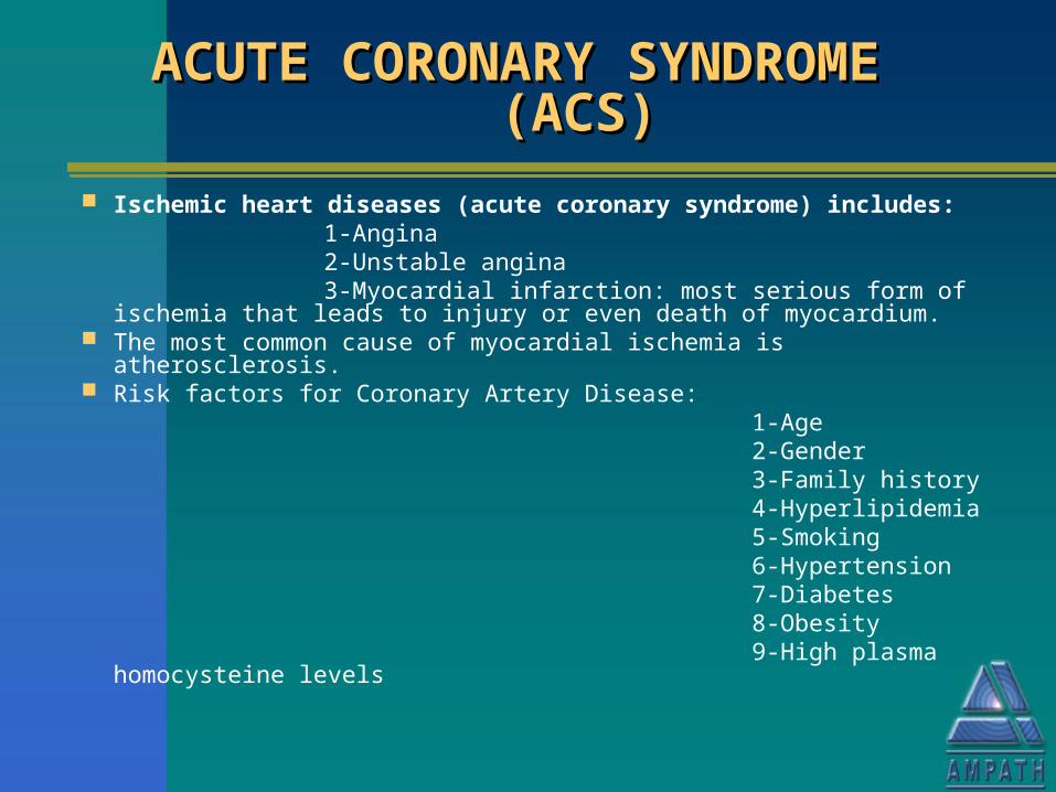
Ischemic heart diseases (acute coronary syndrome) includes: 1-Angina 2-Unstable angina 3-Myocardial infarction: most serious form of ischemia that leads to
injury or even death of myocardium. The most common cause of myocardial ischemia is atherosclerosis. Risk factors for Coronary Artery Disease: 1-Age 2-Gender 3-Family history 4-Hyperlipidemia 5-Smoking 6-Hypertension 7-Diabetes 8-Obesity 9-High plasma homocysteine levels
CRITERIA FOR DIAGNOSIS OF ACSCRITERIA FOR DIAGNOSIS OF ACSCRITERIA FOR DIAGNOSIS OF ACSCRITERIA FOR DIAGNOSIS OF ACS
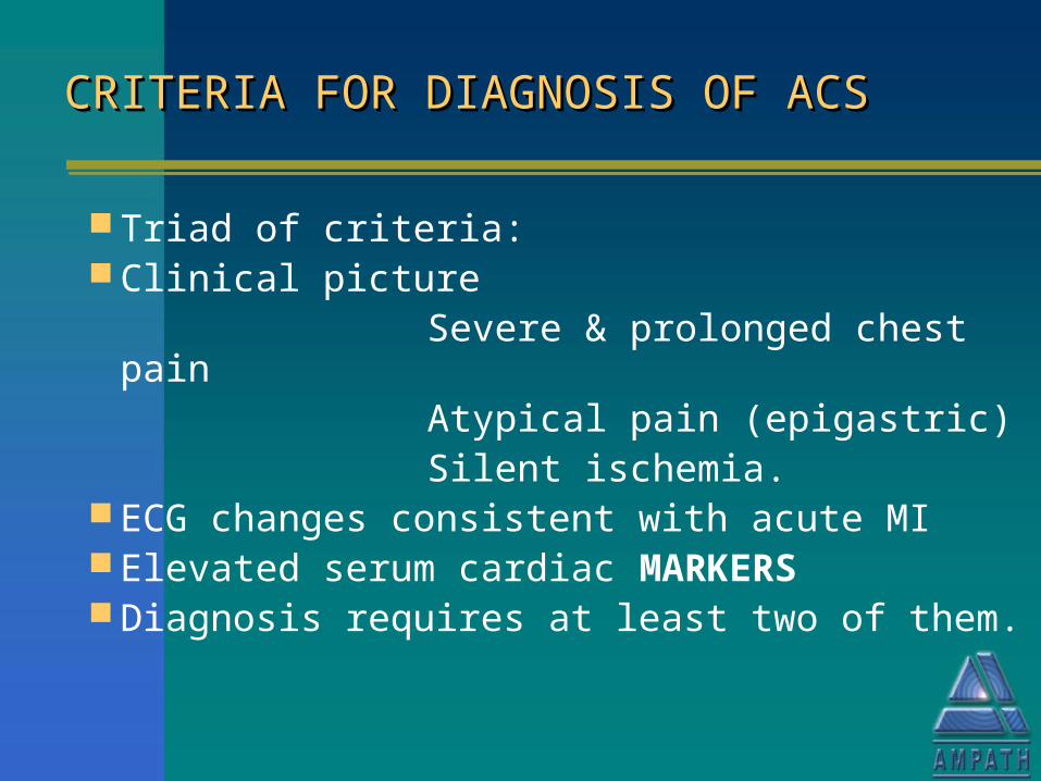
Triad of criteria: Clinical picture Severe & prolonged chest pain Atypical pain (epigastric) Silent ischemia. ECG changes consistent with acute MI Elevated serum cardiac MARKERS Diagnosis requires at least two of them.
CARDIAC MARKERS MUST CARDIAC MARKERS MUST BE:BE:CARDIAC MARKERS MUST CARDIAC MARKERS MUST BE:BE:
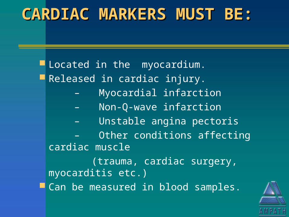
Located in the myocardium. Released in cardiac injury.
– Myocardial infarction
– Non-Q-wave infarction
– Unstable angina pectoris
– Other conditions affecting cardiac muscle
(trauma, cardiac surgery, myocarditis etc.) Can be measured in blood samples.
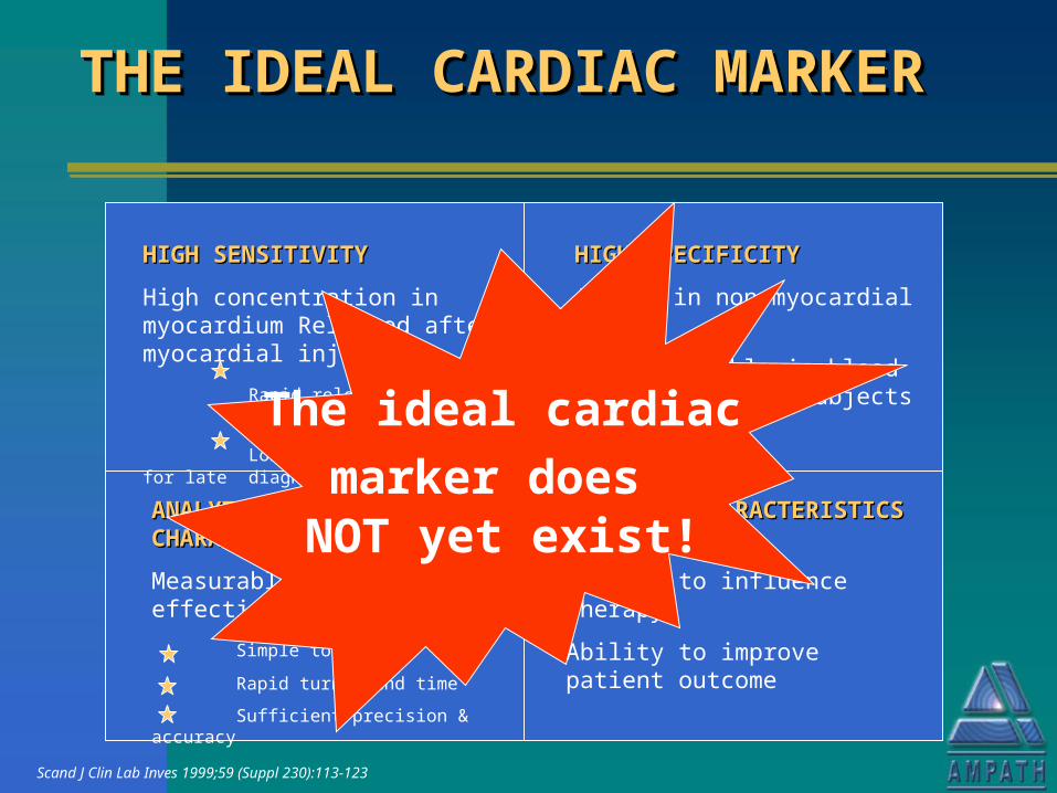
THE IDEAL CARDIAC THE IDEAL CARDIAC MARKERMARKERTHE IDEAL CARDIAC THE IDEAL CARDIAC MARKERMARKER
HIGH SENSITIVITYHIGH SENSITIVITY
High concentration in myocardium Released after myocardial injury:
Rapid release for early diagnosis
Long half-life in blood for late diagnosis
HIGH SPECIFICITYHIGH SPECIFICITY
Absent in non-myocardial tissue
Not detectable in blood of non-diseased subjects
CLINICAL CHARACTERISTICSCLINICAL CHARACTERISTICS fk
Ability to influence therapy
Ability to improve patient outcome
ANALYTICAL ANALYTICAL CHARACTERISTICSCHARACTERISTICS
Measurable by cost-effective method
Simple to perform
Rapid turnaround time
Sufficient precision & accuracy
The ideal cardiacmarker does NOT yet exist!
Scand J Clin Lab Inves 1999;59 (Suppl 230):113-123
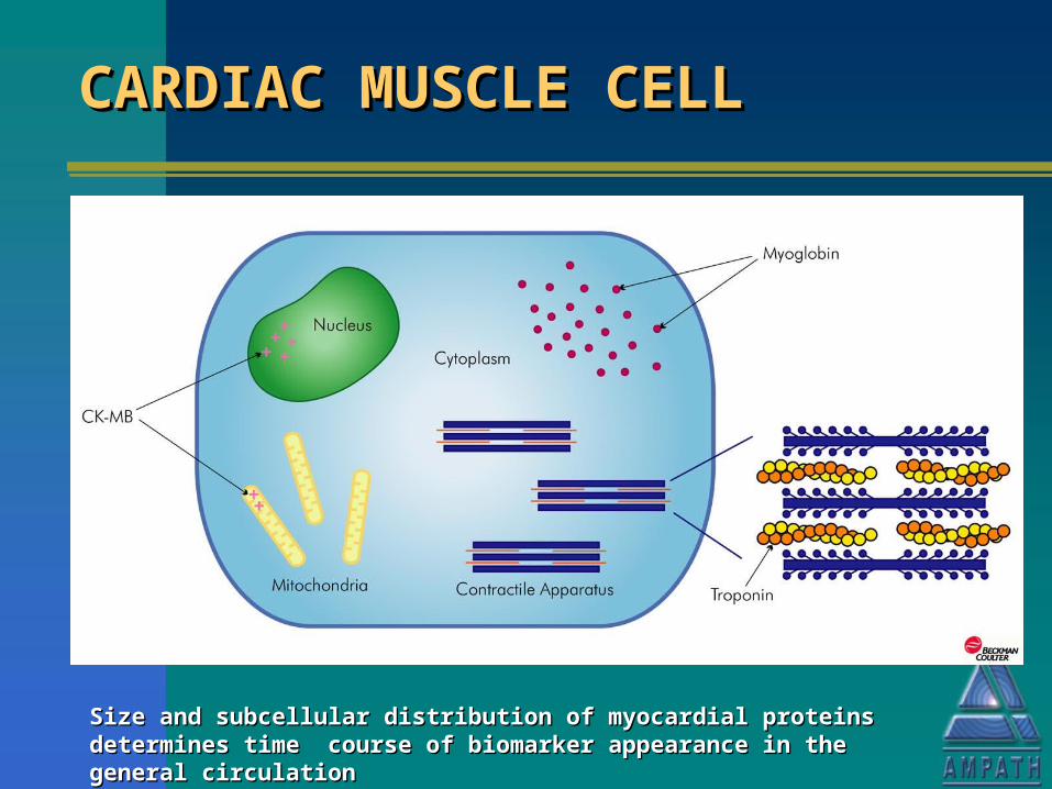
CARDIAC MUSCLE CELLCARDIAC MUSCLE CELLCARDIAC MUSCLE CELLCARDIAC MUSCLE CELL
Size and subcellular distribution of myocardial proteins determines time Size and subcellular distribution of myocardial proteins determines time course of biomarker appearance in the general circulationcourse of biomarker appearance in the general circulation
CLASSIFICATION OF LABORATORY CLASSIFICATION OF LABORATORY TESTS IN CARDIAC DISEASETESTS IN CARDIAC DISEASECLASSIFICATION OF LABORATORY CLASSIFICATION OF LABORATORY TESTS IN CARDIAC DISEASETESTS IN CARDIAC DISEASE
Markers of cardiac tissue damage
Markers of myocardial function
Cardiovascular risk factor markers
Genetic analysis for candidate genes or risk factors
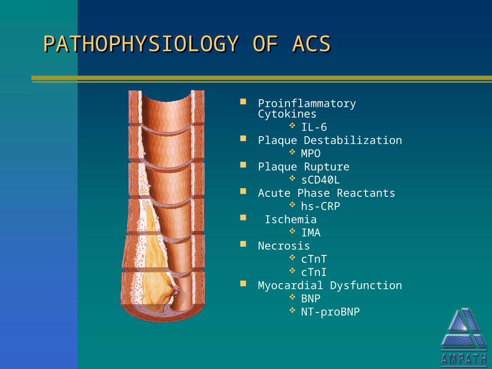
PATHOPHYSIOLOGY OF ACSPATHOPHYSIOLOGY OF ACSPATHOPHYSIOLOGY OF ACSPATHOPHYSIOLOGY OF ACS
Proinflammatory Cytokines IL-6
Plaque Destabilization MPO
Plaque Rupture sCD40L
Acute Phase Reactants hs-CRP
Ischemia IMA
Necrosis cTnT cTnI
Myocardial Dysfunction BNP NT-proBNP
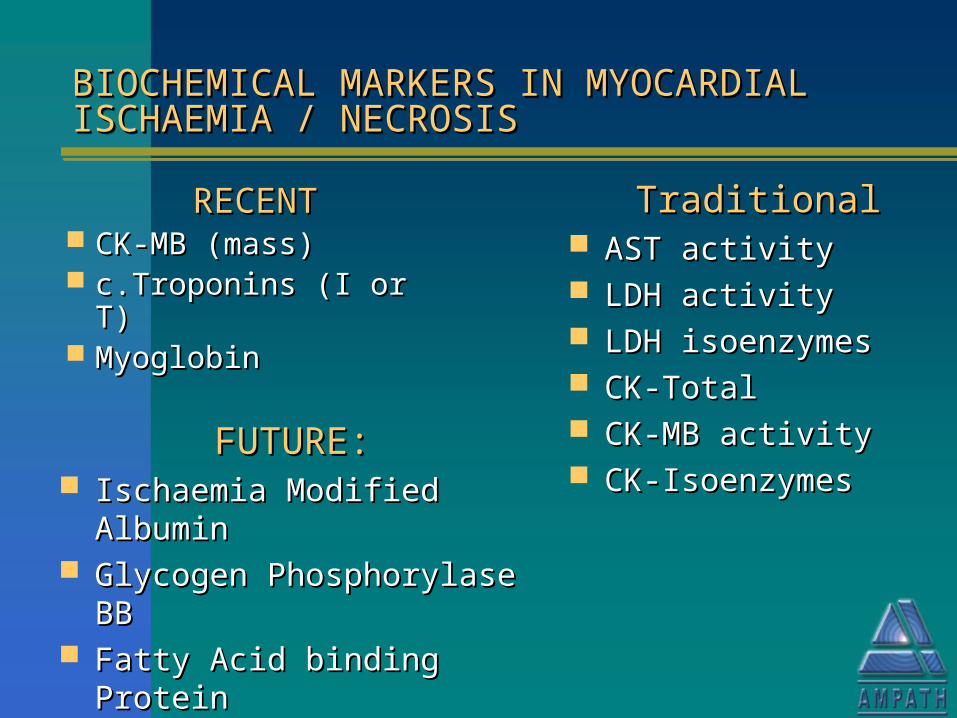
BIOCHEMICAL MARKERS IN MYOCARDIAL BIOCHEMICAL MARKERS IN MYOCARDIAL ISCHAEMIA / NECROSISISCHAEMIA / NECROSISBIOCHEMICAL MARKERS IN MYOCARDIAL BIOCHEMICAL MARKERS IN MYOCARDIAL ISCHAEMIA / NECROSISISCHAEMIA / NECROSIS
RECENTRECENT CK-MB (mass)CK-MB (mass) c.Troponins (I or T)c.Troponins (I or T) MyoglobinMyoglobin
TraditionalTraditional AST activityAST activity LDH activityLDH activity LDH isoenzymesLDH isoenzymes CK-TotalCK-Total CK-MB activityCK-MB activity CK-IsoenzymesCK-Isoenzymes
FUTURE:FUTURE: Ischaemia Modified AlbuminIschaemia Modified Albumin Glycogen Phosphorylase BBGlycogen Phosphorylase BB Fatty Acid binding ProteinFatty Acid binding Protein Highly sensitive CRP.Highly sensitive CRP.
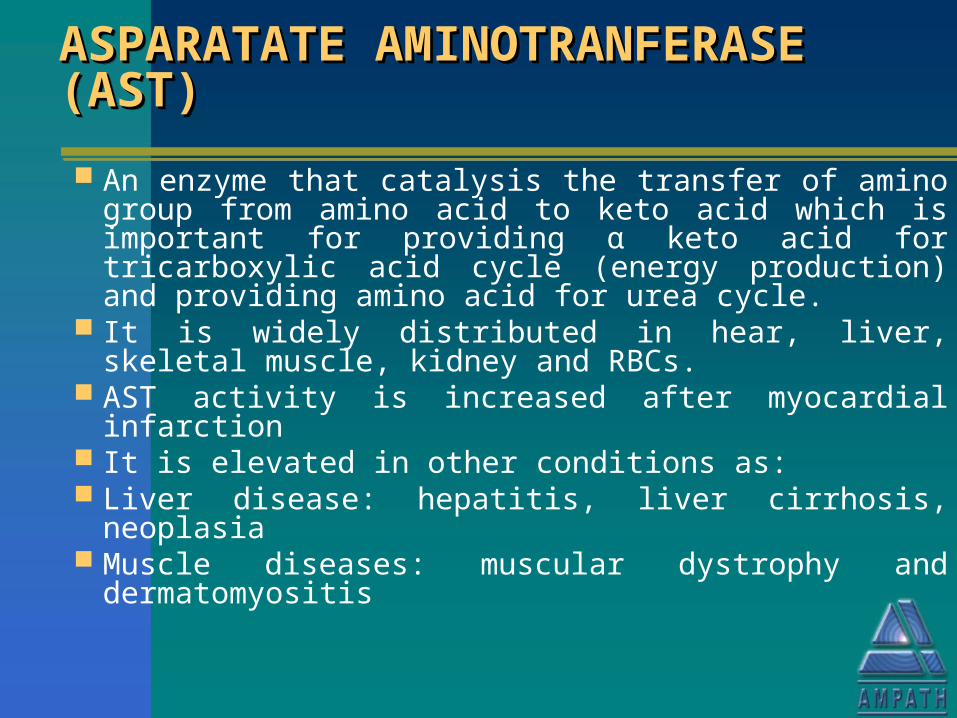
ASPARATATE ASPARATATE AMINOTRANFERASE (AST)AMINOTRANFERASE (AST)ASPARATATE ASPARATATE AMINOTRANFERASE (AST)AMINOTRANFERASE (AST)
An enzyme that catalysis the transfer of amino group from amino acid to keto acid which is important for providing α keto acid for tricarboxylic acid cycle (energy production) and providing amino acid for urea cycle.
It is widely distributed in hear, liver, skeletal muscle, kidney and RBCs.
AST activity is increased after myocardial infarction It is elevated in other conditions as: Liver disease: hepatitis, liver cirrhosis, neoplasia Muscle diseases: muscular dystrophy and
dermatomyositis
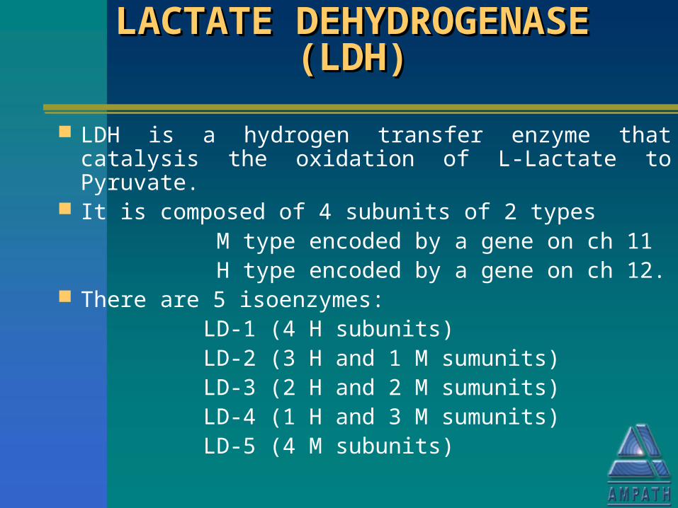
LACTATE DEHYDROGENASE LACTATE DEHYDROGENASE (LDH)(LDH)
LACTATE DEHYDROGENASE LACTATE DEHYDROGENASE (LDH)(LDH)
LDH is a hydrogen transfer enzyme that catalysis the oxidation of L-Lactate to Pyruvate.
It is composed of 4 subunits of 2 types M type encoded by a gene on ch 11 H type encoded by a gene on ch 12. There are 5 isoenzymes: LD-1 (4 H subunits) LD-2 (3 H and 1 M sumunits) LD-3 (2 H and 2 M sumunits) LD-4 (1 H and 3 M sumunits) LD-5 (4 M subunits)
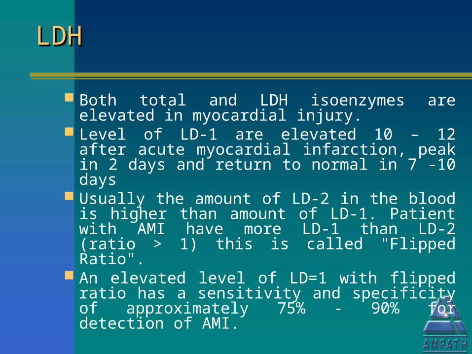
LDHLDHLDHLDH
Both total and LDH isoenzymes are elevated in myocardial injury.
Level of LD-1 are elevated 10 – 12 after acute myocardial infarction, peak in 2 days and return to normal in 7 -10 days
Usually the amount of LD-2 in the blood is higher than amount of LD-1. Patient with AMI have more LD-1 than LD-2 (ratio > 1) this is called "Flipped Ratio".
An elevated level of LD=1 with flipped ratio has a sensitivity and specificity of approximately 75% - 90% for detection of AMI.
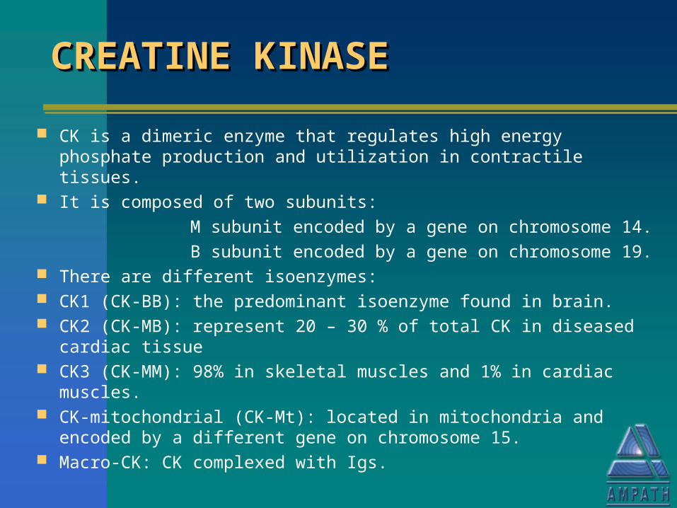
CREATINE KINASECREATINE KINASECREATINE KINASECREATINE KINASE
CK is a dimeric enzyme that regulates high energy phosphate production and utilization in contractile tissues.
It is composed of two subunits:
M subunit encoded by a gene on chromosome 14.
B subunit encoded by a gene on chromosome 19. There are different isoenzymes: CK1 (CK-BB): the predominant isoenzyme found in brain. CK2 (CK-MB): represent 20 – 30 % of total CK in diseased cardiac
tissue CK3 (CK-MM): 98% in skeletal muscles and 1% in cardiac muscles. CK-mitochondrial (CK-Mt): located in mitochondria and encoded by a
different gene on chromosome 15. Macro-CK: CK complexed with Igs.

CREATINE KINASECREATINE KINASECREATINE KINASECREATINE KINASE
NORMAL VALUES:NORMAL VALUES:
Vary according to – age sex race physical condition muscle mass
PATHOLOGICAL INCREASES:
Myocardial infarction or injury Skeletal muscle injury or disease Hypothyroidism IM injections Generalised convulsions Cerebral injury Malignant hyperpyrexia Prolonged hypothermia
CREATINE KINASE: CK-MBCREATINE KINASE: CK-MBCREATINE KINASE: CK-MBCREATINE KINASE: CK-MB
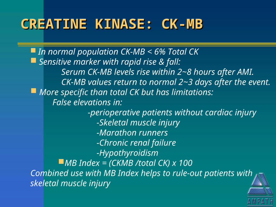
In normal population CK-MB < 6% Total CK Sensitive marker with rapid rise & fall: Serum CK-MB levels rise within 2~8 hours after AMI. CK-MB values return to normal 2~3 days after the event. More specific than total CK but has limitations: False elevations in: -perioperative patients without cardiac injury -Skeletal muscle injury -Marathon runners -Chronic renal failure -Hypothyroidism
MB Index = (CKMB /total CK) x 100Combined use with MB Index helps to rule-out patients with skeletal muscle injury
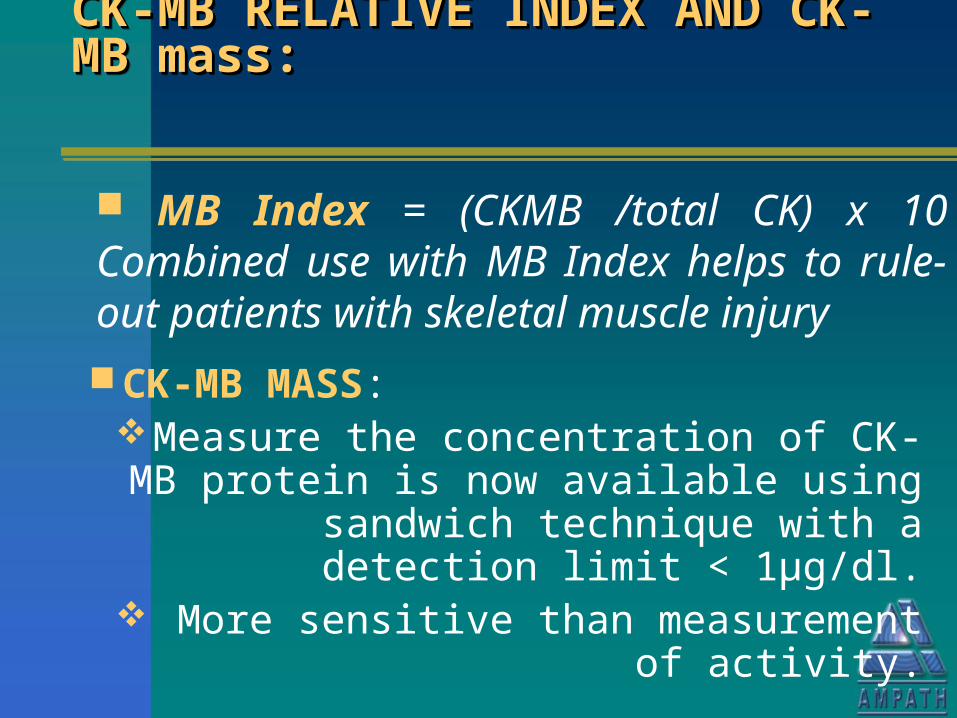
CK-MB RELATIVE INDEX AND CK-MB RELATIVE INDEX AND CK-MB mass: CK-MB mass: CK-MB RELATIVE INDEX AND CK-MB RELATIVE INDEX AND CK-MB mass: CK-MB mass:
CK-MB MASS: Measure the concentration of CK-MB protein is now available using sandwich technique with a detection limit < 1µg/dl. More sensitive than measurement of
activity.
MB Index = (CKMB /total CK) x 10 Combined use with MB Index helps to rule-out patients with skeletal muscle injury
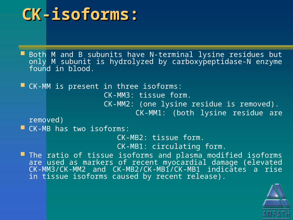
CK-isoforms:CK-isoforms:CK-isoforms:CK-isoforms:
Both M and B subunits have N-terminal lysine residues but only M subunit is hydrolyzed by carboxypeptidase-N enzyme found in blood.
CK-MM is present in three isoforms: CK-MM3: tissue form. CK-MM2: (one lysine residue is removed). CK-MM1: (both lysine residue are removed) CK-MB has two isoforms: CK-MB2: tissue form. CK-MB1: circulating form. The ratio of tissue isoforms and plasma modified isoforms are
used as markers of recent myocardial damage (elevated CK-MM3/CK-MM2 and CK-MB2/CK-MB1/CK-MB1 indicates a rise in tissue isoforms caused by recent release).
MYOGLOBIN (Mb)MYOGLOBIN (Mb)MYOGLOBIN (Mb)MYOGLOBIN (Mb)
Low MW protein Skeletal & cardiac muscle Mb identical Serum levels increase within 2h of muscle damage Peak at 6 – 9h Normal by 24 – 36h Excellent NEGATIVE predictor of myocardial injury
– 2 samples 2 – 4 hours apart with no rise in levels virtually excludes AMI
Rapid, quantitative serum immunoassays
CARDIAC TROPONINSCARDIAC TROPONINSCARDIAC TROPONINSCARDIAC TROPONINS
It consists of 3 subunits troponin C, I, and T. The complex regulates the contraction of
striated muscle.
1. TnC binds to calcium ions.
2. TnI binds to actin and inhibits actin-myosin interaction.
3. TnT binds to tropomyosin, attaching to thin filament.
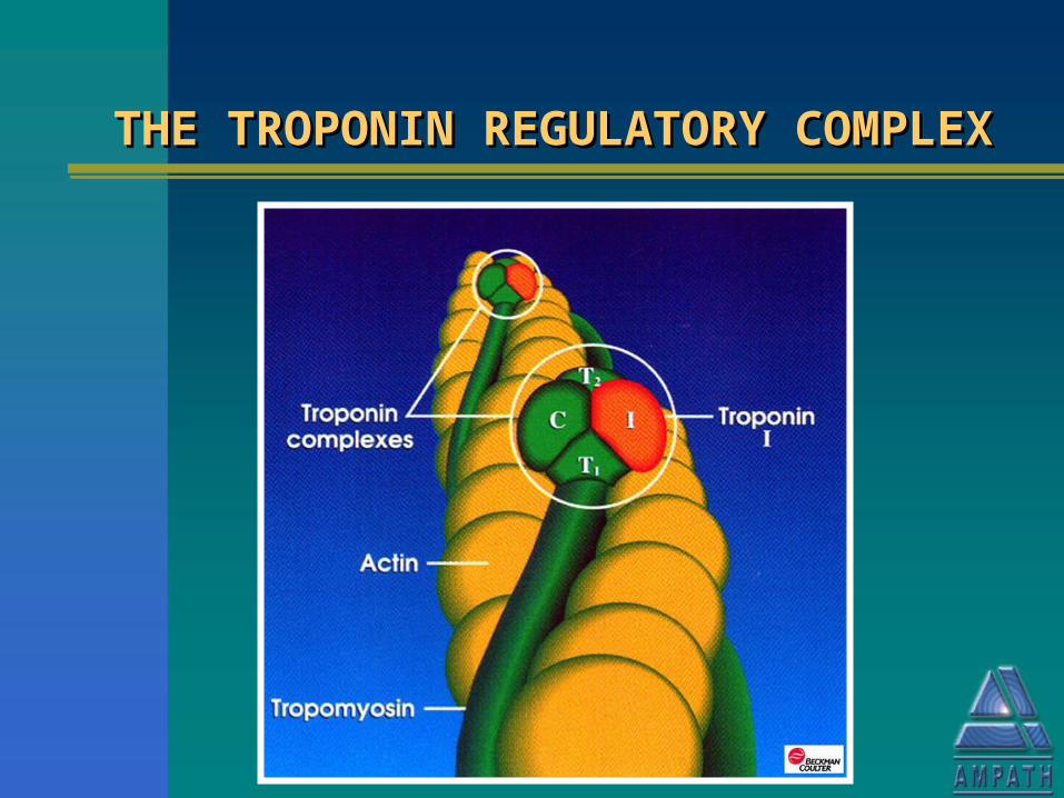
THE TROPONIN REGULATORY THE TROPONIN REGULATORY COMPLEXCOMPLEX
THE TROPONIN REGULATORY THE TROPONIN REGULATORY COMPLEXCOMPLEX
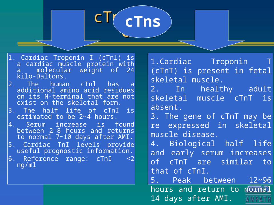
1. Cardiac Troponin I (cTnl) is a cardiac muscle protein with a molecular weight of 24 kilo-Daltons.
2. The human cTnl has a additional amino acid residues on its N-terminal that are not exist on the skeletal form.
3. The half life of cTnI is estimated to be 2~4 hours.
4. Serum increase is found between 2-8 hours and returns to normal 7~10 days after AMI.
5. Cardiac TnI levels provide useful prognostic information.
6. Reference range: cTnI <2 ng/ml
1.Cardiac Troponin T (cTnT) is present in fetal skeletal muscle.2. In healthy adult skeletal muscle cTnT is absent.3. The gene of cTnT may be re expressed in skeletal muscle disease. 4. Biological half life and early serum increases of cTnT are similar to that of cTnI.5. Peak between 12~96 hours and return to normal 14 days after AMI.
cTnI cTnTcTnI cTnT cTnI cTnTcTnI cTnT
cTns
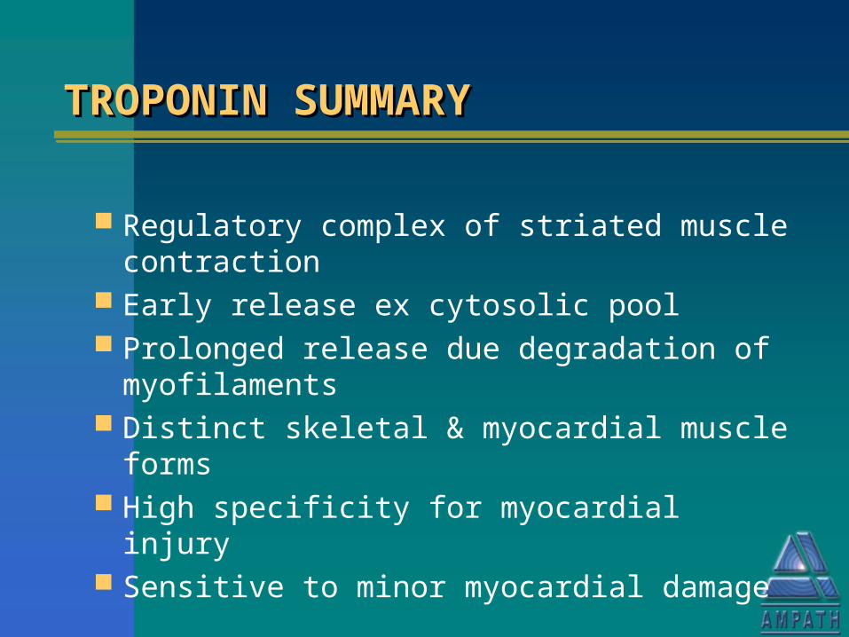
TROPONIN SUMMARYTROPONIN SUMMARYTROPONIN SUMMARYTROPONIN SUMMARY
Regulatory complex of striated muscle contraction
Early release ex cytosolic pool Prolonged release due degradation of
myofilaments Distinct skeletal & myocardial muscle forms High specificity for myocardial injury Sensitive to minor myocardial damage
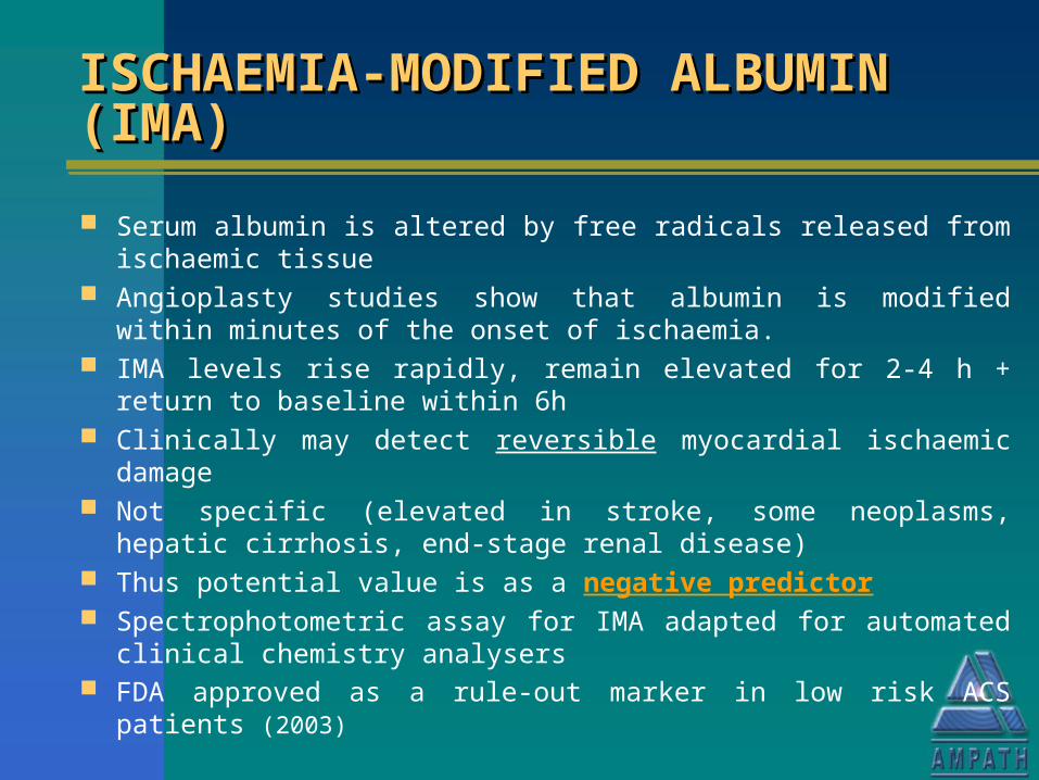
ISCHAEMIA-MODIFIED ALBUMIN ISCHAEMIA-MODIFIED ALBUMIN (IMA)(IMA)ISCHAEMIA-MODIFIED ALBUMIN ISCHAEMIA-MODIFIED ALBUMIN (IMA)(IMA)
Serum albumin is altered by free radicals released from ischaemic tissue
Angioplasty studies show that albumin is modified within minutes of the onset of ischaemia.
IMA levels rise rapidly, remain elevated for 2-4 h + return to baseline within 6h
Clinically may detect reversible myocardial ischaemic damage Not specific (elevated in stroke, some neoplasms, hepatic cirrhosis,
end-stage renal disease) Thus potential value is as a negative predictor Spectrophotometric assay for IMA adapted for automated clinical
chemistry analysers FDA approved as a rule-out marker in low risk ACS patients (2003)
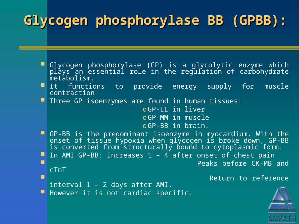
Glycogen phosphorylase BB Glycogen phosphorylase BB (GPBB):(GPBB):
Glycogen phosphorylase BB Glycogen phosphorylase BB (GPBB):(GPBB):
Glycogen phosphorylase (GP) is a glycolytic enzyme which plays an essential role in the regulation of carbohydrate metabolism.
It functions to provide energy supply for muscle contraction Three GP isoenzymes are found in human tissues: o GP-LL in liver o GP-MM in muscle o GP-BB in brain. GP-BB is the predominant isoenzyme in myocardium. With the
onset of tissue hypoxia when glycogen is broke down, GP-BB is converted from structurally bound to cytoplasmic form.
In AMI GP-BB: Increases 1 – 4 after onset of chest pain Peaks before CK-MB and cTnT Return to reference interval 1 – 2 days after
AMI. However it is not cardiac specific.
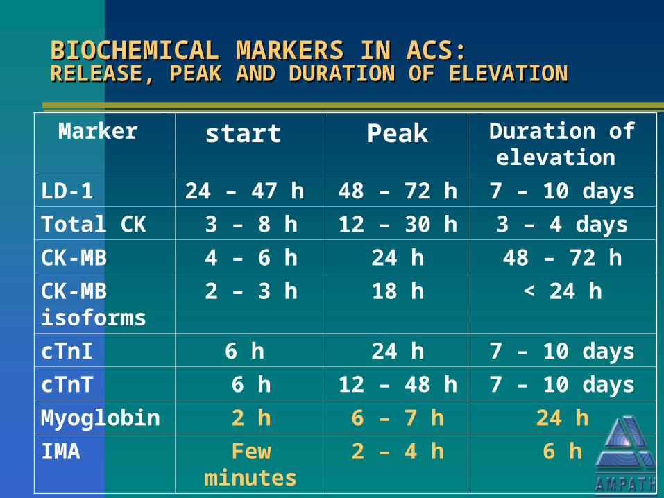
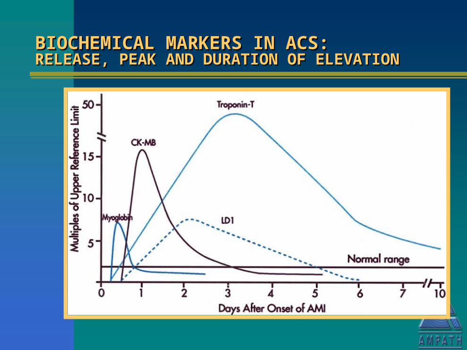
BIOCHEMICAL MARKERS IN ACS: BIOCHEMICAL MARKERS IN ACS: RELEASE, PEAK AND DURATION OF ELEVATIONRELEASE, PEAK AND DURATION OF ELEVATIONBIOCHEMICAL MARKERS IN ACS: BIOCHEMICAL MARKERS IN ACS: RELEASE, PEAK AND DURATION OF ELEVATIONRELEASE, PEAK AND DURATION OF ELEVATION
Marker start Peak Duration of elevation
LD-1 24 – 47 h 48 – 72 h 7 – 10 days
Total CK 3 – 8 h 12 – 30 h 3 – 4 days
CK-MB 4 – 6 h 24 h 48 – 72 h
CK-MB isoforms
2 – 3 h 18 h < 24 h
cTnI 6 h 24 h 7 – 10 days
cTnT 6 h 12 – 48 h 7 – 10 days
Myoglobin 2 h 6 – 7 h 24 h
IMA Few minutes 2 – 4 h 6 h
BIOCHEMICAL MARKERS IN ACS: BIOCHEMICAL MARKERS IN ACS: RELEASE, PEAK AND DURATION OF ELEVATIONRELEASE, PEAK AND DURATION OF ELEVATIONBIOCHEMICAL MARKERS IN ACS: BIOCHEMICAL MARKERS IN ACS: RELEASE, PEAK AND DURATION OF ELEVATIONRELEASE, PEAK AND DURATION OF ELEVATION
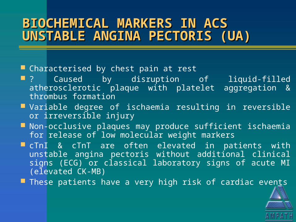
BIOCHEMICAL MARKERS IN ACS BIOCHEMICAL MARKERS IN ACS UNSTABLE ANGINA PECTORIS (UA)UNSTABLE ANGINA PECTORIS (UA)BIOCHEMICAL MARKERS IN ACS BIOCHEMICAL MARKERS IN ACS UNSTABLE ANGINA PECTORIS (UA)UNSTABLE ANGINA PECTORIS (UA)
Characterised by chest pain at rest ? Caused by disruption of liquid-filled atherosclerotic
plaque with platelet aggregation & thrombus formation Variable degree of ischaemia resulting in reversible or
irreversible injury Non-occlusive plaques may produce sufficient ischaemia
for release of low molecular weight markers cTnI & cTnT are often elevated in patients with unstable
angina pectoris without additional clinical signs (ECG) or classical laboratory signs of acute MI (elevated CK-MB)
These patients have a very high risk of cardiac events
CARDIAC TROPONINS IN CARDIAC TROPONINS IN UNSTABLE ANGINA PECTORIS UNSTABLE ANGINA PECTORIS (UA)(UA)
CARDIAC TROPONINS IN CARDIAC TROPONINS IN UNSTABLE ANGINA PECTORIS UNSTABLE ANGINA PECTORIS (UA)(UA)
Does an elevated Troponin level in the absence of other signs reflect irreversible myocardial damage?
– Epidemiological studies– Animal experiments– Clinical trials– Sensitive imaging techniques
Say Say YES!YES!
MIMI must be must be REDEFINED!REDEFINED!
QUESTION:QUESTION:QUESTION:QUESTION:
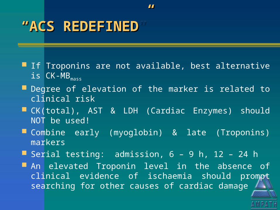
““ACS REDEFINED”ACS REDEFINED”““ACS REDEFINED”ACS REDEFINED”
If Troponins are not available, best alternative is CK-MBmass
Degree of elevation of the marker is related to clinical risk CK(total), AST & LDH (Cardiac Enzymes) should NOT be
used! Combine early (myoglobin) & late (Troponins) markers Serial testing: admission, 6 – 9 h, 12 – 24 h An elevated Troponin level in the absence of clinical
evidence of ischaemia should prompt searching for other causes of cardiac damage
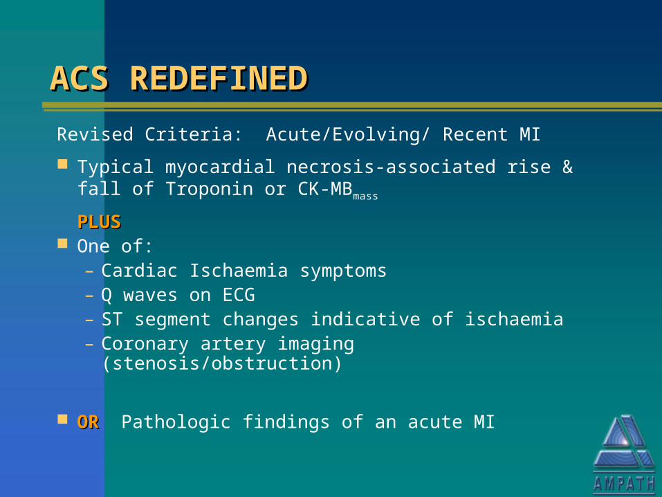
ACS REDEFINEDACS REDEFINEDACS REDEFINEDACS REDEFINED
Revised Criteria: Acute/Evolving/ Recent MI
Typical myocardial necrosis-associated rise & fall of Troponin or CK-MBmass
PLUSPLUS One of:
– Cardiac Ischaemia symptoms– Q waves on ECG– ST segment changes indicative of ischaemia– Coronary artery imaging (stenosis/obstruction)
OROR Pathologic findings of an acute MI
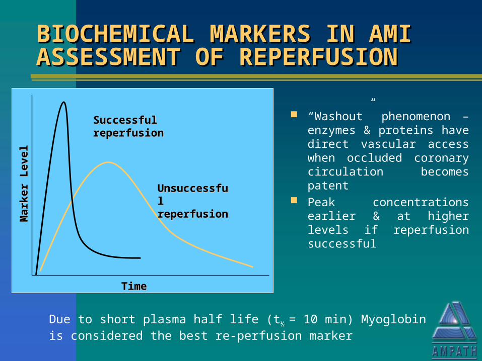
BIOCHEMICAL MARKERS IN AMI BIOCHEMICAL MARKERS IN AMI ASSESSMENT OF REPERFUSIONASSESSMENT OF REPERFUSIONBIOCHEMICAL MARKERS IN AMI BIOCHEMICAL MARKERS IN AMI ASSESSMENT OF REPERFUSIONASSESSMENT OF REPERFUSION
“Washout” phenomenon – enzymes & proteins have direct vascular access when occluded coronary circulation becomes patent
Peak concentrations earlier & at higher levels if reperfusion successful
Due to short plasma half life (t½ = 10 min) Myoglobin is considered the best re-perfusion marker
TimeTime
Mar
ker
Lev
elM
arke
r L
evel
Successful Successful reperfusionreperfusion
Unsuccessful Unsuccessful reperfusionreperfusion
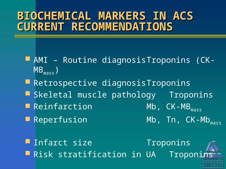
BIOCHEMICAL MARKERS IN ACS BIOCHEMICAL MARKERS IN ACS CURRENT RECOMMENDATIONSCURRENT RECOMMENDATIONSBIOCHEMICAL MARKERS IN ACS BIOCHEMICAL MARKERS IN ACS CURRENT RECOMMENDATIONSCURRENT RECOMMENDATIONS
AMI – Routine diagnosis Troponins (CK-MBmass)
Retrospective diagnosis Troponins Skeletal muscle pathology Troponins Reinfarction Mb, CK-MBmass
Reperfusion Mb, Tn, CK-Mbmass
Infarct size Troponins Risk stratification in UA Troponins
BIOCHEMICAL MARKERS OF BIOCHEMICAL MARKERS OF MYOCARDIAL FUNCTIONMYOCARDIAL FUNCTIONBIOCHEMICAL MARKERS OF BIOCHEMICAL MARKERS OF MYOCARDIAL FUNCTIONMYOCARDIAL FUNCTION
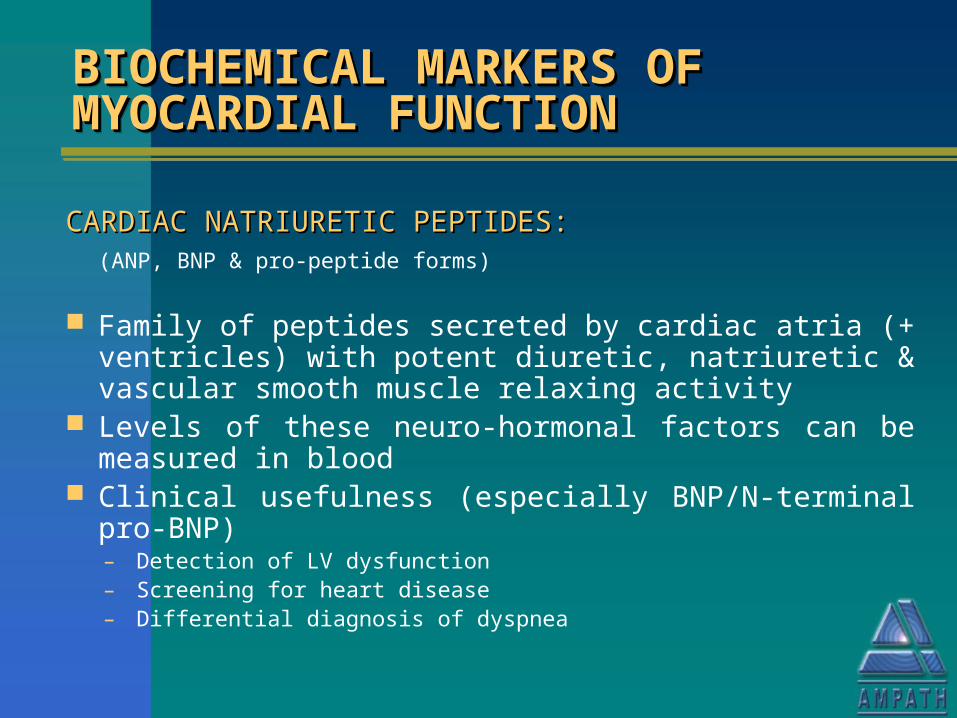
CARDIAC NATRIURETIC PEPTIDES:CARDIAC NATRIURETIC PEPTIDES:(ANP, BNP & pro-peptide forms)
Family of peptides secreted by cardiac atria (+ ventricles) with potent diuretic, natriuretic & vascular smooth muscle relaxing activity
Levels of these neuro-hormonal factors can be measured in blood
Clinical usefulness (especially BNP/N-terminal pro-BNP)– Detection of LV dysfunction– Screening for heart disease– Differential diagnosis of dyspnea
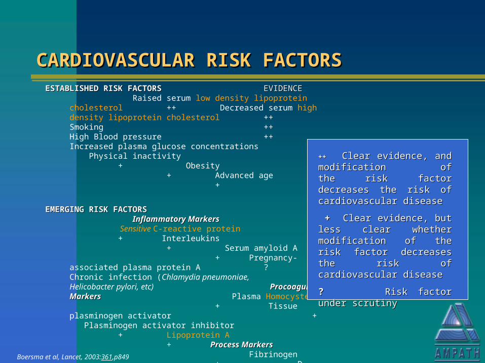
CARDIOVASCULAR RISK FACTORSCARDIOVASCULAR RISK FACTORSCARDIOVASCULAR RISK FACTORSCARDIOVASCULAR RISK FACTORSESTABLISHED RISK FACTORSESTABLISHED RISK FACTORS EVIDENCE EVIDENCE
Raised serum low density lipoprotein cholesterol ++ Decreased serum high density lipoprotein cholesterol ++ Smoking
++ High Blood pressure ++ Increased plasma glucose concentrations + Physical inactivity
+ Obesity+ Advanced age +
EMERGING RISK FACTORSEMERGING RISK FACTORS Inflammatory MarkersInflammatory Markers Sensitive C-reactive protein + Interleukins + Serum amyloid A + Pregnancy-associated plasma protein A ? Chronic infection (Chlamydia pneumoniae, ? Helicobacter pylori, etc) Procoagulant MarkersProcoagulant Markers
Plasma Homocysteine+ Tissue plasminogen activator+ Plasminogen activator inhibitor+ Lipoprotein A+ Process MarkersProcess Markers Fibrinogen+ D-dimer? Coronary artery calcification?
Boersma et al, Lancet, 2003:361,p849
++++ Clear evidence, and modification Clear evidence, and modification of the risk factor decreases the risk of the risk factor decreases the risk of cardiovascular diseaseof cardiovascular disease
++ Clear evidence, but less clear Clear evidence, but less clear whether modification of the risk factor whether modification of the risk factor decreases the risk of cardiovascular decreases the risk of cardiovascular diseasedisease
?? Risk factor under scrutiny Risk factor under scrutiny

GENETIC ANALYSIS OF CANDIDATE GENETIC ANALYSIS OF CANDIDATE GENES OR RISK FACTORS FOR GENES OR RISK FACTORS FOR CARDIOVASCULAR DISEASECARDIOVASCULAR DISEASE
GENETIC ANALYSIS OF CANDIDATE GENETIC ANALYSIS OF CANDIDATE GENES OR RISK FACTORS FOR GENES OR RISK FACTORS FOR CARDIOVASCULAR DISEASECARDIOVASCULAR DISEASE
Recent explosion of genetic analysis & micro-array technology
Common cardiovascular diseases are polygenic. Multiple susceptibility loci interact with lifestyle & environment
Single gene defects may account for some of the cardiomyopathies, inherited cardiac arrhythmias
Possible genetic cardiovascular risk factors under assessment
Technology is still complex & expensive but is developing very rapidly