Embed Size (px)
Citation preview
Hindawi Publishing CorporationEvidence-Based Complementary and Alternative MedicineVolume 2012, Article ID 802625, 8 pagesdoi:10.1155/2012/802625
Research Article
Bioassay-Guided Isolation of Neuroprotective Compounds fromUncaria rhynchophylla against Beta-Amyloid-InducedNeurotoxicity
Yan-Fang Xian,1 Zhi-Xiu Lin,1 Qing-Qiu Mao,1 Zhen Hu,1 Ming Zhao,2 Chun-Tao Che,2 andSiu-Po Ip1
1 School of Chinese Medicine, The Chinese University of Hong Kong, Shatin, Hong Kong2 Department of Medicinal Chemistry and Pharmacognosy, College of Pharmacy, University of Illinois at Chicago, Chicago IL 60607,USA
Correspondence should be addressed to Zhi-Xiu Lin, [email protected] and Siu-Po Ip, [email protected]
Received 27 March 2012; Accepted 12 May 2012
Academic Editor: Karl Wah-Keung Tsim
Copyright © 2012 Yan-Fang Xian et al. This is an open access article distributed under the Creative Commons Attribution License,which permits unrestricted use, distribution, and reproduction in any medium, provided the original work is properly cited.
Uncaria rhynchophylla is a component herb of many Chinese herbal formulae for the treatment of neurodegenerative diseases.Previous study in our laboratory has demonstrated that an ethanol extract of Uncaria rhynchophylla ameliorated cognitive deficitsin a mouse model of Alzheimer’s disease induced by D-galactose. However, the active ingredients of Uncaria rhynchophyllaresponsible for the anti-Alzheimer’s disease activity have not been identified. This study aims to identify the active ingredients ofUncaria rhynchophylla by a bioassay-guided fractionation approach and explore the acting mechanism of these active ingredientsby using a well-established cellular model of Alzheimer’s disease, beta-amyloid- (Aβ-) induced neurotoxicity in PC12 cells.The results showed that six alkaloids, namely, corynoxine, corynoxine B, corynoxeine, isorhynchophylline, isocorynoxeine, andrhynchophylline were isolated from the extract of Uncaria rhynchophylla. Among them, rhynchophylline and isorhynchophyllinesignificantly decreased Aβ-induced cell death, intracellular calcium overloading, and tau protein hyperphosphorylation in PC12cells. These results suggest that rhynchophylline and isorhynchophylline are the major active ingredients responsible for theprotective action of Uncaria rhynchophylla against Aβ-induced neuronal toxicity, and their neuroprotective effect may be mediated,at least in part, by inhibiting intracellular calcium overloading and tau protein hyperphosphorylation.
1. Introduction
Alzheimer’s disease (AD), a neurodegenerative disordercharacterized by a progressive loss of learning, memory, andother cognitive functions, is the most common form ofdementia in the elderly. The pathological hallmarks of AD areextracellular senile plaques and intracellular neurofibrillarytangles [1]. It is well known that deposition of β-amyloid(Aβ) is a pivotal event in initiating the neuronal degenerationof AD [2]. Aβ aggregates into amyloid fibrils, which havebeen reported to be neurotoxic in vitro [3] and in vivo [4]. Inthis connection, the toxic effect of Aβ on a cultured neuronalcells can be used as a screening tool for identifying potentialtherapeutic agents for AD.
Current clinical treatments of AD patients use acetyl-cholinesterase inhibitors (AChEIs) and antagonists of N-methyl-D-aspartate receptors (NMDA) to slow down theprogress of the deterioration of AD. However, effectiveapproaches for delaying the progression of AD are yet to befound to date. Thus, searching for safer, better-tolerated, andeffective drugs for the treatment of AD remains an importantarea of drug discovery.
Traditional Chinese herbal medicine has been practicedin China for thousands of years, and vast experience hasbeen accumulated for using medicinal herbs for clinicaltreatment of diseases. Thus, Chinese herbal medicine maybe a promising source of effective drugs for treating AD.Uncaria rhynchophylla has been extensively used in Chinese
2 Evidence-Based Complementary and Alternative Medicine
herbal medicine to relieve headache, dizziness, tremors,and hypertension-induced convulsion [5–7]. In recent years,Uncaria rhynchophylla has been shown to be effective forinhibiting Aβ fibril formation, disassembling performed Aβfibrils [8] and antiacetylcholinesterase [9]. Previous studyin our laboratory demonstrated that an ethanol extractof Uncaria rhynchophylla significantly reversed cognitivedeficits induced by D-galactose, a mouse model of AD [10].Phytochemical study has shown that alkaloids, terpenoids,and flavonoids are the major chemical ingredients of Uncariarhynchophylla [7]. However, the bioactive principles respon-sible for the protective action of Uncaria rhynchophylla havenot been identified. The present study aims to identify theactive constituents of Uncaria rhynchophylla using bioassay-guided fractionation. Furthermore, the acting mechanismof these active ingredients is explored by using a well-established cellular model of Alzheimer’s disease, beta-amyloid- (Aβ-) induced neurotoxicity in PC12 cells.
2. Materials and Methods
2.1. Chemicals and Reagents. Nerve growth factor (NGF),3-(4,5-dimethylthiazol-2-yl)-2,5-diphenyltetrazolium bro-mide (MTT), and the fragment of β-amyloid peptide(Aβ25−35) were purchased from Sigma-Aldrich (St. Louis,MO, USA). Fura 2-AM, Dulbecco’s modified Eagle’s me-dium (DMEM), horse serum, fetal bovine serum, and apenicillin/streptomycin mixture were purchased from Gib-co-Invitrogen (Grand Island, NY, USA). All other solventsand chemicals used in the study were of analytical grade.
2.2. Plant Material. The dried stem with hooks of Uncariarhynchophylla was purchased from Zhixin PharmaceuticalCo., a GMP-certified supplier of Chinese medicinal herbalmaterials (Guangzhou, China). It was authenticated to bethe dried rhizome of Uncaria rhynchophylla (Miq.) Miq.ex Havil. by Ms. Y. Y. Zong, School of Chinese Medicine,The Chinese University of Hong Kong, Hong Kong, wherea voucher specimen (no. 091220) has been deposited.
2.3. Preparation of Aggregated Aβ25−35. The aggregatedAβ25−35 was prepared according to a method describedpreviously [11]. Briefly, Aβ25−35 was dissolved in steriledistilled water at a concentration of 1 mM and incubatedat 37◦C for 4 days to form the aggregation. It was stored at−20◦C until use.
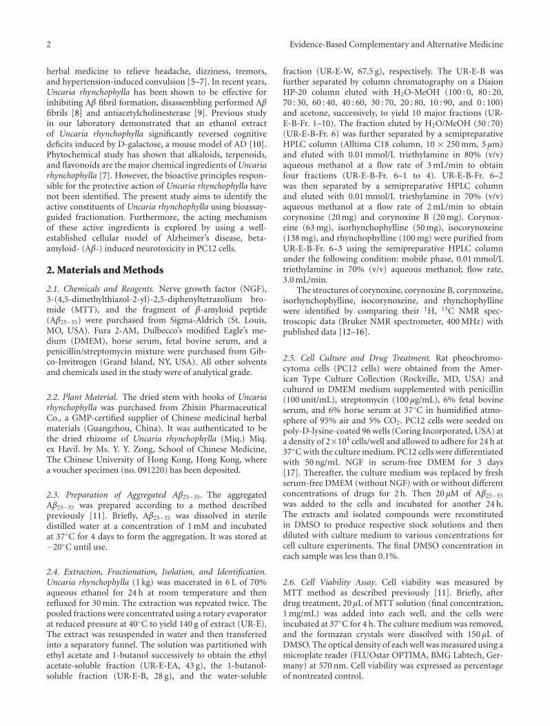
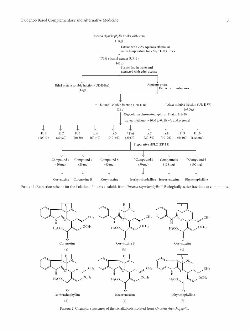
2.4. Extraction, Fractionation, Isolation, and Identification.Uncaria rhynchophylla (1 kg) was macerated in 6 L of 70%aqueous ethanol for 24 h at room temperature and thenrefluxed for 30 min. The extraction was repeated twice. Thepooled fractions were concentrated using a rotary evaporatorat reduced pressure at 40◦C to yield 140 g of extract (UR-E).The extract was resuspended in water and then transferredinto a separatory funnel. The solution was partitioned withethyl acetate and 1-butanol successively to obtain the ethylacetate-soluble fraction (UR-E-EA, 43 g), the 1-butanol-soluble fraction (UR-E-B, 28 g), and the water-soluble
fraction (UR-E-W, 67.5 g), respectively. The UR-E-B wasfurther separated by column chromatography on a DiaionHP-20 column eluted with H2O-MeOH (100 : 0, 80 : 20,70 : 30, 60 : 40, 40 : 60, 30 : 70, 20 : 80, 10 : 90, and 0 : 100)and acetone, successively, to yield 10 major fractions (UR-E-B-Fr. 1–10). The fraction eluted by H2O/MeOH (30 : 70)(UR-E-B-Fr. 6) was further separated by a semipreparativeHPLC column (Alltima C18 column, 10 × 250 mm, 5 μm)and eluted with 0.01 mmol/L triethylamine in 80% (v/v)aqueous methanol at a flow rate of 3 mL/min to obtainfour fractions (UR-E-B-Fr. 6–1 to 4). UR-E-B-Fr. 6–2was then separated by a semipreparative HPLC columnand eluted with 0.01 mmol/L triethylamine in 70% (v/v)aqueous methanol at a flow rate of 2 mL/min to obtaincorynoxine (20 mg) and corynoxine B (20 mg). Corynox-eine (63 mg), isorhynchophylline (50 mg), isocorynoxeine(138 mg), and rhynchophylline (100 mg) were purified fromUR-E-B-Fr. 6–3 using the semipreparative HPLC columnunder the following condition: mobile phase, 0.01 mmol/Ltriethylamine in 70% (v/v) aqueous methanol; flow rate,3.0 mL/min.
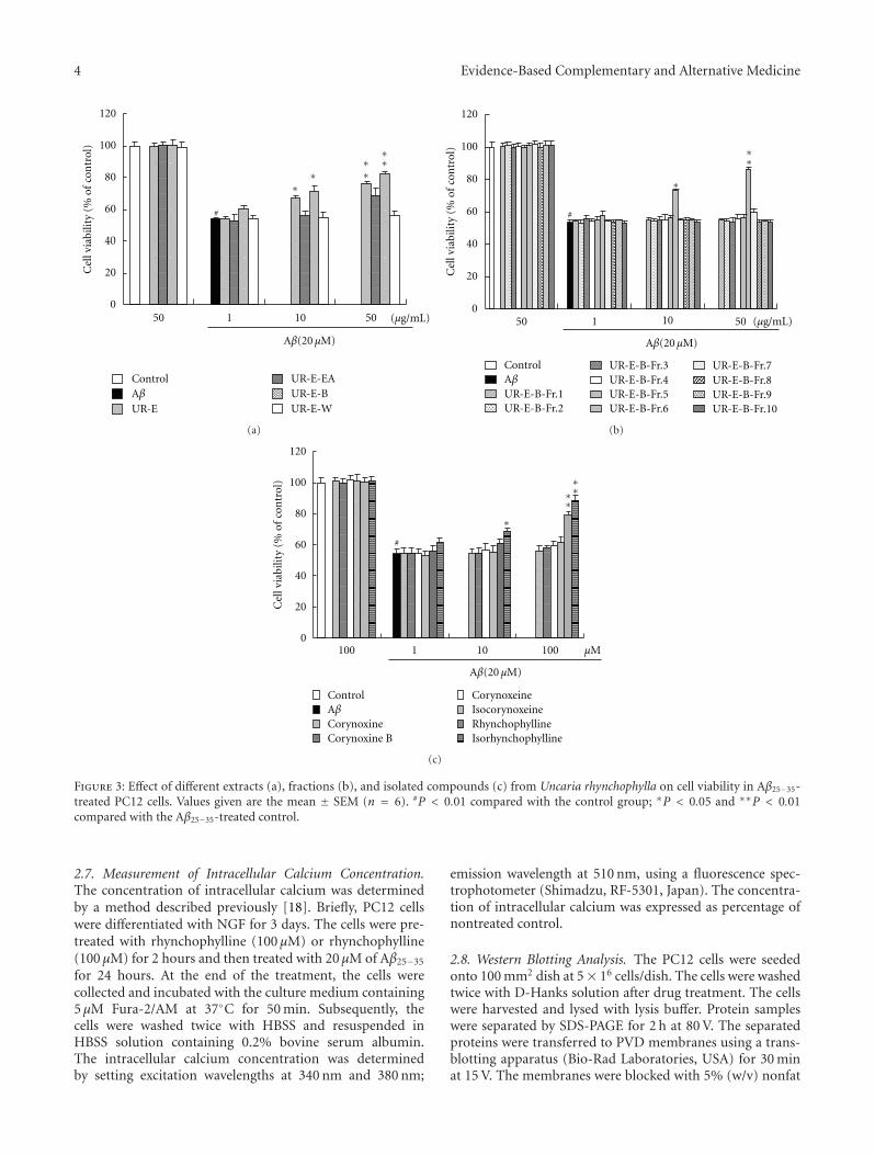
The structures of corynoxine, corynoxine B, corynoxeine,isorhynchophylline, isocorynoxeine, and rhynchophyllinewere identified by comparing their 1H, 13C NMR spec-troscopic data (Bruker NMR spectrometer, 400 MHz) withpublished data [12–16].
2.5. Cell Culture and Drug Treatment. Rat pheochromo-cytoma cells (PC12 cells) were obtained from the Amer-ican Type Culture Collection (Rockville, MD, USA) andcultured in DMEM medium supplemented with penicillin(100 unit/mL), streptomycin (100 μg/mL), 6% fetal bovineserum, and 6% horse serum at 37◦C in humidified atmo-sphere of 95% air and 5% CO2. PC12 cells were seeded onpoly-D-lysine-coated 96 wells (Coring Incorporated, USA) ata density of 2×10⁴ cells/well and allowed to adhere for 24 h at37◦C with the culture medium. PC12 cells were differentiatedwith 50 ng/mL NGF in serum-free DMEM for 3 days[17]. Thereafter, the culture medium was replaced by freshserum-free DMEM (without NGF) with or without differentconcentrations of drugs for 2 h. Then 20 μM of Aβ25−35
was added to the cells and incubated for another 24 h.The extracts and isolated compounds were reconstitutedin DMSO to produce respective stock solutions and thendiluted with culture medium to various concentrations forcell culture experiments. The final DMSO concentration ineach sample was less than 0.1%.
2.6. Cell Viability Assay. Cell viability was measured byMTT method as described previously [11]. Briefly, afterdrug treatment, 20 μL of MTT solution (final concentration,1 mg/mL) was added into each well, and the cells wereincubated at 37◦C for 4 h. The culture medium was removed,and the formazan crystals were dissolved with 150 μL ofDMSO. The optical density of each well was measured using amicroplate reader (FLUOstar OPTIMA, BMG Labtech, Ger-many) at 570 nm. Cell viability was expressed as percentageof nontreated control.
Evidence-Based Complementary and Alternative Medicine 3
Uncaria rhynchophylla hooks with stem
(1 Kg)
Extract with 70% aqueous ethanol atroom temperature for 72 h, 8 L ×3 times
∗70% ethanol extract (UR-E)
(140 g)Suspended in water andextracted with ethyl acetate
Ethyl acetate soluble fraction (UR-E-EA)
(43 g)
Aqueous phaseExtract with n-butanol
∗1-butanol-soluble fraction (UR-E-B)
(28 g)
Water-soluble fraction (UR-E-W)
(67.5 g)
25 g-column chromatography on Diaion HP-20
(water: methanol −10 : 0 to 0 : 10, v/v and acetone)
Fr.1(100 : 0)
Fr.2(80 : 20)
Fr.3(70 : 30)
Fr.4(60 : 40)
Fr.5(40 : 60)
∗Fr.6(30 : 70)
Fr.7(20 : 80)
Fr.8(10 : 90)
Fr.9(0 : 100)
Fr.10(acetone)
Compound 1
(20 mg)
∗Compound 6
(100 mg)
Compound 5
(138 mg)
Compound 2
(20 mg)
Compound 3
(63 mg)
Preparative HPLC (RP-18)
Corynoxine Corynoxine B Corynoxeine Isorhynchophylline Isocorynoxeine Rhynchophylline
∗Compound 4
(50 mg)
Figure 1: Extraction scheme for the isolation of the six alkaloids from Uncaria rhynchophylla. ∗ Biologically active fractions or compounds.
N
N
H
H
CH3
OCH3H3CO
O
O
Corynoxine
(a)
N
N
H
H
CH3
OCH3H3CO
O
O
Corynoxine B
(b)
N
N
H
H
CH2
OCH3H3CO
O
O
Corynoxeine
(c)
N
N
H
H
CH3
OCH3H3CO
O
O
Isorhynchophylline
(d)
N
N
H
H
CH2
OCH3H3CO
O
O
Isocorynoxeine
(e)
N
N
H
H
CH3
OCH3H3CO
O
O
Rhynchophylline
(f)
Figure 2: Chemical structures of the six alkaloids isolated from Uncaria rhynchophylla.
4 Evidence-Based Complementary and Alternative Medicine
Control
UR-E
UR-E-EAUR-E-BUR-E-W
0
20
40
60
80
100
120
Cel
l via
bilit
y (%
of
con
trol
)
10 50150 (μg/mL)
Aβ
∗∗ ∗
∗ ∗∗
#
Aβ(20 μM)
(a)
Control
UR-E-B-Fr.1UR-E-B-Fr.2
UR-E-B-Fr.3UR-E-B-Fr.4UR-E-B-Fr.5UR-E-B-Fr.6
UR-E-B-Fr.7UR-E-B-Fr.8UR-E-B-Fr.9UR-E-B-Fr.10
0
20
40
60
80
100
120
Cel
l via
bilit
y (%
of
con
trol
)
1 10 5050 (μg/mL)
Aβ
∗
∗∗
#
Aβ(20 μM)
(b)
Control
CorynoxineCorynoxine B
CorynoxeineIsocorynoxeineRhynchophyllineIsorhynchophylline
0
20
40
60
80
100
120
Cel
l via
bilit
y (%
of
con
trol
)
100100 1 10
Aβ
∗∗
∗∗
∗
#
μM
Aβ(20 μM)
(c)
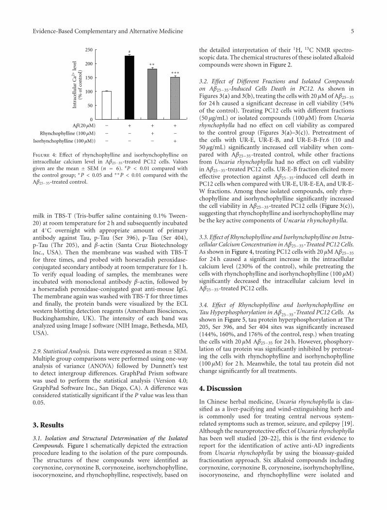
Figure 3: Effect of different extracts (a), fractions (b), and isolated compounds (c) from Uncaria rhynchophylla on cell viability in Aβ25−35-treated PC12 cells. Values given are the mean ± SEM (n = 6). #P < 0.01 compared with the control group; ∗P < 0.05 and ∗∗P < 0.01compared with the Aβ25−35-treated control.
2.7. Measurement of Intracellular Calcium Concentration.The concentration of intracellular calcium was determinedby a method described previously [18]. Briefly, PC12 cellswere differentiated with NGF for 3 days. The cells were pre-treated with rhynchophylline (100 μM) or rhynchophylline(100 μM) for 2 hours and then treated with 20 μM of Aβ25−35
for 24 hours. At the end of the treatment, the cells werecollected and incubated with the culture medium containing5 μM Fura-2/AM at 37◦C for 50 min. Subsequently, thecells were washed twice with HBSS and resuspended inHBSS solution containing 0.2% bovine serum albumin.The intracellular calcium concentration was determinedby setting excitation wavelengths at 340 nm and 380 nm;
emission wavelength at 510 nm, using a fluorescence spec-trophotometer (Shimadzu, RF-5301, Japan). The concentra-tion of intracellular calcium was expressed as percentage ofnontreated control.
2.8. Western Blotting Analysis. The PC12 cells were seededonto 100 mm2 dish at 5× 16 cells/dish. The cells were washedtwice with D-Hanks solution after drug treatment. The cellswere harvested and lysed with lysis buffer. Protein sampleswere separated by SDS-PAGE for 2 h at 80 V. The separatedproteins were transferred to PVD membranes using a trans-blotting apparatus (Bio-Rad Laboratories, USA) for 30 minat 15 V. The membranes were blocked with 5% (w/v) nonfat
Evidence-Based Complementary and Alternative Medicine 5
0
50
100
150
200
250
(% o
f co
ntr
ol)
Intr
acel
lula
r C
a2+le
vel
−−−
−−
+
−+
+−
+
+Rhynchophylline (100 μM)
Isorhynchophylline (100 μM))
Aβ(20 μM)
∗∗
∗∗
∗
#
Figure 4: Effect of rhynchophylline and isorhynchophylline onintracellular calcium level in Aβ25−35-treated PC12 cells. Valuesgiven are the mean ± SEM (n = 6). #P < 0.01 compared withthe control group; ∗P < 0.05 and ∗∗P < 0.01 compared with theAβ25−35-treated control.
milk in TBS-T (Tris-buffer saline containing 0.1% Tween-20) at room temperature for 2 h and subsequently incubatedat 4◦C overnight with appropriate amount of primaryantibody against Tau, p-Tau (Ser 396), p-Tau (Ser 404),p-Tau (Thr 205), and β-actin (Santa Cruz BiotechnologyInc., USA). Then the membrane was washed with TBS-Tfor three times, and probed with horseradish peroxidase-conjugated secondary antibody at room temperature for 1 h.To verify equal loading of samples, the membranes wereincubated with monoclonal antibody β-actin, followed bya horseradish peroxidase-conjugated goat anti-mouse IgG.The membrane again was washed with TBS-T for three timesand finally, the protein bands were visualized by the ECLwestern blotting detection reagents (Amersham Biosciences,Buckinghamshire, UK). The intensity of each band wasanalyzed using Image J software (NIH Image, Bethesda, MD,USA).
2.9. Statistical Analysis. Data were expressed as mean± SEM.Multiple group comparisons were performed using one-wayanalysis of variance (ANOVA) followed by Dunnett’s testto detect intergroup differences. GraphPad Prism softwarewas used to perform the statistical analysis (Version 4.0;GraphPad Software Inc., San Diego, CA). A difference wasconsidered statistically significant if the P value was less than0.05.
3. Results
3.1. Isolation and Structural Determination of the IsolatedCompounds. Figure 1 schematically depicted the extractionprocedure leading to the isolation of the pure compounds.The structures of these compounds were identified ascorynoxine, corynoxine B, corynoxeine, isorhynchophylline,isocorynoxeine, and rhynchophylline, respectively, based on
the detailed interpretation of their 1H, 13C NMR spectro-scopic data. The chemical structures of these isolated alkaloidcompounds were shown in Figure 2.
3.2. Effect of Different Fractions and Isolated Compoundson Aβ25−35-Induced Cells Death in PC12. As shown inFigures 3(a) and 3(b), treating the cells with 20 μM of Aβ25−35
for 24 h caused a significant decrease in cell viability (54%of the control). Treating PC12 cells with different fractions(50 μg/mL) or isolated compounds (100 μM) from Uncariarhynchophylla had no effect on cell viability as comparedto the control group (Figures 3(a)–3(c)). Pretreatment ofthe cells with UR-E, UR-E-B, and UR-E-B-Fr.6 (10 and50 μg/mL) significantly increased cell viability when com-pared with Aβ25−35-treated control, while other fractionsfrom Uncaria rhynchophylla had no effect on cell viabilityin Aβ25−35-treated PC12 cells. UR-E-B fraction elicited moreeffective protection against Aβ25−35-induced cell death inPC12 cells when compared with UR-E, UR-E-EA, and UR-E-W fractions. Among these isolated compounds, only rhyn-chophylline and isorhynchophylline significantly increasedthe cell viability in Aβ25−35-treated PC12 cells (Figure 3(c)),suggesting that rhynchophylline and isorhynchophylline maybe the key active components of Uncaria rhynchophylla.
3.3. Effect of Rhynchophylline and Isorhynchophylline on Intra-cellular Calcium Concentration in Aβ25−35-Treated PC12 Cells.As shown in Figure 4, treating PC12 cells with 20 μM Aβ25−35
for 24 h caused a significant increase in the intracellularcalcium level (230% of the control), while pretreating thecells with rhynchophylline and isorhynchophylline (100 μM)significantly decreased the intracellular calcium level inAβ25−35-treated PC12 cells.
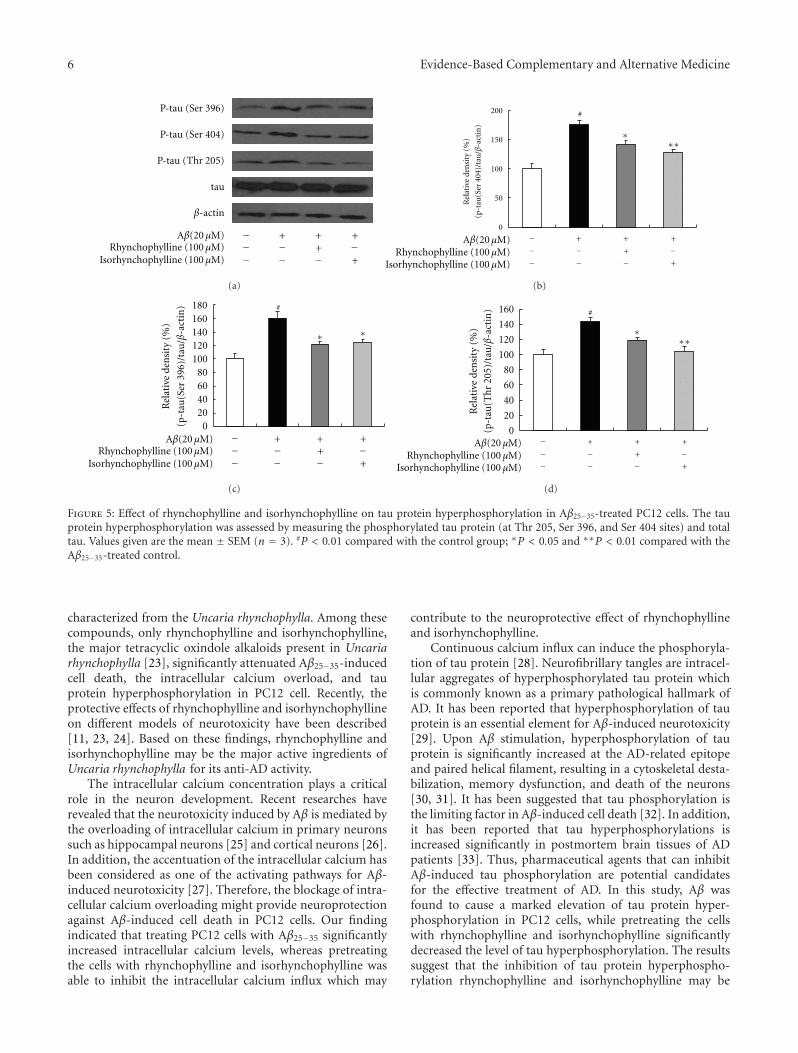
3.4. Effect of Rhynchophylline and Isorhynchophylline onTau Hyperphosphorylation in Aβ25−35-Treated PC12 Cells. Asshown in Figure 5, tau protein hyperphosphorylation at Thr205, Ser 396, and Ser 404 sites was significantly increased(144%, 160%, and 176% of the control, resp.) when treatingthe cells with 20 μM Aβ25−35 for 24 h. However, phosphory-lation of tau protein was significantly inhibited by pretreat-ing the cells with rhynchophylline and isorhynchophylline(100 μM) for 2 h. Meanwhile, the total tau protein did notchange significantly for all treatments.
4. Discussion
In Chinese herbal medicine, Uncaria rhynchophylla is clas-sified as a liver-pacifying and wind-extinguishing herb andis commonly used for treating central nervous system-related symptoms such as tremor, seizure, and epilepsy [19].Although the neuroprotective effect of Uncaria rhynchophyllahas been well studied [20–22], this is the first evidence toreport for the identification of active anti-AD ingredientsfrom Uncaria rhynchophylla by using the bioassay-guidedfractionation approach. Six alkaloid compounds includingcorynoxine, corynoxine B, corynoxeine, isorhynchophylline,isocorynoxeine, and rhynchophylline were isolated and
6 Evidence-Based Complementary and Alternative Medicine
P-tau (Ser 396)
P-tau (Ser 404)
P-tau (Thr 205)
tau
β-actin
Rhynchophylline (100 μM)Isorhynchophylline (100 μM)
−−−
−−
+−+
+−
++
Aβ(20 μM)
(a)
0
50
100
150
200
−−−
−−
+
−+
+−
+
+
Rel
ativ
e de
nsi
ty (
%)
(p-t
au(S
er 4
04)/
tau
/β-a
ctin
)
∗∗∗
#
Rhynchophylline (100 μM)Isorhynchophylline (100 μM)
Aβ(20 μM)
(b)
020406080
100120140160180
Rhynchophylline (100 μM)Isorhynchophylline (100 μM)
−−−
−−
+−+
+−
++
Rel
ativ
e de
nsi
ty (
%)
(p-t
au(S
er 3
96)/
tau
/β-a
ctin
)
∗∗
#
Aβ(20 μM)
(c)
−−−
−−
+
−+
+−
+
+Rhynchophylline (100 μM)Isorhynchophylline (100 μM)
Aβ(20 μM)0
20
40
60
80
100
120
140
160
∗∗∗
#
Rel
ativ
e de
nsi
ty (
%)
(p-t
au(T
hr
205)
/tau
/β-a
ctin
)(d)
Figure 5: Effect of rhynchophylline and isorhynchophylline on tau protein hyperphosphorylation in Aβ25−35-treated PC12 cells. The tauprotein hyperphosphorylation was assessed by measuring the phosphorylated tau protein (at Thr 205, Ser 396, and Ser 404 sites) and totaltau. Values given are the mean ± SEM (n = 3). #P < 0.01 compared with the control group; ∗P < 0.05 and ∗∗P < 0.01 compared with theAβ25−35-treated control.
characterized from the Uncaria rhynchophylla. Among thesecompounds, only rhynchophylline and isorhynchophylline,the major tetracyclic oxindole alkaloids present in Uncariarhynchophylla [23], significantly attenuated Aβ25−35-inducedcell death, the intracellular calcium overload, and tauprotein hyperphosphorylation in PC12 cell. Recently, theprotective effects of rhynchophylline and isorhynchophyllineon different models of neurotoxicity have been described[11, 23, 24]. Based on these findings, rhynchophylline andisorhynchophylline may be the major active ingredients ofUncaria rhynchophylla for its anti-AD activity.
The intracellular calcium concentration plays a criticalrole in the neuron development. Recent researches haverevealed that the neurotoxicity induced by Aβ is mediated bythe overloading of intracellular calcium in primary neuronssuch as hippocampal neurons [25] and cortical neurons [26].In addition, the accentuation of the intracellular calcium hasbeen considered as one of the activating pathways for Aβ-induced neurotoxicity [27]. Therefore, the blockage of intra-cellular calcium overloading might provide neuroprotectionagainst Aβ-induced cell death in PC12 cells. Our findingindicated that treating PC12 cells with Aβ25−35 significantlyincreased intracellular calcium levels, whereas pretreatingthe cells with rhynchophylline and isorhynchophylline wasable to inhibit the intracellular calcium influx which may
contribute to the neuroprotective effect of rhynchophyllineand isorhynchophylline.
Continuous calcium influx can induce the phosphoryla-tion of tau protein [28]. Neurofibrillary tangles are intracel-lular aggregates of hyperphosphorylated tau protein whichis commonly known as a primary pathological hallmark ofAD. It has been reported that hyperphosphorylation of tauprotein is an essential element for Aβ-induced neurotoxicity[29]. Upon Aβ stimulation, hyperphosphorylation of tauprotein is significantly increased at the AD-related epitopeand paired helical filament, resulting in a cytoskeletal desta-bilization, memory dysfunction, and death of the neurons[30, 31]. It has been suggested that tau phosphorylation isthe limiting factor in Aβ-induced cell death [32]. In addition,it has been reported that tau hyperphosphorylations isincreased significantly in postmortem brain tissues of ADpatients [33]. Thus, pharmaceutical agents that can inhibitAβ-induced tau phosphorylation are potential candidatesfor the effective treatment of AD. In this study, Aβ wasfound to cause a marked elevation of tau protein hyper-phosphorylation in PC12 cells, while pretreating the cellswith rhynchophylline and isorhynchophylline significantlydecreased the level of tau hyperphosphorylation. The resultssuggest that the inhibition of tau protein hyperphospho-rylation rhynchophylline and isorhynchophylline may be

Evidence-Based Complementary and Alternative Medicine 7
one of the acting mechanisms for the protective effect ofrhynchophylline and isorhynchophylline against Aβ-inducedneurotoxicity.
5. Conclusions
In summary, our results demonstrated that rhynchophyllineand isorhynchophylline significantly decreased Aβ25−35-induced cell death, calcium overloading, and tau proteinhyperphosphorylation in PC12 cells, suggesting that rhyn-chophylline and isorhynchophylline may be the major activeingredients of Uncaria rhynchophylla for the treatment ofAD, and their neuroprotective effect may be mediated,at least in part, by inhibition of intracellular calciumoverloading and tau protein hyperphosphorylation. Furtherinvestigation on the potential use of rhynchophylline andisorhynchophylline in animal model of AD is warranted.
Acknowledgment
This study was supported by a Direct Grant for Researchfrom The Chinese University of Hong Kong.
References
[1] A. D. Dayan, “Quantitative histological studies on the agedhuman brain—II. Senile plaques and neurofibrillary tanglesin senile dementia (with an appendix on their occurrence incases of carcinoma),” Acta Neuropathologica, vol. 16, no. 2, pp.95–102, 1970.
[2] J. Hardy and D. J. Selkoe, “The amyloid hypothesis ofAlzheimer’s disease: progress and problems on the road totherapeutics,” Science, vol. 297, no. 5580, pp. 353–356, 2002.
[3] W. H. Zheng, S. Bastianetto, F. Mennicken, W. Ma, and S.Kar, “Amyloid β peptide induces tau phosphorylation andloss of cholinergic neurons in rat primary septal cultures,”Neuroscience, vol. 115, no. 1, pp. 201–211, 2002.
[4] Y. Peng, C. Xing, S. Xu et al., “L-3-n-butylphthalide improvescognitive impairment induced by intracerebroventricularinfusion of amyloid-β peptide in rats,” European Journal ofPharmacology, vol. 621, no. 1–3, pp. 38–45, 2009.
[5] C. L. Hsieh, M. F. Chen, T. C. Li et al., “Anticonvulsanteffect of Uncaria rhynchophylla (Miq) Jack, in rats with kainicacid-induced epileptic seizure,” American Journal of ChineseMedicine, vol. 27, no. 2, pp. 257–264, 1999.
[6] C. L. Hsieh, N. Y. Tang, S. Y. Chiang, C. T. Hsieh, and L. Jaung-Geng, “Anticonvulsive and free radical scavenging actions oftwo herbs, Uncaria rhynchophylla (Miq) Jack and Gastrodiaelata Bl., in kainic acid-treated rats,” Life Sciences, vol. 65, no.20, pp. 2071–2082, 1999.
[7] K. Endo, Y. Oshima, and H. Kikuchi, “Hypotensive principlesof Uncaria hooks,” Planta Medica, vol. 49, no. 3, pp. 188–190,1983.
[8] H. Fujiwara, K. Iwasaki, K. Furukawa et al., “Uncaria rhyn-chophylla, a Chinese medicinal herb, has potent antiaggre-gation effects on Alzheimer’s β-amyloid proteins,” Journal ofNeuroscience Research, vol. 84, no. 2, pp. 427–433, 2006.
[9] H. Q. Lin, M. T. Ho, L. S. Lau, K. K. Wong, P. C. Shaw, and D.C. C. Wan, “Anti-acetylcholinesterase activities of traditionalChinese medicine for treating Alzheimer’s disease,” Chemico-Biological Interactions, vol. 175, no. 1–3, pp. 352–354, 2008.
[10] Y. F. Xian, Z. X. Lin, M. Zhao, Q. Q. Mao, S. P. Ip, and C.T. Che, “Uncaria rhynchophylla ameliorates cognitive deficitsinduced by D-galactose in mice,” Planta Medica, vol. 77, no.18, pp. 1977–1983, 2011.
[11] Y. F. Xian, Z. X. Lin, Q. Q. Mao, S. P. Ip, Z. R. Su, and X. P. Lai,“Protective effect of isorhynchophylline against β-amyloid-induced neurotoxicity in PC12 cells,” Cellular and MolecularNeurobiology, vol. 32, no. 3, pp. 353–360, 2012.
[12] J. Zhang, C. J. Yang, and D. G. Wu, “Studies on the chemicalconstituents of Uncaria rhynchophylla (Miq) Jack. (II),” ZhongCao Yao, vol. 29, no. 10, pp. 649–651, 1998.
[13] T. Nozoye, Y. Shibanuma, and A. Shigehisa, “Uncaria alka-loids. XXI. Separation of rhynchophylline and corynoxeine(Japanese),” Yakugaku Zasshi, vol. 95, no. 6, pp. 758–759, 1975.
[14] J. Haginiwa, S. Sakai, and N. Aimi, “Studies of plantscontaining indole alkaloids. (II). The alkaloids of Uncariarhynchophylla Miq,” Yakugaku Zasshi, vol. 93, no. 4, pp. 448–452, 1973.
[15] P. J. Houghton and E. J. Shellard, “The Mitragyna species ofAsia. XXVIII. The alkaloidal pattern in Mitragyna rotundifoliafrom Burma,” Planta Medica, vol. 26, no. 2, pp. 104–112, 1974.
[16] T. Nozoye, “Studies on uncaria alkaloid. XX. Structure ofrhynchophylline. 5. Structure of rhynchophylline and isorhyn-chophylline,” Chemical & Pharmaceutical Bulletin, vol. 6, no.3, pp. 309–312, 1958.
[17] C. P. Hoi, Y. P. Ho, L. Baum, and A. H. L. Chow, “Neu-roprotective effect of honokiol and magnolol, compoundsfrom Magnolia officinalis, on beta-amyloid-induced toxicityin PC12 cells,” Phytotherapy Research, vol. 24, no. 10, pp. 1538–1542, 2010.
[18] A. Berts and K. P. Minneman, “Tyrosine kinase inhibitorsand Ca2+ signaling: direct interactions with fura-2,” EuropeanJournal of Pharmacology, vol. 389, no. 1, pp. 35–40, 2000.
[19] J. S. Shim, H. G. Kim, M. S. Ju, J. G. Choi, S. Y. Jeong, andM. S. Oh, “Effects of the hook of Uncaria rhynchophylla onneurotoxicity in the 6-hydroxydopamine model of Parkinson’sdisease,” Journal of Ethnopharmacology, vol. 126, no. 2, pp.361–365, 2009.
[20] W. Liu, Z. Q. Zhang, X. M. Zhao, and Y. S. Gao, “Protectiveeffect of Uncaria rhynchophylla total alkaloids pretreatmenton hippocampal neurons after acute hypoxia,” ZhongguoZhongyao Zazhi, vol. 31, no. 9, pp. 763–765, 2006.
[21] K. Suk, S. Y. Kim, K. Leem et al., “Neuroprotection bymethanol extract of Uncaria rhynchophylla against globalcerebral ischemia in rats,” Life Sciences, vol. 70, no. 21, pp.2467–2480, 2002.
[22] Z. Kawakami, H. Kanno, Y. Ikarashi, and Y. Kase, “Yok-ukansan, a kampo medicine, protects against glutamatecytotoxicity due to oxidative stress in PC12 cells,” Journal ofEthnopharmacology, vol. 134, no. 1, pp. 74–81, 2011.
[23] D. Yuan, B. Ma, J. Y. Yang et al., “Anti-inflammatory effectsof rhynchophylline and isorhynchophylline in mouse N9microglial cells and the molecular mechanism,” InternationalImmunopharmacology, vol. 9, no. 13-14, pp. 1549–1554, 2009.
[24] H. J. Lu, J. Q. Tan, S. S. Durairajan et al., “Isorhynchop-hylline, a natural alkaloid, promotes the degradation ofalpha-synuclein in neuronal cells via inducing autophagy,”Autophagy, vol. 8, no. 1, pp. 98–108, 2012.
[25] B. L. Kelly and A. Ferreira, “β-amyloid-induced dynamin 1degradation is mediated by N-methyl-D-aspartate receptorsin hippocampal neurons,” Journal of Biological Chemistry, vol.281, no. 38, pp. 28079–28089, 2006.
[26] J. T. T. Zhu, R. C. Y. Choi, H. Q. Xie et al., “Hibifolin, aflavonol glycoside, prevents β-amyloid-induced neurotoxicity
8 Evidence-Based Complementary and Alternative Medicine
in cultured cortical neurons,” Neuroscience Letters, vol. 461, no.2, pp. 172–176, 2009.
[27] D. Sul, H. S. Kim, D. Lee, S. S. Joo, K. W. Hwang, and S. Y.Park, “Protective effect of caffeic acid against beta-amyloid-induced neurotoxicity by the inhibition of calcium influx andtau phosphorylation,” Life Sciences, vol. 84, no. 9-10, pp. 257–262, 2009.
[28] M. J. Berridge, P. Lipp, and M. D. Bootman, “The calciumentry pas de deux,” Science, vol. 287, no. 5458, pp. 1604–1605,2000.
[29] M. Rapoport, H. N. Dawson, L. I. Binder, M. P. Vitek, andA. Ferreira, “Tau is essential to β-amyloid-induced neurotox-icity,” Proceedings of the National Academy of Sciences of theUnited States of America, vol. 99, no. 9, pp. 6364–6369, 2002.
[30] F. M. LaFerla and S. Oddo, “Alzheimer’s disease: Aβ, tau andsynaptic dysfunction,” Trends in Molecular Medicine, vol. 11,no. 4, pp. 170–176, 2005.
[31] M. G. Spillantini and M. Goedert, “Tau protein pathology inneurodegenerative diseases,” Trends in Neurosciences, vol. 21,no. 10, pp. 428–433, 1998.
[32] J. Leschik, A. Welzel, C. Weissmann, A. Eckert, and R.Brandt, “Inverse and distinct modulation of tau-dependentneurodegeneration by presenilin 1 and amyloid-β in culturedcortical neurons: evidence that tau phosphorylation is thelimiting factor in amyloid-β-induced cell death,” Journal ofNeurochemistry, vol. 101, no. 5, pp. 1303–1315, 2007.
[33] X. W. Zhou, X. Li, C. Bjorkdahl et al., “Assessments of theaccumulation severities of amyloid β-protein and hyperphos-phorylated tau in the medial temporal cortex of control andAlzheimer’s brains,” Neurobiology of Disease, vol. 22, no. 3, pp.657–668, 2006.

Submit your manuscripts athttp://www.hindawi.com
Stem CellsInternational
Hindawi Publishing Corporationhttp://www.hindawi.com Volume 2014
Hindawi Publishing Corporationhttp://www.hindawi.com Volume 2014
MEDIATORSINFLAMMATION
of
Hindawi Publishing Corporationhttp://www.hindawi.com Volume 2014
Behavioural Neurology
EndocrinologyInternational Journal of
Hindawi Publishing Corporationhttp://www.hindawi.com Volume 2014
Hindawi Publishing Corporationhttp://www.hindawi.com Volume 2014
Disease Markers
Hindawi Publishing Corporationhttp://www.hindawi.com Volume 2014
BioMed Research International
OncologyJournal of
Hindawi Publishing Corporationhttp://www.hindawi.com Volume 2014
Hindawi Publishing Corporationhttp://www.hindawi.com Volume 2014
Oxidative Medicine and Cellular Longevity
Hindawi Publishing Corporationhttp://www.hindawi.com Volume 2014
PPAR Research
The Scientific World JournalHindawi Publishing Corporation http://www.hindawi.com Volume 2014
Immunology ResearchHindawi Publishing Corporationhttp://www.hindawi.com Volume 2014
Journal of
ObesityJournal of
Hindawi Publishing Corporationhttp://www.hindawi.com Volume 2014
Hindawi Publishing Corporationhttp://www.hindawi.com Volume 2014
Computational and Mathematical Methods in Medicine
OphthalmologyJournal of
Hindawi Publishing Corporationhttp://www.hindawi.com Volume 2014
Diabetes ResearchJournal of
Hindawi Publishing Corporationhttp://www.hindawi.com Volume 2014
Hindawi Publishing Corporationhttp://www.hindawi.com Volume 2014
Research and TreatmentAIDS
Hindawi Publishing Corporationhttp://www.hindawi.com Volume 2014
Gastroenterology Research and Practice
Hindawi Publishing Corporationhttp://www.hindawi.com Volume 2014
Parkinson’s Disease
Evidence-Based Complementary and Alternative Medicine
Volume 2014Hindawi Publishing Corporationhttp://www.hindawi.com