Embed Size (px)
Citation preview

1
Bioassay-Guided Isolation and Characterisation of Antifungal
Metabolites
Studies of Lactic Acid Bacteria and Propionic Acid Bacteria
Jörgen Sjögren Faculty of Natural Resources and Agricultural Sciences
Department of Chemistry Uppsala
Doctoral thesis Swedish University of Agricultural Sciences
Uppsala 2005

2
Acta Universitatis Agriculturae Sueciae
2005:17 ISSN 1652-6880 ISBN 91-576-7016-1 © 2005 Jörgen Sjögren, Uppsala Tryck: SLU Service/Repro, Uppsala 2005

3
Abstract
Sjögren, J., Bioassay-guided isolation and characterisation of antifungal metabolites -Studies of lactic acid bacteria and propionic acid bacteria. Doctor’s dissertation. ISSN 1652-6880, ISBN 91-576-7016-1
A bioassay-guided isolation method was developed with Aspergillus fumigatus and Rhodotorula mucilaginosa as target organisms. The bioassay was a dilution bioassay and was performed after each fractionation step. The isolation procedure was based on fractionation of cell-free supernatants using solid phase extraction (SPE) and reversed-phase high performance liquid chromatography (RP-HPLC). Cell-free supernatant was fractionated on a C18 SPE column, and the 95% aqueous acetonitrile fraction was further fractionated on a preparative HPLC C18 column. Fractions active in the bioassay were further separated using preparative HPLC with a C18 column or a porous graphitized carbon column. In the second HPLC step the conditions were optimised for each active fraction. The method was used for analysis of cell-free supernatants from strains of lactic acid bacteria (LAB), propionic acid bacteria (PAB), and uninoculated sodium lactate (SL) medium. The structures of the isolated antifungal metabolites were characterised using nuclear magnetic resonance (NMR), mass spectrometry (MS) and gas chromatography-mass spectrometry (GC-MS).
The studies of LAB strains resulted in the isolation and identification of several antifungal diketopiperazines (DKPs); cyclo(L-Leu-cis-4-OH-D-Pro), cyclo(L-Leu-trans-4-OH-L-Pro), cyclo(L-Phe-cis-4-OH-D-Pro), cyclo(L-Phe-trans-4-OH-L-Pro) and cyclo(L-Phe-L-Pro), and 3-hydroxy fatty acids (3-OH-FAs); (R)-3-hydroxydecanoic acid, (R)-3-hydroxydodecanoic acid, (R)-3-hydroxytetradecanoic acid and 3-hydroxy-5-cis-dodecenoic acid, and D-3-phenyllactic acid (PLA) and L-3-PLA. The antifungal metabolites isolated or identified from dairy PAB were; cyclo(L-Phe-L-Pro) and cyclo(L-Ile-L-Pro) and D-3-PLA and L-3-PLA. From SL medium, used for cultivation of dairy PAB, seven antifungal peptides, His-Pro-Leu-Pro-Leu, Phe-Leu-Pro-Tyr-Pro, Gly-Pro-Phe-Pro-Ile, Gly-Pro-Phe-Pro-Leu, Gly-Pro-Phe-Pro-Leu-Val, Val-Tyr-Pro-Phe-Pro-Gly-Pro-Ile and Val-Ala-Pro-Phe-Gly-Val-Ala-Val-Phe-Gly were isolated and characterised by FAB-MS/MS. The minimal inhibitory concentration (MIC) against A. fumigatus was 20 mg/ml for cyclo(L-Phe-L-Pro) and cyclo(L-Ile-L-Pro), 25 µg/ml for 3-OH-dodecanoic acid, 100 µg/ml for 3-OH-decanoic acid and 7.5 mg/ml for PLA.
A high performance liquid chromatography electrospray mass spectrometry (HPLC-ES-MS) method was developed for identification of DKPs in a complex cultivation medium. For determination of the absolute configuration of 3-OH-FAs a GC-MS method was developed. The method was based on the analysis of a 3-O-trimethylsilyl N-(S)-phenylethylamide derivatives of the isolated 3-OH-FAs.
Keywords: Propionibacterium, Lactobacillus, Pediococcus, cyclic dipeptides, antifungal activity, silage, yeast, mould.
Author’s adress: Jörgen Sjögren, Department of Chemistry, P.O. Box 7015, SLU, SE-750 07 Uppsala, Sweden, [email protected]

4
‘Would you tell me, please, which way I ought to go from here?’
‘That depends a good deal on where you want to get to,’ said the Cat.
‘I don’t much care where - ’ said Alice.
‘Then it doesn’t matter which way you go,’ said the Cat.
‘ – so long as I get somewhere,’ Alice added as an explanation.
‘Oh, you’re sure to do that,’ said the Cat, ‘if you only walk long enough.’
Lewis Carroll
”Alice’s Adventures in Wonderland”
CELEBRATE, THIS PARTYS OVER IM GOING HOME
An Emotional Fish
”Celebrate”

5
Contents
Introduction 9
Aims 10
Lactic acid bacteria 10
Propionic acid bacteria 11
Isolation of antifungal compounds 12 Bioassay 12 Sample preparation 13 Solid phase extraction 14 Separation 15 Liquid chromatography 16
Structure elucidation 20 Nuclear magnetic resonance spectroscopy 23 Mass spectrometry 29 Absolute configuration 35
Antifungal metabolites 40 Diketopiperazines 41 3-Phenyllactic acid 44 Hydroxy fatty acids 46
Antifungal compounds in the growth medium 47
Conclusions and future perspectives 48
References 51
Acknowledgements 63

6
Appendix Papers I-V
This thesis is based on the following papers, which will be referred to by their Roman numerals.
I. Ström, K., J. Sjögren, A. Broberg & J. Schnürer. 2002. Lactobacillus plantarum MiLAB 393 produces the antifungal cyclic dipeptides cyclo(L-Phe-L-Pro) and cyclo(L-Phe-trans-4-OH-L-Pro) and 3-phenyllactic acid. Applied and Environmental Microbiology 68, 4322-4327.
II. Magnusson, J., K. Ström, S. Roos, J. Sjögren & J. Schnürer. 2003. Broad and complex antifungal activity among environmental isolates of lactic acid bacteria. FEMS Microbiology Letters 219, 129-135.
III. Sjögren, J., J. Magnusson, A. Broberg, J. Schnürer & L. Kenne. 2003 Antifungal 3-hydroxy fatty acids from Lactobacillus plantarum MiLAB 14. Applied and Environmental Microbiology 69, 7554-7557.
IV. Sjögren, J., H. Lind, A. Broberg, J. Schnürer & L. Kenne. Antifungal metabolites from the type strains of five propionibacteria species. Submitted to Applied and Environmental Microbiology.
V. Lind, H., J. Sjögren, A. Broberg, S. Gohil, L. Kenne & J. Schnürer. Antifungal peptides in the sodium lactate medium for dairy propionibacteria. Submitted to Journal of Agricultural and Food Chemistry.
Reprints were made with permissions from the publishers (Elsevier and American Society for Microbiology). My contributions to the papers has been as followed:
I. Did all the chemical related experiments. Planning and writing were done in collaboration with Katrin Ström.
II. Did the isolation and structure elucidation and the writing referring to this. III. Did all the chemical related experiments. Planning and writing were done
in collaboration with Jesper Magnusson. IV. Did all chemical related experiments. Planning and writing were done in
collaboration with Helena Lind. V. Did all the chemical related experiments, FAB-MS/MS together with
Suresh Gohil. Planning and writing were done in collaboration with Helena Lind.

7
Abbreviations and symbols used
3-OH-FA 3-Hydroxy fatty acid APCI Atmospheric-pressure chemical ionisation BSTFA N,O-bis(trimethylsilyl)trifluoroactamide C8 Octyl C18 Octadecyl CD Circular dichroism COSY Correlation spectroscopy D2O Deuterium oxide Da Dalton DKP Diketopiperazine DMSO Dimethyl sulfoxide EI Electron impact EIC Extracted ion chromatogram ES Electrospray ESI Electrospray ionisation FA Fatty acid FAB Fast atom bombardment FID Free induction decay GC Gas chromatography GRAS Generally recognised as safe HMBC Heteronuclear multiple bond correlation HMQC Heteronuclear multiple quantum coherence HPLC High performance liquid chromatography HSQC Heteronuclear single quantum coherence ICP Inductively coupled plasma i.d. Internal diameter IE Ion exchange IEC Ion exchange chromatography IR Infrared J Coupling constant LAB Lactic acid bacteria LC Liquid chromatography LLE Liquid-liquid extraction MALDI Matrix-assisted laser desorption/ionisation MeCN Acetonitrile MeOH Methanol MIC Minimal inhibitory concentration MRS de Man Rogosa Sharpe MS Mass spectrometry MSn Multiple-stage mass spectrometry Mw Molecular weight m/z Mass-to-charge ratio NMR Nuclear magnetic resonance NP Normal-phase ODS Octadecylsilica

8
PAB Propionic acid bacteria PGC Porous graphitized carbon PLA 3-Phenyllactic acid RP Reversed-phase SEC Size exclusion chromatography SFC Supercritical fluid chromatography SL Sodium lactate SPE Solid phase extraction TFA Trifluoroacetic acid TLC Thin layer chromatography TMS Tetramethylsilane TOCSY Total correlation spectroscopy TOF Time-of-flight tR Retention time UV Ultra violet δ Chemical shift

9
Introduction
Fungi cause great losses in agriculture as well as in the forest and food industry. Different chemicals, so called fungicides, have been used to prevent and kill fungi in various environments. Although fungicides have been used with good results the drawback of using fungicides is their potential negative effects on the environment, for instance toxicity to humans, birds and animals, accumulation in soil and water and build-up of resistance in pathogen populations. Today, the public demand has grown for more environmental friendly methods to prevent fungal spoilage of food and feed.
The research program Microbial Antagonism against Fungi (MAaF) started in 1996 at the Swedish University of Agricultural Sciences and has had as its goal to find and characterise microorganisms that could be used for biological control of fungi in different areas. Microorganisms have been used in the service of mankind for thousands of years, e.g. the Sumerians used fermentation as early as 2000 B.C. to make beer and the Egyptians used lactic acid bacteria (LAB) for preservation of milk (1). The discovery of penicillin from Penicillium mould by Fleming in 1928 has had a huge impact on the health of man, and the start of industrial production of penicillin in the 1940’s has indeed shown to what extent mankind can make use of microorganisms. In 1981 Demain (2) predicted, as a result of the revolutionary developments in molecular genetics, that industrial microbiology had the potential to solve major world problems. Twenty-four years later the field still has this great potential and we are just beginning to learn how to utilise the vast natural resources that microorganisms are. Perlman (3) has stated: (i) the microorganism is always right, your friend, and a sensitive partner; (ii) there are no stupid microorganisms; (iii) microorganisms can and will do anything; (iv) microorganisms are smarter, wiser, more energetic than chemists, engineers, and others; and (v) if you take care of your microbial friends, they will take care of your future. A lot has happened in this field during the last two decades, but one explanation to why we have not yet solved major world problems with the aid of microorganisms is that it is very difficult to master and to study microorganisms. Today, it is estimated that less than 1% of the soil microorganisms can be cultivated in the laboratory (4). We also have the ethical aspects (5, 6), for instance, should we be allowed to genetically modify microorganisms? In the MAaF program we have not worked with genetically modified microorganisms, since we think that many microorganisms have the potential to be used without genetic modifications.
Phoebe et al (7) investigated 217 extremophilic microorganisms in their search for antifungal metabolites and the antifungal metabolite, pyochelin, was isolated and characterised. Bode et al (8) varied the growth conditions for the microorganism to increase the number of secondary metabolites from one microbial source. We have chosen a another way in the search for microorganisms with antifungal properties for use in different feed and crop systems. We went looking for nice, friendly and omnipresent bacteria (II). We focused on LAB and propionic acid bacteria (PAB). Both of these groups of bacteria have a long history of use in the preservation of food and feed. We have all probably eaten them

10
several times, since they are present in cheese, yoghurt, sour milk and on fruits. To be able to create a good preservation system by applying microorganisms, we believe that the microorganisms should be isolated from the environment where they are suppose to be used. The microorganisms should furthermore be harmless to humans and animals. Most importantly, their antifungal metabolites, mode of action and possible usefulness, e.g. in silage systems, have to be studied and investigated. This thesis is a part of that investigation and is focused on antifungal metabolites produced by LAB and PAB.
Aims
The goal of this thesis was to isolate antifungal metabolites from LAB and PAB by using bioassay-guided isolation and elucidate their structures. To achieve this goal strains of LAB and PAB had to be selected, and methods for isolation, identification, and absolute configuration determination of metabolites had to be developed during the process. The uninoculated cultivation media for LAB and PAB, de Man Rogosa Sharpe (MRS) broth and sodium lactate (SL) broth, were also investigated to verify that the isolated antifungal metabolites originated from the bacteria.
The specific objectives of this thesis were:
• Develop a bioassay-guided isolation protocol (I). • Isolate and characterise antifungal metabolites from LAB (I–III,
unpublished results). • Isolate and characterise antifungal metabolites from dairy
propionibacteria (IV). • Develop a method for identification of diketopiperazines (DKPs) in MRS
broth (unpublished result). • Develop a method for determination of the absolute configuration of 3-
hydroxy fatty acids (unpublished results). • Investigate the SL medium used for cultivation of dairy propionibacteria
(V).
The literature survey in this thesis, has, as much as possible, been focused on recent publications and has, in the chapters covering the analytical techniques used in this thesis, also focused on review articles.
Lactic acid bacteria
The general description of LAB states that LAB are bacteria that produce lactic acid as the principal or sole end product of sugar fermentation. They are also

11
described as Gram1-positive, non-sporulating and rod- or cocci-shaped bacteria. Lactic acid bacteria inhabit nutrition rich environments, such as humans, animals, food and plants (9). At present, around 20 different genera are included in the group of LAB (10).
Lactic acid bacteria are aerotolerant anaerobes used for the conservation and production of milk products, sausages, sauerkraut and feed. Lactic acid bacteria naturally occur on plants and grass used for fodder and have for a long time been recognised as a natural preservative of silage. Lactic acid bacteria have also been studied for their use as preservatives in food (1) and the antifungal activity of LAB has recently been reviewed by Batish et al (11) and Schnürer & Magnusson (12). Lactic acid bacteria have GRAS (generally recognised as safe) status, which means they are regarded as harmless to humans. There are also reports indicating that LAB could have beneficial properties for human health, so called probiotic effects, however the results are inconsistent (13, 14). LAB in silage may also have beneficial effects on animal performance (15).
In paper II over 1200 isolates of LAB were isolated and screened against Aspergillus fumigatus. The different strains of LAB were mostly isolated from plant materials such as leaves, stems and flowers. In Table 1 presents the LAB species studied in this thesis together with their source of isolation. Table 1. Source of isolation, species identity of Lactobacillus and Pediococcus strains studied in this thesis
Strain Species Source Reference MiLAB 6 L. plantarum Lilac flowers II MiLAB 14 L. plantarum Lilac flowers II, III MiLAB 16 P. pentosaceus Chestnut flowers II MiLAB 24 P. pentosaceus Clover II MiLAB 91 L. sakei Dandelion II MiLAB 170 P. pentosaceus Grass II MiLAB 393 L. plantarum Grassa I Si3 L. coryniformis Grass II aGrass silage, two sorts of grass and <20% clover
Propionic acid bacteria
Propionic acid bacteria include several genera, which are quite different and found in different environments; on skin, in soil or in food. In this thesis we have worked with dairy propionibacteria. Propionibacteria are known for their ability to produce propionic acid from lactic acid and their production of CO2, responsible for the holes in certain cheese types, e.g. Emmentaler (16). They can also produce acetic acid and vitamin B12 (16) and it has been reported that PAB can produce
1 Hans Gram, Danish physician who developed a test to distinguish between bacteria with a single thick cell wall (Gram-positive) and bacteria with thin layers as cell wall (Gram-negative).

12
bacteriocins2 (17). Propionibacteria are slow growing, non-sporulating, Gram-positive, anaerobic bacteria, i.e. they are very similar to LAB. The major difference is that LAB usually grow faster than PAB and this can be a problem when trying to isolate PAB in the presence of LAB. Propionic acid bacteria can be rod-shaped or branched and can occur singularly, in pairs, or in groups.
The probiotic properties of PAB have been investigated (18), and as for LAB, the results cannot clearly prove that PAB are beneficial to the health. However, there are indications in favour of using PAB as probiotics (19). Propionic acid bacteria have been studied as biopresevatives in fermented milk and bread (20) and in silage (21). For our study of propionibacteria we chose the type strains of five different dairy propionibacteria species, which were Propionibacterium jensenii, P. thoenii, P. acidipropionici, P. freudenreichii subsp. shermanii and P. freudenreichii subsp. freudenreichii (IV, V).
Isolation of antifungal metabolites
Bioassay
The bioassay plays a very important part in natural products chemistry research and in this thesis. There are some criteria that the bioassay should fulfil:
1. Relevant target organism/organisms. 2. Reliable and reproducible results. 3. Sensitive. 4. Fast. 5. Simple. 6. Standardized detection method, preferably an instrumental method.
All of the parameters above should be considered when developing or selecting a
bioassay. Especially important is the choice of target organism. The target organism determines which kind of compounds that will be detected and later on, isolated. Therefore, to be able to isolate as many compounds as possible it is best to use several target organisms or to use an organism that is sensitive to many different compounds. Generally, the limiting factor in isolation of natural products is the small amount of test materials available for bioassay and structure analysis. Using several organisms during the isolation procedure requires more material than using one organism. So, usually a compromise between time, target organisms and amount of isolated material given to bioassay has to be done.
In general, bioassays can be divided into either diffusion or dilution methods. In a diffusion assay, e.g. the dual-culture overlay assay (22, II), TLC bioautographic assay (23) or disk diffusion method (24), the active compound has to diffuse into a
2 Peptides or proteins that kill bacteria of another strain or species.

13
medium containing a target organism. In diffusion assays there can be a problem for the active metabolite to diffuse into the medium, and as a result no inhibition is detected. In a dilution bioassay, e.g. the microtiter well plate assay (22, I-V), the same problem does not arise because the metabolite and the target organism are both dissolved in the medium. Instead, solubility problems of the active compounds can occur, however this can be avoided by using a detergent, at a non inhibitory concentration, in the liquid medium (25). Hadacek & Greger (26) conducted a comparison between several antifungal bioassays, both diffusion and dilution assays, and concluded that a microdilution method has the greatest potential to become a standard method in the future.
In this thesis both a diffusion and dilution assay have been used (I-V). The dual-culture overlay assay has been used for the screening of LAB for their antifungal activity (II). In the diffusion bioassay the LAB were screened against five mould species, Aspergillus fumigatus, Aspergillus nidulans, Penicillium commune, Penicillium roqueforti and Fusarium sporotrichioides and three yeasts species, Rhodotorula mucilaginosa, Pichia anomala and Kluyveromyces marxianus. All fungi were selected to represent economically important spoilage organisms in the production of food and feed (27). Based on the findings in paper II interesting LAB strains could be selected for further studies of their antifungal activity (I-III). The microtitre plate well assay was used to follow the antifungal activity during isolation of antifungal metabolites in this thesis (I-V). A. fumigatus was chosen as the target organism due to its sensitivity to different LAB strains (II). In paper V R. mucilaginosa was added as a target organism to study if any differences in the activity pattern, using two target organisms, could be detected between three of the investigated propionibacteria. For determination of minimal inhibitory concentration (MIC) the microtitre plate well assay was used (I). Sample preparation
Isolation of metabolites from plants, animals or microorganisms can be achieved in several ways, but most of them need a sample preparation step. Sample preparation has to be applied before the separation step, which usually is a chromatographic technique, to prevent the equipment from being clogged, contaminated or damaged.
The two main purposes of the sample preparation are isolation and enrichment. In the isolation part the purpose is to separate the analytes of interest from the matrix and an enrichment of the analytes is often necessary, to achieve a good result later in the detection step. The problem is to avoid removing active metabolites. This is very difficult to achieve when you perform a blind isolation, i.e. you don’t know what kind of compound or compounds you want to isolate. Ways to achieve this is to use complementary methods or a general method.
Traditionally, liquid-liquid extraction (LLE) has been used for sample preparation in natural product research. Liquid-liquid extraction refers to a transfer of a solute from one liquid phase to another, e.g. metabolites from a culture supernatant to an organic solvent. Hydrophobic compounds are transferred to the organic solvent and water soluble compounds stay in the water phase. Liquid-liquid
14
extraction is a mature and robust method, however there are some drawbacks such as; it is time consuming, large amounts of hazardous organic solvents are used and it is very difficult to automate. Recently an automated LLE system has been described (28), but automation of LLE is still not a matter of routine. Probably the biggest disadvantage with LLE is that it is not suitable for reversed-phase liquid chromatography (RP-LC) without further reprocessing of the samples. For the interested reader I can recommend the article by Pawliszyn (29) and the reviews by Gilar et al (30) and Raynie (31). All three papers present a broader perspective and more fundamental aspects of sample preparation. Solid phase extraction
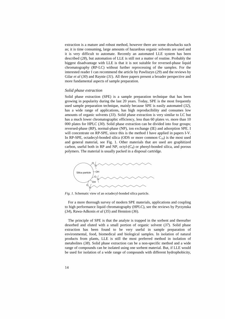
Solid phase extraction (SPE) is a sample preparation technique that has been growing in popularity during the last 20 years. Today, SPE is the most frequently used sample preparation technique, mainly because SPE is easily automated (32), has a wide range of applications, has high reproducibility and consumes low amounts of organic solvents (33). Solid phase extraction is very similar to LC but has a much lower chromatographic efficiency, less than 60 plates vs. more than 10 000 plates for HPLC (30). Solid phase extraction can be divided into four groups; reversed-phase (RP), normal-phase (NP), ion exchange (IE) and adsorption SPE. I will concentrate on RP-SPE, since this is the method I have applied in papers I-V. In RP-SPE, octadecyl-bonded silica (ODS or more common C18) is the most used and general material, see Fig. 1. Other materials that are used are graphitized carbon, useful both in RP and NP, octyl-(C8) or phenyl-bonded silica, and porous polymers. The material is usually packed in a disposal cartridge.
Fig. 1. Schematic view of an octadecyl-bonded silica particle.
For a more thorough survey of modern SPE materials, applications and coupling to high performance liquid chromatography (HPLC), see the reviews by Pyrzynska (34), Rawa-Adkonis et al (35) and Hennion (36).
The principle of SPE is that the analyte is trapped in the sorbent and thereafter desorbed and eluted with a small portion of organic solvent (37). Solid phase extraction has been found to be very useful in sample preparation of environmental, food, biomedical and biological samples. In isolation of natural products from plants, LLE is still the most preferred method in isolation of metabolites (38). Solid phase extraction can be a non-specific method and a wide range of compounds can be isolated using one sorbent material. But, if LLE would be used for isolation of a wide range of compounds with different hydrophobicity,
Silica particle
OSi
OSi
OH
OHO
Si

15
charge etc, then several solvents would need to be used. This can be an advantage when using LLE for isolation of known metabolites or certain types of compounds.
In our studies we wanted a fast, easy, non-hazardous, RP-HPLC compatible and general method, and therefore we used SPE in the sample preparation step. In papers I-V the SPE method for sample preparation was based on disposable 70-ml cartridges packed with 10 g end-capped C18 sorbent. The method involved five steps:
1. Activation of SPE material with 30 ml of MeCN. 2. Equilibration with 30 ml MilliQ-water. The rule is to use the same solvent
composition as in the sample. 3. Sample loading, maximum of 400 ml/10 g cartridge. 4. Washing with 30 ml of 5% aqueous MeCN, to remove hydrophilic
compounds, usually salts, amino acids and carbohydrates originating from the growth medium.
5. Elution with 30 ml 95% aqueous MeCN.
For screening of the antifungal activity of different bacteria, 200 ml of cell-free supernatant was used and for isolation up to 5 l was used. The sample now consisted of two fractions, one hydrophilic, from step 3 and 4 above, and one hydrophobic, from step 5. Both fractions could now be subjected to bioassay, but the hydrophilic fraction would either contain antifungal lactic acid or antifungal propionic acid, depending on which organism was studied. Therefore only the hydrophobic fraction was lyophilized and subjected to bioassay. This bioassay step was later omitted, since it was shown that the antifungal metabolite lactic acid also was present in this fraction (II).
Investigation of the hydrophilic fraction from Lactobacillus plantarum MiLAB 393 resulted only in isolation of the amino acid arginine (large amount) and lactic acid (unpublished results). The hydrophilic fraction was fractionated using preparative size exclusion chromatography (SEC) and then investigated with the methodology that will be described below. The SEC step was very time consuming and labour intense. Due to the fact that we had isolated over 50 interesting active LAB strains in paper II, the rest of the work in this thesis was focused on isolation of antifungal metabolites from the hydrophobic fraction. Separation
In a sample with a complex matrix, a separation step is usually needed before the bioassay, structure elucidation, or identification of metabolites. Chromatography, the most used separation technique, has had a huge impact in the field of separation science since it was developed in 1903 by the Russian botanist Michail Tswett (39). In chromatography, the separation is based on the analytes distribution between a stationary and a mobile phase. Based on the differences in the mobile phase, chromatography is classified into three categories; liquid chromatography, gas chromatography (GC) and supercritical fluid chromatography (SFC).
16
Liquid chromatography
Liquid chromatography is divided into five different categories based on what kind of interaction mechanism the analyte has with the stationary phase; adsorption chromatography (liquid-solid chromatography), partition chromatography (liquid-liquid chromatography), size exclusion chromatography, ion-exchange chromatography (IEC) and affinity chromatography. The power of LC lies in its broad range of compounds that can be separated and its capability of separating water based samples. This makes LC very suitable for separation of compounds in biological systems. The most common method of LC is RP-LC, where the stationary phase is more hydrophobic than the mobile phase. The stationary phase is often C18 chains bound to small porous silica particles as illustrated in Fig. 1. Mobile phases are often water based buffers together with an organic modifier like MeCN or MeOH. When a more hydrophilic stationary phase than the mobile phase is used then it is called normal-phase (NP) LC. The names NP and RP have historical reasons. At the time when LC was being developed, most of the work was performed with more hydrophilic stationary phases than the mobile phase. That was the normal condition, hence the name NP chromatography. Today, the opposite is true for analytical applications and RP-HPLC is the workhorse in most modern laboratories. In preparative applications NP-LC is still widely used, mainly for economical and practical reasons.
The flow of the mobile phase was originally achieved by gravity, but in the 1960´s, the mobile phase was being pumped through the column. To make it possible to distinguish between the two different methods of LC, the new method was called high pressure liquid chromatography. Today the abbreviation HPLC stands for high performance liquid chromatography. I will not go into a detailed discussion of the retention mechanism in RP-HPLC, but for an in depth study I recommend the book by Snyder & Kirkland (40) or more recent reviews (41, 42). An LC method that is widely used in natural products research is thin layer chromatography (TLC). It is a simple, easy, low-tech and low-cost method, but the separation capacity of TLC is rather limited compared to HPLC (40).
I have only used RP-HPLC in all papers so when I refer to HPLC, I mean RP-HPLC. In papers I-IV isolation of antifungal metabolites was performed using multidimensional chromatography (41). In multidimensional chromatography, two HPLC columns with distinct differences in retention mechanism are used. In the first HPLC step a C18 column was used and in the second step a porous graphitized carbon (PGC) column. Before the second HPLC separation, a method development was conducted to find the optimal conditions for the separation of each active fraction. Optimisation of an HPLC separation can be achieved by varying the mobile phase (components or composition), column type (C18, C8, phenyl, graphitized carbon) or temperature (43, 44). Changing the composition (the percentage of solvents) of the mobile phase is usually the simplest thing to start with, however the change in selectivity is not so great. A change in mobile phase components will have a bigger effect on the selectivity, but is also more time consuming. We wanted a method that was compatible both with liquid
17
chromatography mass spectrometry (LC-MS) and nuclear magnetic resonance (NMR), and this narrowed down the selection of mobile phases that could be utilised. No involatile buffers could be used, however volatile additives such as formic acid, trifluoroacetic acid (TFA), ammonia or diethylamine could be used to vary the pH of the mobile phase or to have an ion-pairing effect. Trifluoroacetic acid has ion-pair properties, which can improve the separation in HPLC, especially for peptides and proteins (45). A problem when using TFA in HPLC electrospray ionisation mass spectrometry (ESI-MS) is that the ionisation could be decreased by 30-80% compared to using formic acid (46). In papers I-IV, TFA has been used in the second HPLC step. In paper V formic acid has been used. An effective way to improve the separation of similar compounds, is to use isocratic elution instead of gradient elution. In the second HPLC step an isocratic elution was used in papers I-V for separation of the analytes. To alter the selectivity of the separation another column can be used, and this can be a more powerful way of changing selectivity than changing mobile phase composition (43, 47). We have used a porous graphitized carbon column in the second separation step in papers I-IV. The PGC column can be used both for RP- and NP-HPLC, in the entire pH range and has a stronger hydrophobicity than C18 (48). Due to the strong hydrophobicity of PGC, very polar compounds can be separated using reversed-phase conditions, and this can be difficult using a C18 column (49). The temperature influences retention, system pressure, selectivity and column stability (40, 43). Snyder & Kirkland stated in their book that changing the temperature had little effect on selectivity (40). But today, temperature is recognized as a useful parameter in changing the selectivity in HPLC (50, 51). A change in the temperature is achieved more easily than changing the solvents or the column. In the optimisation of the second HPLC different temperatures, from 20 to 50 °C, were evaluated, however it usually had little effect on the selectivity. Therefore, the separations in papers I-V were all conducted at room temperature, 20–22 °C. The exceptions were in the HPLC-ESI-MS analysis in paper V where the temperature was 35 °C, and for the HPLC-ESI-MS method for identification of DKPs where 50 °C was used (unpublished results).

18
Fig. 2. HPLC chromatogram from analysis of cell-free supernatant of Pedicoccus pentosaceus MiLAB 170 using a Discovery C18 column (100 × 21.2 mm i.d.), flow 10 ml/min, UV detection 210 nm, linear gradient 5% MeCN to 100% MeCN in 10 min, 5 min at 100 % MeCN. Lactic acid and fractions 31-33 had activity against Aspergillus fumigatus in the bioassay.
For isolation of antifungal compounds a crude separation was first achieved using a C18 column and a steep, linear gradient from 5% aqueous MeCN to 100% MeCN in 10 minutes. A preparative C18 column was used for the fractionation and 90 mg lyophilized material from the SPE was injected. All analyses were monitored using an ultraviolet (UV) detector at 210 nm. Fig. 2 shows the chromatogram of cell-free supernatant from P. pentosaceus MiLAB 170 after the SPE. All fractions were collected in a 96 2-ml deep-well plate using a fraction collector. Before bioassay the fractions were dried using a water bath at 40 °C and a gentle stream of air blowing through each well. About 1 g of material was received in the SPE step from 2 l of cell-free supernatant. This material was subsequently injected and fractionated in the first HPLC step. In papers IV and V the same procedure as above was used but with a slight modification. The gradient was 5% aqueous MeCN to 95% MeCN in 10 minutes. The result of the bioassay, a spore germination assay, after the first HPLC of cell-free supernatant from P. pentosaceus MiLAB 170 is shown in Fig. 3.
-50
450
950
1450
1950
2450
0 1 2 3 4 5 6 7 8 9 10 11 12
Time (min)
Detector response (mV)
Lactic acid
Fractions 31-33
-50
450
950
1450
1950
2450
0 1 2 3 4 5 6 7 8 9 10 11 12
Time (min)
Detector response (mV)
Lactic acid
Fractions 31-33

19
Fig. 3. Bioassay against Aspergillus fumigatus of fractions collected after the first HPLC step from fractionation of cell-free supernatant from Pediococcus pentosaceus MiLAB 170. Arrows indicating the direction of collection. Active fractions were 12–15 (lactic acid) and fractions 31–33 (Fig. 2).
After identification of the active fractions, the same fractions from different HPLC runs were pooled together and dried in a vacuum centrifuge. Each active fraction from the first HPLC contained several different compounds as illustrated by the chromatogram after the second HPLC step of fraction 31-33 (Fig. 4).
We have developed a protocol for the method development in the second HPLC step:
1. Change column from C18 to PGC. 2. Add 0.1% formic acid or 0.1% TFA to the mobile phase. 3. Find isocratic condition for optimal resolution. 4. Vary the temperature.
Leira et al (52) have previously described a column-switching HPLC method
where they used an C18 column coupled to a PGC to separate a complex mixture of non-flavonoid polyphenols. The methodology has also been applied for isolation of active constituents in natural materials (53).
1 2 3 4 5 6 7 8 9 10 11 12
A � � � � � � � � � � � �
B � � � � � � � � � � � � C � � � ���� � � ���D � � � � � � � � � � ��E � � � � � � � � � � � � F � � � � � � � � � � � � G � � � � � � � � � � � � H � � � � � � � � � � � �

20
Fig. 4. HPLC chromatogram of fractions 31-33 (10 mg) from P. pentosaceus MiLAB 170. The separation was conducted using a porous graphitized carbon column (100 × 21.2 mm i.d.) equipped with a guard column (10 × 10 mm i.d.), flow 10 ml/min, UV detection 210 nm, isocratic condition 30% MeCN and 70% water with 0.1% trifluoroacetic acid (unpublished results). Identified antifungal metabolites are indicated in the chromatogram.
In paper V, a good separation could not be achieved with the PGC column, instead a C18 column was also used for the second HPLC step. The separation was better but still not all peptides could be separated. TFA was tested as an ion-pairing reagent, but the separation was better with formic acid. A very powerful method that was not used in this thesis, which really can shorten optimisation time in HPLC, is computer simulation. Only after performing a few HPLC experiments, the software can calculate the optimal conditions for a specific separation (54, 55).
Structure elucidation
What is needed for a successful structure elucidation when an active metabolite has been isolated in pure form? What are the problems that have to be solved? Insufficient amounts of material is almost always a problem in the isolation of natural products. Has enough material been isolated for a complete structure elucidation? Usually, this is seldom known in advance, and a reisolation of the active compound is often necessary for the structure elucidation. Hence, it is important to have a reproducible method for the isolation of compounds. A molecular formula, a structural formula and an absolute configuration of the isolated metabolite should be provided for a full structural characterisation. This is
-50
50
150
250
350
450
550
650
0 1 2 3 4 5 6 7 8 9 10 11 12Time (min)
Detector response (mV)
Cyclo(L-Phe-cis-4-OH-D-Pro)
Cyclo(L-Leu-trans-4-OH-L-Pro)
Cyclo(L-Leu-cis-4-OH-D-Pro)
-50
50
150
250
350
450
550
650
0 1 2 3 4 5 6 7 8 9 10 11 12Time (min)
Detector response (mV)
Cyclo(L-Phe-cis-4-OH-D-Pro)
Cyclo(L-Leu-trans-4-OH-L-Pro)
Cyclo(L-Leu-cis-4-OH-D-Pro)

21
illustrated in Fig. 5 using cyclo(L-Leu-trans-4-OH-L-Pro) isolated from P. pentosaceus MiLAB 170 (unpublished results) as an example.
Fig. 5. Cyclo(L-Leu-trans-4-OH-L-Pro) isolated from Pediococcus pentosaceus MiLAB 170 (unpublished results).
According to Hanson (56) the structure elucidation of natural products can be divided into four stages:
1. Preliminary characterisation. Determination of physical constants, molecular weight and identification of functional groups.
2. Structural simplification. Identification of carbon skeleton. 3. Relative stereochemistry. Position of functional groups and their relative
configuration in space. 4. Absolute configuration.
Table 2 summarises different techniques that can be used to achieve structural
information without reference compound or chemical modification. A combination of several techniques is often needed for structure elucidation of an unknown compound. Sometimes this is not enough and chemical modification, e.g. derivatisation, hydrolysis, degradation or oxidation of the sample has to be done to achieve structural information.
To verify that a correct structure has been assigned to a compound, a confirmation of the structure has to be performed. This can be done by comparing NMR data of the isolated compound with literature NMR data or with NMR data from synthesised or purchased compound.
NNH
O
O
H
HH
HO
C11H18N2O3
1346
78 9
10
11
1212

22
Table 2. Summary of the structural information that can be achieved from different techniques without reference compounds
Technique Structural information IR, NMR, UV Functional groups MSn, NMR Building blocks or repeating units (e.g.
amino acids or carbohydrates), structural formula
MS Molecular weight, elemental composition NMR Relative stereochemistry Circular dichroism, specific optical rotation
Absolute configuration
X-ray crystallography (compound must crystallize)
Structure with absolute configuration
A powerful method in the structure elucidation of natural products is to couple
HPLC, MS and NMR. Pullen et al were the first to construct a hyphenated method, which coupled HPLC-NMR-MS for analysis of a mixture (57). During the last decade there have been several publications using coupled HPLC-MS-NMR for analysis of mixtures of natural products (58-65), pharmaceuticals (62, 66, 67), environmental samples (68) and drug metabolites in biological fluids (62, 69). HPLC-NMR-MS in natural product research is a fast and powerful method that provides lots of structural information. HPLC-NMR-MS can be used at an early stage for identification of known compounds before bioassay-guided fractionation starts, so called dereplication (63). There are several operational conflicts to overcome when coupling HPLC with NMR and MS, such as solvent compatibility, instrumental sensitivity and magnetic field effects (70). However, the major obstacles for most research groups is probably the price of this hyphenated instrumentation, location and the expertise needed for operating.
In this thesis off-line HPLC, NMR and MS experiments have been performed. After separation by HPLC the pure compounds are submitted to NMR and MS experiments for structure elucidation. The isolated compounds in this thesis have mainly been identified using NMR experiments in combination with MS experiments (I-IV). In many cases the information from NMR or MS alone is sufficient to elucidate the structure, but in some cases a combination is needed to solve the structure without available standards. From NMR a general skeletal structure was elucidated based on arranging the carbon and hydrogen atoms together with functional groups. By using the molecular weight from MS analysis together with the structural skeleton from NMR, the structures of the unknown metabolites could be determined. In paper V, the structures of the peptides from PAB were determined using FAB-MS/MS, because the structures could not be elucidated by NMR, mainly because the HPLC separation was insufficient to separate the peptides. For determination of the sequence of a peptide, MS methods or an HPLC peptide analysator are much more suitable methods than NMR (71, 72).

23
Nuclear magnetic resonance spectroscopy
Nuclear magnetic resonance was first discovered in 1946 (73, 74). Today, NMR is recognized as the most all-round and informative spectroscopic technique available to a chemist. It was in the 1960´s and 1970´s that the development of NMR really took a huge step forward when superconducting magnets, computers and the Fourier transform technique were introduced (75). The progress and importance of NMR spectroscopy has kept on growing ever since. The main use of NMR spectroscopy is in the determination of unknown structures, but it can also be used for interaction studies (76), chemical exchange experiments (77), imaging (78), and as an HPLC detector (79-80).
In NMR spectroscopy the molecular structure is studied by measuring the interaction of a radio-frequency (rf) generated oscillating electromagnetic field with a nucleus inside a strong magnetic field. The theory in brief is (77, 82): atoms that have a nucleus with a nuclear spin (I) (the most important is 1H and 13C) are studied. These atoms can be oriented in 2I + 1 ways in the presence of a strong magnetic field (behaving like a bar magnet) and there are also 2I + 1 possible energy states. When the nuclear spin is ½, as for 1H and 13C, there are two possible energy levels that the nucleus can adopt. There is a population excess in the lower energy state, which gives rise to a net magnetisation in the applied magnetic field, illustrated in Fig. 6a. When a strong rf pulse is applied to the system the pulse equalises the population difference between the two energy levels, hence no net magnetisation in the z plane remains (Fig. 6b). When the pulse is over, a signal called the free induction decay (FID) can be picked up and thereafter transformed into a spectrum.
Fig. 6. Vector model of NMR. a) Magnetisation vector (M0) at equilibrium due to the excess of spins in the low energy state. b) M0 after a 90°x pulse.
A spectrum according to the description above, would consist of one line from 13C and one from 1H, depending on which nucleus is studied, which would not be very useful in the determination of a structure. However, the nuclei in a molecule experience a magnetic field that is less than the applied magnetic field, i.e. the nuclei are shielded. This shielding originates from the electronic and magnetic environment in the neighbourhood of each nucleus. Therefore all nuclei, if they
b
x y
z
M0
a
x y
z
M0
b
x y
z
M0
a
x y
z
M0
x y
z
M0
a
x y
z
M0
x y
z
M0
a
x y
z
M0
x y
z
M0

24
experience different electronic and magnetic surroundings, will have different signals depending on where they are located in the molecule. The signal in NMR is expressed as chemical shift (δ), which is defined as: δ = (sample [Hz] – TMS [Hz])/operating frequency (Hz) where tetramethylsilane (TMS) is a reference compound. The chemical shift has no unit. It is expressed as parts per million (ppm). By using an internal standard the chemical shift becomes instrument independent, which makes it easy to compare results with literature data, if all other sample parameters are the same.
The sensitivity has generally been regarded as a problem in NMR. The low sensitivity is due to the small energy gap between ground and excited state and the low population difference between the energy states. The kind of nucleus that is studied also effects the sensitivity, e.g. 1H NMR is about 5700 times more sensitive than 13C (83). The sensitivity of 13C studies has been greatly enhanced by using different indirect detection NMR experiments, see below. To improve the sensitivity in NMR stronger superconducting magnets (84), cold probes (85-88) and small sample volumes (89-91) have been used. Today, a sample amount of <100 µg, for small molecules Mw <1000 Da (100 nmol), is sufficient for structural elucidation using NMR. For structure elucidation different NMR experiments are used, and Reynolds & Enríquez (92) discussed in their review how to choose the best pulse sequences, acquisition parameters, processing and probes for structure elucidation of natural products.
The NMR experiments, which were used for the structure elucidation of antifungal metabolites presented in this thesis and in papers I-V, were one dimensional 1H, two-dimensional 1H-1H correlation spectroscopy (COSY, 93), 1H-1H total correlation spectroscopy (TOCSY, 94), 1H-13C heteronuclear multiple bond correlation (HMBC, 95) and 1H-13C heteronuclear multiple quantum coherence (HMQC, 96) or 1H-13C heteronuclear single quantum coherence experiments (HSQC, 97). The most useful information from each different NMR experiment, used in structure elucidation of isolated compounds, is summarised in Table 3. When possible, HSQC should be used instead of HMQC due to higher sensitivity (98-99). All experiments were recorded at 30 °C using dimethyl sulfoxide-d6 (DMSO), D2O, CD3OD or CDCl3 as solvents. The experiments were performed according to Bruker standard pulse sequences. Proton chemical shifts were determined from one-dimensional 1H NMR and from HSQC or HMQC experiments and 13C chemical shifts were determined from HSQC or HMQC and HMBC experiments.

25
Table 3. The structural information that can be achieved from various NMR experiments used in this thesis
NMR experiment Information 1H Identification of kind of protons, number of non-
exchangeable protons. COSY Identification of protons close together (separated by
2 or 3 bonds). TOCSY Identification of spin systems. HSQC or HMQC Identification of connected proton-carbon, CH2
identification, what kind of carbon. HMBC Identification of connected proton-carbon by 2 or 3
bonds, carbons without attached proton.
By using the different NMR experiments on an isolated compound the 1H and
13C signals were assigned. First the number of non-exchangeable protons and functional groups were determined, and then, by using the data from COSY/TOCSY, different pieces (spin systems) of the molecule were linked together. From HSQC or HMQC the carbons were determined and with the aid of the data from HMBC the spin systems could be linked and a structure emerged. The NMR data was compared with data from commercially available compounds or from literature.
ppm
1.01.52.02.53.03.54.04.55.06.0 5.5 ppm
1.0
1.5
2.0
2.5
3.0
3.5
4.0
4.5
5.0
6.0
5.5
H-6 H-5 H-3 H-2H-4
H-7
H-8
H-12
Fig. 7. 1H-1H COSY spectrum of 3-hydroxy-5-cis-dodecenoic acid, CDCl3 used as solvent, temperature 30 °C, 600 MHz instrument equipped with a 2.5 mm microprobe, 64 scans (III).
In Fig. 7 the 1H-1H COSY spectrum illustrates the 1H-1H couplings in 3-hydroxy-5-cis-dodecenoic acid, which was isolated from L. plantarum MiLAB 14 (III). Only 300 µg was used for the structure elucidation and the HSQC spectrum, which was used for the assignment of the carbons in the molecule, is shown in Fig. 8.

26
ppm
1.01.52.02.53.03.54.04.55.06.0 5.5 ppm
10
20
30
40
50
60
70
80
90
100
110
120
130
ppm
1.21.4 ppm
15
20
25
30
35
40C-6
C-5
C-3
C-4
C-2C-7
C-12
C-11
C-9C-8
C-10
Fig. 8. HSQC spectrum of 3-hydroxy-5-cis-dodecenoic acid, CDCl3 used as solvent, temperature 30 °C, 600 MHz instrument equipped with a 2.5 mm microprobe, 64 scans (III). Determination of the configuration of the double bond was achieved by using the coupling constant 3JH5,H6. The literature data for cis 3JH,H is 0-12, typical value of 8, and for trans 3JH,H is 12-18, typical value of 15 (100). The value for 3-hydroxy-5-cis-dodecenoic acid was about 10, which was in agreement with data from the literature on the same compound (101). Furthermore, the signal of the methylene carbon adjacent to the double bond (C-7) had δ 27.5, which is in agreement with literature data, δ 27.3, when adjacent to a cis double bond (102). A methylene carbon, adjacent to a trans double bond, should have δ 32.7 (102). Hence, it was concluded that the double bond had the cis configuration. The 1H chemical shift data from cyclo(L-Leu-trans-4-OH-L-Pro) and cyclo(L-Leu-cis-4-OH-D-Pro) isolated from P. pentosaceus MiLAB 170 are presented in Table 4 (unpublished results). The compounds only differ in the absolute configuration of one of the amino acids, but their 1H NMR spectra are fairly different. The signals that differ the most are signals from H-3, H-8, H-9, H-10 (one of them) and H-11. The difference in chemical shift between the protons in the two molecules is due to the different absolute configuration of the proline residue. In cyclo(L-Leu-cis-4-OH-D-Pro) H-3 gets more shielded, which means that it has a higher electron density, compared to H-3 in cyclo(L-Leu-trans-4-OH-L-Pro). This effect is responsible for the lower chemical shift of the H-3 signal in cyclo(L-Leu-cis-4-OH-D-Pro).

27
Table 4. 1H Chemical shifts for cyclo(L-Leu-trans-4-OH-L-Pro) and cyclo(L-Leu-cis-4-OH-D-Pro) isolated from Pediococcus. pentosaceus MiLAB 170 (unpublished results), solvent CD3OD, temperature 30 °C, 600 MHz instrument
Proton Cyclo(L-Leu-trans-4-OH-L-Pro) Cyclo(L-Leu-cis-4-OH-D-Pro) H-3 4.17 3.88 H-6 3.46
3.66 3.43 3.67
H-7 4.46 4.40 H-8 2.09
2.27 2.20 2.50
H-9 4.54 4.30 H-10 1.52
1.93 1.56 1.66
H-11 1.90 1.78 H-12 0.90
1.00 0.90 1.00
An even more profound effect can be seen when the 1H NMR spectra from
cyclo(L-Phe-cis-4-OH-D-Pro) and cyclo(L-Phe-trans-4-OH-L-Pro) are compared, see Fig. 9. The phenyl ring has a great impact on the chemical shift on H-9 in cyclo(L-Phe-cis-4-OH-D-Pro). The difference in chemical shift for H-9 in the two compounds is 1.5 ppm. The chemical shift for H-9 is δ 4.3 (DMSO-d6) in cyclo(L-Phe-trans-4-OH-L-Pro) and δ 2.8 (CD3OD) in cyclo(L-Phe-cis-4-OH-D-Pro). When CD3OD is used as solvent for cyclo(L-Phe-trans-4-OH-L-Pro) the effect is even more profound and the difference is 1.6 ppm for H-9 (data not shown). In studies of cyclo(L-Phe-L-Pro) it has been shown that the phenyl ring stacks over the DKP ring (103), which is schematically illustrated in Fig. 10 for both DKPs. When H-9 is between the two rings, as in cyclo(L-Phe-cis-4-OH-D-Pro), H-9 gets shielded compared to when H-9 is outside the rings as in cyclo(L-Phe-trans-4-OH-L-Pro).

28
1.01.52.02.53.03.54.0 ppm
1.01.52.02.53.03.54.0 ppm
H-9
a
H-9 bDMSO
MeOH
Fig. 9. 1H NMR spectrum for a) cyclo(L-Phe-cis-4-OH-D-Pro), CD3OD as solvent and b) cyclo(L-Phe-trans-4-OH-L-Pro) DMSO-d6 as solvent, temperature 30 °C for both. H-9 for both compounds is indicated in the spectra.
In total, six different DKPs, cyclo(L-Phe-L-Pro) (I, II, IV), cyclo(L-Phe-trans-4-OH-L-Pro) (I, II), cyclo(L-Phe-cis-4-OH-D-Pro), cyclo(L-Leu-cis-4-OH-D-Pro), cyclo(L-Leu-trans-4-OH-L-Pro) (unpublished results) and cyclo(L-Ile-L-Pro) (IV) and two fatty acids, (R)-3-hydroxydecanoic acid and 3-hydroxy-5-cis-dodecenoic acid (III) and phenyllactic acid (I, II) were characterised using NMR and MS.
Fig. 10. Schematic structures showing the position of H-9 for a) cyclo(L-Phe-cis-4-OH-D-Pro) and b) cyclo(L-Phe-trans-4-OH-L-Pro).
N
NH
O
OH
N
NH
O
O
HH
HO
H
HO
a b
9
9

29
Mass spectrometry
In 1912 J. J. Thompson did the pioneering work in mass spectrometry (104). Fifty years later the coupling with chromatography was utilised and GC-MS had a huge impact on analysing volatiles. In the last two decades MS has been used, often together with HPLC, in analysing different biomolecules such as carbohydrates, peptides and proteins (105, 106). The applications have moved from physics to biology, the same trend as with NMR. A mass spectrometer is a device that separates ions based on their mass-to-charge ratio (m/z). The basic components of a mass spectrometer are the ion source and the mass analyser. The ion sources used in this thesis were electron impact connected to GC, electrospray (ES) coupled to HPLC, and fast atom bombardment (FAB).
Electron impact was devised by Dempster and further developed by Bleakney & Nier (104). In electron impact the sample, in gas phase, is passed through a beam of electrons at ~70 eV. The electrons transfer energy to the sample, which results in molecular ions, fragment ions and neutral fragments: M + e- (~70 eV) → Molecular ions + fragment ions + neutral fragments
Electron impact is well suited to be connected with a gas chromatograph because
in GC the analytes are in gas phase. The fragmentation pattern that EI creates can be used for identification of the analyte. This can be achieved either by studying the fragmentations and thereby solving the structure or by using the EI spectrum as a fingerprint. Electron impact spectra are to a large extent reproducible and therefore can the fingerprint be used for searching in a database, which can lead to identification of the analyte. There are many commercially available databases with EI spectra. Some of the disadvantages with EI are that the molecular ions are seldom observed, EI is limited to volatile and thermally stable molecules (105).
Electrospray was developed in 1984 by Yamashita & Fenn (107, 108). Their work was based on the discoveries of Dole and co-workers (109) 16 years earlier, when they were able to generate gas-phase ions of macromolecules by spraying a solution from the tip of an electrically charged capillary. In ES a diluted sample solution is pumped through a high voltage capillary. The voltage can be either positive or negative depending on what analytes are studied. The electrospray ionisation process is illustrated in Fig. 11. A Taylor cone is created at the capillary tip and multiple charged droplets are formed. By using a heated gas, the solvent of the droplet evaporates which shrinks the droplet and creates an accumulation of the charges in the droplet. This accumulation of charges in the droplet creates an explosion. Eventually, smaller droplets are formed, containing a single charged molecular ion.

30
Fig. 11. Schematic view of the electrospray ionisation process.
The mechanism of the formation of the single charged molecular ions is not fully understood. Today two mechanisms are generally accepted, the charged residue model and the ion evaporation model (110-112). Electrospray is a soft ionisation technique, which means that almost only molecular ions are detected and thus less fragmentation is observed. Electrospray MS is regarded as a rapid, sensitive and accurate technique for the determination of biomolecules. Electrospray is suited for analysing high molecular weight compounds because in ES multiply charged ions can be created. This means that an instrument with a low m/z range can be used to analyse high-mass molecules. For a more detailed survey of using ES-MS I recommend the article by Cech & Enke (113). The ability to analyse compounds in a solution makes ES compatible with separation techniques such as HPLC and CE (114). HPLC coupled to ES-MS (115) has been widely applied in chemistry, pharmaceutical sciences, natural products research and biochemistry (116-118). Disadvantages with ES are difficulties with mixture analysis, the purity of the sample is important to avoid ion suppression, and multiple charging and adduct formation (Na+- and K+- adducts) that can be confusing in the interpretation of the spectrum (105).
In the 1980´s Barber and co-workers utilised the MS technique for a study of non-volatiles and they opened the field for studies of biomolecules when they invented FAB (119-123). In FAB ionisation, the sample is mixed with a non-volatile liquid matrix, usually glycerol, and deposited on a metal probe. The probe is bombarded with a beam of high-energy atoms, usually Xe or Ar. The high-energy atoms eject sample molecules into the gas phase when crashing on the matrix. The sample molecules become charged through reactions with matrix molecules or the sample molecules could already be present as ions in the matrix. This technique is also a rather soft technique, which gives mostly molecular ions and little fragmentation. A disadvantage with this technique is that a high background of matrix ions can complicate the detection of the sample molecules in the spectrum (105). Detailed surveys of EI, ES and FAB and other ionisation techniques, such as matrix-assisted laser desorption/ionisation (MALDI), thermospray, plasma desorption, atmospheric-pressure chemical ionisation (APCI) and inductively
++
+++++
−−
−−
−+
+ ++ +
++
+++
+
+
+
+
+++
dry gas and/or heat
dry gas and/or heat
from HPLC mass analyser
N2
Taylor cone
ESI droplets
Spray needle 2 – 5 kV
++++
++++++++++
−−−−
−−−−
−−++
++ ++++ ++
++++
++++++
++
++
++
++
++++++
dry gas and/or heat
dry gas and/or heat
from HPLC mass analyser
N2
Taylor cone
ESI droplets
Spray needle 2 – 5 kV

31
coupled plasma (ICP) can be found in the book by de Hoffmann (104), or in the reviews by van Berkel (124) and Vestal (125).
After ions have been formed in the ion source, the ions are analysed in the mass analyser. In the mass analyser the ions are separated according to their m/z. There are three main characteristics of a mass analyser, the upper mass limit, the transmission and the resolution (104). The mass limit determines the highest m/z that can be detected, and the transmission is the ratio of how many of the ions created in the ion source reach the detector. The resolution is a measurement of the ability to separate two distinct signals for two ions with a small difference in m/z. The mass analysers that have been used in this thesis are a quadrupole, quadrupole ion trap and a four sector (electric sector and magnetic sector + electric sector and magnetic sector) tandem mass spectrometer. Other important mass analysers are time-of-flight (TOF) and ion cyclotron resonance analysers (104). The quadrupole was coupled to a GC with an EI ion source. The quadrupole ion trap instrument was coupled to an ES ion source and the instrument had the possibility to perform MSn. The sector instrument was coupled to a FAB ion source and the instrument had the possibility to perform MS/MS. Tandem MS (MS/MS) is used to obtain structural information about compounds (126), e.g. sequences of peptides and oligosaccharides. In the first MS-stage the molecular ion of interest is isolated and then fragmented, usually by using an inert gas (collision-induced dissociation, CID). In the second MS-stage the m/z of the fragmentation ions is monitored in a so called daughter ion spectrum. In MSn, which can be done with the quadrupole ion trap, further isolation and fragmentation can be performed. This possibility was not used in this thesis. The advantages of a sector instrument over the ion trap are its higher resolution and accuracy and that high-energy collisions can be used, which gives a more complete sequence information. The drawbacks are that the sector instrument is big, expensive and more difficult to use (105).
When FAB-MS is used a spectrum with more matrix peaks is generated (Fig. 12), compared to when ESI-MS is used (Fig. 13). The matrix peaks can complicate the interpretation of the spectrum. However, these matrix peaks can be subtracted from the spectrum after a blank analysis has been performed.

32
0
200000
400000
600000
800000
1000000
1200000
0 50 100 150 200 250 300 350
m/z
Intensity
(M+H)
245185*93*
75*
57*
120
70
277*337*
369*
(M+Glyc+H)
*
+
+
Fig. 12. FAB mass spectrum of standard cyclo(L-Phe-L-Pro) with glycerol as matrix. Matrix peaks are marked with *.
For the structure elucidation of cyclo(L-Phe-L-Pro) and cyclo(L-Phe-4-trans-OH-L-Pro) in paper I, FAB-MS was used, with glycerol as matrix. In paper II ES-MS was used for the determination of the molecular weight of the isolated compounds from different LAB.
120.2 217.2
245.2
267.0
0.0
0.2
0.4
0.6
0.8
7x10
Intensity
75 100 125 150 175 200 225 250 275m/z
(M+H)+
(M+Na)+
F (M+H-CO)+
Fig. 13. ESI mass spectrum of standard cyclo(L-Phe-L-Pro). F is the immonium ion of Phe.
The investigation of DKPs in MRS medium in paper I was done with HPLC-ES-MS and is illustrated in Fig. 14. The presence of cyclo(L-Leu-cis-4-OH-D-Pro), cyclo(L-Leu-trans-4-OH-L-Pro), cyclo(L-Phe-cis-4-OH-D-Pro), cyclo(L-Phe-trans-4-OH-L-Pro) and cyclo(L-Phe-L-Pro) was confirmed by comparing retention times

33
(tR) with previously identified DKPs and comparing mass spectra. To verify that cyclo(L-Phe-L-Pro) and cyclo(L-Phe-trans-4-OH-L-Pro) were produced by the bacteria, a defined medium with only amino acids was used for cultivation of L. plantarum MiLAB 393 (I). Both cyclo(L-Phe-L-Pro) and cyclo(L-Phe-trans-4-OH-L-Pro) were produced by L. plantarum MiLAB 393 in defined media and it was confirmed by isolation and analysis with FAB-MS (I).
EIC 226-228
EIC 244-246
EIC 260-262
0
1
2
3
4
7x107x10Intensity
0
1
2
3
4
0
1
2
3
4
5
4 6 8 10 12 14 16 18Time [min]
5
5.0
10.3
19.4
13.4
15.7
19 20
6
Cyclo(L-Phe-L-Pro)
Cyclo(L-Phe-cis-4-OH-D-Pro)
Cyclo(L-Phe-trans-4-OH-L-Pro)
Cyclo(L-Leu-trans-4-OH-L-Pro)
Cyclo(L-Leu-cis-4-OH-D-Pro)
Fig. 14. Extracted ion chromatogram (EIC) for MRS broth after SPE. Separation was performed on a Hypercarb PGC (100 × 4.6 mm i.d.) under the following conditions: flow 0.7 ml/min, temp. 50 °C, mobile phase consisted of A; 0.1% formic acid in water and B; 95% MeCN with 0.1% formic acid and a linear gradient from 40% to 70% B in 30 min and 0.4 mg sample was injected.
To optimise the HPLC conditions for the HPLC-ES-MS method for DKPs, both MeOH and MeCN were tested as organic modifiers together with formic acid or TFA. Different temperatures 20-50 °C, and two different column diameters, 4.6 mm and 2.1 mm, were evaluated. When using a column with a small internal diameter, a lower flow rate could be used which is preferable when using ES (127). The optimal conditions for best separation and ionisation were MeOH/water and 0.1% formic acid at 50 °C using the 2.1 mm column. The result of using MeOH instead of MeCN as the organic modifier is in agreement with the result of Giorgianni et al (128). Giorgianni et al (128) reported that MeOH is a better

34
organic modifier than MeCN in LC-MS/MS analysis of peptides. The chromatogram for a mixture of five isolated DKPs is shown in Fig. 15. With this method we have shown that HPLC-ES-MS, using a PGC column, can be used for analysing DKPs.
Time (miTime (mi n)0 2 4 6 8 1 0 1 2 1 4
Detector respons (mAU) Detector respons (mAU)
0
5 0
1 0 0
1 5 0
2 0 0
2 5 0
3 0 0
3 5 0
4 0 0
4.49
8.24
11.21
13.66
Cyclo(L-Leu-cis-OH-D-Pro)
Cyclo(L-Leu-trans-OH-L-Pro)
Cyclo(L-Phe-cis-OH-D-Pro)
Cyclo(L-Phe-trans-OH-L-Pro)
Cyclo(L-Phe-L-Pro)
12.42
Fig. 15. Chromatogram of five isolated DKPs (0.01–0.001 mg/ml) from LAB (unpublished results). Separation was performed on a Hypercarb PGC (100 × 2.1 mm i.d.) under the following conditions: UV-detection at 210 nm, flow 0.4 ml/min, temp. 50 °C, mobile phase consisted of A; 0.1% formic acid in water and B; 95% MeOH with 0.1% formic acid and a linear gradient from 35% to 100% B in 15 min and 20 µl was injected.
In the determination of peptides from SL medium FAB-MS/MS was used. FAB-MS/MS is a established method for determination of the amino acid sequences in peptides (72, 129-131). The FAB-MS/MS spectrum of Gly-Pro-Phe-Pro-Ile is shown in Fig. 16. A differentiation between Leu or Ile could not be achieved from the FAB-MS/MS spectra, due to the lack of secondary fragmentation of Leu and Ile (wn ions). The wn ions were absent since there was no basic amino acid present in the isolated peptides at or near the C-terminus, which seems to be required for the formation of wn ions (132). Identification of Leu or Ile was instead done with GC-MS, see below.

35
100 200 300 400 500
4
6
8
10
12
14
16
x 105
m/z
Intensity
P
F b3302.2
y2229.3
b2155.0
y4473.2
b4399.1
y3376.3
(M+H)
b5512.0
Gly - Pro - Phe - Pro - Ile+
530.3 y1 y2 y3
b1 b4 b3 b2
y4
Fig. 16. FAB-MS/MS spectrum of the (M+H)+ ion (m/z 530.3) of Gly-Pro-Phe-Pro-Ile (V). Peaks labelled P and F denotes immonium ions of Pro and Phe. Absolute configuration
The need for determination of the absolute configuration of a molecule arises from the fact that the spatial arrangements of atoms in a molecule may determine important chemical, physical and biological properties of the molecule. The most powerful technique for the determination of the absolute configuration of an unknown compound is X-ray crystallography. To use X-ray crystallography the compound must form crystals, which is often not the case for natural products. Even if crystals can be formed, this might not be enough and a heavy atom has to be incorporated into the structure. Other methods that can be used are circular dichroism (CD), specific optical rotation, chiral separation or derivatisation of the analyte, but all of these methods usually need reference compounds, which are not always available. Both CD and optical rotation can be used without reference compounds, however this requires a lot of experience. NMR can also be utilised in determination of the absolute configuration, either by using derivatisation with a chiral agent or analysing the analyte in the presence of a chiral solvent or a chiral solvation agent. The former method is the most commonly applied and has recently been reviewed by Seco et al (133). For the determination of the absolute

36
configuration of the isolated compounds in this thesis, various sample derivatisation methods together with chiral or non-chiral GC-MS were utilised.
14.50 15.00 15.50 16.00 16.50 17.00 17.50 18.00 18.500
100000
150000
200000
250000
300000
50000 16.98
17.23Abundance
Time (min)
D-PLA
L-PLA
Ion 120 (119.5 - 120.5)
Fig. 17. Absolute configuration determination of PLA from L. plantarum MiLAB 393 with GC-MS using a chiral GC column, for experimental conditions, see paper I.
To determine the absolute configuration of PLA in paper I and IV, a modified version of a method for determination of urinary organic acids by Heil et al (134) was used. The PLA was analysed as the corresponding methyl ester with GC-MS and the two isomers were separated on a chiral GC column. The analysis of PLA from L. plantarum MiLAB 393 is shown in Fig. 17. The absolute configuration for PLA was determined to be 9 parts L-PLA and 1 part D-PLA. The method did not induce any racemisation of the sample because no L-PLA was detected when D-PLA was analysed (data not shown). In paper IV the absolute configuration of PLA from five type strains of PAB was determined and the results, together with the result from paper I, have been summarised in Table 5. Table 5. L/D-PLA ratios determined from propionibacteria and Lactobacillus plantarum MiLAB 393
Bacteria L/D-PLA isomer ratio Propionibacterium jensenii 3:2 P. thoenii 7:3 P. acidipropionici 2:3 P. freudenreichii subsp. shermanii 9:1 P. freudenreichii subsp. freudenreichii 9:1 L. plantarum MiLAB 393 9:1
There are big differences in the production of L/D isomers of PLA among the
bacteria. The different PAB also produce different amounts of PLA (IV), e.g. P. jensenii produced around 10 times more PLA than P. thoenii. These findings, that different PAB produce different ratios of L/D isomers of PLA, could maybe be used for identification of PAB.

37
0
100000
200000
300000
400000
500000
600000
700000
5 6 7 8 9 10 11 12 13 14 15 16 17 18 19 20 21 22Time (min)
Detector respons
D/L-Leucis-OH-D/L-Pro
D/L-Phe
trans-OH-D/L-Pro
D/L-Pro
Fig. 18. GC-MS analysis of a standard mixture of amino acids used for determination of the absolute configuration of DKPs isolated from LAB (I, unpublished results). The D isomer elutes before the L isomer. For experimental conditions, see paper I.
The absolute configuration of the amino acids of the isolated DKPs and peptides was determined using non-chiral GC-MS, after the amino acids had been derivatised with (S)-2-butanol (I, IV, V). The sample was first hydrolysed with 6 M HCl, then derivatised with (S)-2-butanol to the corresponding ester, followed by an amide formation and an ester formation when hydroxyl is present, with perfluoropropanoic anhydride. The creation of diastereomers made it possible to separate L/D isomers of the amino acids on a non-chiral GC column, because the diastereomers have different chemical properties and are hence, possible to separate. In Fig. 18 the chromatogram of the standards for L/D-Leu, L/D-Pro, trans-4-OH-L-Pro, cis-4-OH-D-Pro and L/D-Phe is shown. These reference compounds were used for the determination of the absolute configuration of the amino acids in the DKPs from P. pentosaceus MiLAB 170 and L. plantarum MiLAB 14 (unpublished results). One D amino acid, cis-4-OH-D-Pro, was determined from the six isolated DKPs and the rest of the amino acids all had the L configuration.
The peptides isolated from the SL medium (V) consisted all of amino acids with the L configuration, but the absolute configuration of His could not be determined with this method, due to the incompatibility of the His with the GC column. With the GC-MS method for absolute configuration of the amino acids we could also distinguish Ile from Leu in the isolated peptides and based on the retention times of the standards and the MS spectra, the sequence of the isolated peptides was completed.

38
Fig. 19. Schematic view, without the stereochemistry, for derivatisation of 3-OH-FAs to 3-methoxy-FA (S)-phenylethylamide according to Gradowska & Larsson (135).
In paper III the absolute configuration of three 3-hydroxy fatty acids (3-OH FAs) from L. plantarum MiLAB 14 was determined using GC-MS. The method was developed by Gradowska & Larsson (135) for determination of the absolute configuration of 2- and 3-hydroxy fatty acids in organic dust. The absolute configuration of the 3-OH FAs was determined by preparation of 3-O-methyl N-(S)-phenylethylamide derivatives of the 3-OH-FAs, followed by GC-MS analysis using a non-chiral GC column. Racemates of 3-OH-decanoic acid, 3-OH-dodecanoic acid and 3-OH-tetradecanoic acid were used as reference compounds, since it has previously been reported that the (R,S) diastereomer of 3-methoxyphenylethylamides elute earlier than the corresponding (S,S) diastereomer (136). A schematic view of the derivatisation procedure, according to Gradowska & Larsson (135), of a 3-OH FAs to 3-methoxy-FA (S)-phenylethylamide is shown in Fig. 19. The 3-OH-FAs were determined to have the (R) configuration (III). The presence of small peaks corresponding to 3-OH-FAs in the (S)-configuration is due to racemisation of the sample in the hydrolysis step. The hydrolysis step is shown in Fig. 20. It can clearly be seen why racemisation of the sample can occur under these conditions.
RCH C
O
OH
1. Esterfication
RCH C
O
OCH3OH OH
2. Ether formation
CH2N2BF3 R
CH CO
OCH3OCH3
3. Hydrolysis
KOHMeOH/H2O 9:1 R
CH CO
OHOCH3
4. 3-Metoxy FA (S)- phenylethylamide formation
SOCl2(S)-Phenylethylamine
HCl in MeOH
RCH C
O
OCH3NH

39
Fig. 20. Racemisation in the hydrolysis step in the derivatisation of 3-OH-FAs to 3-methoxy-FA (S)-phenylethylamide.
The occurrence of the racemisation was proved by using methanol-d4/D2O, instead of using methanol/water in the hydrolysis step (Fig. 21). A sample from L. plantarum MiLAB 14 has been hydrolysed using either methanol-d4/D2O or methanol/water and the rest of the procedure as shown in Fig. 19. When methanol-d4/D2O was used as solvent, two protons were replaced with deuterium, hence the molecular mass of the final compound was 307, compared to 305 when no protons had been exchanged. If racemisation occurred and methanol-d4 was incorporated into the molecule, then 50% of the R and 50% of the S isomer would be detected, and the molecular mass of the molecule would be 310. From the extracted ion chromatogram of ion 310, shown in Fig. 21, it was concluded that a racemisation had occurred. To avoid racemisation of the sample we developed a faster and simpler method (unpublished results). The method was also based on the analysis of a (S)-phenylethylamide derivative, but instead of making an ether of the alcohol, the alcohol was trimethylsilylated with the silylating reagent N,O-bis(trimethylsilyl)trifluoroactamide (BSTFA) and the order of the derivatisations was reversed. First, the 3-OH-FA (S)-phenylethylamide was made according to Gradowska & Larsson (135) and after that the sample was dissolved in 100 µl hexane and 50 µl BSTFA and 10 µl pyridine were added. The sample was heated for 60 min at 80 °C and analysed by GC-MS on a fused-silica capillary column using a temperature gradient (120-260 °C at 10 °C min-1, 260 °C for 8 min, injector 270 °C, interface 270 °C, carrier gas He 1 ml min-1). The extracted ion chromatogram and the mass spectrum for (R)-3-OH-decanoic acid from L.
R CH CH CO
OCH3
OCH3
H
OH-
i ii
R CHHC C
O-
OCH3
OCH3
i 5% ii 95%+D
HC C
OOCH3C
HR
CD3OD
R CD2C C
O
OCH3
OCH3
R CD2C C
OOCH3
OCD3
+D
50% S-form 50% R-form +D
Hydrolysis withCD3OD/D2O
+D
+D
-CH3OD
HH
R CD2C C
OOCH3
OCD3
H

40
plantarum MiLAB 14 is shown in Fig. 22 and no racemisation was detected. The mass spectra for (R)-3-OH-decanoic acid and for the peak after, which should have been (S)-3-OH-decanoic acid if a racemisation had occurred, were not identical. This result proved that no racemisation had occurred.
In paper III 3-hydroxy-5-cis-dodecenoic acid was also isolated, but due to the lack of reference compounds, the absolute configuration of 3-hydroxy-5-cis-dodecenoic acid could not be determined.
10.00 11.00 12.00 13.00 14.00 15.00 16.00 17.00 18.000
5000
10000
15000
20000
Ion 307.00 (306.70 to 307.70)
10.00 11.00 12.00 13.00 14.00 15.00 16.00 17.00 18.000
5000
10000
15000
20000 Ion 310.00 (309.70 to 310.70)
Abundance
Time (min)
a
b
(R)-3-OH-decanoic acid
(R)-3-OH-decanoic acid (S)-3-OH-decanoic acid
(S)-3-OH-deacanoic acid
Fig. 21. Extracted ion chromatogram (EIC) of derivatised (R)-3-OH-decanoic acid showing the incorporation of a) 2 deuterium, Mw = 307 and b) CD3O and two deuterium, Mw 310, see text for details.
Antifungal metabolites
There are several reports of antifungal activity from LAB and the literature has recently been reviewed by Schnürer & Magnusson (12), but reports of identified antifungal metabolites are quite few. The production of the antimicrobial acids lactic, propionic and acetic acid from LAB and PAB has been known for a long time. Their antifungal effects on several spoilage yeasts and moulds in relation to pH have recently been investigated by Lind et al (137).
Other identified antifungal metabolites produced by LAB are pentocin TV35b (138), 3-hydroxypropionaldehyde (139), phenyllactic acid, p-hydroxyphenyllactic acid (I, 140), cyclic dipeptides such as cyclo(Gly-L-Leu) (141), cyclo(L-Phe-L-Pro) and cyclo(L-Phe-trans-4-OH-L-Pro) (I), benzoic acid, 5-methyl-2,4-imidazolidinedione, 4-hydroxy-4-methyl-tetrahydro-2H-pyran-2-one (141), the aliphatic acids formic, acetic, propanoic, butanoic, pentanoic, hexanoic (142) and 3-OH-FAs (III).

41
13.50 14.00 14.50 15.00 15.50
0
10000
20000
30000
40000
50000
60000
70000Ion 363.00 (362.70 to 363.70)Abundance
Time (min)
(R)-3-OH-decanoic acid
14.13
14.28
60 80 100 120 140 160 180 200 220 240 260 280 300 320 340 3600
50000
100000
150000
200000
250000
300000
350000
400000
450000Scan 14.13 min
55
73
105
120
174244
348363
258
m/z
Abundance
20000400006000080000100000120000140000160000180000200000220000240000260000280000300000
60 80 100 120 140 160 180 200 220 240 260 280 300 320 340 360
105
120
132147
244
174274
309372
55
Scan 14.28 min
0
a
b cAbundance
m/z Fig. 22. a) Extracted ion chromatogram of derivatised (R)-3-OH-decanoic acid from L. plantarum MiLAB 14 with novel derivatisation procedure, see text for details. b) Mass spectrum for (R)-3-OH-decanoic acid, tR = 14.13 min and c) mass spectrum for the closest peak eluating after, tR = 14.28 min.
Investigations of antifungal metabolites from PAB are almost non-existing in the literature, except for a few publications on antifungal activity. Schwenninger et al (143) have reported antifungal effects from a mixed culture of P. jensenii and Lactobacillus paracasei subsp paracasei in yoghurt and cheese, but no metabolites were identified, and Meile et al (144) reported several antifungal Propionibacterium from the Swiss dairy Propionibacterium collection. 3-Phenyllactic acid has been reported to be produced by P. freudenreichii (145, IV). In the following chapters I will present the antifungal metabolites that we have isolated from LAB and dairy PAB. The isolated and characterised antifungal metabolites are shown in Fig. 23. Diketopiperazines
Diketopiperazines or cyclic dipeptides, have been known for more than 100 years, e.g. cyclo(Gly-Gly) was synthesised in 1888 (146). Diketopiperazines are common in nature and many DKPs have been isolated through the years (146-148). For a detailed survey on DKPs and related compounds I recommend the article by Sammes (147). The isolated DKPs from different culture media were for a long time regarded as by-products of protein degradation, mainly because many DKPs were identified in peptide and protein hydrolysates as well (146). Nevertheless, today it is a well-known fact that many microorganisms produce DKPs (146, 148).

42
Fig. 23. Isolated and identified antifungal metabolites from lactic acid bacteria and propionic acid bacteria presented in this thesis.
The mechanism for the production of DKPs is not fully understood, but the formation of DKPs can be achieved by multifunctional enzymes, non-ribosomal peptide synthetases (149) and by single enzyme production (150) from amino acids. Diketopiperazines can also be formed from peptides and proteins under basic (151) or acidic (152, 153) conditions, e.g. in peptide synthesis, or by thermolysis (154, 155). Goolcharran & Borchardt (156) have shown that the
NNH
O
O
H
HH
HO
Cyclo(L-Leu-trans-4-OH-L-Pro)
NNH
O
O
H
HH
HO
Cyclo(L-Leu-cis-4-OH-D-Pro)
NNH
O
O
H
H
Cyclo(L-Phe-L-Pro)
NNH
O
O
H
HH
HO
Cyclo(L-Phe-trans-4-OH-L-Pro)
NNH
O
O
H
HH
HO
Cyclo(L-Phe-cis-4-OH-D-Pro)
NNH
O
O
H
H
Cyclo(L-Ile-L-Pro)
O
O
O
OH
OH
OH
O
OH
OHHO
HO
HO H
H
H
O
OHHO
H
L-Phenyllactic acid(S)-Phenyllactic acid
O
OHH OH
D-Phenyllactic acid(R)-Phenyllactic acid
(R)-3-Hydroxydecanoic acid
(R)-3-Hydroxydodecanoic acid
(R)-3-Hydroxytetradecanoic acid
3-Hydroxy-5-cis-dodecenoic acid

43
formation of DKPs, from a dipeptide, is sensitive to the pH of the solution, the buffer species present at high pH, the temperature and the N-terminal amino acid residue. Battersby et al (157) identified cyclo(Phe-Pro) after storage of human growth hormone for 4 days, pH 7.4 in 50 °C and at dry state. This indicates a non-enzymatical degradation of a protein to a DKP under mild conditions. Diketopiperazines have been studied in a variety of foods because of their bitter taste (154). Several DKPs have been identified in beer (158), chicken essence (159), cocoa (154) and roasted coffee (160). During the last 15 years the biological activity of DKPs has increased the interest in DKPs (146). Antimicrobial and antifungal activities have been reported for cyclo(L-Phe-L-Pro) and three other DKPs (161). Brauns et al (162) and Graz et al (163) reported anticancer activity of cyclo(Phe-Pro). Diketopiperazines have also been identified as signal substances in quorum sensing (164-166). Quorum sensing, or auto induction, is a cell-cell communication in bacteria accomplished through the exchange of small molecules. Most of the quorum sensing systems are species- or group-specific (167).
In this work, several DKPs from LAB and PAB have been isolated (Fig. 23), in
total six different DKPs have been isolated, and three of them have not been reported before. In Table 6 the literature data for the isolated DKPs in this work are summarised, showing their origin and biological activity.
None of the isolated DKPs were synthesised by us. For a comprehensive
literature survey on the synthesis of DKPs I recommend the review by Fischer (179). Two DKPs were purchased, cyclo(L-Phe-L-Pro) and cyclo(L-Ile-L-Pro), and their antifungal activity were evaluated (I, IV). The minimal inhibitory concentration (MIC) for cyclo(L-Phe-L-Pro) was determined to be 20 mg/ml at pH 4 against A. fumigatus and P. roquerforti (I) and 20 mg/ml for cyclo(L-Ile-L-Pro) against A. fumigatus (IV). When combining 5 mg/ml of PLA with 10 mg/ml of cyclo(L-Phe-L-Pro) total inhibition was detected, revealing possible synergistic effects (I). The identified antifungal activity of cyclo(L-Phe-L-Pro) is quite moderate when compared to antifungal drugs, e.g. amphotericin B had a MIC of 1 µg/ml at pH 5.5 and for nikkomycin Z the MIC was 512 µg/ml at pH 7.0 (180).
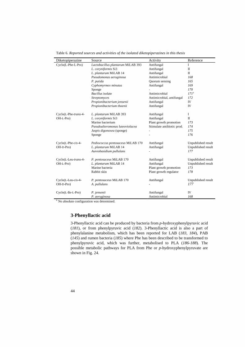
44
Table 6. Reported sources and activities of the isolated diketopiperazines in this thesis
Diketopiperazine Source Activity Reference Cyclo(L-Phe-L-Pro)
Lactobacillus plantarum MiLAB 393 L. coryniformis Si3 L. plantarum MiLAB 14 Pseudomonas aeruginosa P. putida Cyphomyrmex minutus Sponge Bacillus isolate Streptomyces Propionibacterium jensenii Propionibacterium thoenii
Antifungal Antifungal Antifungal Antimicrobial Quorum sensing Antifungal - Antimicrobial Antimicrobial, antifungal Antifungal Antifungal
I II II 168 165 169 170 171a
172 IV IV
Cyclo(L-Phe-trans-4-OH-L-Pro)
L. plantarum MiLAB 393 L. coryniformis Si3 Marine bacterium Pseudoalteromonas luteoviolacea Jaspis digonoxea (sponge) Sponge
Antifungal Antifungal Plant growth promotion Stimulate antibiotic prod. - -
I II 173 174 175 176
Cyclo(L-Phe-cis-4-OH-D-Pro)
Pediococcus pentosaceus MiLAB 170 L. plantarum MiLAB 14 Aureobasidium pullulans
Antifungal Antifungal -
Unpublished result Unpublished result 177
Cyclo(L-Leu-trans-4-OH-L-Pro)
P. pentosaceus MiLAB 170 L. plantarum MiLAB 14 Marine bacteria Rabbit skin
Antifungal Antifungal Plant growth promotion Plant growth regulator
Unpublished result Unpublished result 173 178
Cyclo(L-Leu-cis-4-OH-D-Pro)
P. pentosaceus MiLAB 170 A. pullulans
Antifungal -
Unpublished result 177
Cyclo(L-Ile-L-Pro) P. jensenii P. aeruginosa
Antifungal Antimicrobial
IV 168
a No absolute configuration was determined.
3-Phenyllactic acid
3-Phenyllactic acid can be produced by bacteria from p-hydroxyphenylpyruvic acid (181), or from phenylpyruvic acid (182). 3-Phenyllactic acid is also a part of phenylalanine metabolism, which has been reported for LAB (183, 184), PAB (145) and rumen bacteria (185) where Phe has been described to be transformed to phenylpyruvic acid, which was further, metabolised to PLA (186-188). The possible metabolic pathways for PLA from Phe or p-hydroxyphenylpyruvate are shown in Fig. 24.
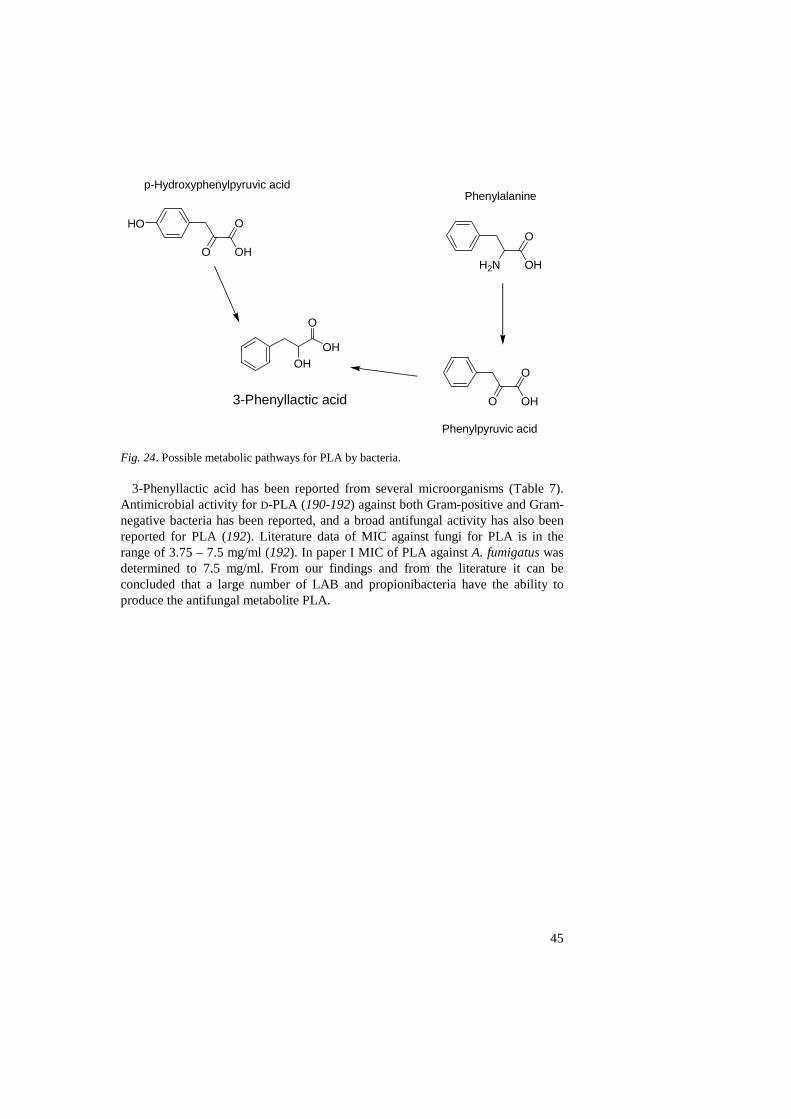
45
Fig. 24. Possible metabolic pathways for PLA by bacteria.
3-Phenyllactic acid has been reported from several microorganisms (Table 7). Antimicrobial activity for D-PLA (190-192) against both Gram-positive and Gram-negative bacteria has been reported, and a broad antifungal activity has also been reported for PLA (192). Literature data of MIC against fungi for PLA is in the range of 3.75 – 7.5 mg/ml (192). In paper I MIC of PLA against A. fumigatus was determined to 7.5 mg/ml. From our findings and from the literature it can be concluded that a large number of LAB and propionibacteria have the ability to produce the antifungal metabolite PLA.
OH
O
OH
3-Phenyllactic acid
Phenylalaninep-Hydroxyphenylpyruvic acid
O
O
OH
Phenylpyruvic acid
H2N
O
OHO
O
OH
HO

46
Table 7. Microorganisms that produce 3-phenyllactic acid
Compound Microorganism Reference Phenyllactic acid Lactobacillus plantarum MiLAB 393
L. coryniformis Si3 L. plantarum MiLAB 14 L. plantarum L. rhamnosus L. alimentarius L. fermentum L. sanfranciscensis L. acidophilus L. brevis L. hilgardii Weissella confusa Enterococcus faecium Leuconostoc mesenteroides L. citreum Geotrichum candidum Propionibacterium jensenii P. thoenii P. acidipropionici P. freudenreichii subsp. shermanii P. freudenreichii subsp. freudenreichii P. freudenreichii
I II Unpublished result 140 184 184 184 184 184 184 184 184 184 184 184 189 IV IV IV IV IV 145
Hydroxy fatty acids
Hydroxy fatty acids are abundant in plants, animals and fungi (193, 194). In bacteria OH-FAs can be present in lipopolysaccharides (195) or in poly-hydroxyalkanoic acids (196). On the other hand, Gram-positive bacteria, such as LAB, have no lipopolysaccharides and there are no reports on polyhydroxyalkanoic acids from LAB. Fatty acids from LAB in fermented milk have been reported by Rao & Reddy (197) and cellular FAs, mainly saturated and monounsaturated FAs, have been used for the classification of LAB (198-200), but reports on OH-FAs from LAB are few. However, Lee et al (201) identified 2-hydroxyhexadecanoic acid and 3-hydroxyheptadecanoic acid from different Leuconostoc strains. Reports have shown that LAB can metabolise unsaturated FAs into OH-FAs (202, 203) indicating metabolic pathways for the hydroxylation of FAs, however the exact role of 3-OH-FAs in LAB metabolism remains to be elucidated.
There are several reports on biological activity for FAs, e.g. antifungal (204-206) and antimicrobial activity (207-210). However, there are only a few reports on biological activity for OH-FAs, antifungal (211, 212), antiviral (213) and antimicrobial activities (214) for OH-FAs.
The antifungal saturated 3-OH-FAs and 3-hydroxy-5-cis-dodecenoic acid from L. plantarum MiLAB 14 (III) are presented in Fig. 23. All 3-OH-FAs have previously been isolated and reported, 3-hydroxydecanoic acid from ant (215), milk (216) and from lake sediment (217). 3-Hydroxydodecanoic and 3-

47
hydroxytetradecanoic acid have also been isolated from milk (216) and lake sediment (217), while 3-hydroxy-5-cis-dodecenoic acid has been isolated from Serratia marcescencs (218), without determination of the absolute configuration, and from Phytium ultimum (101). The identified saturated 3-OH-FAs were potent against a broad spectrum of yeast and moulds (III). The MIC values for the compounds were in the range of 10 – 100 µg/ml. The MIC for 3-hydroxytetradecanoic acid could not be determined due to the low solubility in water. These values show that 3-OH-FAs are much more potent than PLA and DKPs, and almost as potent as antifungal drugs (180).
Antifungal compounds in the growth medium
In the search for antifungal metabolites from microorganisms cell-free supernatant of microorganism grown in a liquid medium is often analysed. The growth medium is usually quite complex and the content is not fully characterised. The broth used for LAB in papers I-III was MRS broth (219), which contains undefined compounds from yeast extract, a peptic digest of meat proteins (peptone) and a meat extract (Lab-Lemco). Thus, the medium itself might contain several compounds with antifungal properties. Therefore, it is very important to evaluate the growth medium separately as well as with the bacterium present. In 2002 there were two reports that identified acetic acid in MRS medium as an antifungal agent, which contributed to the antifungal activity in the presence of LAB (220, 221). In our studies of LAB (I-III) we had no problems with acetic acid from the MRS, because we used two fractionation steps, SPE and HPLC, before we investigated the antifungal activity. Hence, no acetic or lactic acid was present in the fractions with activity (II). All of the DKPs isolated from LAB presented in this thesis (I-III, unpublished results) were also detected in the MRS broth. However, the fractions that correspond to the DKPs had no activity when the MRS broth was analysed (I). In paper I we showed that L. plantarum MiLAB 393 produced the isolated DKPs when the bacterium was cultivated in a defined medium (222) that only contained amino acids. The defined medium was analysed with the HPLC-ESI-MS method for DKPs, described above, and no DKPs were detected (data not shown).
The medium used for cultivation of propionibacteria was an SL medium (137), which contained undefined compounds from yeast extract and from a pancreatic digest of casein (tryptone). Casein is the main protein of milk. During the study of five type strains of PAB (IV) several antifungal peptides were isolated (V). In total, seven peptides were isolated and characterised by FAB-MS/MS (Table 8). All these peptides were detected in the SL medium (V) and were thus not produced by the bacteria.

48
Table 8. Antifungal peptides identified in SL medium and their possible origin (V)
Molecular mass Sequence Posssible origin 575 His-Pro-Leu-Pro-Leu Unknown 635 Phe-Leu-Pro-Tyr-Pro Casein 529 Gly-Pro-Phe-Pro-Ile Casein 529 Gly-Pro-Phe-Pro-Leu
Unknown
628 Gly-Pro-Phe-Pro-Leu-Val Unknown 889 Val-Tyr-Pro-Phe-Pro-Gly-Pro-Ile
Casein
963 Val-Ala-Pro-Phe-Gly-Val-Ala-Val-Phe-Gly Unknown
The activity of the peptides was low and the MIC values were > 10 mg/ml (V).
The exact values for the MIC determination were not investigated because of the low antifungal potency of the peptides. Three of the seven peptides, Gly-Pro-Phe-Pro-Ile (223, Swiss-Prot no.: P02666), Phe-Leu-Pro-Tyr-Pro (224, Swiss-Prot no.: P02668) and Val-Tyr-Pro-Phe-Pro-Gly-Pro-Ile (223, Swiss-Prot no.: P02666) could originate from the casein in the medium. This was determined by comparing the protein sequence for casein (223, 224) with the sequences of the isolated peptides. The other peptides did not match any of the protein sequences for casein or for Saccharomyces cerevisiae, and therefore the origin of the other peptides could not be determined (V). One of the peptides, Gly-Pro-Phe-Pro-Ile from casein, has previously been isolated and reported to inhibit cathepsin B (225). None of the other peptides have previously been isolated or characterised.
Conclusions and future perspectives
In this thesis several antifungal strains of LAB and strains of dairy propionibacteria have been studied and in total twelve antifungal metabolites have been characterised (I – IV, unpublished results). The compounds were low-molecular weight compounds and the majority of them have never before been reported to be produced by LAB or PAB.
In Paper I a method for isolating and characterising antifungal metabolites was reported. The methodology of using bioassay-guided fractionation is not new, however the combination of fungal bioassay and the use of modern separation techniques, has not been used previously for the study of metabolites from LAB and propionibacteria. A patent application was filed concerning the use of L. plantarum MiLAB 393 in the preservation of straw or green fodder (226) and the right to use it has been purchased by Medipharm AB. Medipharm AB has developed a biopreservative product, Feedtech 3000®, containing L. plantarum MiLAB 393 (Kerstin Holmgren, Medipharm AB, personal communication) that will be launched in Europe this year.
In paper II, several LAB strains were also shown to produce PLA, cyclo(L-Phe-L-Pro) and cyclo(L-Phe-trans-4-OH-L-Pro) and from P. pentosaceus MiLAB 170

49
additional DKPs were isolated and characterised (unpublished results). The most potent compounds isolated from LAB were 3-OH-FAs, which were identified from L. plantarum MiLAB 14 (III). This strain of L. plantarum also produced several DKPs and PLA (unpublished results).
An HPLC-ESI-MS method for the determination of DKPs was developed (unpublished results) and used for the detection of DKPs in MRS medium (I) and SL medium (IV). However, the amounts of DKPs in the cultivation media were not enough for inhibition of A. fumigatus. In paper I we showed that L. plantarum MiLAB 393 produced DKPs when we used a defined medium without any DKPs present.
An existing GC-MS method for the determination of the absolute configuration of 2- and 3-OH-FAs was shown to racemise the sample slightly (5%) during the sample derivatisation (I). The method was modified so that no racemisation would occur (unpublished results).
In paper IV the production of antifungal metabolites from the type strains of five dairy propionibacteria species was studied. The production of PLA was investigated and, surprisingly, four of the propionibacteria produced PLA at different L/D-isomer ratios. The two DKPs that were isolated and characterised were cyclo(L-Phe-L-Pro) and cyclo(L-Ile-L-Pro), the latter DKP has not been isolated previously by us, and none of the isolated DKPs have previously been reported to be produced by propionibacteria.
In paper V we investigated the antifungal compounds present in the SL medium. Some of the active fractions were active against many different fungi and from these fractions seven antifungal peptides were isolated and characterised. The most potent peptide was His-Pro-Leu-Pro-Leu, which inhibited A. fumigatus at the same concentration as cyclo(L-Phe-L-Pro) (I) and cyclo(L-Ile-L-Pro) (IV).
For many years the pharmaceutical industry has been screening natural products for potential drug compounds. During the period 1981–2002, 49% of all new chemical entities was derived, isolated from or in other ways related to metabolites from a natural product (227). However, in the last decade many pharmaceutical companies have downsized their research of natural products (228, 229). I hope that this thesis has shown some of the possibilities that microorganisms, in this study LAB and PAB, offer. The use of microorganisms in agriculture, forestry and food industry has a great potential. However, a lot of research is needed:
• Investigation of microorganisms and determination of active metabolites against e.g. fungi, bacteria, viruses and cancer has to continue.
• Studies of the mechanism and the mode of action for microorganisms and their active metabolites.
• Investigation of possible synergistic effects between active metabolites produced, e.g. synergistic effects between the DKPs, lactic acid, phenyllactic acid and 3-OH-FAs produced by L. plantarum MiLAB 14.

50
• Evaluation of the possibility of using one or several of the studied bacteria in food and feed systems.
• In vivo studies to investigate if the evaluated antifungal LAB and propionibacteria strains in this thesis are active in vivo and produce antifungal metabolites.
• In vivo studies to investigate if there are negative effects, and in that case, the nature of those negative effects when using microorganisms in vivo.

51
References
1. Ross, R.P., Morgan, S. & Hill, C. 2002. Preservation and fermentation: past, present and future. International Journal of Food Microbiology 79, 3-16.
2. Demain, A.L. 1981. Industrial microbiology. Science 214, 987-995. 3. Perlman, D. 1980. Some problems on the new horizons of applied microbiology.
Developments in Industrial Microbiology 21, xv-xxii. 4. Torsvik, V. & Øvreås, L. 2002. Microbial diversity and function in soil: from genes to
ecosystems. Current Opinion in Microbiology 5, 240-245. 5. Donaghy, K.P. 2001. Some moral, ethical and transethical issues raised by
biotechnology and how we might deliberate about them. American Behavioral Scientist 44, 1267-1282.
6. Singer, P.A & Daar, A.S. 2000. Avoiding Frankendrugs. Nature Biotechnology 18, 1225.
7. Phoebe, C.H., Combie, J., Albert, F.G., van Tran, K., Cabrera, J., Correira, H.J., Guo, Y., Lindermuth, J., Rauert, N., Galbraith, W. & Selitrennikoff, C.P. 2001. Extremophilic organisms as an unexplored source of antifungal compounds. The Journal of Antibiotics 54, 56-65.
8. Bode, H.B., Bethe, B., Höfs, R. & Zeeck, A. 2002. Big effects from small changes: possible ways to explore nature´s chemical diversity. ChemBioChem 3, 619-627.
9. Tannock, G.W. 2004. A special fondness for lactobacilli. Applied and Environmental Microbiology 70, 3189-3194.
10. Axelsson, L.T. 2004. Lactic acid bacteria: classification and physiology, p. 1-66. Lactic acid bacteria-Microbiology and functional aspects, edited by Salminen, S., Von Wright, A. & Ouwehand, A. 3rd edition. Marcel Dekker Inc. New York, USA.
11. Batish, V.K., Roy, U., Lal, R. & Grover, S. 1997. Antifungal attributes of lactic acid bacteria-a review. Critical Reviews in Biotechnology 17, 209-225.
12. Schnürer, J. & Magnusson, J. 2005. Antifungal lactic acid bacteria as biopreservatives. Trends in Food Science & Technology, in press, 1-9.
13. Reid, G. 1999. The scientific basis for probiotic strains of Lactobacillus. Applied and Environmental Microbiology 65, 3763-3766.
14. Simmering, R. & Blaut, M. 2001. Pro- and prebiotics-the tasty guardian angels? Applied Microbiology and Biotechnology 55, 19-28.
15. Weinberg, Z.G. & Muck, R.E. 1996. New trends and opportunities in the development and use of inoculants for silage. FEMS Microbiology Reviews 19, 53-68.
16. Piveteau, P. 1999. Metabolism of lactate and sugars by dairy propionibacteria: a review. Lait 79, 23-41.
17. Holo, H., Faye, T., Brede, D.A., Nilsen, T., Ødegård, I., Langsrud, T., Brendehaug, J. & Nes, I.F. 2002. Bacteriocins of propionic acid bacteria. Lait 82, 59-68.
18. Mantere-Alhonen, S. 1995. Propionibacteria used as probiotics-a review. Lait 75, 447-452.
19. Chaia, A.P., Zárate, G. & Oliver, G. 1999. The probiotic properties of propionibacteria. Lait 79, 175-185.
20. Suomalainen, T.H. & Mäyrä-Mäkinen, A.M. 1999. Propionic acid bacteria as protective cultures in fermented milks and breads. Lait 79, 165-174.
21. Merry, R.J. & Davies, D.R. 1999. Propionibacteria and their role in the biological control of aerobic spoilage in silage. Lait 79, 149-164.
22. Magnusson, J. & Schnürer, J. 2001. Lactobacillus coryniformis subsp. coryniformis strain Si3 produces a broad-spectrum proteinaceous antifungal compound. Applied and Environmental Microbiology 67, 1-5.
23. Rahilson, L., Hamburger, M., Monid, M., Frenk, E. & Hostettmann, K. 1994. Antifungal tests in phytochemical investigations: comparison of bioautographic methods using phytopathogenic and human pathogenic fungi. Planta Medica 60, 41-44.

52
24. Fukai, T., Yonekawa, M., Hou, A-J., Nomura, T., Sun, H-D. & Uno, J. 2003. Antifungal agents from the roots of Cudrania cochinchinensis against Candida, Cryptococcus and Aspergillus species. Journal of Natural Products 66, 1118-1120.
25. Engelmeier, D., Hadacek, F., Pacher, T., Vajrodaya, S. & Greger, H. 2000. Cyclopenta[b]benzofurans from Aglaia species with pronounced antifungal activity against rice blast fungus (Pyricularia grisea). Journal of Agricultural and Food Chemistry 48, 1400-1404.
26. Hadacek, F. & Greger, H. 2000. Testing antifungal natural products: methodologies, comparability of results and assay choice. Phytochemical analysis 11, 137-147.
27. Pitt, J.J. & Hocking, A.D. 1999. Fungi and food spoilage. 2nd edition. Aspen Publications. New York, USA.
28. Peng, S.X., Branch, T., M. & King, S.L. 2001. Fully automated 96-well liquid-liquid extraction for analysis of biological samples by liquid chromatography with tandem mass spectrometry. Analytical Chemistry 73, 708-714.
29. Pawliszyn, J. 2003. Sample preparation: Quo Vadis? Analytical Chemistry 75, 2543-2558.
30. Gilar, M., Bouvier, E.S.P. & Compton, B.J. 2001. Advances in sample preparation in electromigration, chromatographic and mass spectrometric separation methods. Journal of Chromatography A 909, 111-135.
31. Raynie, D.E. 2004. Modern extraction techniques. Analytical Chemistry 76, 4659-4664.
32. Rossi, D.T. & Zhang, N. 2000. Automating solid-phase extraction: current aspects and future prospects. Journal of Chromatography A 885, 97-113.
33. Smith, R.M. 2003. Before the injection-modern methods of sample preparation for separation techniques. Journal of Chromatography A 1000, 3-27.
34. Pyrzynska, K. 2003. Novel selective sorbents for solid-phase extraction. Chemia Analityczna 48, 781-795.
35. Rawa-Adkonis, M., Wolska, L. & Namiesnik J. 2003. Modern techniques of extraction of organic analytes from environmental matrices. Critical Reviews in Analytical Chemistry 33, 199-248.
36. Hennion, M-C. 1999. Solid-phase extraction: method development, sorbents, and coupling with liquid chromatography. Journal of Chromatography A 856, 3-54.
37. Thurman, E. M. 1998. Solid-phase extraction: principles and practice. John Wiley & Sons, Inc. New York, USA.
38. Nyiredy, S. 2004. Separation strategies of plant constituents-current status. Journal of Chromatography B 812, 35-51.
39. Ettre, L.S. 2003. M.S. Tswett and the invention of chromatography. LC-GC Europe 16, 632-640.
40. Snyder, L.R. & Kirkland, J.J. 1979. Introduction to modern liquid chromatography. 2nd edition. John Wiley & Sons, Inc. New York, USA.
41. Dorsey, J.G., Cooper, W.T., Siles, B.A., Foley, J.P. & Barth, H.G. 1998. Liquid chromatography: theory and methodology. Analytical Chemistry 70, 591R-644R.
42. Vailaya, V. & Horváth, C. 1998. Retention in reversed-phase chromatography: partition or adsorption? Journal of Chromatography A 829, 1-27.
43. Snyder, L.R. 1997. Changing reversed-phase high performance liquid chromatography selectivity. Which variables should be tried first? Journal of Chromatography B 689, 105-115.
44. Snyder, L.R., Kirkland, J.J. & Glajch, J.L. 1997. Practical HPLC method development. 2nd edition. John Wiley & Sons, Inc. New York, USA.
45. Guo, D., Mant, C.T. & Hodges, R.T. 1987. Effects of ion-pairing reagents on the prediction of peptide retention in reversed-phase high-performance liquid chromatography. Journal of Chromatography 386, 205-222.
46. Gustavsson, S.Å., Samskog, J., Markides, K.E. & Långström, B. 2001. Studies of signal suppression in liquid chromatography-electrospray ionization mass spectrometry using volatile ion-pairing reagents. Journal of Chromatography A 937, 41-47.

53
47. Zhao, J. & Carr, P.W. 1999. An approach to the concept of resolution optimization through changes in the effective chromatographic selectivity. Analytical Chemistry 71, 2623-2632.
48. Forgács, E. 2002. Retention characteristics and practical applications of carbon sorbents. Journal of Chromatography A 975, 229-243.
49. Hanai, T. 2003. Separation of polar compounds using carbon columns. Journal of Chromatography A 989, 183-196.
50. Dolan, J.W. 2002. Temperature selectivity in reversed-phase high performance liquid chromatography. Journal of Chromatography A 965, 195-205.
51. Wolcott, R.G., Dolan, J.W., Snyder, L.R., Bakalyar, S.R., Arnold, M.A. & Nichols, J.A. 2000. Control of column temperature in reversed-phase liquid chromatography. Journal of Chromatography A 869, 211-230.
52. Leira, E., Botana, A. & Cela, R. 1996. Resolution of complex mixtures of non-flavonoid polyphenols by column-switching high-performance liquid chromatography using octadecylsilica and graphitized carbon columns. Journal of Chromatography A 724, 67-78.
53. Wong, V. & Shalliker, R.A. 2004. Isolation of the active constituents in natural materials by 'heart-cutting' isocratic reversed-phase two-dimensional liquid chromatography. Journal of Chromatography A 1036, 15-24.
54. Molnar, I. 2002. Computerized design of separation strategies by reversed-phase liquid chromatography: development of DryLab software. Journal of Chromatography A 965, 175-194.
55. Rieger, H.J. & Molnar, I. 2002. Advanced high-performance liquid chromatography method development. Discovering unexpected choices in chromatography. Journal of Chromatography A 948, 43-49.
56. Hanson, J.R. 2003. Natural products: the secondary metabolites. The Royal Society of Chemistry. Cambridge, UK.
57. Pullen, F.S., Swanson, A.G., Newman, M.J. & Richards, D.S. 1995. On-line liquid chromatography/nuclear magnetic resonance mass spectrometry-a powerful spectroscopic tool for the analysis of mixtures of pharmaceutical interest. Rapid Communications in Mass Spectrometry 9, 1003-1006.
58. Hostettmann, K. & Wolfender, J-L. 1997. The search for biologically active secondary metabolites. Pesticide Science 51, 471-482.
59. Wolfender, J-L., Rodriguez, S. & Hostettmann, K. 1998. Liquid chromatography coupled to mass spectrometry and nuclear magnetic resonance spectroscopy for the screening of plant consituents. Journal of Chromatography A 794, 299-316.
60. Hansen, S.H., Jensen, A.G., Cornett, C., Bjørnsdottir, I., Taylor, S., Wright, B. & Wilson, I. D. 1999. High-performance liquid chromatography on-line coupled to high-field NMR and mass spectrometry for structure elucidation of constituents of Hypericum perforatum L. Analytical Chemistry 71, 5235-5241.
61. Lommen, A., Godejohann, M., Venema, D.P., Hollman, P.C.H. & Spraul, M. 2000. Application of directly coupled HPLC-NMR-MS to the identification and conformation of quercetin glycosides and phloretin glycosides in apple peel. Analytical Chemistry 72, 1793-1797.
62. Wilson, I.D. 2000. Multiple hyphenation of liquid chromatography with nuclear magnetic resonance spectroscopy, mass spectrometry and beyond. Journal of Chromatography A 892, 315-327.
63. Sandvoss, M., Weltring, A., Preiss, A., Levsen, K. & Wuensch, G. 2001. Combination of matrix solid-phase dispersion extraction and direct on-line liquid chromatography-nuclear magnetic resonance spectroscopy-tandem mass spectrometry as a new efficient approach for the rapid screening of natural products: application to the total asterosaponin fraction of the starfish Asterias rubens. Journal of Chromatography A 917, 75-86.

54
64. Queiroz, E.F., Wolfender, J-L., Atindehou, K.K., Traore, D. & Hostettmann, K. 2002.
On-line identification of the antifungal constituents of Erythrina vogelii by liquid chromatography with tandem mass spectrometry, ultraviolet absorbance detection and nuclear magnetic resonance spectrometry combined with liquid chromatographic micro-fractionation. Journal of Chromatography A 974, 123-134.
65. Wolfender, J-L., Ndjoko, K. & Hostettmann, K. 2003. Liquid chromatography with ultraviolet absorbance-mass spectrometric detection and with nuclear magnetic resonance spectroscopy: a powerful combination for the on-line structural investigation of plant metabolites. Journal of Chromatography A 1000, 437-455.
66. Holt, R.M., Newman, M.J., Pullen, F.S., Richards, D.S. & Swanson, A.G. 1997. High-performance liquid chromatography/NMR spectroscopy/mass spectrometry: further advances in hyphenated technology. Journal of Mass Spectrometry 32, 64-70.
67. Dear, G.J., Plumb, R.S., Sweatman, B.C., Parry, P.S., Roberts, A.D., Lindon, J.C., Nicholson, J.K. & Ismail, I.M. 2000. Mass directed peak selection, an efficient method of drug metabolite identification using directly coupled liquid chromatography-mass spectrometry-nuclear magnetic resonance spectroscopy. Journal of Chromatography B 748, 295-309.
68. Levsen, K., Preiss, A. & Godejohann, M. 2000. Application of high-performance liquid chromatography coupled to nuclear magnetic resonance and high-performance liquid chromatography coupled to mass spectrometry to complex environmental samples. Trends in Analytical Chemistry 19, 27-48.
69. Shockcor, J.P., Unger, S.E., Wilson, I.D., Foxall, P.J.D., Nicholson, J.K. & Lindon, J.C. 1996. Combined HPLC, NMR spectroscopy, and ion-trap mass spectrometry with application to the detection and characterization of xenobiotic and endogenous metabolites in human urine. Analytical Chemistry 68, 4431-4435.
70. Down, S. 2004. LC-NMR/MS. Spectroscopy Europe 16, 8-14. 71. Bailey, P.D. 1997. An introduction to peptide chemistry. John Wiley & Sons, Ltd.
Chichester, England. 72. Das, P.R. & Pramanik, B.N. 1998. Fast atom bombardment mass spectrometric
characterization of peptides. Molecular Biotechnology 9, 141-154. 73. Bloch, F., Hansen, W.W. & Packard, M. 1946. Nuclear induction. Physical Review 69,
127. 74. Purcell, E.M., Torrey, H.C. & Pound, R.V. 1946. Resonance absorption by nuclear
magnetic moments in a solid. Physical Review 69, 37-38. 75. Shoolery, J.N. 1995. The development of experimental and analytical high resolution
NMR. Progress in Nuclear Magnetic Spectroscopy 28, 37-52. 76. Lepre, C.A., Moore, J.M. & Peng, J.W. 2004. Theory and applications of NMR-based
screening in pharmaceutical research. Chemical Reviews 104, 3641-3675. 77. Friebolin, H. 1993. Basic one- and two-dimensional NMR spectroscopy. 2nd edition.
VCH Publishers. New York, USA. 78. Hills, B. 1998. Magnetic resonance imaging in food science, p. 179-204. John Wiley
& Sons, Inc. New York, USA. 79. Bobzin, S.C., Yang, S. & Kasten, T.P. 2000. Application of liquid chromatography-
nuclear magnetic resonance spectroscopy to the identification of natural products. Journal of Chromatography B 748, 259-267.
80. Albert, K. 1999. Liquid chromatography-nuclear magnetic resonance spectroscopy. Journal of Chromatography A 856, 199-211.
81. Griffiths, L. 1995. Optimization of NMR and HPLC conditions for LC-NMR. Analytical Chemistry 67, 4091-4095.
82. Claridge, T.D.W. 1999. High-resolution NMR techniques in organic chemistry. Elsevier Science Ltd. Oxford, UK.
83. Shoolery, J.N. 1984. Recent developments in 13C- and proton-NMR. Journal of Natural Products 47, 226-259.
84. Street, A. 2003. Superconducting magnets: at heart of NMR. Spectroscopy Europe 15, 10-13.

55
85. Serber, Z., Richter, C., Moskau, D., Böhlen, J-M., Gerfin, T., Marek, D., Häberli, M., Baselgia, L., Laukien, F., Stern, A.S., Hoch, J.C. & Dötsch, V. 2000. New carbon-detected protein NMR experiments using CryoProbes. Journal of the American Chemical Society 122, 3554-3555.
86. Logan, T.M., Murali, N., Wang, G. & Jolivet, C. 1999. Application of a high-resolution superconducting NMR probe in natural product structure determination. Magnetic Resonance in Chemistry 37, 762-765.
87. Hajduk, P.J., Gerfin, T., Boehlen, J-M., Häberli., Marek, D. & Fesik, S.W. 1999. High-throughput nuclear magnetic resonance-based screening. Journal of Medicinal Chemistry 42, 2315-2317.
88. Styles, P. & Soffe, N.F. 1984. A high-resolution NMR probe in which the coil and preamplifier are cooled with liquid helium. Journal of Magnetic Resonance 60, 397-404.
89. Lacey, M.E., Subramanian, R., Olson, D.L., Webb, A.G. & Sweedler, J.V. 1999. High-resolution NMR spectroscopy of sample volumes from 1 nL to 10 µL. Chemical Reviews 99, 3133-3152.
90. Reynolds, W.F., Yu, M. & Enriquez, R.G. 1997. Investigating the sensitivity limits of 13C-detected 1H-13C chemical shift correlation sequences with modern microprobe and microtube technology. Magnetic Resonance in Chemistry 35, 614-618.
91. Olson, D.L., Peck, T.L., Webb, A.G., Magin, R.L. & Sweedler, J.V. 1999. High-resolution microcoil 1H-NMR for mass-limited, nanoliter-volume samples. Science 270, 1967-1970.
92. Reynolds, W.F. & Enriquez, R.G. 2002. Choosing the best pulse sequences, acquisition parameters, postacquisition processing strategies, and probes for natural product structure elucidation by NMR spectroscopy. Journal of Natural Products 65, 221-244.
93. Aue, W.P., Bartholdi, E. & Ernst, R.R. 1976. Two-dimensional spectroscopy. Application to nuclear magnetic resonance. Journal of Chemical Physics 64, 2229-2246.
94. Braunschweiler, L. & Ernst, R.R. 1983. Coherence transfer by isotropic mixing: application to proton correlation spectroscopy. Journal of Magnetic Resonance 53, 521-528.
95. Bax, A. & Summers, M.F. 1986. 1H and 13C assignment from sensitivity-enhanced detection of heteronuclear multiple-bond connectivity by 2D multiple quantum NMR. Journal of the American Chemical Society 108, 2093-2094.
96. Bax, A. & Subramanian, S. 1986. Sensitivity-enhanced two-dimensional heteronuclear shift correlation NMR spectroscopy. Journal of Magnetic Resonance 67, 565-569.
97. Bodenhausen, G. & Ruben, D.J. 1980. Natural abundance nitrogen-15 NMR by enhanced heteronuclear spectroscopy. Chemical Physics Letters 69, 185-189.
98. Reynolds, W.F., McLean, S., Tay, L-L., Yu, M., Enriquez, R.G., Estwick, D.M. & Pascoe, K.P. 1997. Comparison of 13C resolution and sensitivity of HSQC and HMQC sequences and application of HSQC-based sequences to the total 1H and 13C- spectral assignment of clionasterol. Magnetic Resonance in Chemistry 35, 455-462.
99. Kövér, K.E., Prakash, O. & Hruby, V.J. 1993. Improved 2D inverse proton detected C, H correlation NMR techniques for the total assignment of carbon resonance of a highly delta opioid receptor agonist peptide. Magnetic Resonance in Chemistry 31, 231-237.
100. Williams, D.H. & Fleming, I. 1989. Spectroscopic methods in organic chemistry. 4th edition. McGraw-Hill Book Company Europe. Berkshire, England.
101. Ichihara, A., Hashimoto, M. & Sakamura, S. 1985. (3R,5Z)-(−)-3-Hydroxy-5-dodecenoic acid, a phytotoxic metabolite of Pythium ultimum. Agricultural and Biological Chemistry 49, 2207-2209.
102. Gunstone, F.D., Pollard, M.R., Scrimgeour, C.M. & Vedanayagam, H.S. 1977. Fatty acids. Part 50. 13C Nuclear magnetic resonance studies of olefinic fatty acids esters. Chemistry and Physics of Lipids 18, 115-129.

56
103. Budesínský, M., Symershý, J., Jecný, J., van Hecke, J., Hosten, N., Anteunis, M. &
Borremans, F. 1992. Cyclo(L-propyl-L-N-methylphenylalanyl) conformation in solution and in the crystal. International Journal of Peptide and Protein Research 39, 123-130.
104. de Hoffmann, E., Charette, J. & Stroobant, V. 1999. Mass spectrometry-principles and application. John Wiley & Sons, Ltd. Chichester, England.
105. Siuzdak, G. 1996. Mass spectrometry for biotechnology. Academic Press. San Diego, USA.
106. Krishnamurthy, T., Ross, P.L., Goode, M.T., Menking, D.L., Rajamani, U. & Heroux, K. 1997. Biomolecules and mass spectrometry. Journal of Natural Toxins 6, 121-162.
107. Yamashita, M. & Fenn, J.B. 1984. Electrospray ion source. Another variation of the free-jet theme. Journal of Physical Chemistry 88, 4451-4459.
108. Yamashita, M. & Fenn, J.B. 1984. Negative ion production with the electrospray ion source. Journal of Physical Chemistry 88, 4471-4475.
109. Dole, M., Mack, L.L., Hines, R.L., Mobley, R.C., Ferguson, L.D. & Alice, M.B. 1968. Molecular beams of macroions. The Journal of Chemical Physics 49, 2240-2249.
110. Cole, R.B. 2000. Some tenets pertaining to electrospray ionization mass spectrometry. Journal of Mass Spectrometry 35, 763-772.
111. Kebarle, P. 2000. A brief overview of the present status of the mechanisms involved in electrospray mass spectrometry. Journal of Mass Spectrometry 35, 804-817.
112. Gaskell, S.J. 1997. Electrospray: principles and practice. Journal of Mass Spectrometry 32, 677-688.
113. Cech, N.B. & Enke, C.G. 2001. Practical implications of some recent studies in electrospray ionization fundamentals. Mass Spectrometry Reviews 20, 362-387.
114. Tomer, K.B. 2001. Separations combined with mass spectrometry. Chemical Reviews 101, 297-328.
115. Whitehouse, C.M., Dreyer, R.N., Yamashita, M. & Fenn, J.B. 1985. Electrospray interface for liquid chromatographs and mass spectrometers. Analytical Chemistry 57, 675-679.
116. Niessen, W.M.A. 2003. Progress in liquid chromatography-mass spectrometry instrumentation and its impact on high-throughput screening. Journal of Chromatography A 1000, 413-436.
117. Lim, C-K. & Lord, G. 2002. Current developments in LC-MS for pharmaceutical analysis. Biological and Pharmaceutical Bulletin 25, 547-557.
118. He, X-G. 2000. On-line identification of phytochemical constituents in botanical extracts by combined high-performance liquid chromatographic-diode array detection-mass spectrometric techniques. Journal of Chromatography A 880, 203-232.
119. Barber, M., Bordoli, R.S., Sedgwick, R.D. & Tyler, A.N. 1981. Fast atom bombardment of solids (F.A.B.): a new ion source for mass spectrometry. Journal of the Chemical Society. Chemical Communications, 325-327.
120. Barber, M., Bordoli, R.S., Sedgwick, R.D. & Tyler, A.N. 1981. Fast atom bombardment mass spectrometry of bradykinin and related oligopeptides. Biomedical Mass Spectrometry 8, 337-342.
121. Barber, M., Bordoli, R.S., Garner, G.V., Gordon, D.B., Sedgwick, R.D., Tetler, L.W. & Tyler, A.N. 1981. Fast-atom-bombardment mass spectra of enkephalins. Biochemical Journal 197, 401-404.
122. Barber, M., Bordoli, R.S., Sedgwick, R.D. & Tyler, A.N. 1981. Fast atom bombardment of solids as an ion source in mass spectrometry. Nature 293, 270-275.
123. Barber, M., Bordoli, R.S., Sedgwick, R.D. & Tyler, A.N. 1981. Fast atom bombardment mass spectrometry of cobalamines. Biomedical Mass Spectrometry 8, 492-495.
124. van Berkel, G.J. 2003. An overview of some recent developments in ionization methods for mass spectrometry. European Journal of Mass Spectrometry 9, 539-562.
125. Vestal, M.L. 2001. Methods of ion generation. Chemical Reviews 101, 361-375. 126. de Hoffmann. E. 1996. Tandem mass spectrometry: a primer. Journal of Mass
Spectrometry 31, 129-137.
57
127. Sandra, P., Vanhoenacker, G., Lynen, F., Li, L. & Schelfaut, M. 2001. Considerations on column selection and operating conditions for LC-MS. LC-GC Europe 14, Guide to LC-MS, 2-14.
128. Giorgianni, F., Cappiello, A., Beranova-Giogianni, S., Palma, P., Trufelli, H. & Desiderio, D. M. 2004. LC-MS/MS analysis of peptides with methanol as organic modifier: improved limits of detection. Analytical Chemistry 76, 7028-7038.
129. Carr, S.A., Hemling, M.E., Bean, M.F. & Roberts, G.D. 1991, Integration of mass spectrometry in analytical biotechnology. Analytical Chemistry 63, 2802-2824.
130. Williams, D.H., Bradley, C.V., Santikarn, S. & Bojesen, G. 1982. Fast-atom bombardment mass spectrometry. A new technique for the determination of molecular weights and amino acid sequences of peptides. Biochemical Journal 201, 105-117.
131. Williams, D.H., Bojesen, G., Auffret, A.D. & Taylor, C.E. 1981. Study of difficult peptides from Paracoccus cytochrome c-550 and a dolphin cytochrome c. FEBS Letters 128, 37-39.
132. Johnson, R.S., Martin, S.A., Bieman, K., Stults, J.T. & Watson, J.T. 1987. Novel fragmentation process of peptides by collision-induced decomposition in a tandem mass spectrometer: differentiation of leucine and isoleucine. Analytical Chemistry 59, 2621-2625.
133. Seco, J.M., Quiñoá, E. & Riguera, R. 2004. The assignment of absolute configuration by NMR. Chemical Reviews 104, 17-117.
134. Heil, M., Podebrad, F., Beck, T., Mosandl, A., Sewell, A. C. & Böhles, H. 1998. Enantioselective multidimensional gas chromatography-mass spectrometry in the analysis of urinary organic acids. Journal of Chromatography B 714, 119-126.
135. Gradowska, W. & Larsson, L. 1994. Determination of absolute configuration of 2- and 3-hydroxy fatty acids in organic dust by gas chromatography-mass spectrometry. Journal of Microbiological Methods 20, 55-67.
136. Rietschel, E.T., Lüderitz, O. & Volk, W.A. 1975. Nature, type of linkage, and absolute configuration of (hydroxy) fatty acids in lipopolysaccharides from Xanthomonas sinensis and related strains. Journal of Bacteriology 122, 1180-1188.
137. Lind, H., Jonsson, H. & Schnürer, J. 2005. Antifungal effect of dairy propionibacteria-contribution of organic acids. International Journal of Food Microbiology 98, 157-165.
138. Okkers, D.J., Dicks, L.M.T., Silvester, M., Joubert, J.J. & Odendaal, H.J. 1999. Characterization of pentocin TV35b, a bacteriocin-like peptide isolated from Lactobacillus pentosus with fungistatic effect on Candida albicans. Journal of Applied Microbiology 87, 726-734.
139. Talarico, T.L., Casa, I.A., Chung, T.C. & Dobrogosz, W.J. 1988. Production and isolation of reuterin, a growth inhibitor produced by Lactobacillus reuteri. Antimicrobial Agents and Chemotherapy 32, 1854-1858.
140. Lavermicocca, P., Valerio, F., Evidente, A., Lazzaroni, S., Corsetti, A. & Gobbetti, M. 2000. Purification and characterization of novel antifungal compounds from the sourdough Lactobacillus plantarum strain 21B. Applied and Environmental Microbiology 66, 4084-4090.
141. Niku-Paavola, M-L., Laitila. A., Mattila-Sandholm, T. & Haikara, A. 1999. New types of antimicrobial compounds produced by Lactobacillus plantarum. Journal of Applied Microbiology 86, 29-35.
142. Corsetti, A., Gobbetti, M., Rossi, J. & Damiani, P. 1998. Antimould activity of sourdough lactic acid bacteria: identification of a mixture of organic acids produced by Lactobacillus sanfrancisco CB1. Applied Microbiology and Biotechnology 50, 253-256.
143. Miescher Schwenninger, S. & Meile, L. 2004. A mixed culture of Propionibacterium jensenii and Lactobacillus paracasei subsp. paracasei inhibits food spoilage yeasts. Systematic and Applied Microbiology 27, 229-237.
144. Meile, L., Dasen, G., Miescher, S., Stierli, M. & Teuber, M. 1999. Classification of propionic acid bacteria and approaches to applied genetics. Lait 79, 71-78.
145. Thierry, A. & Maillard, M-B. 2002. Production of cheese flavor compounds derived from amino acid catabolism by Propionbacterium freudenreichii. Lait 82, 17-32.

58
146. Prasad, C. 1995. Bioactive cyclic dipeptides. Peptides 16, 151-164. 147. Sammes, P.G. 1975. Naturally occuring 2,5-dioxopiperazines and related compounds,
p. 51-118. Fortschritte Der Organischer Naturstoffe 32. Edited by Zechmeister, L., Herz, W., Grisebach, H. & Kirby, G.W. Springer Verlag. Wien, Austria.
148. Bycroft, B.W. & Higton, A.A. 1985. Amino acids, peptides and proteins, p. 254-260, Natural products. Edited by Thomson, R.H. Blackie & Sons Ltd. Glasgow, UK.
149. Schwarzer, D., Mootz, H.D. & Marahiel, M.A. 2001. Exploring the impact of different thioesterase domains for the design of hybrid peptide synthetases. Chemistry and Biology 8, 997-1010.
150. Lautru, S., Gondry, M., Genet, R. & Pernodet, J-L. 2002. Chemistry and Biology 9, 1355-1364.
151. Khosla, M.C., Seby, R.R. & Bumpus, F.M. 1972. Failure sequence in solid-phase peptide synthesis due to presence of an N-alkylamino acid. Journal of the American Chemical Society 94, 4721-4724.
152. Suzuki, K., Sasaki, Y., Endo, N. & Mihara, Y. 1981. Acetic acid catalyzed diketopiperazine synthesis. Chemical and Pharmaceutical Bulletin 29, 233-237.
153. Gisin, B.F. & Merrifield, R.B. 1972. Carboxyl-catalyzed intramolecular aminolysis. A side reaction in solid-phase peptide synthesis. Journal of the American Chemical Society 94, 3102-3106.
154. Pickenhagen, W., Dietrich, P., Keil, B., Polonsky, J., Nouaille, F. & Lederer, E. 1975. Identification of the bitter principle of cocoa. Helvetica Chimica Acta 58, 1078-1086.
155. Steinberg, S.M. & Bada, J.L. 1983. Peptide decomposition in the neutral pH region via formation of diketopiperazines. Journal of Organic Chemistry 48, 2295-2298.
156. Goolcharra, C. & Borchardt, R.T. 1998. Kinetics of diketopiperazine formation using model peptides. Journal of Pharmaceutical Sciences 87, 283-288.
157. Battersby, J.E., Hancock, W.S., Canovan-Davis, E., Oeswein, J. & O´Connor, B. 1994. Diketopiperazine formation and N-terminal degradation in recombinant human growth hormone. International Journal of Peptide and Protein Research 44, 215-222.
158. Gautschi, M., Schmid, J.P., Peppard, T.L., Ryan, T.P., Tuorto, R.M. & Yang, X. 1997. Chemical characterization of diketopiperazines in beer. Journal of Agricultural and Food Chemistry 45, 3183-3189.
159. Chen, Y-H., Liou, S-E. & Chen, C-C. 2004. Two-step mass spectrometric approach for the identification of diketopiperazines in chicken essence. European Food Research and Technology 218, 589-597.
160. Ginz, M. & Engelhardt, U.H. 2000. Identification of proline-based diketopiperazines in roasted coffee. Journal of Agricultural and Food Chemistry 48, 3528-3532.
161. Graz, M., Hunt, A., Jamie, H., Grant, G. & Milne, P. 1999. Antimicrobial activity of selected cyclic dipeptides. Pharmazie 54, 772-775.
162. Brauns, S.C., Milne, P., Naudé, R. & van de Venter, M. 2004. Selected cyclic dipeptides inhibit cancer cell growth and induce apoptosis in HT-29 colon cancer cells. Anticancer Research 24, 1713-1720.
163. Graz, C.J.M., Grant, G.D., Brauns, S.C., Hunt, A., Jamie, H. & Milne, P. 2000. Journal of Pharmacy and Pharmacology 52, 75-82.
164. Daniels, R., Vanderleyden, J. & Michiels, J. 2004. Quorum sensing and swarming migration in bacteria. FEMS Microbiology Reviews 28, 261-289.
165. Degrassi, G., Aguilar, C., Bosco, M., Zahariev, S., Pongor, S. & Venturi, V. 2002. Plant growth-promoting Pseudomonas putida WC358 produces and secretes four cyclic dipeptides: cross-talk with quorum sensing bacterial sensors. Current Microbiology 45, 250-254.
166. Holden, M.T.G., Chhabra, S.R., de Nys, R., Stead, P., Bainton, N.J., Hill, P.J., Manefield, M., Kumar, N., Labatte, M., England, D., Rice, S., Givskov, M., Salmond, G.P.C., Stewart, G.S.A.B., Bycroft, B.W., Kjelleberg, S.A. & Williams, P. 1999. Quorum-sensing cross talk: isolation and chemical characterization of cyclic dipeptides from Pseudomonas aeruginosa and other Gram-negative bacteria. Molecular Microbiology 33, 1254-1266.

59
167. Taga, M.E. & Bassler, B.L. 2003. Chemical communication among bacteria. Proceedings of the National Academy of Sciences of the United States of America 100, 14549-14554.
168. Jayatilake, G.S., Thornton, M.P., Leonard, A.C., Grimwade, J.E. & Baker, B.J. 1996. Metabolites from an Antarctic sponge-associated bacterium, Pseudomonas aeruginosa. Journal of Natural Products 59, 293-296.
169. Wang, Y., Mueller, U.G. & Clardy, J. 1999. Antifungal diketopiperazines from symbiotic fungus of fungus-growing ant. Journal of Chemical Ecology 25, 935-941.
170. Adamczeski, M., Reed, A.R. & Crews, P. 1995. New and known diketopiperazines from the Caribbean sponge, Calyx CF. Podatypa. Journal of Natural Products 58, 201-208.
171. Trischman, J.A., Oeffner, R.E., de Luna, M. G. & Kazaoka, M. 2004. Competitive induction and enhancement of indole and a diketopiperazine in marine bacteria. Marine Biotechnology 6, 215-220.
172. Rhee, K-H., Choi, K-H., Kim, C-J. & Kim, C-H. 2001. Identification of Streptomyces sp. AMLK-335 producing antibiotic substance inhibitory to vancomycin-resistant Enterococci. Journal of Microbiology and Biotechnology 11, 469-474.
173. Cronan Jr, J.M., Davidson, T.R., Singleton, F.L., Colwell, R.R. & Cardellina II, J.H. 1998. Plant growth promoters isolated from a marine bacterium associated with Palythoa sp. Natural Products Letters 11, 271-278.
174. Jiang, Z., Boyd, K.G., Mearns-Spragg, A., Adams, D.R., Wright, P.C. & Burgess, J.G. 2000. Two diketopiperazines and one halogenated phenol from culture of the marine bacterium, Pseudoalteromonas luteoviolacea. Natural Products Letters 14, 435-440.
175. Rudi, A., Kashman, Y., Benayahu, Y. & Schleyer, M. 1994. Amino acid derivatives from the marine sponge Jaspis digonoxea. Journal of Natural Products 57, 829-832.
176. Adamczeski, M., Quiñoà, E. & Crews, P. 1989. Novel sponge-derived amino acids 5. Structures, stereochemistry, and synthesis of several new heterocycles. Journal of the American Chemical Society 111, 647-654.
177. Shigemori, H., Tenma, M., Shimazaki, K. & Kobayashi, J. 1998. Three new metabolites from the marine yeast Aureobasidium pullulans. Journal of Natural Products 61, 696-698.
178. Ienaga, K., Nakamura, K. Goto, T. 1987. Bioactive compounds produced in animal skin tissues (I); two diketopiperadine plant growth regulators containing hydroxyproline isolated from rabbit skin tissue extract. Tetrahedron Letters 28, 1285-1286.
179. Fischer, P.M. 2003. Diketopiperazines in peptides and combinatorial chemistry. Journal of Peptide Science 9, 9-35.
180. Frändberg, E., Peterson, C., Lundgren, L.N. & Schnürer, J. 2000. Streptomyces halstedii K122 produces the antifungal compounds bafilomycin B1 and C1. Canadian Journal of Microbiology 46, 753-758.
181. Khan, R.I., Onodera, R., Amin, M.R. & Mohammed, N. 2002. Aromatic amino acid biosynthesis and production of related compounds from p-hydroxyphenylpyruvic acid by rumen bacteria, protozoa and their mixture. Amino Acids 22, 167-177.
182. Amin, M.R. & Onodera, R. 1997. Synthesis of phenylalanine and production of other related compounds from phenylpyruvic acid and phenylacetic acid by ruminal bacteria, protozoa, and their mixture in vitro. Journal of General and Applied Microbiology 43, 9-15.
183. Christensen, J.E., Dudley, E.G., Pederson, J.A. & Steele, J.L. 1999. Peptidases and amino acid catabolism in lactic acid bacteria. Antonie van Leeuwenhoek 76, 217-246.
184. Valerio, F., Lavermicocca, P., Pascale, M. & Visconti, A. 2004. Production of phenyllactic acid by lactic acid bacteria: an approach to the selection of strains contributing to food quality and preservation. FEMS Microbiology Letters 233, 289-295.

60
185. Amin, M.R. & Onodera, R. 1997. In vitro metabolism of phenylalanine by ruminal
bacteria, protozoa, and their mixture. Journal of General and Applied Microbiology 43, 1-7.
186. Kieronczyk, A., Skeie, S., Langsrud, T. & Yvon, M. 2003. Cooperation between Lactococcus lactis and nonstarter lactobacilli in the formation of cheese aroma from amino acids. Applied and Environmental Microbiology 69, 734-739.
187. Yvon, M. & Rijnen, L. 2001. Cheese flavour formation by amino acid catabolism. International Dairy Journal 11, 185-201.
188. Yvon, M., Thirouin, S., Rijnen, L., Fromentier, D. & Gripon, J.C. 1997. An aminotransferase from Lactococcus lactis initiates conversion of amino acids to cheese flavour compounds. Applied and Environmental Microbiology 63, 414-419.
189. Dieuleveux, V., van der Pyl, D., Chataud, J. & Guéguen, M. 1998. Purification and characterization of anti-Listeria compounds produced by Geotrichum candidum. Applied and Environmental Microbiology 64, 800-803.
190. Dieuleveux, V. & Guéguen, M. 1998. Antimicrobial effects of D-3-phenyllactic acid on Listeria monocytogenes in TSB-YE medium, milk and cheese. Journal of Food Protection 61, 1281-1285.
191. Dieuleveux, V., Lemarinier, S. & Guéguen, M. 1998. Antimicrobial spectrum and target site of D-3-phenyllactic acid. International Journal of Food Microbiology 40, 177-183.
192. Lavermicocca, P., Valerio, F. & Visconti, A. 2003. Antifungal activity of phenyllactic acid against molds isolated from bakery products. Applied and Environmental Microbiology 69, 634-640.
193. Kock, J.L.F., Strauss, C.J., Pohl, C.H. & Nigam, S. 2003. The distribution of 3-hydroxy oxylipins in fungi. Prostaglandins and other Lipid Mediators 71, 85-96.
194. van Dyk, M. S., Kock, J.L.F. & Botha, A. 1994. Hydroxy long-chain fatty acids in fungi. World Journal of Microbiology and Biotechnology 10, 495-504.
195. Alexander, C. & Zähringer, U. 2002. Chemical structure of Lipid A-the primary immunomodulatory center of bacterial lipopolysaccharides. Trends in Glycoscience and Glycotechnology 14, 69-86.
196. Steinbüchel, A. & Valentin, H.E. 1995. Diversity of bacterial polyhydroxyalkanoic acids. FEMS Microbiology Letters 128, 219-228.
197. Rao, D.R. & Reddy, J.C. 1984. Effects of lactic fermentation of milk on milk lipids. Journal of Food Science 49, 748-750.
198. Gilarová, R., Voldrich, M., Demnerová, K., Cerovský, M. & Dobiás, J. 1994. Cellular fatty acids analysis in the identification of lactic acid bacteria. International Journal of Food Microbiology 24, 315-319.
199. Decallonne, J., Delmee, M., Wauthoz, P., El Lioui, M. & Lambert, R. 1991. A rapid procedure for the identification of lactic acid bacteria based on the gas chromatographic analysis of the cellular fatty acids. Journal of Food Protection 54, 217-224.
200. Rizzo, A.F., Korkeala, H. & Mononen, I. 1987. Gas chromatography analysis of cellular fatty acids and neutral monosaccharides in the identification of lactobacilli. Applied and Environmental Microbiology 53, 2883-2888.
201. Lee, J.S., Chun, C.O., Kim, H.J., Joo, Y.J., Lee, H.J., Park, C.S., Ahn, J.S., Park, Y.H. & Mheen, T.I. 1996. Analysis of cellular fatty acid methyl esters (FAMEs) for the identification of Leuconostoc strains isolated from kimchi. Journal of Microbiology 34, 225-228.
202. Wanikawa, A., Shoji, H., Hosoi, K. & Nakagawa, K-I. 2002. Stereospecifity of 10-hydroxystearic acid formation of 10-ketostearic acid by lactic acid bacteria. Journal of the American Society of Brewing Chemists 60, 14-20.
203. Yamada, Y., Uemura, H., Nakaya, H., Sakata, K., Takatori, T., Nagao, M., Iwase, H. & Iwadate, K. 1996. Production of hydroxy fatty acid (10-hydroxy-12(Z)-octadecenoic acid) by Lactobacillus plantarum from linoleic acid and its cardiac effects to guinea pig papillary muscles. Biochemical and Biophysical Research Communications 226, 391-395.

61
204. Walters, D., Raynor, L., Mitchell, A., Walker, R. & Walker, K. 2004. Antifungal effects of four fatty acids against plant pathogenic fungi. Mycopathologia 157, 87-90.
205. Avis, T.J. & Bélanger, R.R. 2002. Mechanisms and means of detection of biocontrol activity of Pseudozyma yeasts against plant-pathogenic fungi. FEMS Yeast Research 1447, 1-4.
206. Bergsson, G., Arnfinnsson, J., Steingrímsson, Ó. & Thormar, H. 2001. In vitro killing of Candida albicans by fatty acids and monoglycerides. Antimicrobial Agents and Chemotherapy 45, 3209-3212.
207. Bergsson, G., Arnfinnsson, J., Steingrímsson, Ó. & Thormar, H. 2002. Bactericidal effects of fatty acids and monoglycerides on Helicobacter pylori. International Journal of Antimicrobial Agents 20, 258-262.
208. Dawson, P.L., Carl, G.D., Acton, J.C. & Han, I.Y. 2002. Effect of lauric acid and nisin-impregnated soy-based films on the growth of Listeria monocytogenes on turkey bologna. Poultry Science 81, 721-726.
209. Lee, J-Y., Kim, Y-S. & Shin, D-H. 2002. Antimicrobial synergistic effect of linolenic acid and monoglyceride against Bacillus cereus and Staphylococcus aureus. Journal of Agricultural and Food Chemistry 50, 2193-2199.
210. Woolford, M.K. 1975. Microbiological screening of the straight chain fatty acids (C1-C12) as potential silage additives. Journal of the Science of Food and Agriculture 26, 219-228.
211. Granér, G., Hamberg, M. & Meijer, J. 2003. Screening of oxylipins for control of oilseed rape (Brassica napus) fungal pathogens. Phytochemistry 63, 89-95.
212. Hou, C.T. & Forman III, R.J. 2000. Growth inhibition of plant pathogenic fungi by hydroxy fatty acids. Journal of Industrial Microbiology and Biotechnology 24, 275-276.
213. Harper, D.R., Gilbert, R.L., O´Connor, T.J., Kinchington, D., Mahmood, N., McIlhinney, R.A.J. & Jeffries, D.J. 1996. Antiviral activity of 2-hydroxy fatty acids. Antiviral Chemistry and Chemotherapy 7, 138-141.
214. Mundt, S., Kreitlow, S. & Jansen, R. 2003. Fatty acids with antibacterial activity from the cyanobacterium Oscillatoria redekei HUB 051. Journal of Applied Phycology 15, 263-267.
215. Schildknecht, H. & Koob, K. 1971. Myrmicacin, the first insect herbicide. Angewandte Chemie. International Edition 10, 124-125
216. Parks, O.W. 1977. Isolation and characterization of nonesterified 3-hydroxy acids in milk. Journal of Dairy Science 60, 718-720.
217. Matsumoto, G.I., Watanuki, K. & Torii, T. 1987. Hydroxy acids in Antarctic lake sediments and their geochemical significance. Advances in Organic Geochemistry 13, 785-790.
218. Bishop, D.G. & Still, J.L. 1962. Fatty acid metabolism in Serratia marcescens. II. The occurence of hydroxy acids. Biochemical and Biophysical Research Communications 7, 337-341.
219. De Man, J.C., Rogosa, M. & Sharpe, M.E. 1960. A medium for the cultivation of lactobacilli. Journal of Applied Bacteriology 23, 130-135.
220. Stiles, J., Penkar, S., Plocková, M., Chumchalová, J. & Bullerman, L.B. 2002. Antifungal activity of sodium acetate and Lactobacillus rhamnosus. Journal of Food Protection 65, 1188-1191.
221. Cabo, M.L., Braber, A.F. & Koenraad, P.M.F.J. 2002. Apparent antifungal activity of several lactic acid bacteria against Penicillium discolor is due to acetic acid in the medium. Journal of Food Protection 65, 1309-1316.
222. Møretrø, T., Hagen, B.F. & Axelsson, L. 1998. A new, completely defined medium for meat lactobacilli. Journal of Applied Microbiology 85, 715-722.
223. Jimenez-Flores, R., Kang, Y.C. & Richardson, T. 1987. Cloning and sequence analysis of bovine β-casein cDNA. Biochemical and Biophysical Research Communications 142, 617-621.
224. Mercier, J-C., Brignon, G. & Ribadeau-Dumas, B. 1973. Structure primaire de la caséine κB bovine. European Journal of Biochemistry 35, 222-235.

62
225. Lee, H.S. & Lee, K.J. 2000. Cathepsin B inhibitory peptide derived from β-casein. Peptides 21, 807-809.
226. Ström, K., Sjögren, J., Magnusson, J., Broberg, A. & Schnürer, J. New bacterial strain and the use thereof. European Patent Application EP 1386543 A1. Medipharm AB, Sverige.
227. Newman, D.J., Cragg, G.M. & Snader, K.M. 2003. Natural products as sources of new drugs over the period 1981-2002. Journal of Natural Products 66, 1022-1037.
228. Butler, M.S. 2004. The role of natural product chemistry in drug discovery. Journal of Natural Products 67, 2141-2153.
229. Wildman, H.G. 2003. The fall and rise of natural products screening for drug discovery. Fungal Diversity 13, 221-231.

63
Acknowledgements
Det finns många människor som har gjort det möjligt för mig att slutföra denna avhandling. Det finns några som jag särskilt vill lyfta fram och tacka:
min huvudhandledare Lennart Kenne, för att du så generöst har delat med dig av ditt stora kunnande inom kärnmagnetisk resonans (NMR) och naturproduktkemi. Men kanske framför allt för att du har glimten i ögat och kan blanda allvar med lek.
min biträdande (praktiska) handledare Anders Broberg, för att du alltid har
ställt upp för mig genom att vara här och ta dig tid att förklara och diskutera allt som har dykt upp under doktorandperioden. Utan dig skulle jag vara en mycket tröttare man och avhandlingen skulle inte blivit lika bra.
min biträdande (mikrobiologiska) handledare Johan Schnürer, som alltid ser
det positiva i allt, även när vi kemister tycker allt är nattsvart och opublicerbart. min kollega, rumskompis, före detta granne och vän Anton Pohanka, för att du
genom dina kritiska ögon ser verkligheten när jag svävar i det blå, ser det biologiska när jag bara ser kemi. Glömmer aldrig upp-och-ned-hänga-ut-över-balkongen-upplevelsen och roffling en lördagkväll i Borlänge!
mikrobiologen och LHC-fantasten Katrin Ström för ett roligt och givande
sammarbete som ledde fram till den första artikeln. Din sprudlande energi var omöjlig att värja sig mot och du lärde mig att prata, och så smått förstå, ”mikrobiologiska”.
mikrobiologen och bara-ta-en-öl-mannen Jesper Magnusson för ett gott och kul
sammarbete som gav oss båda en behövlig artikel. Men roligast var nog Marockotrippen och bilturen till Atlasbergen, med getter i träd, gräs överallt och vackra arabiskor.
mikrobiologen och fäboresenären Helena Lind som gav mig en härlig aha-
upplevelse när du förklarade skillnaden mellan PAB och propionobacterium. Efter så mycket slit och jobb med dessa bakterier så blev det till slut två (!) artiklar.
Suresh Gohil för all hjälp med massspektrometriska analyser under åren,
framför allt för arbetet med peptiderna i sista arbetet. Barbro Söderberg för språkgranskningen av avhandlingen. Agneta Walhagen, AstraZeneca Lund, som så generöst har lånat och skänkt
utrustning till min forskning. Mest tacksam är jag över det självförtroende som du gav mig genom att stötta mig och tro på mig under tiden i din grupp.

64
Det finns också många andra på institutionen för kemi och på SLU som har bidragit med smått och stort under dessa fem år:
Rolf Andersson för din hjälpsamhet med allt som hade att göra med NMR. Lena Andersson som har haft koll på allt administrativt under åren som lön, semester, fakturor och intyg. Thomas Norberg för granskning av avhandlingen och för en rolig och lärorik tid med att undervisa studenter i naturproduktkemi. Bernt Andersson som har varit en ovärderlig hjälp på kurslab.
alla i forskningsprogrammet Mikrobiell antagonism mot svamp (MAaF), Jan,
Berndt, Anki, Riccardo, Lillian, Elna, Kristof, Maria, Jamshid, Anna, Rimvis, Audrius, Ulrika, Lisa, Jonas, Marianne m.fl. för fruktsamma vetenskapliga diskussioner i Enaforsholm, Marocko, Wik, Wattholma, och Ekenäs.
Daniel Lundberg för att du har fört liv och kommit med massor av nya idéer
och uppslag rörande allt mellan himmel och jord. Och, inte att förglömma, din givmildhet med Erasurelåtar och Lindemän.
nurvarande och gamla kollegor på institutionen för kemi, Lasse, Per, Nils, Lars,
Johan, Somer, Ianric, Elena, Jan E, Ingmar, Corine, Susanna, Birgit, Semiha, Ulrika, Joris, Gerald, Kersti, Stickan, Gunnar, Anders S………
Cristian Marcu för all hjälp med datorrelaterat strul. Stiftelsen för miljöstrategisk forskning (MISTRA) som har finansierat min
forskning. Dessutom finns det en hel del människor som har gjort min tid här i Uppsala
möjlig och minnesvärd: Kristina Burman och Adrian Brunkhorst för att ni ingav mig mod att våga fly
södern. Jag har alltid lika skoj när vi träffas och jag glömmer aldrig kvällen på medicinska föreningen. Ni båda kommer alltid att ha en speciell plats i mitt hjärta.
innebandy och partygänget Bagarn Boys (Malle, Benny, Mats, Jolle, Tobbe,
Niclas, Dennis, Näsan…) med supportrar (Erkan, Salle) som har fått mig att koppla av från forskningen, antigen genom att avnjuta en och annan öl på nationerna eller genom att försöka fånga en hård, hal, vit boll.
Kaspervakterna Mikaela, Petra o Parham, Anton o Eva, Fredrik o Emilia, och
systrarna Arnqvist, men framför allt Benny som har dragit det tyngsta lasset. Sen har vi dem som gjorde att jag blev den jag är: N3B årgång 1991 med Mary och Bogge i spetsen (och alla ni runtomkring
denna krets av härligt, galna människor). Ace of grades!

65
Sen finns det två saker som förtjänar ett extra stort tack:
min silverfärgade DBS som har varit med mig sedan 1983 (fast det är inte många orginaldelar kvar förutom ramen) och som alltid, nästan alltid, har fraktat mig dit jag vill.
min vita stiftpenna som jag införskaffade 1991 när jag började plugga på universitetet i Linköping. Med den har jag skrivit alla mina tentor. Hur det har gått? Det får ni fråga pennan om.
Jag är även tacksam för allt ekonomiskt stöd som mina föräldrar har gett mig
under alla år av studier. Tack Annika. Efter fem långa år så blev det en tunn bok. Jag tillägnar den till Alexandra och
Erik som har varit tvungna att stå ut med att ha en pappa på distans i fem år, men nu är det slut. Nu kommer jag hem.