Embed Size (px)
Citation preview
JOURNAL OF VIROLOGY, Sept. 1988, p. 3388-33980022-538X/88/093388-11$02.00/0Copyright © 1988, American Society for Microbiology
Binding of Cellular Proteins to the Regulatory Region ofBK Virus DNA
RHEA-BETH MARKOWITZ* AND WILLIAM S. DYNAN
Department of Chemistry and Biochemistry, Campus Box 215, University of Colorado, Boulder, Colorado 80309-0215
Received 15 March 1988/Accepted 7 June 1988
The human papovavirus BK has a noncoding regulatory region located between the divergently transcribedearly and late coding regions. Many strains of BK virus (BKV) have direct DNA sequence repeats in theregulatory region, although the number and extent of these repeats varies widely between independent isolates.Until recently, little was known about the individual functional elements within the BKV regulatory region, andthe biological significance of the variable repeat structure has been unclear. To characterize the interactionbetween sequences in the BKV regulatory region and host cell transcription factors, we have carried out DNaseI footprinting and competitive binding experiments on three strains of BKV, including one strain that does notcontain direct sequence repeats. We have used relatively crude fractions from HeLa cell nuclear extracts, aswell as DNA affinity-purified preparations of proteins. Our results demonstrate that BK(Dunlop), BK(WW),and BK(MM) each contain multiple binding sites for a factor, NF-BK, that is a member of the nuclear factor1 family of transcription factors. We predict the presence of three to eight binding sites for NF-BK in the otherstrains of BKV for which a DNA sequence is available. This suggests that the binding of this protein is likelyto be required for biological activity of the virus. In addition to NF-BK sites, BK(WW) and BK(MM) eachcontain a single binding site for transcription factor Spl, and BK(Dunlop) contains two binding sites fortranscription factor AP-1. The AP-1 sites in BK(Dunlop) span the junction of adjacent direct repeats,suggesting that repeat formation may be an important mechanism for de novo formation of binding sites notpresent in a parental strain.
BK virus (BKV) is a human papovavirus first isolated in1971 from the urine of a renal allograft patient (19). Anumber of different strains of BKV have subsequently beenisolated, in most cases from the urine of renal allograftrecipients or patients suffering from Wiskott-Aldrich syn-drome, an X-linked recessive disorder characterized bydefects in cellular and humoral immunity (4). ComparativeDNA sequence analysis shows that the BKV isolates haveextensive homologies in their protein-coding region, both toeach other and to simian virus 40 (SV40) (25, 27, 56, 71).However, these studies also indicate that the noncodingregulatory regions of BKV and SV40 are quite different insequence.
In both BKV and SV40, this noncoding regulatory regionis located between the early and late transcription units, nearthe origin of viral DNA replication. In SV40, this region isabout 350 base pairs (bp) in size and contains most or all ofthe cis-acting elements required for initiation of viral RNAsynthesis. There are several repeated sequence motifs, in-cluding the 21-bp repeats and the 72-bp repeats. The latterhave been shown to possess enhancer function, that is, theyincrease the level of transcription from the early promoterregardless of orientation and position (3, 18, 34, 44).
In BKV, the noncoding regulatory region varies in sizebetween isolates. Repeated sequences are present in thisregion of most viral strains. For example, the widely usedBK(Dunlop) strain contains a triplication of a 68-bp blockwith an 18-bp deletion within the middle repeat (56). Usingthe chloramphenicol acetyltransferase assay, Rosenthal etal. (54) demonstrated that these BK(Dunlop) repeats possesstranscriptional enhancer function. Studies using deletionmutants constructed in vitro demonstrate that one copy ofthe BK(Dunlop) 68-bp block is not sufficient for early
* Corresponding author.
transcription, but that activity is restored when certainsequences within the 68-bp block are repeated (64). A recentcomprehensive deletion analysis of BK(prototype), which issimilar to BK(Dunlop), showed a progressive stepwise de-cline in early gene expression, as sequences within the 68-bpblocks were deleted (11).The repeat structure varies widely between BKV strains
and in some cases is quite complex. Recently, however, a
new strain, BK(WW), was obtained by isolation and molec-ular cloning of viral DNA directly from patient urine (55).This differs from the procedures used to isolate most otherBKV strains, which generally involved passage of the virusin cell culture prior to cloning. BK(WW) is distinguishedfrom most other BKV strains by the absence of repeatedDNA sequences. The workers who isolated this strain havepointed out that the regulatory regions of many other BKVstrains can be derived from BK(WW) by postulating a smallnumber of duplication and deletion events. Such eventsmight have occurred either in nature or during passage of thevirus in culture. Specific examples of how this might haveoccurred with BK(prototype), BK(Dunlop), and BK(MM)will be presented in the Results section.The transcriptional enhancer activity associated with the
BKV regulatory region is almost certainly dependent on thebinding of specific host cell transcription factors, as has beenshown with other papovaviruses. A prior report describedtwo recognition sites in the regulatory region of BK(Dunlop)for the TGGCA-binding protein, now thought to be function-ally the same as nuclear factor 1 (NF1) (39, 46). Little isknown about other proteins that may bind to this region,however, and comparative studies of different strains havenot been carried out. There has been considerable specula-tion in the literature about the DNA sequences similar to theadenovirus Ela and SV40 enhancer core motifs which are
present in BKV (23, 67), but it is not known if these actually
3388
Vol. 62, No. 9
PROTEINS THAT BIND TO THE BKV ENHANCER 3389
correspond to protein-binding sites or constitute functionalunits of the enhancer.We have screened protein fractions derived from HeLa
cell nuclear extracts for the presence of proteins that recog-nize specific DNA sequences in BKV. This approach hasbeen used in SV40 to identify many different proteins thatbind the enhancer region (68). Initially, we carried outDNase I footprinting experiments with relatively crudeheparin-agarose step-gradient fractions. Subsequently, theidentity of specific proteins was confirmed by footprintingwith DNA affinity-purified protein preparations and by com-petitive binding experiments.A surprisingly simple picture emerged from these studies.
The enhancer region of each of the three strains BK(Dun-lop), BK(WW), and BK(MM) contained three to six bindingsites for a protein we refer to as NF-BK, which is a memberof the NF1 family of transcription factors. This was the onlyprotein we detected that recognized sites in all three strains,suggesting that it is essential for biological activity of theenhancer region. An additional recognition site for thetranscription factor Spl was detected in two strains, and twosites for the transcription factor AP-1 were detected in athird strain.
MATERIALS AND METHODS
Preparation of radiolabeled DNA probes. (i) BK(Dunlop)strain. The 216-bp HaeIII DNA fragment containing theBK(Dunlop) regulatory region was inserted via BamHIlinkers into the BamHI site of pUC19 to create the plasmidpDun/Bam. To prepare the probe for DNase I footprinting,the EcoRI-HindIII fragment of pDun/Bam containing theBK(Dunlop) sequences was excised, treated with calf intes-tinal phosphatase (Boehringer Mannheim Biochemicals),and purified by preparative gel electrophoresis. This frag-ment was then radiolabeled with T4 polynucleotide kinaseand [y-32P]ATP (crude grade, 7,000 Ci/mmol; ICN Pharma-ceuticals Inc.). To prepare a late-strand probe that wassingly end labeled at the HindIII site, the DNA was digestedwith KpnI to remove a small fragment of labeled DNA fromthe opposite end. Similarly, to prepare an early-strand probelabeled at the EcoRI site, the DNA was digested with PstI.After the final restriction endonuclease digestion, the DNAwas extracted with PCIA (phenol-chloroform-isoamyl alco-hol, 25:24:1 [vol/vol]), extracted with CIA (chloroform-isoamyl alcohol, 24:1 [vol/vol]), and precipitated from etha-nol.
(ii) BK(WW) strain. The plasmid pHS Bam (55) containeda 426-bp HhaI-SstI fragment encompassing the BK(WW)regulatory region. This fragment, which as the result of thecloning procedure was flanked by BamHI sites, was excisedwith BamHI and inserted into pUC19 at the BamHI site tocreate pWW/Bam-9 and pWW/Bam-1. These plasmids con-tain the WW regulatory sequences in the orientations anal-ogous to pDun/Bam and pMM/Bam, respectively. Early-strand probes were generated from the EcoRI-end-labeled,PstI-digested fragment from pWW/Bam-9 or the HindIll-end-labeled, KpnI-digested fragment from pWW/Bam-1.
(iii) BK(MM) strain. The 291-bp HaeIII fragment contain-ing the BK(MM) regulatory region was inserted via BamHIlinkers into the BamHI site of pUC19 to create the plasmidpMM/Bam. Radiolabeled DNA probes were prepared asdescribed above. The BK(MM) fragment was inserted in anorientation opposite to that of the BK(Dunlop) fragment.Therefore, a late-strand probe that was singly end labeled atthe EcoRI site was prepared by PstI digestion, and an
early-strand probe that was singly end labeled at the HindIIIsite was prepared by KpnI digestion.DNase I footprinting. DNase I footprinting was performed
essentially as described (16, 48), except that all bindingreactions contained 40 p.g of poly(dI-dC) alternating co-polymer (Pharmacia, Inc.) per ml and 40 ,ug of pentadeoxy-nucleotide mixture [pd(N)5] oligonucleotide mixture (Phar-macia) per ml. Binding reactions were carried out on ice.Samples were then warmed to 20°C and treated with DNaseI. Reactions were terminated as described, extracted withPCIA and CIA, precipitated with ethanol, and electropho-resed on 8% denaturing polyacrylamide gels (41). Sequencemarkers were prepared by the method of Maxam and Gilbert(41). In competitive binding experiments, the competitorDNA was mixed with the radiolabeled probe DNA, poly(dI-dC) copolymer, and pd(N)5 oligonucleotide prior to theaddition of protein.
Preparation of DNA-binding proteins. Nuclei were pre-pared as described previously (16) using HeLa cells from 12to 24 liters of suspension culture (approximately 5 x 10⁹ to 1x 10¹⁰ cells total). The nuclei were suspended in 2 packed-cell volumes of extraction buffer, and after centrifugation,the resulting supernatant was applied directly to a column ofheparin-agarose prepared as described previously (10) (1-mlbed volume per liter of original culture). The column waseluted with a 0.15 to 0.4 M KCl step gradient, and protein-containing fractions were pooled. Protein concentrationswere determined by the method of Bradford (5).DNA affinity columns were prepared as described by
Kadonaga and Tjian (33). Two complementary oligonucleo-tides containing the desired consensus sequences were syn-thesized by the ,-cyanoethyl phosphoramidite method withan Applied Biosystems model 380B DNA synthesizer andwere purified by preparative electrophoresis on a 20% poly-acrylamide gel containing 8 M urea. The two oligonucleo-tides were dissolved in a solution of 50 mM Tris chloride [pH7.6], 10 mM MgCl2, 5 mM dithiothreitol, 0.1 mM spermidine,and 0.1 mM EDTA and were annealed and radiolabeled asdescribed previously (33). The ligation reaction was carriedout by incubating 440 p.g of annealed oligonucleotide in a1-ml reaction containing 66 mM Tris chloride (pH 7.5), 5 mMMgCl2, 5 mM dithiothreitol, and 4 mM ATP for 24 h at 25°C.The extent of ligation was analyzed by polyacrylamide gelelectrophoresis. Following purification by extraction withPCIA and CIA, and precipitation from ethanol, the oligonu-cleotide ligation products were dissolved in water and cou-pled to cyanogen bromide-activated Sepharose CL2B300(Sigma Chemical Co.) as described. For NF-BK purification,the complementary oligonucleotides GATCTGGAATGCAGCCAA and GATCTTGGCTGCATTCCA were used. ForAP-1 purification, the complementary oligonucleotides GATCATGGTTGCTGACTAATTGAGA and GATCTCTCAATTAGTCAGCAACCAT were used. For Spl purification,the complementary oligonucleotides GATCGGGGCGGGGC and GATCGCCCCGCCCC were used.
Heparin-agarose 0.15 to 0.4 M KCl-step gradient fractionswere mixed with 200 ,ug of poly(dI-dC), diluted to a conduc-tivity equivalent to column buffer containing 0.15 M KCl,and subjected to chromatography on the DNA affinity col-umns. The DNA affinity column (1-ml bed volume per 8liters of original culture) was equilibrated with buffer Z (25mM HEPES(K+) (N-2-hydroxyethylpiperazine-N'-2-eth-anesulfonic acid) [pH 7.8], 12.5 mM MgCl2, 1 mM dithio-threitol, 20% [vol/vol] glycerol, 0.1% [vol/vol] Nonidet P-40)containing 0.15 M KCl. The extract was cycled over thecolumn four times by gravity at a flow rate of approximately
VOL. 62, 1988

3390 MARKOWITZ AND DYNAN
BK(WW)
BK(Dunlop)
BK(MM)
OQRI cEz GD cgx; ( 1DHha I4- ~~~~~~~~~~~~~~~~~~~~AUGP,
eary RNA
ORI Hae 'II H®e IIIae inearlR-AUGearly RNA
ORI Hae III <D (i)(2D 9:w Oi3 He III
early RNA
Key:
P blockblock ;*i.i;*
R block
I-100 bp
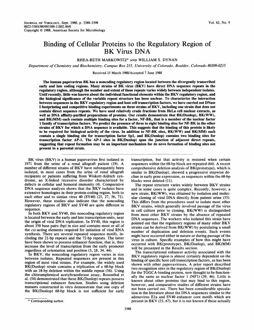
FIG. 1. Sequence relationship of BK(WW), BK(Dunlop), and BK(MM). Stippled boxes represent the regulatory region of BK(WW),which has been divided arbitrarily into three sequence blocks, P, Q, and R (see Key). Single lines represent early-region DNA to the left andlate-region DNA to the right of the regulatory region. Binding sites in BK(WW) for NF-BK (Ni, N4, N5, and N6), Spl (Si), and an unknownfactor (L1) are indicated. The AUG codon for the agnoprotein, the presumptive initiation site for early RNA, and the replication origin regionare marked. Restriction sites used in subcloning procedures are indicated (see Materials and Methods). BK(Dunlop) could have arisen fromBK(WW) by a deletion of both the Q and R blocks and 4 bp of flanking DNA, triplication of the P block, and subsequent deletion of 18 bpwithin the second of the three repeated P blocks. Binding sites in BK(Dunlop) for NF-BK (Ni, N2, and N3) and AP-1 (Ai and A2) and the18-bp deletion within the middle P block in BK(Dunlop) (A) are indicated. BK(MM) could have arisen from BK(WW) by a series ofduplication and deletion events as described in the text. Binding sites in BK(MM) for NF-BK (Ni, N4, N7, N8, N9, and N10) and Spl (Si)are indicated. The small open boxes within the BK(MM) sequence indicate the insertions of unique DNA.
5 column volumes per h. Fractions were eluted with 5 ml ofbuffer Z containing 0.15 M KCI at a flow rate of 2 columnvolumes per h, followed by a 30-ml gradient of 0.15 to 1.75 MKCI at a flow rate of 3 column volumes per h. Fractions werecollected and assayed for binding activity by DNase Ifootprinting. Typically, heparin-agarose fractions were firstpassed over the AP-1 DNA affinity column. The AP-1-depleted flowthrough from this column was passed over theSpl affinity column, and the AP-1- and Spl-depleted flow-through from this column was passed over the NF-BKcolumn. Specific DNA binding activity was detected infractions eluting from the respective affinity columns atapproximately 0.25 to 0.4 M KCI. NF-BK and AP-1 prepa-rations were purified approximately 100-fold, and Spl prep-arations were purified approximately 30-fold.
RESULTS
Sequence relationship of BK(WW), BK(Dunlop), BK(pro-totype), and BK(MM). As discussed previously, a compari-son of the BK(WW) regulatory region with those of BK(pro-totype), BK(Dunlop), BK(MM), and other BKV isolatessuggests that the latter strains could have been derived fromBK(WW) by a small number of sequence duplications anddeletions (55, 56, 71). To illustrate how this might haveoccurred and to facilitate subsequent discussion of thebinding sites for cellular factors, we have arbitrarily dividedthe WW sequence into three blocks, labeled P, Q, and R inFig. 1. These blocks contain 68, 39, and 63 bp of DNA,respectively. BK(Dunlop) could have arisen from BK(WW)by deletion of the Q and R blocks and 4 bp of flanking DNA,triplication of the P block, and subsequent deletion of 18 bpwithin the second of the three repeated P blocks. BK(pro-totype) (not shown) could have arisen in a similar way butthe deletion contains only the R block; the Q block remains.BK(WW) reportedly will not grow in cultured cells (55);BK(prototype) grows but is unstable (56); BK(Dunlop)grows relatively well. This suggests that duplication of the P
block favors viral growth in culture and that the Q and Rblock sequences are nonessential.The generation of BK(MM) from BK(WW) is more com-
plicated but can be readily visualized. The first step couldhave involved the deletion of the rightmost 55 bp of the Rblock, together with 12 bp of the adjacent unique sequence.A unit encompassing 53 bp of the P block and 34 bp of the Qblock may then have been duplicated, with 4 bp of uniquesequence inserted between the repeats. Another duplicationencompassing 36 bp of the P block and 25 bp of the Q blockmay then have occurred, with 2 bp inserted between theduplicated segments, to give the final structure as shown.
Binding of cellular proteins to the regulatory region ofBK(Dunlop) DNA. To develop a better understanding of thefunction of the different sequence blocks, we constructed amap of the binding sites for various host cell proteins thatbind to this region using the technique of DNase I footprint-ing. Because BK(Dunlop) is the best-characterized of theBKV laboratory strains, we began our studies using thisDNA. DNase I footprinting probes were prepared for bothstrands of DNA as described in Materials and Methods.Heparin-agarose fractions of HeLa cell nuclear extractswere used as a source of DNA-binding proteins. Previousstudies have shown that fractions prepared as described (seeMaterials and Methods) are enriched for a broad spectrum ofDNA-binding proteins and transcription factors (16, 48).We identified five sites within the BK(Dunlop) regulatory
DNA that were protected by heparin-agarose fractions ofHeLa cell nuclear extract (Fig. 2A, lanes 2 through 4; Fig.2B, lanes 3 through 5). Three of these sites, Ni, N2, and N3,are located within the triplicated P blocks. Ni and N3 areidentical in sequence and show complete protection. N2differs slightly in sequence from Ni and N3 because of thepreviously mentioned deletion internal to the middle P blockand shows only partial protection. Ni and N3 coincide withsites identified by Nowock et al. (46) as binding regions for apartially purified TGGCA-binding protein. It has been sug-
J. VIROL.
PROTEINS THAT BIND TO THE BKV ENHANCER 3391
B NF-H-A AP-1 BK
G O O 0 OM
N3 ._
N2
Ali
NI
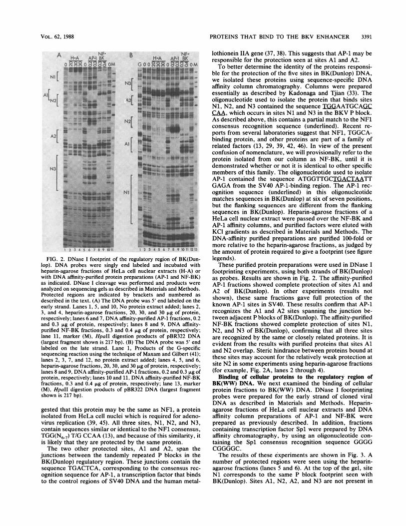
FIG. 2. DNase I footprint of the regulatory region of BK(Dun-lop). DNA probes were singly end labeled and incubated withheparin-agarose fractions of HeLa cell nuclear extracts (H-A) orwith DNA affinity-purified protein preparations (AP-1 and NF-BK)as indicated. DNase I cleavage was performed and products wereanalyzed on sequencing gels as described in Materials and Methods.Protected regions are indicated by brackets and numbered asdescribed in the text. (A) The DNA probe was 5' end labeled on theearly strand. Lanes 1, 5, and 10, No protein extract added; lanes 2,3, and 4, heparin-agarose fractions, 20, 30, and 30 ,ug of protein,respectively; lanes 6 and 7, DNA affinity-purified AP-1 fractions, 0.2and 0.3 ,ug of protein, respectively; lanes 8 and 9, DNA affinity-purified NF-BK fractions, 0.3 and 0.4 ,ug of protein, respectively;lane 11, marker (M), HpaII digestion products of pBR322 DNA(largest fragment shown is 217 bp). (B) The DNA probe was 5' endlabeled on the late strand. Lane 1, Products of the G-specificsequencing reaction using the technique of Maxam and Gilbert (41);lanes 2, 3, 7, and 12, no protein extract added; lanes 4, 5, and 6,heparin-agarose fractions, 20, 30, and 30 ,ug of protein, respectively;lanes 8 and 9, DNA affinity-purified AP-1 fractions, 0.2 and 0.3 pg ofprotein, respectively; lanes 10 and 11, DNA affinity-purified NF-BKfractions, 0.3 and 0.4 ,ug of protein, respectively; lane 13, marker(M), HpaII digestion products of pBR322 DNA (largest fragmentshown is 217 bp).
gested that this protein may be the same as NF1, a proteinisolated from HeLa cell nuclei which is required for adeno-virus replication (39, 45). All three sites, Ni, N2, and N3,contain sequences similar or identical to the NF1 consensus,TGG(N6-7) T/G CCAA (13), and because of this similarity, itis likely that they are protected by the same protein.The two other protected sites, Al and A2, span the
junctions between the tandemly repeated P blocks in theBK(Dunlop) regulatory region. These junctions contain thesequence TGACTCA, corresponding to the consensus rec-
ognition sequence for AP-L, a transcription factor that bindsto the control regions of SV40 DNA and the human metal-
lothionein IIA gene (37, 38). This suggests that AP-1 may beresponsible for the protection seen at sites Al and A2.To better determine the identity of the proteins responsi-
ble for the protection of the five sites in BK(Dunlop) DNA,we isolated these proteins using sequence-specific DNAaffinity column chromatography. Columns were preparedessentially as described by Kadonaga and Tjian (33). Theoligonucleotide used to isolate the protein that binds sitesNi, N2, and N3 contained the sequence TGGAATGCAGCCAA, which occurs in sites Ni and N3 in the BKV P block.As described above, this contains a partial match to the NF1consensus recognition sequence (underlined). Recent re-ports from several laboratories suggest that NF1, TGGCA-binding protein, and other proteins are part of a family ofrelated factors (13, 29, 39, 42, 46). In view of the presentconfusion of nomenclature, we will provisionally refer to theprotein isolated from our column as NF-BK, until it isdemonstrated whether or not it is identical to other specificmembers of this family. The oligonucleotide used to isolateAP-1 contained the sequence ATGGTTGCTGACTAATTGAGA from the SV40 AP-1-binding region. The AP-1 rec-ognition sequence (underlined) in this oligonucleotidematches sequences in BK(Dunlop) at six of seven positions,but the flanking sequences are different from the flankingsequences in BK(Dunlop). Heparin-agarose fractions of aHeLa cell nuclear extract were passed over the NF-BK andAP-1 affinity columns, and purified factors were eluted withKCl gradients as described in Materials and Methods. TheDNA-affinity purified preparations are purified 100-fold ormore relative to the heparin-agarose fractions, as judged bythe amount of protein required to give a footprint (see figurelegends).These purified protein preparations were used in DNase I
footprinting experiments, using both strands of BK(Dunlop)as probes. Results are shown in Fig. 2. The affinity-purifiedAP-1 fractions showed complete protection of sites Al andA2 of BK(Dunlop). In other experiments (results notshown), these same fractions gave full protection of theknown AP-1 sites in SV40. These results confirm that AP-1recognizes the Al and A2 sites spanning the junction be-tween adjacent P blocks of BK(Dunlop). The affinity-purifiedNF-BK fractions showed complete protection of sites Ni,N2, and N3 of BK(Dunlop), confirming that all three sitesare recognized by the same or closely related proteins. It isevident from the results with purified proteins that sites Aland N2 overlap. Steric hindrance between proteins bound atthese sites may account for the relatively weak protection atsite N2 in some experiments using heparin-agarose fractions(for example, Fig. 2A, lanes 2 through 4).
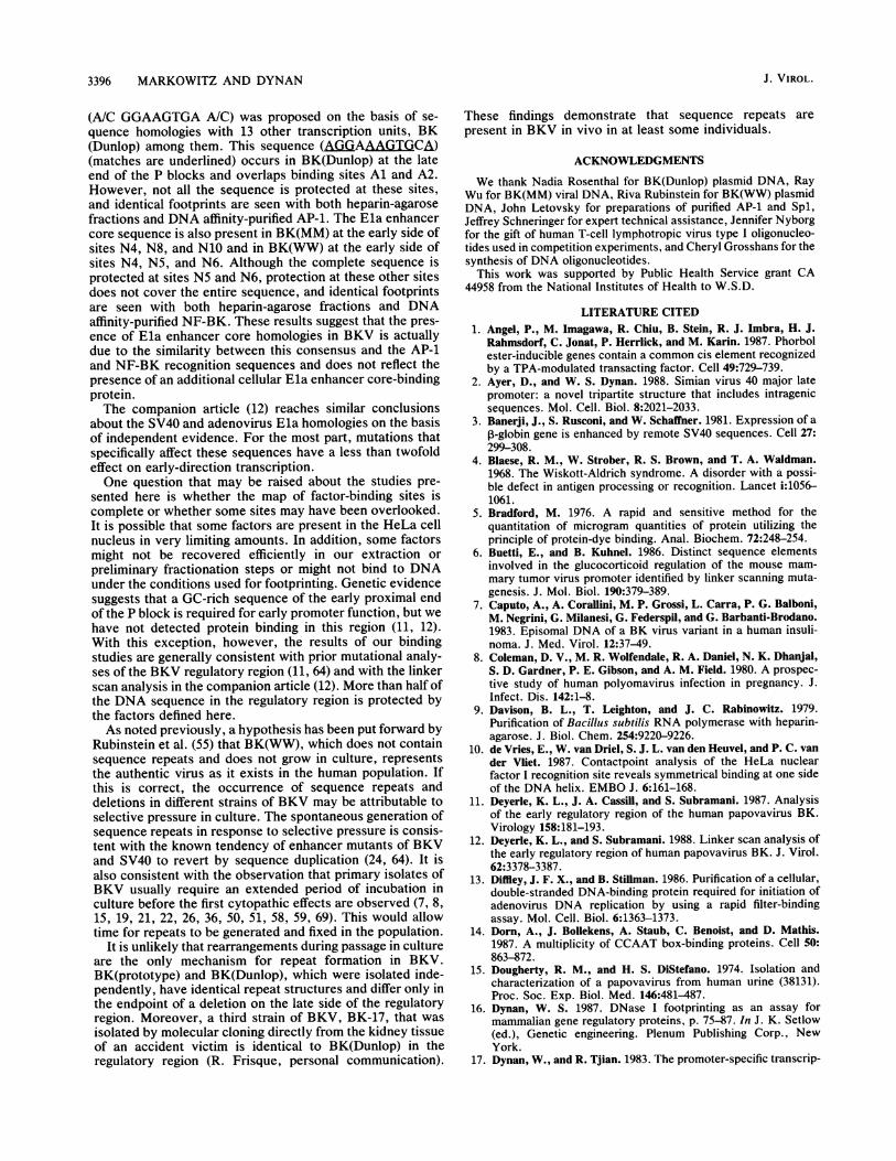
Binding of cellular proteins to the regulatory region ofBK(WW) DNA. We next examined the binding of cellularprotein fractions to BK(WW) DNA. DNase I footprintingprobes were prepared for the early strand of cloned viralDNA as described in Materials and Methods. Heparin-agarose fractions of HeLa cell nuclear extracts and DNAaffinity column preparations of AP-1 and NF-BK wereprepared as previously described. In addition, fractionscontaining transcription factor Spl were prepared by DNAaffinity chromatography, by using an oligonucleotide con-taining the Spl consensus recognition sequence GGGGCGGGGC.The results of these experiments are shown in Fig. 3. A
number of protected regions were seen using the heparin-agarose fractions (lanes 5 and 6). At the top of the gel, siteNi corresponds to the same P block footprint seen withBK(Dunlop). Sites Al, N2, A2, and N3 are not present in
A NF-H-A AP-1 BK
o Z OM
Nl 133,
N4,'235
:z
N3 *
I st wU *s_
w isiSi i
VOL. 62, 1988

3392 MARKOWITZ AND DYNAN
TABLE 1. Recognition sequences for NF-BK in BK(Dunlop),BK(WW), and BK(MM)a
Site SequenceN .TGGAATGCAGCCAAAN2.GGGAATGCAGCCAAAN4.TGGGCAGC C/A GCCAGTN5.TGGAAACTGGCCAAAN6.TGGCTGCTTTCCACTN9.TGAAACCATGCCAAAL .TGGCCTTGTCCCCAG
Consensus...... TGGAA T/A G/C C/T A/T GCCAAA
N5 |GX
Li
XX ~~-4h.
1 2 3 4 5 6 7 8 910 11121314
FIG. 3. DNase I footprint of the regulatory region of BK(WW).
The DNA probe was 5' end labeled on the early strand. For
experimental details, see the legend to Fig. 2 and Materials and
Methods. Protected regions are indicated by brackets and numbered
as described in the text. Asterisks indicate a possible Spi recogni-tion sequence where no binding was detected. Lane i, Products of
the Maxam-Gilbert G-specific sequencing reaction; lane 2, productsof the Maxam-Gilbert G- and A-specific sequencing reaction; lanes
3, 4, and 9, no protein extract added; lanes 5 and 6, heparin-agarose
fractions, 30 ~Lg of protein; lanes 7 and 8, DNA affinity-purified AP-i
fractions, 0.2 j±g of protein; lanes and ii, DNA affinity-purifiedNF-BK fractions, 0.3 and 0.4 ~±g of protein, respectively; lanes i2
and i3, DNA affinity-purified Spi fractions, i.2 and 2.0 p.g of
protein, respectively; lane i4, marker (M), HpaII digestion productsof pBR322 DNA (largest fragment shown is 242 bp).
BK(WW); these sites arose in BK(Dunlop) as the result of
the P block tniplication. The next three protected sites that
are seen in BK(WW) have therefore been labeled N4, N5,
and N6. N4 spans the junction between the P and Q blocks,
N5 is within the R block, and N6 spans the righthand
junction of the R block. All three sites contain significantmatches to the P block NF-BK recognition site (Table 1),
suggesting that all are recognized by the same protein. The
final protected site in BK(WW) DNA, labeled Li, is located
to the right of the R block, immediately upstream from the
start codon for the BKV agnoprotein. This site has some
a The following NF-BK recognition sites have been omitted because theirsequence is identical to sites already listed: N3, N7, N8 and N10.
similarity to the NF-BK recognition site in the P block but ismore divergent than the sequences in sites N2, N4, N5, andN6 (Table 1).
Affinity-purified NF-BK fractions gave full protection ofsites Ni, N4, N5, and N6 of BK(WW), confirming that thesesites are recognized by the same or closely related proteins.By contrast, site Li showed only limited protection withpurified NF-BK fractions, suggesting that NF-BK has someaffinity for these sequences but may not account for the fullprotection seen with heparin-agarose fractions.We noticed that the BK(WW) regulatory region contained
a sequence GGGGCGGGGT that is a good match to theconsensus recognition sequence for the transcription factorSpl. This sequence is located at the junction between the Qand R blocks. Although this region was not significantlyprotected by heparin-agarose fractions, we did observecomplete protection with affinity-purified Spl, and havelabeled the site Si (Fig. 3, lanes 12 and 13). Because there isboth a match to the consensus and observed protection withpurified protein, we believe this is a bona fide Spl recogni-tion site. The relative lack of protection with heparin-agarose fractions may reflect a low concentration of Spl inthis particular preparation. This was the only site observedfor Spl; we did not detect protection of a putative Spl site(AGGGAGGGAGC) in the P block (Fig. 3, asterisks) usingeither heparin-agarose or DNA affinity-purified fractions.We also tested BK(WW) DNA with affinity-purified AP-1.
As expected, no protection of BK(WW) DNA was observedwith AP-1 fractions (Fig. 3, lanes 7 and 8). The AP-1 sitesthat were seen with BK(Dunlop) DNA spanned the junctionbetween the triplicated P blocks, and this triplication is notpresent in BK(WW).
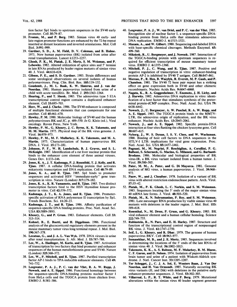
Binding of cellular proteins to the regulatory region ofBK(MM) DNA. DNase I footprinting experiments withBK(MM) DNA are shown in Fig. 4. As in the previousexperiments, we used heparin-agarose fractions of HeLa cellnuclear extracts, as well as NF-BK, AP-1, and Spl fractionspurified by DNA affinity column chromatography.
Six regions were protected by heparin-agarose fractions(Fig. 4A, lane 5; Fig. 4B, lanes 5 and 6). Site Ni (Fig. 4Bonly; not shown in Fig. 4A) is the same P block footprintseen in the other strains. Site N4 is at the junction of the Pand Q blocks and corresponds to the N4 site in BK(WW)DNA. Sites N7, N8, and N10 arise as the results of dupli-cations of portions of the P and Q blocks. N9, however, isunique to BK(MM) and arises as the result of a novel Q-Pjunction containing a 2-bp insert. This unusual creation of aprotein-binding site across a repeat junction is analogous tothe situation in BK(Dunlop), where novel AP-1 sites werecreated at the P-P junctions.
Ni [
N4 [
Si [
J. VIROL.

PROTEINS THAT BIND TO THE BKV ENHANCER 3393
B
N1O[N9 [N8 [
N7
N47
at..,
NIl
1 '2 3 4 5 6 7 B 9 10 11 12 131415
FIG. 4. DNase 1 footprint of the regulatory region of BK(MM).For experimental details, see the legend to Fig. 2 and Materials andMethods. (A) The DNA probe was 5' end labeled on the earlystrand. Lane 1, Products of the Maxam-Gilbert G-specific sequenc-ing reaction; lane 2, products of the Maxam-Gilbert G- and A-specific sequencing reaction; lanes 3, 4, 6, and 11, no protein extractadded; lane 5, heparin-agarose fractions, 20 jig of protein; lanes 7and 8, DNA affinity-purified NF-BK fractions, 0.3 and 0.4 ,ug ofprotein, respectively; lanes 9 and 10, DNA affinity-purified Splfractions, 2.5 and 3.8 ,ug of protein, respectively; lane 12, marker(M), HpaII digestion products of pBR322 DNA (largest fragmentshown is 242 bp). (B) The DNA probe was 5' end labeled on the latestrand. Lane 1, Products of the Maxam-Gilbert G-specific sequenc-ing reaction; lane 2, products of the Maxam-Gilbert G- and A-specific sequencing reaction; lanes 3, 4, 9, and 14, no protein extractadded; lanes 5 and 6, heparin-agarose fractions, 20 and 30 ,ug ofprotein, respectively; lanes 7 and 8, DNA affinity-purified AP-1fractions, 0.2 jig of protein; lanes 10, 11, and 12, DNA affinity-purified NF-BK fractions, 3, 5, and 10 ,ul, respectively; the highestamount tested (10 ,ul) contained less than 0.3 ,ug of protein; lane 13,DNA affinity-purified Spl fractions, 1.2 ,ug of protein; lane 15,marker (M), HpaII digestion products of pBR322 DNA (largestfragment shown is 309 bp).
All six of the sites protected by heparin-agarose fractionsin BK(MM) DNA contain close matches to the NF-BKconsensus recognition sequence (Table 1). We tested theBK(MM) probes in DNase I footprinting experiments usingaffinity-purified NF-BK and found, as expected, full protec-tion of sites Ni, N4, N7, N8, N9 and N10, with essentiallythe same boundaries as seen with the heparin-agarose frac-tions.BK(MM) contains an Spl consensus recognition sequence
coinciding with the single Q-R junction present in this strain.This sequence is protected by purified Sp-1 (Fig. 4A, lanes 9
and 10; Fig. 4B, lane 13). This site is analogous to the singleSpl site in BK(WW) and therefore has been labeled Si.
Competitive binding experiments. Experiments with frac-tions purified by DNA sequence affinity chromatography,together with sequence comparisons, strongly suggests thatthe observed footprints at sites Ni through N10 are attrib-utable to a single factor, NF-BK. To confirm this, we carriedout competitive binding experiments using double-strandedoligonucleotides containing either the NF-BK recognitionsequence from site Ni or a heterologous sequence from thepromoter region of an unrelated virus. In these experimentscompetitor oligonucleotide was mixed with probe DNA,heparin-agarose fractions were added, complexes were al-lowed to form, and DNase I footprinting was carried out asbefore.
Figure 5A shows a competitive binding experiment usingBK(Dunlop) probe. Significant inhibition of NF-BK bindingat sites Ni and N3 is evident with 20 nM specific competitor,whereas no inhibition is seen with the heterologous oligonu-cleotide at concentrations as high as 310 nM. Inhibition ofbinding to site N2 is more difficult to see, because protectionis only partial even in the absence of competitor. However,it appears that inhibition occurs with 6 to 20 nM of thespecific competitor, and that there is no inhibition with theheterologous competitor. These results suggest that, asexpected, sites Ni, N2, and N3 of BK(Dunlop) are protectedby the same or closely related protein species.
In contrast, the binding of AP-1 at sites Al and A2 ofBK(Dunlop) DNA is not inhibited by any of the concentra-tions of the NF-BK oligonucleotide that we tested. Someinhibition of AP-1 binding was seen at the highest concen-tration tested of the heterologous oligonucleotide, probablybecause this oligonucleotide contains a sequence(TGiACGTG) that partially matches the AP-1 consensusrecognition sequence. This result confirms that sites Ai andA2 are protected by a protein that is different from the onethat binds to sites Ni, N2, and N3.
Competitive binding studies were extended to BK(WW)DNA (Fig. 5B). In this case, binding to four protectedregions, Ni, N4, N5, and N6, was inhibited by the NF-BKoligonucleotide. There was no inhibition with the heterolo-gous oligonucleotide. As with BK(Dunlop), these resultssuggest that the same protein is binding to all sites. Therewas some inhibition of binding to site Li with the NF-BKoligonucleotide, but the inhibition was not complete, even at120 nM. This is consistent with the results of the bindingexperiments using purified factors and suggests that protec-tion in the Li region arises predominantly from the bindingof a different factor.
Competitive binding studies with BK(MM) DNA areshown in Fig. SC. Here, there are six protected regions, Ni,N4, N7, N8, N9, and N10. Binding to sites Ni, N4, N7, andN8 is inhibited by 20 nM NF-BK oligonucleotide, whereasthere is no inhibition by 310 nM heterologous oligonucleo-tide. Again, this suggests that these sites are protected by asingle factor, NF-BK. Some inhibition of binding is evidentat sites N9 and N10. These are more difficult to see becauseprotection of these sites with heparin-agarose fractions in theabsence of competitor is not complete (Fig. 5, lanes 3 and 4;Fig. 4A, lane 5). However, some inhibition occurs at 20 nMspecific competitor at site N9 (note the disappearance of thehypersensitive band just above the protected region) and siteN10. No inhibition of protection is seen with the heterolo-gous oligonucleotide at 310 nM. Similar results were ob-tained in several independent experiments, suggesting that
A
N4 [Si [N7[
NB[
N9
-
ew-*2356 B901
VOL. 62, 1988

3394 MARKOWITZ AND DYNAN
Specific .nM Nnsi>ecific'nM)CC oo:> -4 88C oc oo2 Cm
w --~~~~-~lb
l.
lb.Ilb d
23 4 5 6 -78 ' )0171 12 U3141516 1718S 19
B Specific:%M) NwsoecIficKnM0 02
GGc C o onC, 04° 2>! oo°_---m
N4 [ I6* -S . _
[N5 stss ""§
I 2 34A 6 7 89 10 1112 13 A14116 1718 19
C
NU
N44
NI
IM
FIG. 5. Competitive binding to NF-BK sites in the regulatory region of BK virus. Competitor DNA was added in the amounts indicatedto a mixture containing singly end-labeled DNA probes, poly(dI-dC) copolymer, and pd(N)5 oligonucleotides as described in the text andMaterials and Methods. The double-stranded oligonucleotide containing the NF-BK recognition sequence from site Ni (5' TGGAATGCAGCCAA 3') was used as the specific competitor. A double-stranded oligonucleotide containing a sequence present in the human T-celllymphotropic virus type 1 promoter (5' TGGGCTAGGCCCTGACGTGTCCCCCTGAAGACAAA 3') was used as the nonspecific competitor.Heparin-agarose fractions (30 ,ug) of HeLa cell nuclear extracts was added to the DNA mixture, DNase I cleavage was performed, andproducts were analyzed on sequencing gels as described in Materials and Methods. Protected regions are indicated by brackets andenumerated as described in the text. (A) The DNA probe was 5' end labeled on the late strand of BK(Dunlop). Lanes 1, 2, 10, and 18, Noprotein extract added (control [C]); lanes 3 through 9, NF-BK-specific competitor DNA was added in the following concentrations: 0, 0, 2,6, 20, 60, and 120 nM, respectively; lanes 11 through 17, nonspecific competitor DNA was added in the following concentrations: 0, 0, 2, 20,110, 180, and 310 nM, respectively; lane 19, marker (M), HpaII digestion products of pBR322 DNA (largest fragment shown is 201 bp). (B)The DNA probe was 5' end labeled on the early strand of BK(WW). Lane 1, Products of the Maxam-Gilbert G-specific sequencing reaction;lane 2, products of the Maxam-Gilbert G- and A-specific sequencing reaction; lanes 3, 4, and 12, no protein extract added (control [C]); lanes5 through 11, NF-BK-specific competitor DNA was added in the following concentrations: 0, 0, 2, 6, 20, 60, and 120 nM, respectively; lanes13 through 18, nonspecific competitor DNA was added in the following concentrations: 0, 0, 2, 20, 110, and 180 nM, respectively; lane 19,marker (M), HpaII digestion products of pBR322 DNA (largest fragment shown is 309 bp). (C) The DNA probe was 5' end labeled on the earlystrand of BK(MM). Lanes 1, 2, 10, and 17, No protein extract added (control [C]); lanes 3 through 9, NF-BK-specific competitor DNA wasadded in the following concentrations: 0, 0, 2, 6, 20, 60, and 120 nM, respectively; lanes 11 through 16, nonspecific competitor DNA wasadded in the following concentrations: 0, 0, 2, 20, 110, and 180 nM, respectively; lane 18, marker (M), HpaII digestion products of pBR322DNA (largest fragment shown is 309 bp).
protection at N9 and N10 is due, in part, to the binding ofNF-BK, but that other proteins may also contribute.
DISCUSSIONOne of the ongoing questions with BKV is to what degree
the enhancer region is similar to that of the related andbetter-characterized virus SV40. The present results showthe differences between the two systems. The distinguishingfeature of the enhancer region in BKV is the presence ofmultiple binding sites for NF-BK; these were the onlyprotein recognition sites that we detected in all three BKVstrains tested. The ubiquity of NF-BK binding sites in BKVstands in contrast to SV40, where NFl-related protein-binding sites have not been reported in the enhancer region.Although different strains of BK virus have undergone
complex rearrangements, multiple binding sites for NF-BK
are always present. Where sites have been lost throughdeletions, new sites have been formed by duplications and inone case, by a duplication coupled with a small insertion.BK(WW), which does not contain repeated sequences in theregulatory region, has four NF-BK sites: Ni within the Pblock, N4 at the P-Q junction, N5 within the R block, and N6at the R block-late region junction. BK(Dunlop) has adeletion that results in the loss of sites N4, N5, and N6.However, the P block triplication has resulted in the gener-ation of two additional NF-BK binding sites, N2 and N3.BK(MM) has a deletion, relative to BK(WW), that results inthe loss of sites N5 and N6, but duplications have resulted inthe formation of four additional sites, N7, N8, N9, and N10.We can predict the location ofNF-BK sites in a number of
other BKV strains on the basis of our results with BK(WW),BK(Dunlop), and BK(MM). Thus, we expect that BK(pro-
A
AiN[N2[
Al
Ni
J. VIROL.

PROTEINS THAT BIND TO THE BKV ENHANCER 3395
totype) contains the same NF-BK sites as BK(Dunlop) plusan N4-like site at the P-Q junction. BK(IR), isolated from apancreatic adenoma of beta islet cells (7, 49), contains a totalof eight NF-BK binding sites: three Ni-like binding siteswithin the P block, three N4-like sites at P-Q junctions, oneN5-like site within the R block, and one N6-like site at the Rblock-late region junction. BK(pm526), a small-plaque vari-ant isolated from a BK(prototype)-induced hamster pineocy-toma, contains three potential NF-BK binding sites, oneNl-like site within the P block and two N4-like sites at P-Qjunctions. BK(pm527), a different small-plaque variant, con-tains five potential NF-BK sites, including three N4-likesites and two Ni-like sites (61-65).There are a number of lines of evidence that suggest that
the NF-BK sites are important for growth in culture. Allstrains of BKV apparently contain multiple sites. Often,there are many sites within relatively short stretches ofDNA, for example, eight sites within 320 bp in BK(IR) andsix sites within 370 bp in BK(MM). In enhancer-defectivemutants containing only a single P block, duplication of aregion encompassing the Ni site was observed (64). A role ofNF-BK binding in early transcription of BKV is also consis-tent with recent findings showing that the binding of thesame or a related protein is essential for mouse mammarytumor virus promoter function (6, 35, 42).A systematic linker scan mutational analysis of the BKV
early enhancer is found in the companion article (12). Toeliminate the effect of sequence redundancy, these workersintroduced their mutations into a viral background that hasonly a single P block. The background is identical toBK(WW), except that the R block is missing. This analysisshows a correlation between early transcriptional activityand the presence of sequences in the Ni and N4 sites (12[note that the N4 site overlaps the genetically defined celement]). This is further evidence that the NF-BK sitesdefined by footprinting are functional elements of the BKVenhancer.The recognition sequence for NF-BK (Table 1) is a subset
of the recognition sequence for NF1, TGG(N6-7) T/G CCAA(10, 13, 40). NF1 was originally described as a host proteinrequired for adenovirus replication (45). NF1 appears to befunctionally equivalent to the TGGCA-binding protein of thelaboratory of Nowock and Sippel (39, 46, 47). Other workhas suggested that NF1 is identical to the CCAAT transcrip-tion factor (29, 31) that binds in the upstream region ofseveral promoters, although this remains controversial, anda recent study showed that there are a "multiplicity ofCCAAT box binding proteins" that bind to these sequencesin different promoters (14, 42). In view of this complexity,we have chosen to refer to the BKV-binding protein asNF-BK, until its identity is better established.The presence of two binding sites for the transcription
factor AP-1 that cross the junction between adjacent Pblocks in BK(Dunlop) was surprising. Apparently, thesebinding sites were created at the time the P block triplicationwas formed. The possibility that functionally importantelements span repeat junctions has been largely overlookedin prior studies of the BKV regulatory region, perhapsbecause this phenomenon is not known to occur in the SV40enhancer.The three AP-1 recognition sites in SV40, none of which
are located at repeat junctions, are believed to contribute toinduction of SV40 RNA synthesis by tetradecanoyl phorbolacetate (1, 38). The AP-1-binding sites in BKV may havesimilar functions. Studies with deletion mutants of BK(pro-totype) indicate that there is some loss of early gene expres-
sion when an AP-1 recognition sequence (site A2 in ournomenclature) is removed (11). AP-1 recognition sites areclearly not essential for viral viability, however, since theyare not present in all strains.BK(WW) and BK(MM) have a single binding site for the
transcription factor Spl (17) at the junction of the Q and Rblocks. This site is completely protected by affinity-purifiedSpl, and has a 10 of 10 bp match to a known Spl site in theIE3 promoter of herpes simplex virus (30). This site is notpresent in BK(Dunlop) and is partially deleted in BK(pro-totype). In addition to the Spl site at the Q-Rjunction, thereis a sequence AGGGiAGGAGC at the early-proximal end ofthe P block that has an 8 of 10 match (underlined) to the Splrecognition consensus (32). Although there is genetic evi-dence that the region containing this potential Spl site maybe important for early promoter function (11, 12), we havenever detected binding at this site by affinity-purified Splfractions in any of the strains tested. This suggests eitherthat it is a very weak Spl site or that it is a recognition sitefor a different protein.We have also observed protection of a late-proximal
region in BK(WW) that overlaps the start codon for agno-protein. This site, which we call Li, is protected by heparin-agarose fractions. Although there is some protection at Liwith purified NF-BK, competitive binding experiments sug-gest that protection at Li arises primarily from a differentfactor. The Li sequence is also present in the viral DNA ofBK(Dunlop) and BK(MM), although it was not present in thesubcloned probes used in our binding experiments. In astudy using deletion mutants, deletion of Li sequences hadno effect on early gene expression (11), suggesting that thefunction of this site, if any, might be related to late geneexpression. In SV40, there is evidence that sequences nearand within the late coding region affect late gene expression(2, 20, 52, 53, 57, 60).There has been considerable speculation about the role of
motifs homologous to the SV40 enhancer core (67) and theEla enhancer core (23) that are present within the regulatoryregion of BKV. The SV40 enhancer core sequence, TGGAAAGT, was originally defined by correlating loss ofenhancer function with specific point mutations locatedwithin the 72-bp repeats and is now known to be the bindingsite for one or more cellular proteins (28, 43, 70). On thebasis of the presence of similar sequences within Moloneymurine sarcoma virus, BK(Dunlop), and polyomavirus, theSV40 enhancer core consensus (G)TGG A/T A/i A/T (G)was proposed (67). In the light of current knowledge, how-ever, this fairly degenerate consensus may not be meaning-ful. It has been pointed out by Weber et al. (66) that the TGGA/T A/T A/T G motif occurs in at least 38 places within theSV40 genome. Moreover, the sequence in BK(Dunlop)matches (underlined) SV40 in only four of the eight positions(ATGGTTTG).Our experiments show no evidence that the SV40 en-
hancer core homology in BKV is recognized by cellularproteins. Although the homology overlaps the late end of theNF-BK binding sites within the P block (sites Ni, N2, N3,N7, and N9), only part of the sequence is protected. Inaddition, identical patterns of protection are seen with crudeheparin-agarose fractions and DNA affinity-purified NF-BK,suggesting that NF-BK is the only factor binding in thisregion.A second enhancer motif thought to be present in BKV,
the Ela enhancer core, was originally defined by deletionmutants affecting expression of the adenovirus type 5 earlyregion 1A (Ela) transcription unit (23). A consensus sequence
VOL. 62, 1988
3396 MARKOWITZ AND DYNAN
(A/C GGAAGTGA A/C) was proposed on the basis of se-quence homologies with 13 other transcription units, BK(Dunlop) among them. This sequence (AGGAAAGTGCA)(matches are underlined) occurs in BK(Dunlop) at the lateend of the P blocks and overlaps binding sites Al and A2.However, not all the sequence is protected at these sites,and identical footprints are seen with both heparin-agarosefractions and DNA affinity-purified AP-1. The Ela enhancercore sequence is also present in BK(MM) at the early side ofsites N4, N8, and N10 and in BK(WW) at the early side ofsites N4, N5, and N6. Although the complete sequence isprotected at sites N5 and N6, protection at these other sitesdoes not cover the entire sequence, and identical footprintsare seen with both heparin-agarose fractions and DNAaffinity-purified NF-BK. These results suggest that the pres-ence of Ela enhancer core homologies in BKV is actuallydue to the similarity between this consensus and the AP-1and NF-BK recognition sequences and does not reflect thepresence of an additional cellular Ela enhancer core-bindingprotein.The companion article (12) reaches similar conclusions
about the SV40 and adenovirus Ela homologies on the basisof independent evidence. For the most part, mutations thatspecifically affect these sequences have a less than twofoldeffect on early-direction transcription.One question that may be raised about the studies pre-
sented here is whether the map of factor-binding sites iscomplete or whether some sites may have been overlooked.It is possible that some factors are present in the HeLa cellnucleus in very limiting amounts. In addition, some factorsmight not be recovered efficiently in our extraction orpreliminary fractionation steps or might not bind to DNAunder the conditions used for footprinting. Genetic evidencesuggests that a GC-rich sequence of the early proximal endof the P block is required for early promoter function, but wehave not detected protein binding in this region (11, 12).With this exception, however, the results of our bindingstudies are generally consistent with prior mutational analy-ses of the BKV regulatory region (11, 64) and with the linkerscan analysis in the companion article (12). More than half ofthe DNA sequence in the regulatory region is protected bythe factors defined here.As noted previously, a hypothesis has been put forward by
Rubinstein et al. (55) that BK(WW), which does not containsequence repeats and does not grow in culture, representsthe authentic virus as it exists in the human population. Ifthis is correct, the occurrence of sequence repeats anddeletions in different strains of BKV may be attributable toselective pressure in culture. The spontaneous generation ofsequence repeats in response to selective pressure is consis-tent with the known tendency of enhancer mutants of BKVand SV40 to revert by sequence duplication (24, 64). It isalso consistent with the observation that primary isolates ofBKV usually require an extended period of incubation inculture before the first cytopathic effects are observed (7, 8,15, 19, 21, 22, 26, 36, 50, 51, 58, 59, 69). This would allowtime for repeats to be generated and fixed in the population.
It is unlikely that rearrangements during passage in cultureare the only mechanism for repeat formation in BKV.BK(prototype) and BK(Dunlop), which were isolated inde-pendently, have identical repeat structures and differ only inthe endpoint of a deletion on the late side of the regulatoryregion. Moreover, a third strain of BKV, BK-17, that wasisolated by molecular cloning directly from the kidney tissueof an accident victim is identical to BK(Dunlop) in theregulatory region (R. Frisque, personal communication).
These findings demonstrate that sequence repeats arepresent in BKV in vivo in at least some individuals.
ACKNOWLEDGMENTS
We thank Nadia Rosenthal for BK(Dunlop) plasmid DNA, RayWu for BK(MM) viral DNA, Riva Rubinstein for BK(WW) plasmidDNA, John Letovsky for preparations of purified AP-1 and Spl,Jeffrey Schneringer for expert technical assistance, Jennifer Nyborgfor the gift of human T-cell lymphotropic virus type I oligonucleo-tides used in competition experiments, and Cheryl Grosshans for thesynthesis of DNA oligonucleotides.
This work was supported by Public Health Service grant CA44958 from the National Institutes of Health to W.S.D.
LITERATURE CITED1. Angel, P., M. Imagawa, R. Chiu, B. Stein, R. J. Imbra, H. J.
Rahmsdorf, C. Jonat, P. Herrlick, and M. Karin. 1987. Phorbolester-inducible genes contain a common cis element recognizedby a TPA-modulated transacting factor. Cell 49:729-739.
2. Ayer, D., and W. S. Dynan. 1988. Simian virus 40 major latepromoter: a novel tripartite structure that includes intragenicsequences. Mol. Cell. Biol. 8:2021-2033.
3. Banerji, J., S. Rusconi, and W. Schaffner. 1981. Expression of aP-globin gene is enhanced by remote SV40 sequences. Cell 27:299-308.
4. Blaese, R. M., W. Strober, R. S. Brown, and T. A. Waldman.1968. The Wiskott-Aldrich syndrome. A disorder with a possi-ble defect in antigen processing or recognition. Lancet i:1056-1061.
5. Bradford, M. 1976. A rapid and sensitive method for thequantitation of microgram quantities of protein utilizing theprinciple of protein-dye binding. Anal. Biochem. 72:248-254.
6. Buetti, E., and B. Kuhnel. 1986. Distinct sequence elementsinvolved in the glucocorticoid regulation of the mouse mam-mary tumor virus promoter identified by linker scanning muta-genesis. J. Mol. Biol. 190:379-389.
7. Caputo, A., A. Corallini, M. P. Grossi, L. Carra, P. G. Balboni,M. Negrini, G. Milanesi, G. Federspil, and G. Barbanti-Brodano.1983. Episomal DNA of a BK virus variant in a human insuli-noma. J. Med. Virol. 12:37-49.
8. Coleman, D. V., M. R. Wolfendale, R. A. Daniel, N. K. Dhanjal,S. D. Gardner, P. E. Gibson, and A. M. Field. 1980. A prospec-tive study of human polyomavirus infection in pregnancy. J.Infect. Dis. 142:1-8.
9. Davison, B. L., T. Leighton, and J. C. Rabinowitz. 1979.Purification of Bacillus subtilis RNA polymerase with heparin-agarose. J. Biol. Chem. 254:9220-9226.
10. de Vries, E., W. van Driel, S. J. L. van den Heuvel, and P. C. vander Vliet. 1987. Contactpoint analysis of the HeLa nuclearfactor I recognition site reveals symmetrical binding at one sideof the DNA helix. EMBO J. 6:161-168.
11. Deyerle, K. L., J. A. Cassill, and S. Subramani. 1987. Analysisof the early regulatory region of the human papovavirus BK.Virology 158:181-193.
12. Deyerle, K. L., and S. Subramani. 1988. Linker scan analysis ofthe early regulatory region of human papovavirus BK. J. Virol.62:3378-3387.
13. Diffley, J. F. X., and B. Stillman. 1986. Purification of a cellular,double-stranded DNA-binding protein required for initiation ofadenovirus DNA replication by using a rapid filter-bindingassay. Mol. Cell. Biol. 6:1363-1373.
14. Dorn, A., J. Bollekens, A. Staub, C. Benoist, and D. Mathis.1987. A multiplicity of CCAAT box-binding proteins. Cell 50:863-872.
15. Dougherty, R. M., and H. S. DiStefano. 1974. Isolation andcharacterization of a papovavirus from human urine (38131).Proc. Soc. Exp. Biol. Med. 146:481-487.
16. Dynan, W. S. 1987. DNase I footprinting as an assay formammalian gene regulatory proteins, p. 75-87. In J. K. Setlow(ed.), Genetic engineering. Plenum Publishing Corp., NewYork.
17. Dynan, W., and R. Tjian. 1983. The promoter-specific transcrip-
J. VIROL.
PROTEINS THAT BIND TO THE BKV ENHANCER 3397
tion factor Spl binds to upstream sequences in the SV40 earlypromoter. Cell 35:79-87.
18. Fromm, M., and P. Berg. 1983. Simian virus 40 early- andlate-region promoter functions are enhanced by the 72-bp repeatinserted at distant locations and inverted orientations. Mol. CellBiol. 3:991-999.
19. Gardner, S. D., A. M. Field, D. V. Coleman, and B. Hulme.1971. New human papovavirus (BK) isolated from urine afterrenal transplantation. Lancet i:1253-1257.
20. Ghosh, P. K., M. Piatak, J. E. Mertz, S. M. Weisman, and P.Lebowitz. 1982. Altered utilization of splice sites and 5' terminiin late RNAs produced by leader region mutants of simian virus40. J. Virol. 44:610-624.
21. Gibson, P. E., and S. D. Gardner. 1983. Strain differences andsome serological observations on several isolates of humanpolyomaviruses. Prog. Clin. Biol. Res. 105:119-132.
22. Goudsmit, J., M. L. Baak, K. W. Slaterus, and J. van derNoordaa. 1981. Human papovavirus isolated from urine of achild with acute tonsillitis. Br. Med. J. 293:1363-1364.
23. Hearing, P., and T. Shenk. 1983. The adenovirus type 5 ElAtranscriptional control region contains a duplicated enhancerelement. Cell 33:695-703.
24. Herr, W., and J. Clarke. 1986. The SV40 enhancer is composedof multiple functional elements that can compensate for oneanother. Cell 45:461-470.
25. Howley, P. M. 1980. Molecular biology of SV40 and the humanpolyomaviruses BK and JC, p. 489-550. In G. Klein (ed.), Viraloncology. Raven Press, New York.
26. Howley, P. M., G. Khoury, J. C. Byrne, K. K. Takemoto, andM. M. Martin. 1975. Physical map of the BK virus genome. J.Virol. 16:959-973.
27. Howley, P. M., M. F. Mullarkey, K. K. Takemoto, and M. A.Martin. 1975. Characterization of human papovavirus BKDNA. J. Virol. 15:173-181.
28. Johnson, P. F., W. H. Landschulz, B. J. Graves, and S. L.McKnight. 1987. Identification of a rat liver nuclear protein thatbinds to the enhancer core element of three animal viruses.Genes Dev. 1:133-146.
29. Jones, K. A., J. T. Kadonaga, P. J. Rosenfeld, T. J. Kelly, and R.Tjian. 1987. A cellular DNA-binding protein that activateseukaryotic transcription and DNA replication. Cell 48:79-89.
30. Jones, K. A., and R. Tjian. 1985. Spl binds to promotersequences and activated HSV "immediate-early" gene tran-scription in vitro. Nature (London) 317:179-182.
31. Jones, K. A., K. R. Yamamoto, and R. Tjian. 1985. Two distincttranscription factors bind to the HSV thymidine kinase pro-moter in vitro. Cell 42:559-572.
32. Kadonaga, J. T., K. A. Jones, and R. Tjian. 1986. Promoter-specific activation of RNA polymerase II transcription by Spl.Trends Biochem. Sci. 11:20-23.
33. Kadonaga, J. T., and R. Tjian. 1986. Affinity purification ofsequence-specific DNA binding proteins. Proc. Natl. Acad. Sci.USA 83:5889-5893.
34. Khoury, G., and P. Gruss. 1983. Enhancer elements. Cell 33:313-314.
35. Kuhnel, B., E. Buetti, and H. Diggelman. 1986. Functionalanalysis of the glucocorticoid regulatory elements present in themouse mammary tumor virus long terminal repeat. J. Mol. Biol.190:367-378.
36. Lecatsas, G., and J. A. L. Van Wyk. 1978. DNA viruses in urineafter renal transplantation. S. Afr. Med. J. 53:787-788.
37. Lee, W., A. Haslinger, M. Karin, and R. Tjian. 1987. Activationof transcription by two factors that bind promoter and enhancersequences of the human metallothionein gene and SV40. Nature(London) 325:368-372.
38. Lee, W., P. Mitchell, and R. Tjian. 1987. Purified transcriptionfactor AP-1 binds to TPA-inducible enhancer elements. Cell 49:741-752.
39. Leegwater, P. A. J., P. C. van der Vliet, R. A. W. Rupp, J.Nowock, and A. E. Sippel. 1986. Functional homology betweenthe sequence-specific DNA-binding proteins nuclear factor Ifrom HeLa cells and the TGGCA protein from chicken liver.EMBO J. 5:381-386.
40. Leegwater, P. A. J., W. van Driel, and P. C. van der Vliet. 1985.Recognition site of nuclear factor I, a sequence-specific DNA-binding protein from HeLa cells that stimulates adenovirusDNA replication. EMBO J. 4:1515-1521.
41. Maxam, A., and W. Gilbert. 1980. Sequencing end-labeled DNAwith base-specific chemical cleavages. Methods Enzymol. 65:499-560.
42. Miksicek, R., U. Borgmeyer, and J. Nowock. 1987. Interaction ofthe TGGCA-binding protein with upstream sequences is re-quired for efficient transcription of mouse mammary tumorvirus. EMBO J. 6:1355-1360.
43. Mitchell, P. J., C. Wang, and R. Tjian. 1987. Positive andnegative regulation of transcription in vitro: enhancer-bindingprotein AP-2 is inhibited by SV40 T antigen. Cell 50:847-861.
44. Moreau, P., R. Hen, B. Wasylyk, R. Everett, M. P. Gaub, and P.Chambon. 1981. The SV40 72 base pair repeat has a strikingeffect on gene expression both in SV40 and other chimericrecombinants. Nucleic Acids Res. 9:6047-6068.
45. Nagata, K., R. A. Guggenheimer, T. Enomoto, J. H. Lichy, andJ. Hurwitz. 1982. Adenovirus DNA replication in vitro: identi-fication of a host factor that stimulates synthesis of the preter-minal protein-dCMP complex. Proc. Natl. Acad. Sci. USA 79:6438-6442.
46. Nowock, J., U. Borgmeyer, A. W. Puschel, R. A. W. Rupp, andA. E. Sippel. 1985. The TGGCA protein binds to the MMTV-LTR, the adenovirus origin of replication, and the BK virusenhancer. Nucleic Acids Res. 13:2045-2061.
47. Nowock, J., and A. E. Sippel. 1982. Specific protein-DNAinteraction at four sites flanking the chicken lysozyme gene. Cell30:607-615.
48. Nyborg, J., W. S. Dynan, I. S. Y. Chen, and W. Wachsman.1988. Binding of host cell factors to DNA sequences in theHTLV-I LTR: implications for viral gene expression. Proc.Natl. Acad. Sci. USA 85:1457-1461.
49. Pagnani, M., M. Negrini, P. Reschiglian, A. Corailini, P. G.Balboni, S. Scherneck, G. Macino, G. Milanesi, and G. Barbanti-Brodano. 1986. Molecular and biological properties of BKvirus-IR, a BK virus variant isolated from a human tumor. J.Virol. 59:500-505.
50. Pater, M. M., A. Pater, and G. Di Mayorca. 1981. Genomeanalysis of MG virus, a human papovavirus. J. Virol. 39:968-972.
51. Pauw, W., and J. Choufoer. 1978. Isolation of a variant of BKvirus with altered restriction endonuclease pattern. Arch. Virol.57:35-42.
52. Piatak, M., P. K. Ghosh, L. C. Norkin, and S. M. Weissman.1983. Sequences locating the 5' ends of the major simian virus40 mRNA late forms. J. Virol. 48:503-520.
53. Piatak, M., K. N. Subramanian, P. Roy, and S. M. Weissman.1981. Late messenger RNA production by viable simian virus 40mutants with deletions in the leader region. J. Mol. Biol. 153:589-618.
54. Rosenthal, N., M. Dress, P. Gruss, and G. Khoury. 1983. BKviral enhancer element and a human cellular homolog. Science222:749-755.
55. Rubinstein, R., N. Pare, and E. H. Harley. 1987. Structure andfunction of the transcriptional control region of nonpassagedBK virus. J. Virol. 61:1747-1750.
56. Seif, I., G. Khoury, and R. Dhar. 1979. The genome of humanpapovavirus BKV. Cell 18:963-977.
57. Somasekhar, M. B., and J. E. Mertz. 1985. Sequences involvedin determining the locations of the 5' ends of the late RNAs ofsimian virus 40. J. Virol. 56:1002-1013.
58. Takemoto, K. K., A. S. Rabson, M. F. Mularkey, R. M. Blaese,C. F. Garon, and D. Nelson. 1974. Isolation of papovavirus frombrain tumor and urine of a patient with Wiskott-Aldrich syn-drome. J. Natl. Cancer Inst. 53:1205-1207.
59. Ter Schegget, J., C. J. A. Sol, E. Wouters Baan, J. Van DerNoordaa, and H. Van Ormondt. 1985. Naturally occurring BKvirus variants (JL and Dik) with deletions in the putative earlyenhancer-promoter sequences. J. Virol. 53:302-305.
60. Viliarreal, L. P., R. T. White, and P. Berg. 1979. Mutationalalterations within the simian virus 40 leader segment generate
VOL. 62, 1988
3398 MARKOWITZ AND DYNAN
altered 16S and 19S mRNAs. J. Virol. 29:209-219.61. Watanabe, S., S. Kotake, A. Nozawa, T. Muto, and S. Uchida.
1982. Tumorigenicity of human BK papovavirus plaque isolates,wild-type and plaque morphology mutant, in hamsters. Int. J.Cancer 29:583-586.
62. Watanabe, S., and K. Yoshiike. 1982. Change of DNA near theorigin of replication enhances the transforming capacity ofhuman papovirus BK. J. Virol. 42:973-985.
63. Watanabe, S., and K. Yoshiike. 1985. Decreasing the number of68-base-pair tandem repeats in the BK virus transcriptionalcontrol region reduces plaque size and enhances transformingcapacity. J. Virol. 55:823-825.
64. Watanabe, S., and K. Yoshiike. 1986. Evolutionary changes oftranscriptional control region in a minute-plaque viable deletionmutant of BK virus. J. Virol. 59:260-266.
65. Watanabe, S., K. Yoshiike, A. Nozawa, Y. Yuasa, and S. Uchida.1979. Viable deletion mutant of human papovavirus BK thatinduces insulinomas in hamsters. J. Virol. 32:934-942.
66. Weber, F., J. de Villiers, and W. Schaffner. 1984. An SV40"enhancer trap" incorporates exogenous enhancers or gener-
ates enhancers from its own sequences. Cell 36:938-992.67. Weiher, H., M. Konig, and P. Gruss. 1983. Multiple point
mutations affecting the simian virus 40 enhancer. Science 219:626-631.
68. Wildeman, A. G., M. Zenke, C. Schatz, M. Wintzerith, T.Gundstrom, H. Matthes, K. Takahashi, and P. Chambon. 1986.Specific protein binding to the simian virus 40 enhancer in vitro.Mol. Cell. Biol. 6:2098-2105.
69. Wright, P. J., G. Bernhardt, E. 0. Major, and G. di Mayorca.1976. Comparison of the serology, transforming ability, andpolypeptide composition of human papovaviruses isolated fromurine. J. Virol. 17:762-775.
70. Xiao, J.-H., I. Davidson, M. Macchi, R. Rosales, M. Vigneron,A. Staub, and P. Chambon. 1987. In vitro binding of severalcell-specific and ubiquitous nuclear proteins to the GT-I motif ofthe SV40 enhancer. Genes Dev. 1:794-807.
71. Yang, R. C. A., and R. Wu. 1979. BK virus DNA: completenucleotide sequence of a human tumor virus. Science 206:456-462.
J. VIROL.


![Zipcode RNA-Binding Proteins and Membrane Trafficking ... · Zipcode RNA-Binding Proteins and Membrane Trafficking Proteins Cooperate to Transport Glutelin mRNAs in Rice Endosperm[OPEN]](https://img.dokumen.tips/doc/110x75/5fedaa08e6ee6243c45b24a5/zipcode-rna-binding-proteins-and-membrane-trafficking-zipcode-rna-binding-proteins.jpg)