Embed Size (px)
Citation preview
Behavioral/Systems/Cognitive
Trpv1 Reporter Mice Reveal Highly Restricted BrainDistribution and Functional Expression in ArteriolarSmooth Muscle Cells
Daniel J. Cavanaugh,1* Alexander T. Chesler,2,3* Alexander C. Jackson,3 Yaron M. Sigal,2,3 Hiroki Yamanaka,1
Rebecca Grant,4 Dajan O’Donnell,4 Roger A. Nicoll,3 Nirao M. Shah,1 David Julius,2,3 and Allan I. Basbaum1
Departments of 1Anatomy, 2Physiology, and 3Cellular and Molecular Pharmacology, University of San Francisco, San Francisco, California 94158, and4AstraZeneca R&D Montreal, St. Laurent, Quebec H4S 1Z9, Canada
The heat and capsaicin receptor, TRPV1, is required for the detection of painful heat by primary afferent pain fibers (nociceptors), but theextent to which functional TRPV1 channels are expressed in the CNS is debated. Because previous evidence is based primarily on indirectphysiological responses to capsaicin, here we genetically modified the Trpv1 locus to reveal, with excellent sensitivity and specificity, thedistribution of TRPV1 in all neuronal and non-neuronal tissues. In contrast to reports of widespread and robust expression in the CNS,we find that neuronal TRPV1 is primarily restricted to nociceptors in primary sensory ganglia, with minimal expression in a few discretebrain regions, most notably in a contiguous band of cells within and adjacent to the caudal hypothalamus. We confirm hypothalamicexpression in the mouse using several complementary approaches, including in situ hybridization, calcium imaging, and electrophysi-ological recordings. Additional in situ hybridization experiments in rat, monkey, and human brain demonstrate that the restrictedexpression of TRPV1 in the CNS is conserved across species. Outside of the CNS, we find TRPV1 expression in a subset of arteriolarsmooth muscle cells within thermoregulatory tissues. Here, capsaicin increases calcium uptake and induces vasoconstriction, an effectthat likely counteracts the vasodilation produced by activation of neuronal TRPV1.
IntroductionTRPV1 is a nonselective cation channel that is activated by nox-ious heat, protons, the vanilloid compounds capsaicin (CAP) andresiniferatoxin (RTX), and membrane-derived lipids, includinganandamide (Caterina et al., 1997; Caterina and Julius, 2001). Stud-ies of Trpv1 knock-out mice demonstrated a critical contribution ofTRPV1 to the cellular and behavioral responses to noxious heat(Caterina et al., 2000; Davis et al., 2000). Although expression ofTRPV1 was originally reported to be restricted to primary afferentnociceptors of the dorsal root ganglia (DRGs), trigeminal ganglia(TG), and nodose ganglia (Szallasi et al., 1995; Caterina et al., 1997;Tominaga et al., 1998), subsequent studies argued for a much widerdistribution, both in the CNS and in non-neuronal tissues.
Expression of TRPV1 in the CNS has been reported using avariety of methods, including pharmacological characterization(Steenland et al., 2006), immunohistochemistry (Toth et al.,
2005; Cristino et al., 2006), in situ hybridization (Mezey et al.,2000), radioligand binding (Acs et al., 1996; Roberts et al., 2004),and reverse transcription (RT)-PCR (Mezey et al., 2000). Despitethis abundance of studies, the existence of TRPV1 in the brainremains a controversial topic, mainly because the extent and lo-calization of TRPV1 varies considerably among studies, eventhose using very similar assays. For example, RTX binding studiesreported results ranging from a lack of binding in the CNS (Szal-lasi et al., 1995) to widespread binding throughout the brain(Roberts et al., 2004). In part, the lack of consensus reflects thelimitations of traditional approaches to determining gene expres-sion, including variable sensitivity, poor signal-to-noise, and lackof specificity.
There is also controversy as to the functional contribution ofTRPV1 in the CNS. For example, several groups reported altera-tions in hippocampal synaptic function in Trpv1 knock-out mice(Marsch et al., 2007; Gibson et al., 2008). However, these reportsdid not provide anatomical or biophysical evidence of TRPV1expression in these cells. Furthermore, other groups found noeffect of TRPV1 agonists on hippocampal synaptic function (Ko-falvi et al., 2006; Benninger et al., 2008). These results illustratethe importance of definitively resolving the distribution ofTRPV1. This is especially true because TRPV1 expression in theCNS has important implications for the potential side effects ofTRPV1 agonists and antagonists being developed for the treat-ment of chronic pain.
Here we used gene targeting to modify the Trpv1 genetic locusand generated two lines of reporter mice, which allow for a highly
Received Dec. 10, 2010; revised Jan. 20, 2011; accepted Feb. 7, 2011.This work was supported by National Institutes of Health (NIH) Grants NS14627 (A.I.B.), R01NS049488,
DP1OD006425 (N.M.S.), and R37NS047723 (D.J.), by support from the Pell Family Foundation to A.I.B., and byNational Institute of Mental Health (NIMH) grants to R.A.N. A.T.C. was supported by an NIH postdoctoral traininggrant from the UCSF Cardiovascular Research Institute. A.C.J. was supported by a Ruth L. Kirschstein NationalResearch Service Award from NIMH (F32MH081430). We thank Cindy Yang for the IRES-mycCre construct and NidhiNuwal for help with rat hippocampal cultures. R.G. and D.O. are employees of AstraZeneca and declare that they havecompeting financial interest.
*D.J.C. and A.T.C. are joint first authors.Correspondence should be addressed to Allan I. Basbaum at the above address. E-mail: [email protected]:10.1523/JNEUROSCI.6451-10.2011
Copyright © 2011 the authors 0270-6474/11/315067-11$15.00/0
The Journal of Neuroscience, March 30, 2011 • 31(13):5067–5077 • 5067
sensitive readout of TRPV1 expression patterns. Consistent withour early reports (Tominaga et al., 1998), we demonstrate robustexpression of TRPV1 in primary afferent neurons. However, withthe exception of very low-level expression in a few discrete brainareas, most notably within and adjacent to the caudal hypothal-amus, we could not confirm previous findings of a widespreadTRPV1 distribution in the CNS. Calcium imaging, whole-cellrecording, and in situ hybridization experiments demonstratedthat the expression patterns revealed in the reporter mice re-flected functional TRPV1 expression that is conserved acrossmultiple mammalian species. Finally, the sensitivity and cellularresolution of our genetic marking strategy revealed functionalTRPV1 expression in a subset of arteriolar smooth muscle cells(SMCs) in thermoregulatory tissues.
Materials and MethodsAnimals and injectionsAnimal experiments were approved by the Institutional Animal Care andUse Committee and conducted in accordance with the National Insti-tutes of Health Guide for the Care and Use of Laboratory Animals and therecommendations of the International Association for the Study of Pain.
A genomic clone containing the last exon of the Trpv1 gene (WellcomeTrust Sanger Institute; Geneservice, Ltd.) was used to generate a TRPV1-targeting vector. We used site-directed mutagenesis to engineer an AscIsite 3 bp downstream of the TRPV1 stop codon, and constructs contain-ing IRES-PLAP-IRES-nlacZ (TRPV1 PLAP-nlacZ mice) or IRES-mycCre(TRPV1 Cre mice) were inserted into this targeting vector. Homologousrecombinant embryonic stem cell clones were used in blastocyst injec-tions to obtain chimeric mice. R26R-lacZ (Soriano, 1999), R26R-EYFP(Srinivas et al., 2001), and Trpv1 knock-out mice (Caterina et al., 2000)were described previously.
Intrathecal injections were performed as described previously (Ca-vanaugh et al., 2009). For RTX injections, mice were anesthetized with 1.5%isoflurane and injected subcutaneously with escalating doses of RTX on con-secutive days (30, 70, 100, and 200 mg/ml), followed 7 d later with 200mg/ml. Histology was performed 7–10 d after this final RTX injection.
Placental alkaline phosphatase and lacZ detectionAdult mice (6 – 8 weeks old) were perfused with 10 ml of HEPES-bufferedsaline followed by 20 ml of ice-cold 3.7% formaldehyde. Brain, spinalcord (SC), peripheral tissue, DRGs, and TG were dissected out, postfixed3– 4 h [for placental alkaline phosphatase (PLAP) staining] or 1.5 h [fornuclear lacZ (nlacZ) staining] at 4°C, and cryoprotected overnight in30% sucrose. For brain and SC, 40 – 80 �m sections were processed asfree-floating sections. For DRGs, TG, and tongue, 14 �m cryostat sec-tions were processed on slides. Ear, dura, and cremaster muscle wereprocessed as whole mounts. PLAP and nlacZ staining was performed asdescribed previously (Shah et al., 2004).
ImmunohistochemistryAfter nlacZ histochemistry, tissue was processed for immunohistochem-istry as described previously (Tominaga et al., 1998). Primary antiserawere as follows: guinea pig anti-TRPV1 [1:1000; Julius Laboratory, Uni-versity of California at San Francisco (UCSF), San Francisco, CA], mouseanti-reelin (1:500; Millipore), and mouse anti-smooth muscle actin(SMA; 1:800; Sigma).
RT-PCRAdult mice were perfused with 10 ml of PBS followed by 10 ml of RNAlater (Ambion). TG, liver, bladder, and cremaster were dissected out andplaced in RNA later on ice. For brain regions, 500 �m brain sections werecut with a McIlwain Tissue Chopper into ice-cold PBS, and brain regionswere microdissected out with sharp tissue forceps and put into RNA lateron ice. To avoid contamination, separate forceps were used for dissectionof each tissue/brain region.
RNA isolation was performed with TRIzol reagent (Sigma) according tothe manufacturer’s specifications. One microgram of each RNA was reversetranscribed with the SuperScript II Reverse Strand Synthesis System (Invit-
rogen) using an oligo(dT) primer. Control reactions omitted reverse tran-scriptase. PCR amplification (94°C for 5 min; 35 cycles of 94°C for 30 s, 58°Cfor 30 s, 72°C for 1 min; 72°C for 10 min) was performed with 1 �l of cDNAusing the following primer pair: 5�-gtttgtggacagctacagtg-3� and 5�-gaagccacatactccttgcg-3�. Control PCR amplification (94°C for 5 min; 25 cy-cles of 94°C for 30 s, 58°C for 30 s, 72°C for 1 min; 72°C for 10 min) wasperformed with a glyceraldehyde 3-phosphate dehydrogenase (GAPDH)primer pair (5�-accacagtccatgccatcac-3� and 5�-tccaccaccctgttgctgta-3�) toensure cDNA integrity. Amplification products were visualized on a 1%agarose gel with ethidium bromide and sequenced to confirm identity.
Mouse in situ hybridizationFor in situ hybridization histochemistry (ISHH), mice were killed bydecapitation under deep anesthesia. Brain and L4/5 DRGs were dissectedout, rapidly frozen in powdered dry ice, and cut on a cryostat (5 �m forDRG and 10 �m for brain). Sections were thaw mounted onto slides andfixed in 4% formaldehyde, pH 7.4, for 20 min. ISSH was performed asdescribed previously (Kobayashi et al., 2005) with an 35S-labeled cRNAprobe directed against bases 1155–1908 of the full-length TRPV1 cDNA(antisense or sense; 1 � 107 cpm/ml). For autoradiography, sections werecoated with Kodak Nuclear Track Emulsion Type NTB (Carestream Health)diluted 1:1 with distilled water at 45°C and exposed for 2 months in light-tight boxes at 4°C. After development in D-19 (Carestream Health) andfixation in 24% sodium thiosulfate, the sections were stained with hema-toxylin–eosin (H&E), air dried, cleared in xylene, and coverslipped.
For combined immunohistochemistry with ISHH, mice were deeplyanesthetized with sodium pentobarbital and perfused transcardially with20 ml of phosphate buffer (PB), followed by 50 ml of 4% formaldehyde.DRGs were dissected out and postfixed in the same fixative at 4°C over-night, followed by immersion in 20% sucrose in PB at 4°C for 3 d. Tissuewas frozen in powdered dry ice and cut on a cryostat at a 5 �m thickness.Sections were preincubated in TBS containing 5% normal goat serum(NGS) for 30 min, followed by incubation in guinea pig anti-TRPV1(1:1000; Julius Laboratory, UCSF) in TBS containing 5% NGS for 24 h at4°C, washed in TBS, and processed for DAB immunohistochemistry asdescribed previously (Tominaga et al., 1998). Sections were then fixed in4% formaldehyde for 5 min and immediately processed for ISHH, asdescribed above, but emulsion was stopped after 3 weeks.
Rat, monkey, and human in situ hybridizationFrozen human tissues (with postmortem delay of 7 h) were acquiredfrom the Brain and Tissue Bank for Developmental Disorders (Univer-sity of Maryland, Baltimore, MD). Monkey tissues were obtained froman adult male Macaca fascicularis (ITR Laboratories Canada). Rat brainwas acquired from adult male Sprague Dawley rats (Charles River).
Brains were rapidly dissected, snap frozen at �40°C in isopentane for20 s, and stored at �80°C. Frozen tissues were cryosectioned at 14 �mand mounted onto Superfrost Plus slides (VWR Canada). Slides werestored at �80°C until further use.
Species-specific antisense riboprobes for rat, monkey, and humanTRPV1 were transcribed in vitro using SP6 RNA polymerase (Promega)and radiolabeled with 35S-UTP and 35S-CTP (�800 Ci/mmol; GEHealthcare). After transcription, the TRPV1 DNA templates were di-gested with DNase I (Promega) and subsequently purified using G-50Sepharose microspin columns (GE Healthcare). The quality of labeledriboprobes was verified by polyacrylamide-urea gel electrophoresis andscintillation counting.
For ISHH, tissue sections were fixed with 4% paraformaldehyde,rinsed three times in 2� SSC, equilibrated in 0.1 M triethanolamine(TEA), and treated with 0.25% acetic anhydride in 0.1 M TEA. After arinse in 2� SSC and dehydration through an ethanol series (50 –100%),hybridization was performed in buffer containing 75% formamide(Sigma), 600 mM NaCl, 10 mM Tris-HCl, pH 7.5, 1 mM EDTA, 1� Den-hardt’s solution (Sigma), 50 �g/ml denatured salmon sperm DNA(Sigma), 10% dextran sulfate (Sigma), 20 mM dithiothreitol, and [ 35S]-labeled cRNA probe (20 � 10 6 cpm/ml) at 55°C overnight in chambershumidified with 75% formamide. After hybridization, slides were rinsedtwice in 2� SSC at room temperature, treated with 20 �g/ml RNase IA inRNase buffer (25 mM NaCl, 5 mM Tris-HCl, pH 7.5, 0.5 mM EDTA, pH
5068 • J. Neurosci., March 30, 2011 • 31(13):5067–5077 Cavanaugh et al. • Reporter Mice Reveal Restricted TRPV1 Distribution

8.0) for 45 min at 37°C, and washed to a final stringency of 0.1� SSC at70°C. Sections were then dehydrated and exposed to Kodak BiomaxMR-2 film. After exposure to film, the slides were dipped in Kodak NTB2emulsion and exposed for 8 weeks at 4°C before development and coun-terstaining with H&E. Film autoradiograms were digitized with a high-resolution Microimager digital camera (Xillix) via the MCID imageanalysis system (Imaging Research). Bright- and dark-field photomicro-graphs of emulsion-dipped tissue sections were captured using a micro-scope (DMRBE/DM 4000B; Leica) equipped with a DFC490 digitalcamera (Leica). Digital images were transferred to Adobe Photoshop 7.0for minimal image processing.
DRG calcium imagingDRG neurons were isolated from 6-week-old mice, dissociated, and sub-jected to ratiometric calcium imaging the next day as described previ-ously (Caterina et al., 2000). Cells were stimulated with 1 �M CAP,followed by 150 mM K � (HiK) Ringer’s solution, fixed, and stained fornlacZ histochemistry. All neurons that responded to HiK solution wereincluded for analysis.
Hypothalamic slice and acutely dissociated neuron preparation,Ca 2� imaging, and whole-cell recordingTwo hundred micrometer coronal slices of caudal hypothalamus werecut from TRPV1 Cre/R26R-EYFP mice [age, postnatal day 21 (P21) toP26] using a DTK-100 Microslicer (Ted Pella). Slices were cut in anice-cold, high-sucrose solution consisting of (in mM) 87 NaCl, 1.25NaH2PO4, 25 NaHCO3, 2.5 KCl, 0.5 CaCl2, 7 MgCl2, 25 glucose, and 75sucrose, saturated with 95% O2/5% CO2. Freshly cut slices were placed inan incubating chamber containing artificial CSF (ACSF; in mM: 125NaCl, 2.5 KCl, 25 NaHCO3, 1.25 Na2PO4, 15 glucose, 2 CaCl2, and 1MgCl2), saturated with 95% O2/5% CO2, and allowed to recover at 35°Cfor �30 min and then 30 min to 1 h at room temperature.
Ca2� imaging. Slices were loaded with 10 �m Fura-2-AM (Invitrogen)and 0.2% pluronic acid at 22–25°C for 60 min in ACSF, saturated with 95%O2/5% CO2. For imaging, slices were perfused with ACSF, saturated with95% O2/5% CO2 and containing TTX (500 nM), picrotoxin (100 �M), APV(100 �M), and 2,3-dioxo-6-nitro-1,2,3,4-tetrahydrobenzo[f]quinoxaline-7-sulfonoamide (NBQX; 10 �M). CAP (1–10 �M) and HiK solution (140 mM)were added to the same supplemented ACSF solution and applied using abath perfusion system. Image acquisition and analysis were performed asdetailed above for DRG neurons.
Whole-cell recording. After recovery, slices were transferred to a sub-mersion chamber on an upright Olympus BX51 microscope and per-fused with ACSF, saturated with 95% O2/5% CO2. Enhanced yellowfluorescent protein-positive (EYFP�) cells in the supramammillary nu-cleus (SuM) and posterior hypothalamus were identified by epifluores-cence microscopy and recorded with 3–5 M� borosilicate glass pipettes,filled with an internal solution consisting of (in mM) 140 CsMeSO3, 10HEPES, 10 EGTA, 2 NaCl, 2 Mg-ATP, 1 QX-314, 5 TEA-Cl, and 1 CaCl2,pH 7.3. After the formation of a seal and achieving whole-cell configura-tion, membrane potential was held in voltage-clamp mode at �60 mV,and stable baseline holding current was established. The bath solutionwas then switched to a control ACSF solution containing TTX (500 nM),picrotoxin (100 �M), APV (100 �M), and NBQX (10 �M) for severalminutes, followed by ACSF containing 10 �M CAP for �5 min, and thenback to ACSF to wash out responses. In some cases, 20 �M RutheniumRed (RR; final concentration) was added directly to the bath.
Ca2� imaging of acutely dissociated cells. Coronal slices of caudal hypo-thalamus were cut as described and transferred to a dissociation solutionconsisting of 2.5 mg/ml protease XXIII (Sigma) dissolved in the sucrosecutting solution. Slices were incubated at 35°C for 10 min, rinsed severaltimes in cold sucrose solution, and placed in cold (4°C) sucrose solutioncontaining 1 mg/ml trypsin inhibitor (Sigma) and 1 mg/ml bovine serumalbumin (Sigma). The SuM was isolated with iridectomy scissors andFigure 1. Primary afferent expression of reporter molecules in TRPV1 PLAP-nlacZ mice. A,
Schematic showing the Trpv1 locus of TRPV1 PLAP-nlacZ mice. B, nlacZ is present in nuclei ofDRG neurons. C, PLAP is present both in cell bodies (arrowheads) and axonal processes(arrows) of DRG neurons. D, PLAP staining in primary afferent terminals in the SC dorsalhorn. E, nlacZ staining (magenta) in the DRG section shows near-complete overlap withTRPV1 immunoreactivity (green). F–H, Cultured adult DRG neurons were imaged withFura-2-AM dye at baseline (F ) and following stimulation with 1 �M CAP (G) and HiK (H ).
4
I, nlacZ staining in these cells. J, The 340/380 ratios of cells as numbered in I. Scale bars: B, C, E,I, 100 �m; D, 200 �m.
Cavanaugh et al. • Reporter Mice Reveal Restricted TRPV1 Distribution J. Neurosci., March 30, 2011 • 31(13):5067–5077 • 5069
gently triturated using a series of fine-bore Pasteur pipettes. Dissociatedneurons were imaged as described for DRG neurons.
Hippocampal slice imagingTwo hundred micrometer coronal slices of P19 hippocampus were cutfrom wild-type mice as detailed above for caudal hypothalamic slices.Slices were loaded with 100 �m Fura-2-AM (Invitrogen) and 0.2% plu-ronic acid at 22–25°C for 60 min in ACSF, saturated with 95% O2/5%CO2, and calcium imaging was performed as detailed above for hypotha-lamic slices.
Arteriole calcium imagingThird-order arterioles were dissected out of ears, bathed in ice-cold phys-iological salt solution (in mM: 137 NaCl, 5.6 KCl, 1 MgCl2, 0.42Na2HPO4, 0.44 NaH2P04, 4.2 NaHCO3, 10 HEPES, and 1 mg/ml BSA,pH 7.4), cut into �2 mm pieces, and cultured in poly-D-lysine-coatedwells in low-glucose DMEM with 10% fetal bovine serum and penicillin–streptomycin. The next day, arterioles were subjected to ratiometric cal-cium imaging as described above. Cells were stimulated with 1 and 10 �M
CAP, followed by 10 �M RR to block responses, and finally with HiK�Ringer’s solution.
ResultsTRPV1 reporter miceWe used gene targeting to create a line of mice (TRPV1 PLAP-nlacZ)in which PLAP and nlacZ are expressed under the control of theTRPV1 promoter. We inserted an IRES-PLAP-IRES-nlacZ cas-sette immediately 3� of the TRPV1 stop codon, which permits thetranscription and translation of PLAP and nlacZ in cells express-ing TRPV1, without disrupting the Trpv1 coding region (Fig.1A). In addition to providing a very sensitive detection method,
background levels of PLAP and nlacZ are negligible in the mouse(Shah et al., 2004), which allows for discrete localization ofTRPV1.
As expected, TRPV1 PLAP-nlacZ mice robustly expressed nlacZin nuclei of primary afferent neurons of the DRG and TG (Fig.1B). PLAP histochemistry labeled both cell bodies and axonalprocesses in the DRG (Fig. 1C), as well as primary afferent termi-nals of the SC dorsal horn (Fig. 1D). The SC staining was elimi-nated by intrathecal injection of high-dose CAP (data notshown), which selectively destroys TRPV1 fibers (Cavanaugh etal., 2009), indicating that the PLAP staining indeed arises fromTRPV1 nociceptors.
nlacZ specifically marks CAP-responsive, TRPV1�DRG neuronsTo establish the specificity of nlacZ expression for TRPV1 neu-rons, we investigated the CAP responsiveness of nlacZ� DRGneurons cultured from adult TRPV1 PLAP-nlacZ mice, using live-cell calcium imaging (Fig. 1F–J). Importantly, we found that�99% of nlacZ� neurons responded to 1 �M CAP and �90% ofCAP-responsive neurons were nlacZ� (n � 568). We also com-pared the expression of nlacZ and TRPV1 in DRG sections (Fig.1E). Consistent with the calcium imaging results, we found that�95% of TRPV1-immunoreactive neurons were nlacZ�. Theseresults demonstrate that nlacZ histochemistry provides a sensi-tive and accurate correlate of functional TRPV1 expression, invivo as well as in vitro.
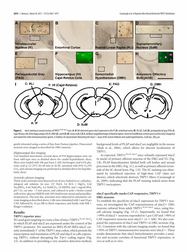
Figure 2. nlacZ staining in coronal sections of TRPV1 PLAP-nlacZ mice. A–H, We observed sparse nlacZ expression in the IF (A), entorhinal cortex (B), RLi (C), SuM (D), periaqueductal gray (PAG; E),Cajal-Retzius cells of the hippocampus (HI; F), DMH (G), and AOB (H). Insets in A, C, D, E, and G are magnified images of dashed regions. Insets in F and H show sections processed for nlacZ (magenta)and stained for reelin immunoreactivity (green). I, Outlines of coronal brains illustrating the nlacZ� areas of the rostral midbrain and caudal hypothalamus. Scale bar, 200 �m.
5070 • J. Neurosci., March 30, 2011 • 31(13):5067–5077 Cavanaugh et al. • Reporter Mice Reveal Restricted TRPV1 Distribution
Highly restricted expression of TRPV1 in the adult brainWe found no PLAP� cell bodies in the brain of TRPV1 PLAP-nlacZ
mice. We did, however, find limited nlacZ expression in a fewbrain areas, primarily along midline structures of the rostral mid-brain and caudal hypothalamus (Fig. 2). Prominent amongthese brain areas were the intrafascicular nucleus (IF) (Fig. 2A)and SuM (Fig. 2D), as well as the dorsomedial hypothalamus(DMH) and posterior hypothalamus (PH) (Fig. 2G). We alsodetected scattered nlacZ� cells in the entorhinal cortex (Fig. 2B),the rostral linear raphe nucleus (RLi) (Fig. 2C) and the periaque-ductal gray (Fig. 2E). Finally, nlacZ expression marked a discretesubset of (nonpyramidal) hippocampal neurons (Fig. 2F) thatcolabeled for reelin, a marker of Cajal-Retzius cells (Fig. 2F, in-set), as well as reelin� cells in the accessory olfactory bulb (AOB)
(Fig. 2H). It is likely that our failure to detect PLAP in these brainregions reflects the greater sensitivity of nlacZ versus PLAPhistochemistry.
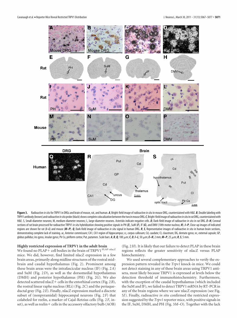
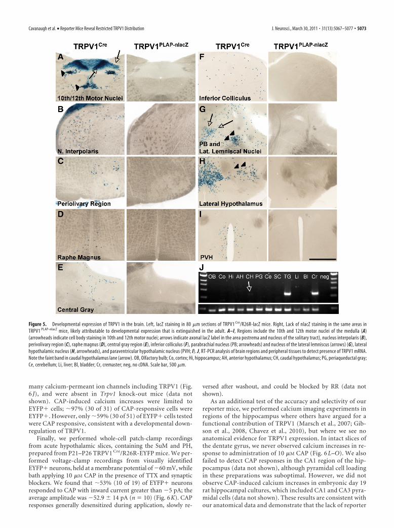
We used several complementary approaches to verify the ex-pression pattern revealed in the Trpv1 knock-in mice. We couldnot detect staining in any of these brain areas using TRPV1 anti-sera, most likely because TRPV1 is expressed at levels below thedetection threshold of immunohistochemistry. Furthermore,with the exception of the caudal hypothalamus (which includedthe SuM and IF), we failed to detect TRPV1 mRNA by RT-PCR inany of the brain regions where we saw nlacZ expression (see Fig.5J). Finally, radioactive in situ confirmed the restricted expres-sion suggested by the Trpv1 reporter mice, with positive signals inthe IF, SuM, DMH, and PH (Fig. 3M–O). Together with the lack
Figure 3. Radioactive in situ for TRPV1 in DRGs and brain of mouse, rat, and human. A, Bright-field image of radioactive in situ in mouse DRG, counterstained with H&E. B, Double labeling withTRPV1 antibody (brown) and radioactive in situ probe (black) shows complete colocalization between the two in mouse DRG. C, Bright-field image of radioactive in situ in rat DRG, counterstained withH&E. S, Small-diameter neurons; M, medium-diameter neurons; L, large-diameter neurons. Asterisks indicate negative cells. D, Dark-field image of radioactive in situ in rat DRG. E–H, Coronalsections of rat brain processed for radioactive TRPV1 in situ hybridization showing positive signals in PH (E), SuM (F), IF (G), and DMV (10th motor nucleus; H). I–P, Close-up images of indicatedregions are shown for rat (I–L) and mouse (M–P). Q, Dark-field image of radioactive in situ signal in human DRG. R, S, Representative images of radioactive in situ in human brain sections,demonstrating complete lack of staining. ac, Anterior commissure; CA1, CA1 region of hippocampus; cc, corpus callosum; Cd, caudate; CI, claustrum; DG, dentate gyrus; ec, external capsule; GP,globus pallidus; ins gyrus, insular gyrus; Pir Cx, piriform cortex; Put, putamen. Scale bars: A, B, Q, 100 �m; C, D, I–L, 50 �m; E–H, 2 mm; M–P, 25 �m; R, S, 5 mm.
Cavanaugh et al. • Reporter Mice Reveal Restricted TRPV1 Distribution J. Neurosci., March 30, 2011 • 31(13):5067–5077 • 5071
of PLAP staining, these results indicate that TRPV1 in the brain islimited to very low-level expression in a small subset of cells of afew discrete brain regions.
Restricted expression of TRPV1 in CNS is conservedacross speciesThe distribution of TRPV1 in the DRG of the mouse differs fromthat of the rat (Woodbury et al., 2004), which suggests that brainexpression patterns might also differ across species. To investi-gate this, we performed radioactive in situ hybridization in sev-eral species, including rat, monkey, and human. Importantly,using several different high-affinity probes directed against ratTRPV1, we found a near-identical pattern of CNS staining com-pared with that obtained in the mouse. Most notably, specificsignals were found in caudal hypothalamic regions, including thePH, SuM, and IF (Fig. 3E–L). Positive signals were also found inthe dorsal motor nucleus of the vagus (DMV), mesencephalictrigeminal nucleus, parabrachial nucleus, RLi, and lateral hypo-thalamus of the rat (data not shown). In monkey (data notshown) and human brain sections (Fig. 3R–S), we could not de-tect any TRPV1� signal; however, we did not have access tosections that included the areas of the caudal hypothalamus thathad positive signals in rat and mouse. Nevertheless, the lack ofsignal in other brain areas, despite the strong specific signal inDRG neurons (Fig. 3Q), is consistent with the highly restrictedpattern we observed in mouse and rat, thus demonstrating thatthis property is conserved across multiple mammalian species.
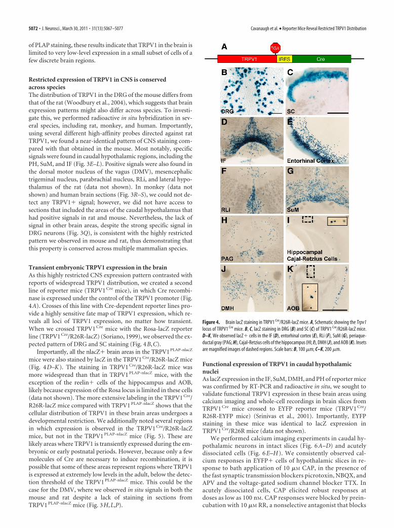
Transient embryonic TRPV1 expression in the brainAs this highly restricted CNS expression pattern contrasted withreports of widespread TRPV1 distribution, we created a secondline of reporter mice (TRPV1 Cre mice), in which Cre recombi-nase is expressed under the control of the TRPV1 promoter (Fig.4A). Crosses of this line with Cre-dependent reporter lines pro-vide a highly sensitive fate map of TRPV1 expression, which re-veals all loci of TRPV1 expression, no matter how transient.When we crossed TRPV1 Cre mice with the Rosa-lacZ reporterline (TRPV1 Cre/R26R-lacZ) (Soriano, 1999), we observed the ex-pected pattern of DRG and SC staining (Fig. 4B,C).
Importantly, all the nlacZ� brain areas in the TRPV1PLAP-nlacZ
mice were also stained by lacZ in the TRPV1 Cre/R26R-lacZ mice(Fig. 4D–K). The staining in TRPV1 Cre/R26R-lacZ mice wasmore widespread than that in TRPV1 PLAP-nlacZ mice, with theexception of the reelin� cells of the hippocampus and AOB,likely because expression of the Rosa locus is limited in these cells(data not shown). The more extensive labeling in the TRPV1 Cre/R26R-lacZ mice compared with TRPV1 PLAP-nlacZ shows that thecellular distribution of TRPV1 in these brain areas undergoes adevelopmental restriction. We additionally noted several regionsin which expression is observed in the TRPV1 Cre/R26R-lacZmice, but not in the TRPV1 PLAP-nlacZ mice (Fig. 5). These arelikely areas where TRPV1 is transiently expressed during the em-bryonic or early postnatal periods. However, because only a fewmolecules of Cre are necessary to induce recombination, it ispossible that some of these areas represent regions where TRPV1is expressed at extremely low levels in the adult, below the detec-tion threshold of the TRPV1 PLAP-nlacZ mice. This could be thecase for the DMV, where we observed in situ signals in both themouse and rat despite a lack of staining in sections fromTRPV1 PLAP-nlacZ mice (Fig. 3H,L,P).
Functional expression of TRPV1 in caudal hypothalamicnucleiAs lacZ expression in the IF, SuM, DMH, and PH of reporter micewas confirmed by RT-PCR and radioactive in situ, we sought tovalidate functional TRPV1 expression in these brain areas usingcalcium imaging and whole-cell recordings in brain slices fromTRPV1 Cre mice crossed to EYFP reporter mice (TRPV1 Cre/R26R-EYFP mice) (Srinivas et al., 2001). Importantly, EYFPstaining in these mice was identical to lacZ expression inTRPV1 Cre/R26R mice (data not shown).
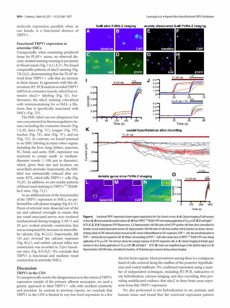
We performed calcium imaging experiments in caudal hy-pothalamic neurons in intact slices (Fig. 6 A–D) and acutelydissociated cells (Fig. 6 E–H ). We consistently observed cal-cium responses in EYFP� cells of hypothalamic slices in re-sponse to bath application of 10 �M CAP, in the presence ofthe fast synaptic transmission blockers picrotoxin, NBQX, andAPV and the voltage-gated sodium channel blocker TTX. Inacutely dissociated cells, CAP elicited robust responses atdoses as low as 100 nM. CAP responses were blocked by prein-cubation with 10 �M RR, a nonselective antagonist that blocks
Figure 4. Brain lacZ staining in TRPV1 Cre/R26R-lacZ mice. A, Schematic showing the Trpv1locus of TRPV1 Cre mice. B, C, lacZ staining in DRG (B) and SC (C) of TRPV1 Cre/R26R-lacZ mice.D–K, We observed lacZ� cells in the IF (D), entorhinal cortex (E), RLi (F), SuM (G), periaque-ductal gray (PAG; H), Cajal-Retzius cells of the hippocampus (HI; I), DMH (J), and AOB (K). Insetsare magnified images of dashed regions. Scale bars: B, 100 �m; C–K, 200 �m.
5072 • J. Neurosci., March 30, 2011 • 31(13):5067–5077 Cavanaugh et al. • Reporter Mice Reveal Restricted TRPV1 Distribution
many calcium-permeant ion channels including TRPV1 (Fig.6 J), and were absent in Trpv1 knock-out mice (data notshown). CAP-induced calcium increases were limited toEYFP� cells; �97% (30 of 31) of CAP-responsive cells wereEYFP�. However, only �59% (30 of 51) of EYFP� cells testedwere CAP responsive, consistent with a developmental down-regulation of TRPV1.
Finally, we performed whole-cell patch-clamp recordingsfrom acute hypothalamic slices, containing the SuM and PH,prepared from P21–P26 TRPV1 Cre/R26R-EYFP mice. We per-formed voltage-clamp recordings from visually identifiedEYFP� neurons, held at a membrane potential of �60 mV, whilebath applying 10 �M CAP in the presence of TTX and synapticblockers. We found that �53% (10 of 19) of EYFP� neuronsresponded to CAP with inward current greater than �5 pA; theaverage amplitude was �52.9 14 pA (n � 10) (Fig. 6K). CAPresponses generally desensitized during application, slowly re-
versed after washout, and could be blocked by RR (data notshown).
As an additional test of the accuracy and selectivity of ourreporter mice, we performed calcium imaging experiments inregions of the hippocampus where others have argued for afunctional contribution of TRPV1 (Marsch et al., 2007; Gib-son et al., 2008, Chavez et al., 2010), but where we see noanatomical evidence for TRPV1 expression. In intact slices ofthe dentate gyrus, we never observed calcium increases in re-sponse to administration of 10 �M CAP (Fig. 6 L–O). We alsofailed to detect CAP responses in the CA1 region of the hip-pocampus (data not shown), although pyramidal cell loadingin these preparations was suboptimal. However, we did notobserve CAP-induced calcium increases in embryonic day 19rat hippocampal cultures, which included CA1 and CA3 pyra-midal cells (data not shown). These results are consistent withour anatomical data and demonstrate that the lack of reporter
Figure 5. Developmental expression of TRPV1 in the brain. Left, lacZ staining in 80 �m sections of TRPV1 Cre/R26R-lacZ mice. Right, Lack of nlacZ staining in the same areas inTRPV1 PLAP-nlacZ mice, likely attributable to developmental expression that is extinguished in the adult. A–I, Regions include the 10th and 12th motor nuclei of the medulla (A)(arrowheads indicate cell body staining in 10th and 12th motor nuclei; arrows indicate axonal lacZ label in the area postrema and nucleus of the solitary tract), nucleus interpolaris (B),perivolivary region (C), raphe magnus (D), central gray region (E), inferior colliculus (F), parabrachial nucleus (PB; arrowheads) and nucleus of the lateral lemniscus (arrows) (G), lateralhypothalamic nucleus (H, arrowheads), and paraventricular hypothalamic nucleus (PVH; I). J, RT-PCR analysis of brain regions and peripheral tissues to detect presence of TRPV1 mRNA.Note the faint band in caudal hypothalamus lane (arrow). OB, Olfactory bulb; Co, cortex; Hi, hippocampus; AH, anterior hypothalamus; CH, caudal hypothalamus; PG, periaqueductal gray;Ce, cerebellum; Li, liver; Bl, bladder; Cr, cremaster; neg, no cDNA. Scale bar, 500 �m.
Cavanaugh et al. • Reporter Mice Reveal Restricted TRPV1 Distribution J. Neurosci., March 30, 2011 • 31(13):5067–5077 • 5073
molecule expression parallels what, inour hands, is a functional absence ofTRPV1.
Functional TRPV1 expression inarteriolar SMCsUnexpectedly, when examining peripheraltissue for PLAP� axons, we observed dis-crete, striated staining running in proximityto blood vessels (Fig. 7A,C,E,F). We foundcomparable patterns of nlacZ staining (Fig.7B,D,G), demonstrating that the PLAP de-rived from TRPV1� cells that are intrinsicto these tissues. In agreement with this ob-servation, RT-PCR analysis revealed TRPV1mRNA in cremaster muscle, which has ex-tensive nlacZ� labeling (Fig. 5J). Fur-thermore, the nlacZ staining colocalizedwith immunostaining for �-SMA, a fila-ment that is specifically associated withSMCs (Fig. 7D).
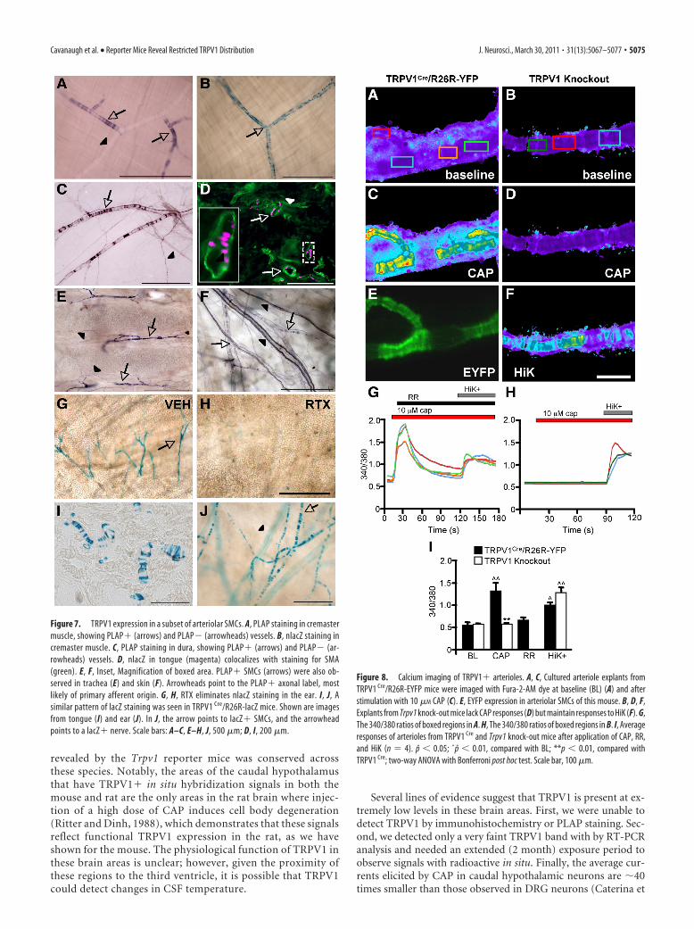
The SMC label was not ubiquitous butwas concentrated in thermoregulatory tis-sues, including the cremaster muscle (Fig.7A,B), dura (Fig. 7C), tongue (Fig. 7D),trachea (Fig. 7E), skin (Fig. 7F), and ear(Fig. 7G). In contrast, we found minimalto no SMC labeling in many other organs,including the liver, lung, kidney, pancreas,SC, brain, and aorta. SMC expression wasrestricted to certain small- to medium-diameter vessels (�100 �m in diameter),which, given their size and location, aremost likely arterioles. Importantly, the SMClabel was substantially reduced after sys-temic RTX, which kills TRPV1� cells (Fig.7G,H). In addition, we saw similar patternsof blood vessel staining in TRPV1Cre/R26R-lacZ mice. (Fig. 7I,J).
As an additional test of the functionalityof the TRPV1 expression in SMCs, we per-formed live-cell calcium imaging (Fig. 8A–F).Pieces of arteriole were dissected out of theear and cultured overnight to ensure thatany vessel-associated nerves were renderednonfunctional during imaging. CAP (1 and10 �M) evoked arteriole constriction thatwas accompanied by increases in intracellu-lar calcium (Fig. 8C,G,I). Importantly, RR(10 �M) reversed the calcium responses(Fig. 8G,I), and neither calcium influx norconstriction was recorded in Trpv1 knock-out mice (Fig. 8D,H,I). We conclude thatTRPV1 is functional and mediates vesselconstriction in arteriolar SMCs.
DiscussionTRPV1 in the CNSTo unequivocally resolve the disagreement as to the extent of TRPV1expression outside of the primary afferent nociceptor, we used agenetic approach to label TRPV1� cells with excellent sensitivityand precision. In contrast to previous reports, we conclude thatTRPV1 in the CNS is limited to very low-level expression in a few
discrete brain regions. Most prominent among these is a contiguousband of cells centered along the midline of the posterior hypothala-mus and rostral midbrain. We confirmed expression using a num-ber of independent techniques, including RT-PCR, radioactive insitu hybridization, calcium imaging, and slice recording, thus pro-viding multifaceted evidence that nlacZ in these brain areas repre-sents bona fide TRPV1 expression.
We also performed in situ hybridization in rat, primate, andhuman tissue and found that the restricted expression pattern
Figure 6. Functional TRPV1 expression in brain regions implicated in the Trpv1 knock-in mice. A–H, Calcium imaging of SuM neuronsin slices (A–D) and acutely dissociated cultures (E–H) from TRPV1 Cre/R26R-EYFP mice during application of 10�M CAP (B, F) and high K �
ACSF (C, G). D, H, Endogenous EYFP fluorescence. I, J, Representative 340/380 ratios of the EYFP-positive cells from slices (normalized tobaseline; I) and acutely dissociated neurons (J). Representative 340/380 ratios of cells from another well (J, bottom) are shown, demon-strating a block of CAP-induced calcium increases by RR. Arrows in D and H point to CAP-responsive, EYPF� cells; the arrowhead points toEYFP� cell that did not respond to CAP. K, Whole-cell recordings of EYFP� SuM cells in brain slices of TRPV1 Cre/R26R-EYFP mice duringapplication of 10 �M CAP. The red trace shows the average response of all CAP-responsive cells. L–N, Calcium imaging of dentate gyrusneurons in slices during application of 10 �M CAP (M) and high K � ACSF (N). Insets are magnified images of the dashed region in L. O,Representative 340/380 ratios, normalized to baseline, of 30 dentate gyrus neurons during calcium imaging.
5074 • J. Neurosci., March 30, 2011 • 31(13):5067–5077 Cavanaugh et al. • Reporter Mice Reveal Restricted TRPV1 Distribution
revealed by the Trpv1 reporter mice was conserved acrossthese species. Notably, the areas of the caudal hypothalamusthat have TRPV1� in situ hybridization signals in both themouse and rat are the only areas in the rat brain where injec-tion of a high dose of CAP induces cell body degeneration(Ritter and Dinh, 1988), which demonstrates that these signalsreflect functional TRPV1 expression in the rat, as we haveshown for the mouse. The physiological function of TRPV1 inthese brain areas is unclear; however, given the proximity ofthese regions to the third ventricle, it is possible that TRPV1could detect changes in CSF temperature.
Several lines of evidence suggest that TRPV1 is present at ex-tremely low levels in these brain areas. First, we were unable todetect TRPV1 by immunohistochemistry or PLAP staining. Sec-ond, we detected only a very faint TRPV1 band with by RT-PCRanalysis and needed an extended (2 month) exposure period toobserve signals with radioactive in situ. Finally, the average cur-rents elicited by CAP in caudal hypothalamic neurons are �40times smaller than those observed in DRG neurons (Caterina et
Figure 7. TRPV1 expression in a subset of arteriolar SMCs. A, PLAP staining in cremastermuscle, showing PLAP� (arrows) and PLAP� (arrowheads) vessels. B, nlacZ staining incremaster muscle. C, PLAP staining in dura, showing PLAP� (arrows) and PLAP� (ar-rowheads) vessels. D, nlacZ in tongue (magenta) colocalizes with staining for SMA(green). E, F, Inset, Magnification of boxed area. PLAP� SMCs (arrows) were also ob-served in trachea (E) and skin (F). Arrowheads point to the PLAP� axonal label, mostlikely of primary afferent origin. G, H, RTX eliminates nlacZ staining in the ear. I, J, Asimilar pattern of lacZ staining was seen in TRPV1 Cre/R26R-lacZ mice. Shown are imagesfrom tongue (I) and ear (J). In J, the arrow points to lacZ� SMCs, and the arrowheadpoints to a lacZ� nerve. Scale bars: A–C, E–H, J, 500 �m; D, I, 200 �m.
Figure 8. Calcium imaging of TRPV1� arterioles. A, C, Cultured arteriole explants fromTRPV1 Cre/R26R-EYFP mice were imaged with Fura-2-AM dye at baseline (BL) (A) and afterstimulation with 10 �M CAP (C). E, EYFP expression in arteriolar SMCs of this mouse. B, D, F,Explants from Trpv1 knock-out mice lack CAP responses (D) but maintain responses to HiK (F). G,The 340/380 ratios of boxed regions in A. H, The 340/380 ratios of boxed regions in B. I, Averageresponses of arterioles from TRPV1 Cre and Trpv1 knock-out mice after application of CAP, RR,and HiK (n � 4). p 0.05; ˆp 0.01, compared with BL; **p 0.01, compared withTRPV1 Cre; two-way ANOVA with Bonferroni post hoc test. Scale bar, 100 �m.
Cavanaugh et al. • Reporter Mice Reveal Restricted TRPV1 Distribution J. Neurosci., March 30, 2011 • 31(13):5067–5077 • 5075
al., 2000). Based on the single-channel conductance of TRPV1 at�60 mV (Tominaga et al., 1998), we estimate that the smallestCAP-induced currents (�5 pA) that we observed in EYFP� cellsof the caudal hypothalamus arise from just a few functionalchannels, highlighting the remarkable sensitivity of the Trpv1knock-in mice.
Several groups used Trpv1 knock-out mice to suggest a func-tional contribution of this channel outside of the DRG, includingurothelial cells of the bladder (Birder et al., 2002), osmosensitiveneurons of the supraoptic and paraventricular regions of the hy-pothalamus (Ciura and Bourque, 2006; Sharif-Naeini et al., 2006,2008), striatal neurons (Maccarrone et al., 2008), dentate gyrusneurons (Chavez et al., 2010), medium spiny neurons of the nu-cleus accumbens (Greuter et al., 2010), and hippocampal pyra-midal cells (Marsch et al., 2007; Gibson et al., 2008). However,despite using what we consider to be the most sensitive measureyet for detecting TRPV1, our analysis of Trpv1 reporter mice doesnot support the conclusions of these reports. We did not detectlacZ in any of these cell populations, even in the TRPV1 Cre/R26R-lacZ mice, where embryonic expression greatly exceedsthat observed in the adult and in which only a few molecules ofCre expression are sufficient to drive recombination. In agree-ment with the lack of reporter molecule expression, CAP did notelicit calcium increases in either intact hippocampal slices or incultured hippocampal neurons. Our observations are thus moreconsistent with studies that found no evidence for TRPV1 inseveral candidate regions (Kofalvi et al., 2006; Benninger et al.,2008; Taylor et al., 2008; Everaerts et al., 2010).
One caveat is that the use of the internal ribosome entry site inour reporter constructs could have led to spurious reporter mol-ecule expression in areas that lack Trpv1 transcription, or to ar-tificially low expression in certain brain areas that do expressTRPV1. However, given the consistency of our results using mul-tiple independent assays, and across various species, we do notbelieve this to be the case. It is also possible that our reporters arenot expressed in cells that transcribe alternate isoforms of Trpv1that lack the final exon, although such splice variants have notbeen reported. Finally, although many of the reports of func-tional TRPV1 in the brain argue for a postsynaptic localization,some of the phenotypes described in these knock-out paperscould reflect a loss of presynaptic TRPV1 that arises from axonalprojections of neurons that we demonstrate do express TRPV1.For example, SuM neurons are known to project heavily to thehippocampus (Vertes, 1992).
TRPV1 in arteriolar SMCsClassic studies have shown that CAP-evoked release of neuropep-tides from the peripheral nociceptor terminals promotes vasodi-lation. More recently, a direct action of CAP on vascular smoothmuscle was reported, but whether this mechanism is widespreador regionally restricted has not been determined (Donnerer andLembeck, 1982; Duckles, 1986; Kark et al., 2008). AlthoughTRPV1 immunoreactivity has been observed in rat SMCs (Karket al., 2008) we did not see similar staining in the mouse (data notshown), likely because of high nonspecific antibody staining inperipheral tissues. On the other hand, PLAP and nlacZ providedstrong signals in SMCs with minimal background. Importantly,nlacZ staining was reduced in SMCs after systemic RTX injection,confirming functional TRPV1 expression. Finally, calcium imag-ing of isolated arterioles from the ear showed that geneticallymarked SMCs respond to CAP application, leading to arterioleconstriction. Similar responses could not be evoked in Trpv1
knock-out mice, demonstrating that the direct CAP effects onSMCs require TRPV1.
Interestingly, the SMC labeling was restricted to a subset ofarterioles in thermoregulatory tissues, including the tongue, skin,trachea, and cremaster. Whereas activation of TRPV1 on sensorynerve endings leads to local vasodilation, vascular TRPV1 medi-ates vasoconstriction. Thus, activation of TRPV1 on SMCs couldcounteract nerve-related changes in vascular tone in response tophysiological TRPV1 agonists, such as heat and pH. TRPV1 ex-pression in SMCs may therefore contribute to the thermoregula-tory effects induced by TRPV1 antagonists, many of which arebeing developed to treat chronic pain conditions (Gavva, 2008).On the other hand, the limited TRPV1 expression in the brainindicates that untoward side effects of these drugs are unlikely tobe CNS mediated.
ReferencesAcs G, Palkovits M, Blumberg PM (1996) Specific binding of [3H]resinifera-
toxin by human and rat preoptic area, locus ceruleus, medial hypothalamus,reticular formation and ventral thalamus membrane preparations. Life Sci59:1899–1908.
Benninger F, Freund TF, Hajos N (2008) Control of excitatory synaptictransmission by capsaicin is unaltered in TRPV1 vanilloid receptorknockout mice. Neurochem Int 52:89 –94.
Birder LA, Nakamura Y, Kiss S, Nealen ML, Barrick S, Kanai AJ, Wang E, RuizG, De Groat WC, Apodaca G, Watkins S, Caterina MJ (2002) Alteredurinary bladder function in mice lacking the vanilloid receptor TRPV1.Nat Neurosci 5:856 – 860.
Caterina MJ, Julius D (2001) The vanilloid receptor: a molecular gateway tothe pain pathway. Annu Rev Neurosci 24:487–517.
Caterina MJ, Schumacher MA, Tominaga M, Rosen TA, Levine JD, Julius D(1997) The capsaicin receptor: a heat-activated ion channel in the painpathway. Nature 389:816 – 824.
Caterina MJ, Leffler A, Malmberg AB, Martin WJ, Trafton J, Petersen-ZeitzKR, Koltzenburg M, Basbaum AI, Julius D (2000) Impaired nociceptionand pain sensation in mice lacking the capsaicin receptor. Science288:306 –313.
Cavanaugh DJ, Lee H, Lo L, Shields SD, Zylka MJ, Basbaum AI, Anderson DJ(2009) Distinct subsets of unmyelinated primary sensory fibers mediatebehavioral responses to noxious thermal and mechanical stimuli. ProcNatl Acad Sci U S A 106:9075–9080.
Chavez AE, Chiu CQ, Castillo PE (2010) TRPV1 activation by endogenousanandamide triggers postsynaptic long term depression in the dentategyrus. Nat Neurosci 13:1511–1518.
Ciura S, Bourque CW (2006) Transient receptor potential vanilloid 1 is re-quired for intrinsic osmoreception in organum vasculosum lamina ter-minalis neurons and for normal thirst responses to systemichyperosmolality. J Neurosci 26:9069 –9075.
Cristino L, de Petrocellis L, Pryce G, Baker D, Guglielmotti V, Di Marzo V(2006) Immunohistochemical localization of cannabinoid type 1 and va-nilloid transient receptor potential vanilloid type 1 receptors in the mousebrain. Neuroscience 139:1405–1415.
Davis JB, Gray J, Gunthorpe MJ, Hatcher JP, Davey PT, Overend P, HarriesMH, Latcham J, Clapham C, Atkinson K, Hughes SA, Rance K, Grau E,Harper AJ, Pugh PL, Rogers DC, Bingham S, Randall A, Sheardown SA(2000) Vanilloid receptor-1 is essential for inflammatory thermal hyper-algesia. Nature 405:183–187.
Donnerer J, Lembeck F (1982) Analysis of the effects of intravenously in-jected capsaicin in the rat. Naunyn Schmiedebergs Arch Pharmacol320:54 –57.
Duckles SP (1986) Effects of capsaicin on vascular smooth muscle. NaunynSchmiedebergs Arch Pharmacol 333:59 – 64.
Everaerts W, Vriens J, Owsianik G, Appendino G, Voets T, De Ridder D,Nilius B (2010) Functional characterization of transient receptor poten-tial channels in mouse urothelial cells. Am J Physiol Renal Physiol298:F692–F701.
Gavva NR (2008) Body-temperature maintenance as the predominantfunction of the vanilloid receptor TRPV1. Trends Pharmacol Sci29:550 –557.
Gibson HE, Edwards JG, Page RS, Van Hook MJ, Kauer JA (2008) TRPV1
5076 • J. Neurosci., March 30, 2011 • 31(13):5067–5077 Cavanaugh et al. • Reporter Mice Reveal Restricted TRPV1 Distribution
channels mediate long-term depression at synapses on hippocampal in-terneurons. Neuron 57:746 –759.
Greuter BA, Brasnjo G, Malenka RC (2010) Postsynaptic TRPV1 triggerscell type-specific long-term depression in the nucleus accumbens. NatNeurosci 13:1519 –1525.
Kark T, Bagi Z, Lizanecz E, Pasztor ET, Erdei N, Czikora A, Papp Z, Edes I,Porszasz R, Toth A (2008) Tissue-specific regulation of microvasculardiameter: opposite functional roles of neuronal and smooth musclelocated vanilloid receptor-1. Mol Pharmacol 73:1405–1412.
Kobayashi K, Fukuoka T, Obata K, Yamanaka H, Dai Y, Tokunaga A, Nogu-chi K (2005) Distinct expression of TRPM8, TRPA1, and TRPV1 mR-NAs in rat primary afferent neurons with a�/c-fbers and colocalizationwith trk receptors. J Comp Neurol 493:596 – 606.
Kofalvi A, Oliveira CR, Cunha RA (2006) Lack of evidence for functionalTRPV1 vanilloid receptors in rat hippocampal nerve terminals. NeurosciLett 403:151–156.
Maccarrone M, Rossi S, Bari M, De Chiara V, Fezza F, Musella A, Gasperi V,Prosperetti C, Bernardi G, Finazzi-Agro A, Cravatt BF, Centonze D(2008) Anandamide inhibits metabolism and physiological actions of2-arachidonoylglycerol in the striatum. Nat Neurosci 11:152–159.
Marsch R, Foeller E, Rammes G, Bunck M, Kossl M, Holsboer F, Ziegl-gansberger W, Landgraf R, Lutz B, Wotjak CT (2007) Reduced anxiety,conditioned fear, and hippocampal long-term potentiation in transientreceptor potential vanilloid type 1 receptor-deficient mice. J Neurosci27:832– 839.
Mezey E, Toth ZE, Cortright DN, Arzubi MK, Krause JE, Elde R, Guo A,Blumberg PM, Szallasi A (2000) Distribution of mRNA for vanilloidreceptor subtype 1 (VR1), and VR1-like immunoreactivity, in the centralnervous system of the rat and human. Proc Natl Acad Sci U S A97:3655–3660.
Ritter S, Dinh TT (1988) Capsaicin-induced neuronal degeneration: silverimpregnation of cells bodies, axons, and terminals in the central nervoussystem of the adult rat. J Comp Neurol 271:79 –90.
Roberts JC, Davis JB, Benham CD (2004) [3H]Resiniferatoxin autoradiog-raphy in the CNS of wild-type and TRPV1 null mice defines TRPV1(VR-1) protein distribution. Brain Res 995:176 –183.
Shah NM, Pisapia DJ, Maniatis S, Mendelsohn MM, Nemes A, Axel R (2004)Visualizing sexual dimorphism in the brain. Neuron 43:313–319.
Sharif-Naeini R, Witty MF, Seguela P, Bourque CW (2006) An N-terminalvariant of Trpv1 channel is required for osmosensory transduction. NatNeurosci 9:93–98.
Sharif-Naeini R, Ciura S, Bourque CW (2008) TRPV1 gene required forthermosensory transduction and anticipatory secretion from vasopressinneurons during hyperthermia. Neuron 58:179 –185.
Soriano P (1999) Generalized lacZ expression with the ROSA26 Cre re-porter strain. Nat Genet 21:70 –71.
Srinivas S, Watanabe T, Lin CS, William CM, Tanabe Y, Jessell TM, Costan-tini F (2001) Cre reporter strains produced by targeted insertion ofEYFP and ECFP into the ROSA26 locus. BMC Dev Biol 1:4.
Steenland HW, Ko SW, Wu LJ, Zhuo M (2006) Hot receptors in the brain.Mol Pain 2:34.
Szallasi A, Nilsson S, Farkas-Szallasi T, Blumberg PM, Hokfelt T, LundbergJM (1995) Vanilloid (capsaicin) receptors in the rat: distribution in thebrain, regional differences in the spinal cord, axonal transport to theperiphery, and depletion by systemic vanilloid treatment. Brain Res703:175–183.
Taylor AC, McCarthy JJ, Stocker SD (2008) Mice lacking the transient re-ceptor vanilloid potential 1 channel display normal thirst responses andcentral Fos activation to hypernatremia. Am J Physiol Regul Integr CompPhysiol 294:R1285–R1293.
Tominaga M, Caterina MJ, Malmberg AB, Rosen TA, Gilbert H, Skinner K,Raumann BE, Basbaum AI, Julius D (1998) The cloned capsaicin recep-tor integrates multiple pain-producing stimuli. Neuron 21:531–543.
Toth A, Boczan J, Kedei N, Lizanecz E, Bagi Z, Papp Z, Edes I, Csiba L,Blumberg PM (2005) Expression and distribution of vanilloid receptor1 (TRPV1) in the adult rat brain. Brain Res Mol Brain Res 135:162–168.
Vertes RP (1992) PHA-L analysis of projections from the supramammillarynucleus in the rat. J Comp Neurol 326:595– 622.
Woodbury CJ, Zwick M, Wang S, Lawson JJ, Caterina MJ, Koltzenburg M,Albers KM, Koerber HR, Davis BM (2004) Nociceptors lacking TRVP1and TRPV2 have normal heat responses. J Neurosci 24:6410 – 6415.
Cavanaugh et al. • Reporter Mice Reveal Restricted TRPV1 Distribution J. Neurosci., March 30, 2011 • 31(13):5067–5077 • 5077


![Behavioral/Systems/Cognitive ... · Behavioral/Systems/Cognitive AcuteCocaineInducesFastActivationofD1Receptorand ProgressiveDeactivationofD2ReceptorStriatalNeurons: InVivoOpticalMicroprobe[Ca2]](https://img.dokumen.tips/doc/110x75/6013f75e26e57852b94803cb/behavioralsystemscognitive-behavioralsystemscognitive-acutecocaineinducesfastactivationofd1receptorand.jpg)