Embed Size (px)
Citation preview
1
BCH 405 – REGULATION OF METABOLIC PROCESSES SYNOPSIS The relationship of Krebs cycle to protein, carbohydrate, lipid and nucleic acid metabolism. Integration of metabolic pathways illustration of regulation of linear and branched metabolic pathways using specific example. Turnover rates and metabolic pools catabolite repression, end-product repression, the lactose and arabinose operons. Identification of different regulatory mechanisms in metabolic pathways. INTRODUCTION Metabolism can be defined as the sum of all the enzyme-catalysed reactions that take place in cells. Metabolism is highly coordinated, purposeful activity in which many sets of interrelated multi-enzyme systems participate, exchanging both matter and energy between the cell and its environment. Metabolism has four specific functions:
(1) To obtain chemical energy from fuel molecules or from absorbed sunlight.
(2) To convert exogenous nutrients into the building blocks, or
precursors, of macro-molecular cell components.
(3) To assemble such building blocks into proteins, nucleic acids, lipids and other cell components.
(4) To form and degrade bio-molecules required in specialised functions of cells.
Metabolism can be divided into 2 phases – Catabolism and Anabolism. Catabolism is the degradative phase of metabolism in which complex nutrient molecules (lipids, carbohydrates and proteins) are broken down to simpler end-products such as C02, H20 and NH3 and is accompanied by the synthesis of ATP. Anabolism refers to biosynthetic processes in which simple precursor molecules are enzymatically converted into the molecular components of cells, such as nucleic acids, proteins, lipids and polysaccharides. Biosynthesis requires the input of ATP which is provided by catabolism. There is also often a requirement for reducing power in the form of NADPH. It is seen from all these considerations that whether it is catabolism or anabolism, a metabolic pathway consists of a series of enzyme catalysed
2
reactions that convert a substrate into a product. The individual reactions of many such pathways have been elucidated using the following techniques:
(1) Identification of intermediates which accumulate upon the addition of specific inhibitors of the pathway;
(2) The addition of possible intermediates of the pathway (since their
conversion to the product confirms the role).
(3) The addition of radioactively labeled substrate (or intermediates) and the study of the distribution of the label in intermediates and products.
(4) The separation of the enzymes of the pathway and the elucidation of the chemistry of each reaction in isolation.
The results of these investigations have now been collated into metabolic maps. Stages of Metabolism. Although metabolism involves hundreds of different compounds and enzymes, 3 stages of metabolism are recognized. Draw Diagram Stage 1: The nutrient macro-molecules are broken down into their respective building blocks – Proteins will yield amino acids, polysaccharides give rise to carbohydrate units that are convertible to glucose and lipids are broken down into glycerol and fatty acids and other components. In stage II of metabolism, the many different products of stage 1 are collected and converted/degraded again into simpler metabolic intermediates. Thus, hexoses, pentoses and glycerol are degraded via the 3-carbon intermediate pynuvic acid to yield a single Z-carbon species, acetyl CoA. The various fatty acids and amino acids are broken down to form acetyl CoA and a few other end products. The combustion of the acetyl groups of acetyl CoA by the nitric acid
3
cycles and oxidative PO4 lation to produce C02 and H20 represent stage 3 of catabolism. C02 and H20 are the ultimate waste products of aerobic catabolism. Biosynthesis also takes place in 3 stages. Small precursor molecules are generated in stage 3, then converted in stage 2 into building block molecules, which are finally assembled into macro-molecules in stage 1, For e.g. biosyn of proteins begins in stage 3 with the formation of certain -Keto acids, which are precursors of the -amino acids. In stage 2, the -Keto acids are ammated by amino-group donors to form -amino acids. Finally in stage 1, the amino acids are assembled into polypeptide chains. Catabolic pathways have diffuse beginning (starting from many fuel molecules) but converge into a final route in stage 3. Anabolic pathways diverge; they start from a few precursors in stage 3 and as they proceed through stage 2 and stage 1, they branch and diverge, leading to formation of many different kinds of biomolecules. Converging Pathways of Diverging pathways of
Catabolism Biosynthesis Differences between catabolism and anabolism
(1) They are not the reverse of each other (2) They are independently regulated
4
(3) They often take place in different locations in the cells (compartmentalization β-oxidation-mutochondria syn of fatty acids – cytosol.
Cellular regulation of metabolic pathways Metabolic pathways can be linear, e.g. glycolysis or can be cyclic, e.g. TCA. In general, the rate of catabolism is controlled not by the conc. of nutrients available in the environment of the cell, but by the cell’s need for energy in the form of ATP. Similarly, the rate of biosynthesis of cell components is also adjusted to immediate needs. However, the regulation of a metabolic pathway may occur at several levels.
(1) The reaction rate of each enzymatic reaction si a function of the pH and the intracellular concs. of its substrates products and cofactor which are pry-elements in the regulation of enzyme activity.
(a) Substrate availability – Any metabolic pathway could in theory at
least, be regulated very simply by the availability of substrate. A reduction insubstrate conc. will decrease the activity of the enzyme (provided it is not saturated with substrate) and this could result in a decreased flux through the pathway. Similarly, an increase in (S) could stimulate the path-way. In general however, the constancy of the internal environment of the animal and the cell, as regards the substrates of metabolic pathways implies that such regulatory mechanisms are not common in higher animals. However, a typical, e.g. of control by substrate availability is that by plasma conc of fatty acids. The conc of plasma fatty acids appears to play a fundamentally impt. role in the regulation of their oxidation by various tissues and in turn their oxidation can modify the rate of carbohydrate utilization by the animal. IN such a situation, if the conc. of the fatty acid is known to be regulatory, emphasis then shifts from the metabolic pathway that accounts for plasma fatty acid conc./transport to the factors responsible for changes in fatty acid conc.
(b) Cofactor availability – Some what similar to control by substrate
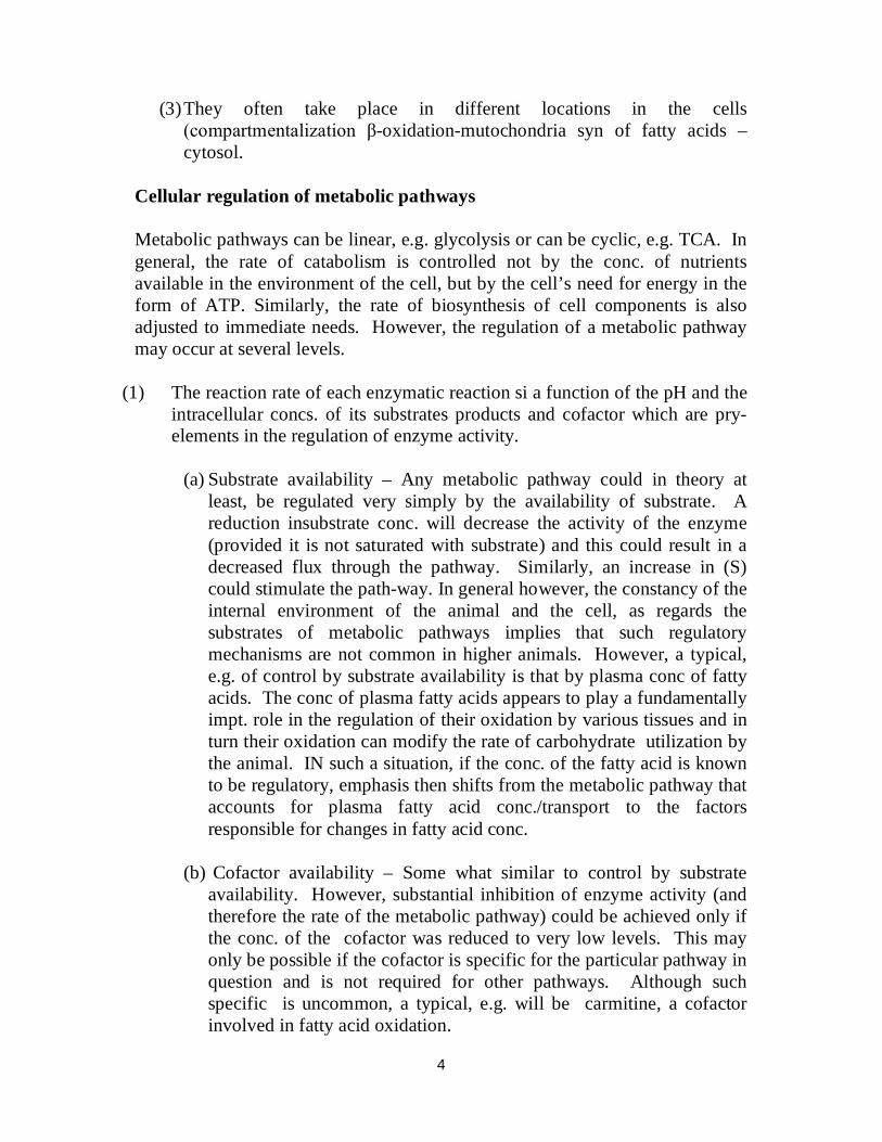
availability. However, substantial inhibition of enzyme activity (and therefore the rate of the metabolic pathway) could be achieved only if the conc. of the cofactor was reduced to very low levels. This may only be possible if the cofactor is specific for the particular pathway in question and is not required for other pathways. Although such specific is uncommon, a typical, e.g. will be carmitine, a cofactor involved in fatty acid oxidation.
5
Fatty acyl – CoA Camitime acyl Transferase
Fatty acids are activated by an enzyme, fatty acyl-CoA synthetase to produce fatty acyl-CoA, a reaction that occurs in the cytoplasm. The β-oxidation of fatty acid occurs inside the mitochondrion. Therefore, the fatty acyl-CoA has to traverse the mitochondrial membranes. The inner mitochondrial membrane is not permeable to fatty acul-CoA; to overcome this barrier, fatty acyl-CoA is converted into fatty acyl-carmitone by the enzyme carmitine acyltransferase. Fatty acyl-carnitine is able to traverse the embrane and on getting into the mitochondrion, is converted back to fatty acyl-CoA and thus provides substrate for β-oxidation. Thus, variations in (carnitone) could regulate the rate of fatty acid oxidation without affecting other metabolic processes. Another, e.g. of control by cofactor availability is the regulation of e-transport and oxidative phosphonylation in the nito-chondria by adenine nucleotides.
6
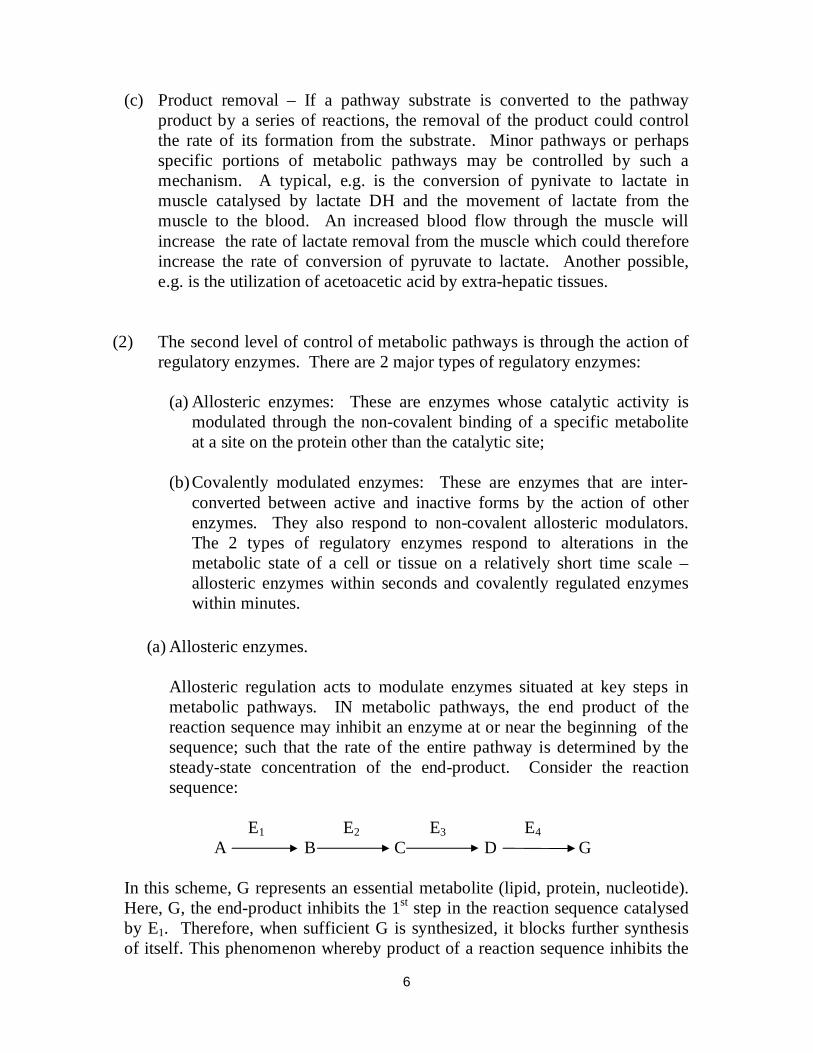
(c) Product removal – If a pathway substrate is converted to the pathway product by a series of reactions, the removal of the product could control the rate of its formation from the substrate. Minor pathways or perhaps specific portions of metabolic pathways may be controlled by such a mechanism. A typical, e.g. is the conversion of pynivate to lactate in muscle catalysed by lactate DH and the movement of lactate from the muscle to the blood. An increased blood flow through the muscle will increase the rate of lactate removal from the muscle which could therefore increase the rate of conversion of pyruvate to lactate. Another possible, e.g. is the utilization of acetoacetic acid by extra-hepatic tissues.
(2) The second level of control of metabolic pathways is through the action of regulatory enzymes. There are 2 major types of regulatory enzymes:
(a) Allosteric enzymes: These are enzymes whose catalytic activity is
modulated through the non-covalent binding of a specific metabolite at a site on the protein other than the catalytic site;
(b) Covalently modulated enzymes: These are enzymes that are inter-
converted between active and inactive forms by the action of other enzymes. They also respond to non-covalent allosteric modulators. The 2 types of regulatory enzymes respond to alterations in the metabolic state of a cell or tissue on a relatively short time scale – allosteric enzymes within seconds and covalently regulated enzymes within minutes.
(a) Allosteric enzymes.
Allosteric regulation acts to modulate enzymes situated at key steps in metabolic pathways. IN metabolic pathways, the end product of the reaction sequence may inhibit an enzyme at or near the beginning of the sequence; such that the rate of the entire pathway is determined by the steady-state concentration of the end-product. Consider the reaction sequence: E1 E2 E3 E4
A B C D G In this scheme, G represents an essential metabolite (lipid, protein, nucleotide). Here, G, the end-product inhibits the 1st step in the reaction sequence catalysed by E1. Therefore, when sufficient G is synthesized, it blocks further synthesis of itself. This phenomenon whereby product of a reaction sequence inhibits the
7
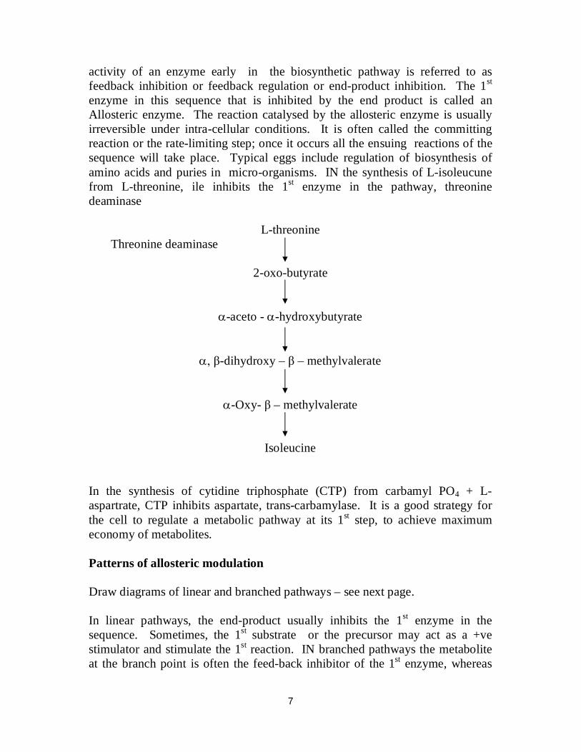
activity of an enzyme early in the biosynthetic pathway is referred to as feedback inhibition or feedback regulation or end-product inhibition. The 1st enzyme in this sequence that is inhibited by the end product is called an Allosteric enzyme. The reaction catalysed by the allosteric enzyme is usually irreversible under intra-cellular conditions. It is often called the committing reaction or the rate-limiting step; once it occurs all the ensuing reactions of the sequence will take place. Typical eggs include regulation of biosynthesis of amino acids and puries in micro-organisms. IN the synthesis of L-isoleucune from L-threonine, ile inhibits the 1st enzyme in the pathway, threonine deaminase
L-threonine Threonine deaminase
2-oxo-butyrate
-aceto - -hydroxybutyrate
, β-dihydroxy – β – methylvalerate
-Oxy- β – methylvalerate
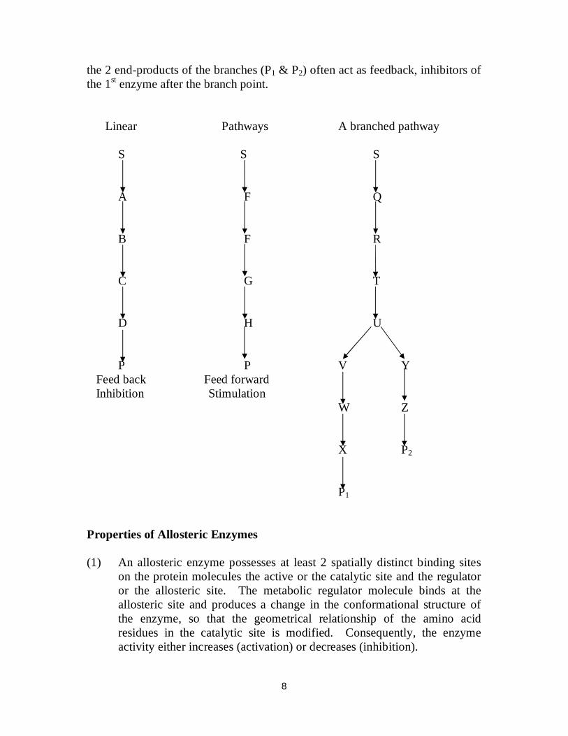
Isoleucine In the synthesis of cytidine triphosphate (CTP) from carbamyl PO4 + L-aspartrate, CTP inhibits aspartate, trans-carbamylase. It is a good strategy for the cell to regulate a metabolic pathway at its 1st step, to achieve maximum economy of metabolites. Patterns of allosteric modulation Draw diagrams of linear and branched pathways – see next page. In linear pathways, the end-product usually inhibits the 1st enzyme in the sequence. Sometimes, the 1st substrate or the precursor may act as a +ve stimulator and stimulate the 1st reaction. IN branched pathways the metabolite at the branch point is often the feed-back inhibitor of the 1st enzyme, whereas
8
the 2 end-products of the branches (P1 & P2) often act as feedback, inhibitors of the 1st enzyme after the branch point. Linear Pathways A branched pathway S S S A F Q B F R C G T D H U P P V Y Feed back Feed forward Inhibition Stimulation W Z X P2 P1 Properties of Allosteric Enzymes (1) An allosteric enzyme possesses at least 2 spatially distinct binding sites
on the protein molecules the active or the catalytic site and the regulator or the allosteric site. The metabolic regulator molecule binds at the allosteric site and produces a change in the conformational structure of the enzyme, so that the geometrical relationship of the amino acid residues in the catalytic site is modified. Consequently, the enzyme activity either increases (activation) or decreases (inhibition).
9
(2) Allosteric enzymes show 2 different types of control – heterotropic and homotropic depending on the nature of the modulating molecule. Heterotropci enzymes are stimulated or inhibited by an effector or modulator molecule other than their substrates, e.g. threonine deaminase the substrate is threamine and the modulator is iso-leucine. When the modulator promotes the binding of substrate to the allosteric enzyme, the modulator is said to be a +ve effector or allosteric activator whereas when the modulator diminishes the binding of substrate, it is called a –ve effector or allosteric inhibitor, +ve effectors increase the number of binding sites for substrate whereas –ve effectors decrease the number of binding sites for the substrate.
In homotropic enzymes, the substrate also functions as the modulator. Homotropic enzymes contain 2 or more binding sites for the substrate modulation depends on how many of the substrate sites are bound.
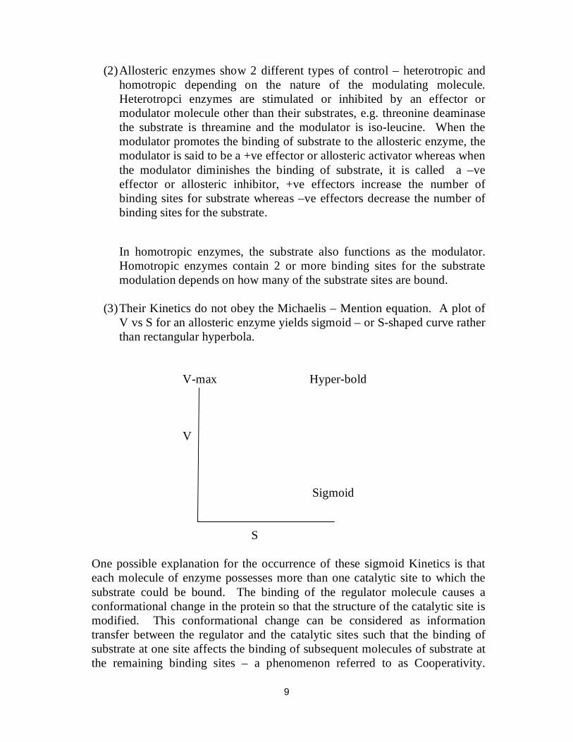
(3) Their Kinetics do not obey the Michaelis – Mention equation. A plot of
V vs S for an allosteric enzyme yields sigmoid – or S-shaped curve rather than rectangular hyperbola.
V-max Hyper-bold V Sigmoid
S One possible explanation for the occurrence of these sigmoid Kinetics is that each molecule of enzyme possesses more than one catalytic site to which the substrate could be bound. The binding of the regulator molecule causes a conformational change in the protein so that the structure of the catalytic site is modified. This conformational change can be considered as information transfer between the regulator and the catalytic sites such that the binding of substrate at one site affects the binding of subsequent molecules of substrate at the remaining binding sites – a phenomenon referred to as Cooperativity.
10
When the binding of the regulator results in more substrates being bound, then it is +ve cooperativity and the reverse as –ve cooperativity.
(4) Inhibition of a regulatory enzyme does not conform to any normal
inhibition pattern and the inhibitor does not bear any obvious structural relationship to the substrate. The enzyme exhibits extreme specificity with regard to the regulator molecule.
(5) Allosteric enzymes have an oligomeric organization. They are composed
of more than one polypeptide chain and have more than one S-binding site per enzyme molecule.
(6) Treatment of the allosteric enzyme with agents or conditions that exert a mild denaturing effect can result in loss of sensitivity to the effects of the regulatory molecule without changing the catalytic activity. This phenomenon is referred to as desensitization and this can be effected by high or low pH mercurials (such as mercuric chloride) urea or by gentle heating. Desensitisation causes dissociation of the ntive enzyme into its component sub-units and this prevents interaction between the regulator and catalytic sites.
Unlike most enzymes, many allosteric enzymes undergo reversible inactivation at OoC.
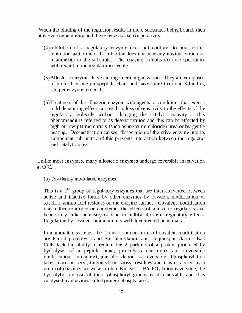
(b) Covalently modulated enzymes. This is a 2nd group of regulatory enzymes that are inter-converted between active and inactive forms by other enzymes by covalent modification of specific amino acid residues on the enzyme surface. Covalent modification may either reinforce or counteract the effects of allosteric regulators and hence may either intensify or tend to nullify allosteric regulatory effects. Regulation by covalent modulation is well documented in animals. In mammalian systems, the 2 most common forms of covalent modification are Partial proteolysis and Phosphorylation and De-phosphorylation. B/C Cells lack the ability to reunite the 2 portions of a protein produced by hydrolysis of a peptide bond, proteolysis constitutes an irreversible modification. In contrast, phosphorylation is a reversible. Phosphorylation takes place on seryl, threomyl, or tyrosyl residues and it is catalysed by a group of enzymes known as protein Kinases. B/c PO4 lation is versible, the hydrolytic removal of these phosphoryl groups is also possible and it is catalysed by enzymes called protein phosphatases.
11
ATP ADP Mg2+ Kinase Phosphorylation
Enz-Ser-OH Enz – Ser – O – PO3
2- Phosphatase
Mg2+ Pi Dephosphorylation H20 The activities of protein Kinases and protein Phosphatases are themselves regulated; if not, their concerted action would be both thermodynamically and biologically unproductive. A classical example of an enzyme regulated by covalent modification of its activity is glycogen phosphorylase of animal tissues which catalyses the breakdown of glyzogen. (Glucose)n + Pi (Glucose)n-1
+ Glucose -1- P04 Glycogen Shortened
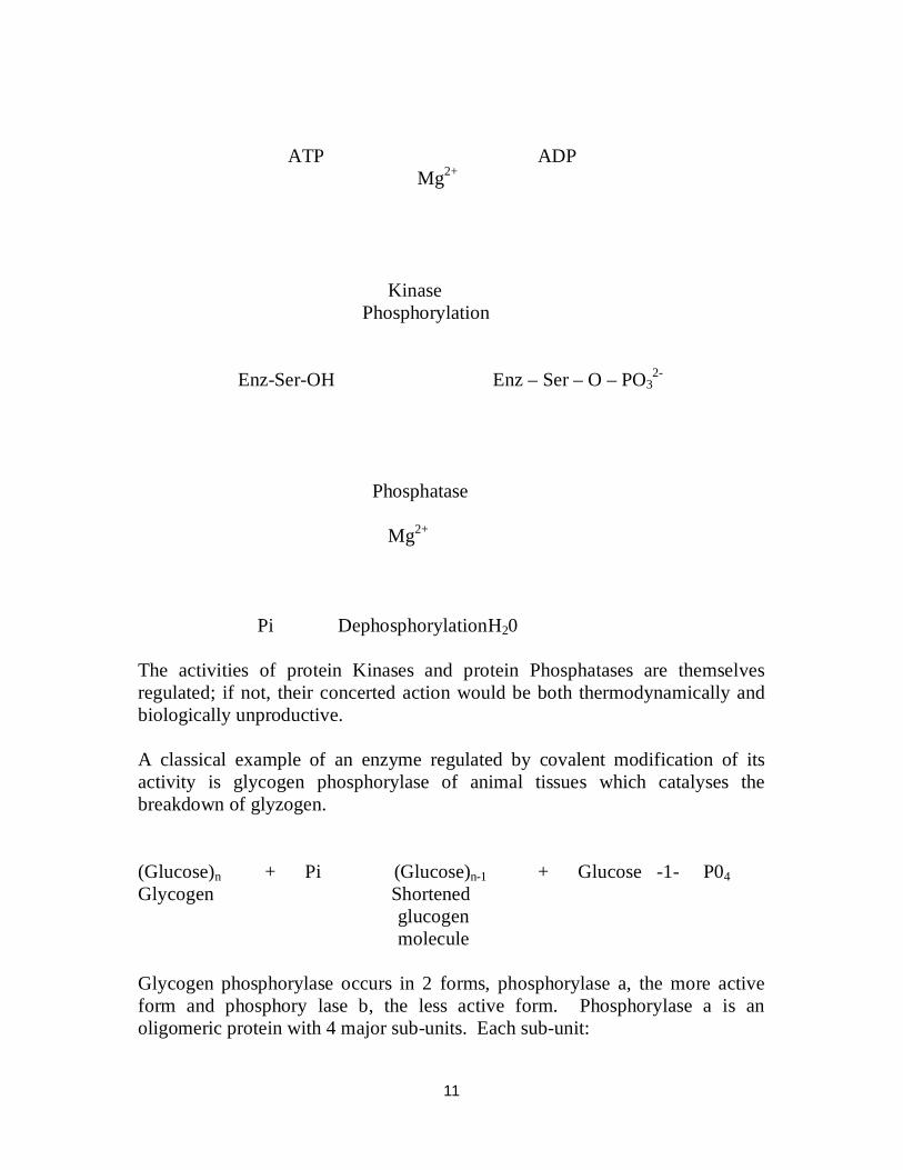
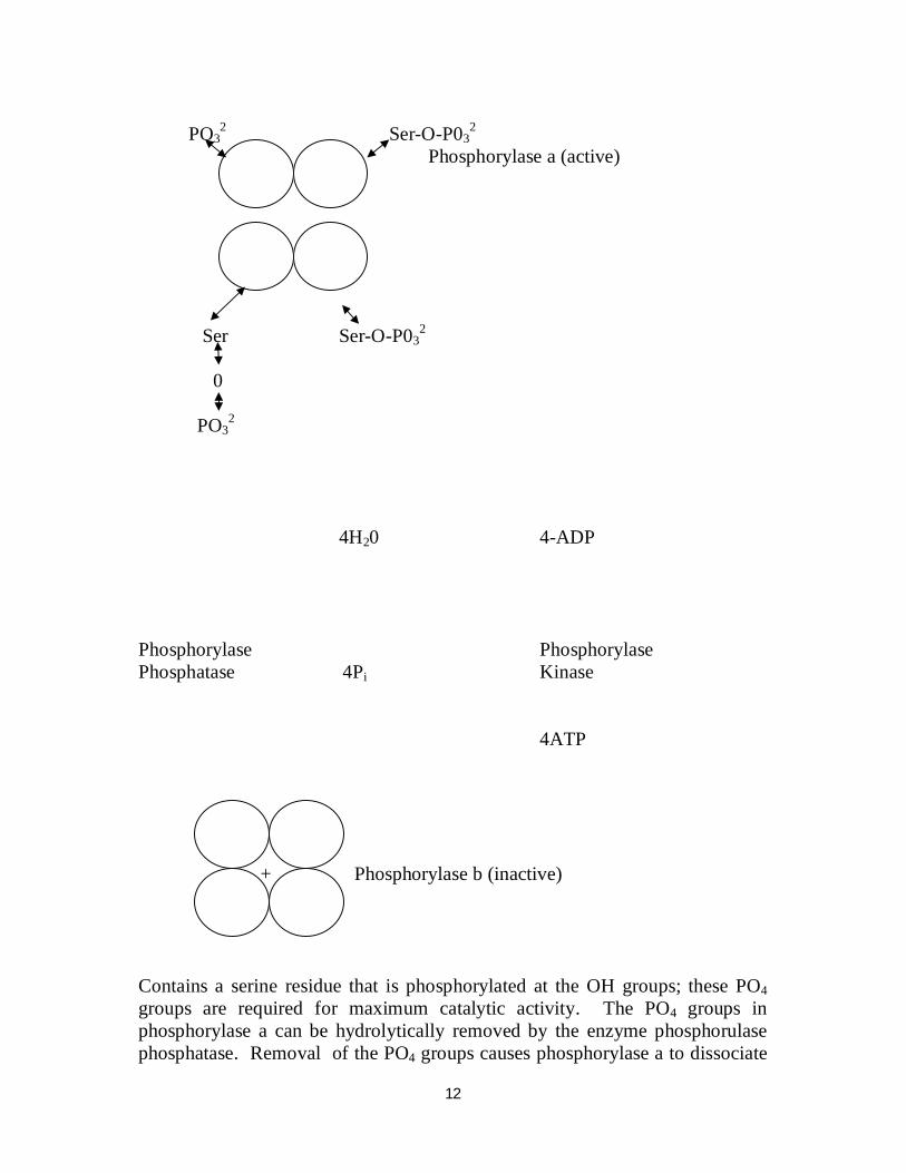
glucogen molecule Glycogen phosphorylase occurs in 2 forms, phosphorylase a, the more active form and phosphory lase b, the less active form. Phosphorylase a is an oligomeric protein with 4 major sub-units. Each sub-unit:
12
PO3
2 Ser-O-P032
Phosphorylase a (active) Ser Ser-O-P03
2 0 PO3
2
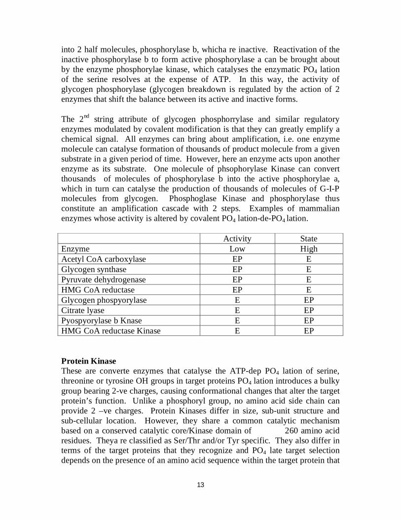
4H20 4-ADP Phosphorylase Phosphorylase Phosphatase 4Pi Kinase 4ATP + Phosphorylase b (inactive) Contains a serine residue that is phosphorylated at the OH groups; these PO4 groups are required for maximum catalytic activity. The PO4 groups in phosphorylase a can be hydrolytically removed by the enzyme phosphorulase phosphatase. Removal of the PO4 groups causes phosphorylase a to dissociate
13
into 2 half molecules, phosphorylase b, whicha re inactive. Reactivation of the inactive phosphorylase b to form active phosphorylase a can be brought about by the enzyme phosphorylae kinase, which catalyses the enzymatic PO4 lation of the serine resolves at the expense of ATP. In this way, the activity of glycogen phosphorylase (glycogen breakdown is regulated by the action of 2 enzymes that shift the balance between its active and inactive forms. The 2nd string attribute of glycogen phosphorrylase and similar regulatory enzymes modulated by covalent modification is that they can greatly emplify a chemical signal. All enzymes can bring about amplification, i.e. one enzyme molecule can catalyse formation of thousands of product molecule from a given substrate in a given period of time. However, here an enzyme acts upon another enzyme as its substrate. One molecule of phsophorylase Kinase can convert thousands of molecules of phosphorylase b into the active phosphorylae a, which in turn can catalyse the production of thousands of molecules of G-I-P molecules from glycogen. Phosphoglase Kinase and phosphorylase thus constitute an amplification cascade with 2 steps. Examples of mammalian enzymes whose activity is altered by covalent PO4 lation-de-PO4 lation. Activity State Enzyme Low High Acetyl CoA carboxylase EP E Glycogen synthase EP E Pyruvate dehydrogenase EP E HMG CoA reductase EP E Glycogen phospyorylase E EP Citrate lyase E EP Pyospyorylase b Knase E EP HMG CoA reductase Kinase E EP Protein Kinase These are converte enzymes that catalyse the ATP-dep PO4 lation of serine, threonine or tyrosine OH groups in target proteins PO4 lation introduces a bulky group bearing 2-ve charges, causing conformational changes that alter the target protein’s function. Unlike a phosphoryl group, no amino acid side chain can provide 2 –ve charges. Protein Kinases differ in size, sub-unit structure and sub-cellular location. However, they share a common catalytic mechanism based on a conserved catalytic core/Kinase domain of 260 amino acid residues. Theya re classified as Ser/Thr and/or Tyr specific. They also differ in terms of the target proteins that they recognize and PO4 late target selection depends on the presence of an amino acid sequence within the target protein that
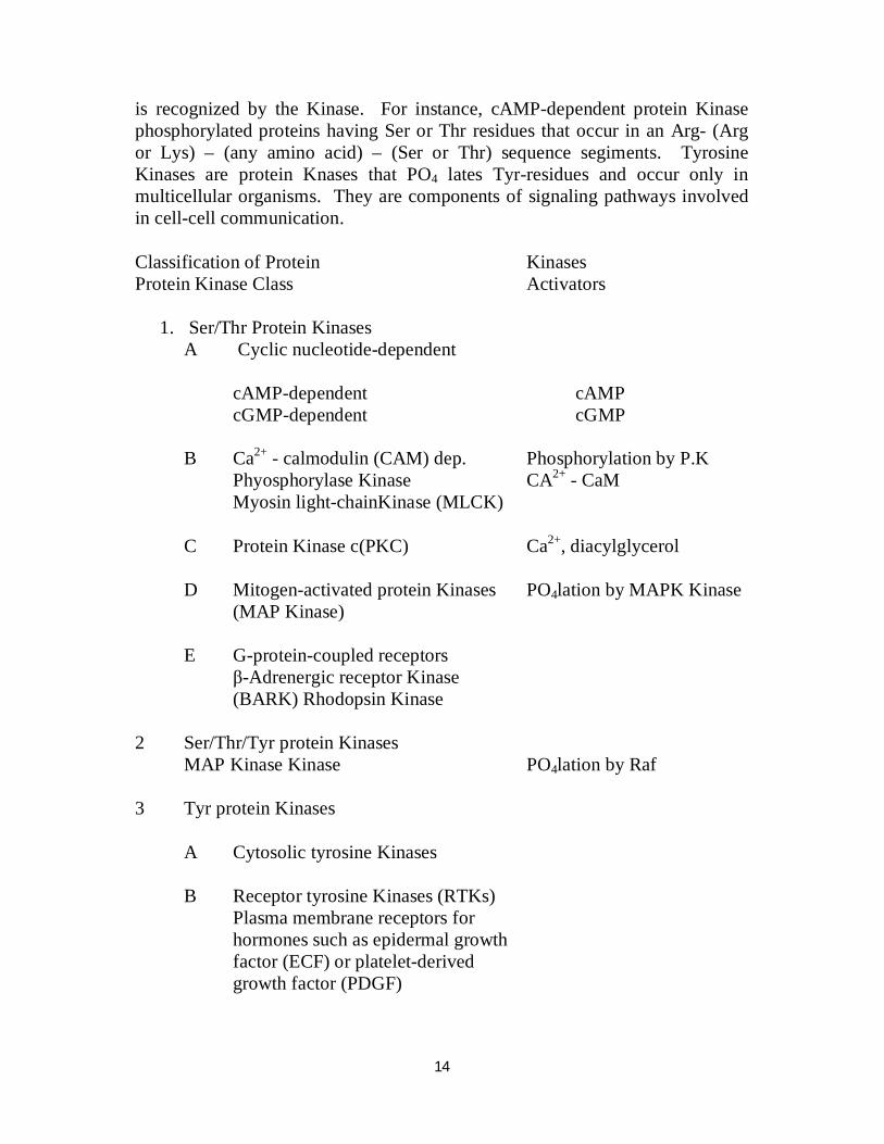
14
is recognized by the Kinase. For instance, cAMP-dependent protein Kinase phosphorylated proteins having Ser or Thr residues that occur in an Arg- (Arg or Lys) – (any amino acid) – (Ser or Thr) sequence segiments. Tyrosine Kinases are protein Knases that PO4 lates Tyr-residues and occur only in multicellular organisms. They are components of signaling pathways involved in cell-cell communication. Classification of Protein Kinases Protein Kinase Class Activators
1. Ser/Thr Protein Kinases A Cyclic nucleotide-dependent
cAMP-dependent cAMP cGMP-dependent cGMP
B Ca2+ - calmodulin (CAM) dep. Phosphorylation by P.K Phyosphorylase Kinase CA2+ - CaM Myosin light-chainKinase (MLCK) C Protein Kinase c(PKC) Ca2+, diacylglycerol D Mitogen-activated protein Kinases PO4lation by MAPK Kinase (MAP Kinase) E G-protein-coupled receptors β-Adrenergic receptor Kinase (BARK) Rhodopsin Kinase 2 Ser/Thr/Tyr protein Kinases MAP Kinase Kinase PO4lation by Raf 3 Tyr protein Kinases A Cytosolic tyrosine Kinases B Receptor tyrosine Kinases (RTKs) Plasma membrane receptors for hormones such as epidermal growth factor (ECF) or platelet-derived growth factor (PDGF)
15
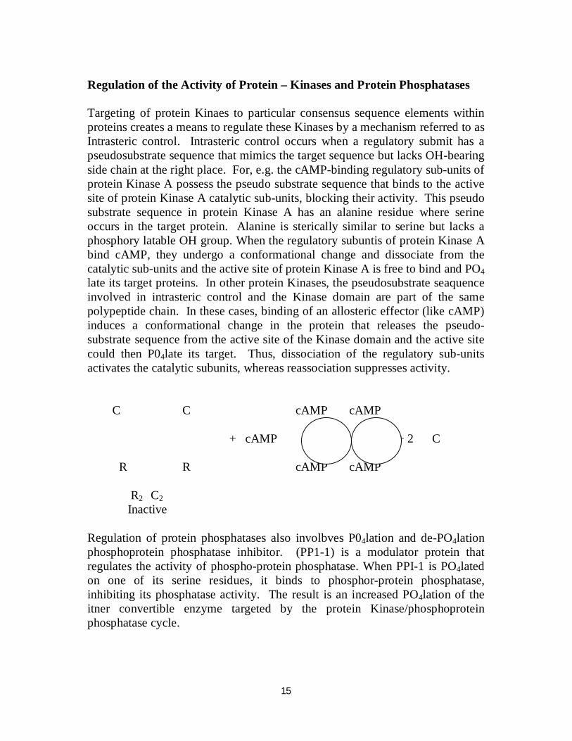
Regulation of the Activity of Protein – Kinases and Protein Phosphatases Targeting of protein Kinaes to particular consensus sequence elements within proteins creates a means to regulate these Kinases by a mechanism referred to as Intrasteric control. Intrasteric control occurs when a regulatory submit has a pseudosubstrate sequence that mimics the target sequence but lacks OH-bearing side chain at the right place. For, e.g. the cAMP-binding regulatory sub-units of protein Kinase A possess the pseudo substrate sequence that binds to the active site of protein Kinase A catalytic sub-units, blocking their activity. This pseudo substrate sequence in protein Kinase A has an alanine residue where serine occurs in the target protein. Alanine is sterically similar to serine but lacks a phosphory latable OH group. When the regulatory subuntis of protein Kinase A bind cAMP, they undergo a conformational change and dissociate from the catalytic sub-units and the active site of protein Kinase A is free to bind and PO4 late its target proteins. In other protein Kinases, the pseudosubstrate seaquence involved in intrasteric control and the Kinase domain are part of the same polypeptide chain. In these cases, binding of an allosteric effector (like cAMP) induces a conformational change in the protein that releases the pseudo-substrate sequence from the active site of the Kinase domain and the active site could then P04late its target. Thus, dissociation of the regulatory sub-units activates the catalytic subunits, whereas reassociation suppresses activity. C C cAMP cAMP + cAMP + 2 C R R cAMP cAMP R2 C2 Inactive Regulation of protein phosphatases also involbves P04lation and de-PO4lation phosphoprotein phosphatase inhibitor. (PP1-1) is a modulator protein that regulates the activity of phospho-protein phosphatase. When PPI-1 is PO4lated on one of its serine residues, it binds to phosphor-protein phosphatase, inhibiting its phosphatase activity. The result is an increased PO4lation of the itner convertible enzyme targeted by the protein Kinase/phosphoprotein phosphatase cycle.
16
Minor Covalent Modification of Enzyme Activity Adenylation An alternative mechanism of covalent modification by ATP involves adenylation transfer to an AMP adenylyl group from ATP to the enzyme with the accompanying formation of inorganic pyro PO4. While this is not known to occur in mammalian systems, adenylation is responsible for regulation of the glutamine synthelase and RNA polymerase in E. coli, e.g. glutamine synthetase.
ATP + glutamate + NH3 GS ADP + glutamine + P1 Glutamine synthetase is converted from its relatively active form to the less active form by the transfer of 12 mol of AMP from ATP to specific tyrosine residues in each of the 12 sub-units of the enzyme, to yield covalently adenylyl derivatives of the phenolic OH groups of the tyrosines. The enzyme may also be enzymatically de-adenylated to its active form. Importance of PO4lation/De-PO4 lation. A typical mammalian cell possesses thousands of PO4lated proteins and several hundred protein Kinases and protein phosphatases that catalyse their inter-conversion. The abundance of many protein Kinases in cells is an indication of the great importance of protein PO4lation in cellular regulation. Exactly 113 protein Kinase genes have been recognized in yeast while 868 putative protein Kinae genes have been identified in the human genome. The case of inter-conversion of enzymes between their phosphor- and dephospho-forms accounts for the frequency of PO4lation – dePO4-lation as a mechanism for the control of metabolic pathways. PO4lation-de P04lation permits the functional properties of the affected enzyme to be altered only for as long as it serves a specific need. Once the need has passed, the enzyme can be converted back to its original form, ready to respond to the next stimulatory event. Another factor underlying the widespread use of protein PO4lation-de-PO4lation lies in the chemical properties of the phosphoryl group itself. In order to alter an enzyme functional properties, any modification of its chemical structure must influence the protein’s 3-dimensional structure. The high charge density of protein-bound phosphoryl group – generally -2 at physiologic pH – and their propensity to form salt bridges with arginy residues make their potent agents for modifying protein structure and function PO4lation generally targets amino acids distant from the catalytic site itself. Consequent conformational changes then influence an enzyme’s intrinsic catalytic efficiency or other properties.
17
PO4lation and de-PO4lation provide for short-term readily reversible regulation of metabolite flow in response to specific physiologic signals. They are both under direct neural and hormonal control. PO4lation can also alter the location of an enzyme within the cell. It can alter the enzyme’s susceptibility to proteolytic degradation, or responsiveness to regulation by allosteric ligands. The third level at which metabolic regulation is exerted is through the genetic control of the rate of enzyme synthesis or degradation. The rate of a given metabolic sequence must depend on the conc. of the vetuse form of the enzyme in a sequence which in turn is the result of a balance between the rate of its synthesis and the rate of its breakdown. Body proteins are continuously synthesized and degraded – a process referred to as Protein Turnover. While the steady state concs of some enzymes and other proteins remain essentially, or constitutive, over time, the concs of many enzymes are influenced by a wide range of physiologic, hormonal or dietary factors. Control of Enzyme Synthesis. The amounts of enzyme synthesized by a cell are determined by transcription regulation if the gene encoding a particular enzyme protein is turned on or off, changes in the amount of enzyme activity soon follow. Induction – activation of enzyme synthesis and repression which is the shut-down of enzyme synthesis, are important mechanisms for the regulation of metabolism. By controlling the amount of an enzyme that is present at any moment, cells can either activate or terminate various metabolic routes. Inducible enzymes of humans include tryptophan pyrrolase, threonine dehydrase tyrosine--Ketoglutarate amino transferase, enzymes of the urea cycle, HMG-CoA reductase and cytochrome P450. Both induction and repression involve cis elements, specific DNA sequences located upstream of regulated genes (Operons) and trans-acting regulation proteins, e.g. Dietary Cholesterol decreases hepatic synthesis of cholesterol. This feedback regulation does not involve feedback inhibition, rather, HMG-CoA reductase, the rate-limiting enzyme of cholesterogenesis is affected. Regulation in response to dietary cholesterol involves curtailment by cholesterol or a cholesterol metabolite of the expression of the gene that encodes HMG-CoA reductase. Control of Enzyme Degradation Enzyme/protein degradation poses a real hazard to cellular processes. To control this hazard, protein degradation is compartmentalized, either in
18
macromolecular structures known as Proteasomes or in degradative organelles such as lysosomes. Protein degradation within lysosomes is largely non-selective, selection occurs during lysosomal uptake. Proteasomes are found in encaryotic as well as prokaryotic cells. Regulation of protein levels through degradation is an essential cellular mechanism. It is both rapid and irreversible. In many animals, many proteins are degraded by the ubiquitin-proteasome pathway. Eukaryotic cells contain 2 forms of proteasomes – the 20S proteasomes and its larger counterpart, the 26S proteasome. The eukaryotic 26S proteasome is made up of a 20S proteasome and 2 additional substructures known as 19S regulators (also called 19S caps or PA 700). Overall the 26S proteasomes has 2 copies each of 32 to 34 distinct sub-units, 14 in the 20S core and 18 to 20 in the cap structures. The 20S are contains 7 different kinds of -subunits and 7 different kinds of β-sub units. The sub-units are arranged in the form of a hollow cylinders. 19S 20S 19S The active sites of the proteolytic sub-units face the interior of the cylinder, thus preventing indiscriminate degradation of cellular proteins. The most common mechanism by which proteins are labeled for proteasome degradation is Ubiquitination – the covalent attachment/ligation of one or more ubiquitin molecules to the protein. Ubiquitin is a small, approximately 75 residue protein that is highly conserved among eukaryotes. Three additional proteins are involved in ubiquitination. They are E1, E2, and E3 ligases.
19
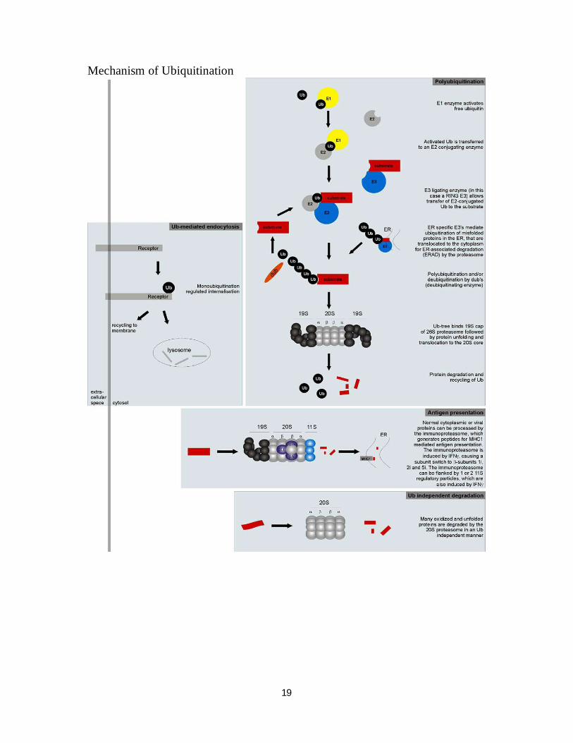
Mechanism of Ubiquitination
20
(1) E1 Ubiquitin – activating enzyme E1 (Ubiquiting-activating enzyme) becomes attached through a thioester bond to the C-terminal Glu residue of ubiquitin through an ATP-driven formation of an activated ubiquitin-adenylate intermediate. Ubiquitin is then transferred from E1 to an SH group on E2, the ubiquitin carrier protein, a family of at least 7 different small proteins, many of which are heat shock proteins. In protein degradation, E2-S-ubiquinin transfers ubiquitin to free amino groups on proteins selected by E3
, the ubiquitin-protein ligase. Upon binding a protein substrate,
21
F3 catalyses the transfer of ubiquitin from E2S-ubiquitin to free amino groups (usually Lys-E-NH2) on the protein. More than one ubiquitin may be attached to a protein substrate. Ubiquitin has 7 Lysine residues at positions 6, 11, 27, 29, 33, 48 and 63. E3 plays a central role in recognizing and selecting proteins for degradation. E3 selects proteins by the nature of the N-terminal amino acid. Proteins must have a free -amino acid terminus to be susceptible. Proteins having either Met, Ser, Ala, Thr, Val, Gly or Cys at the amino terminus are resistant to the ubiquitin-mediated degradation pathway. However, proteins, yaving Arg, Lys, His, Phe, Lyr, Try, Leu, Asn, Gln, Asp or Glu – N terminal are susceptible. Most proteins with susceptible N-terminal residues are not normal intra-cellular proteins but tend to be secreted proteins in which the susceptible residue has been exposed by action of a signal peptidase. The ubiquitin-proteasone pathway is thus responsible both for the regulated degradation of selected cellular proteins, e.g;. cyclins, in response to specific intra or extra-cellular signals and for the removal of defective or aberrant protein species. The key to the versatility and selectivity of the ubiquitin proteasome system resides in both the variety of intra-cellular E3 ligases and their ability to discriminate between different physical or conformational states of a target protein. Thus, the ubiquitin – proteasome pathway can selectively degrade proteins whose physical integrity and functional competency has been compromised by the loss of or damage to a prosthetic group, oxidation of systems or histidine residues, or deamidation of asparagines or glutamine residues. Recognition by proteolytic enzymes also can be regulated by covalent modifications such as PO4lation, binding of substrates or allosteric effectors or association with membranes oligonucleotides or other proteins. A growing body of evidence suggests that dysanctions of the ubiquitin-proteasome pathway contribute to the accumulation of aberrantly folded protein species xteristic of several neurodegenerative diseases.
(4) Metabolic regulation can also be effected by hormones. Hormones are chemical messengers elaborated by endocrine glands that pass via the blood to certain target tissues where they stimulate or inhibit specific metabolic activities. For instance, both insulin and glucagon regulate the metabolism of glucose.
Other ways of Regulating Metabolic pathways
(1) Covalent activation of zymogens. Most proteins become fully active when their synthesis is completed and they spontaneously fold into their nature, 3-dimensional conformation/structures. Some proteins, however, are synthesized and secreted as in active precursors
22
known as Proproteins. The proteins of enzymes are termed Proenzymes or Zymogens. They acquire full activity only upon specific proteolytic cleavage of one or several of their peptide bonds. Unlike allosteric regulation or covalent modification, zymogen activation is an irreversible process. Activation of enzymes and other physiologically proteins by specific proteolysis is a strategy frequently exploited by biological systems to switch on processes at the appro-priate time and place. Proteins synthesized as proproteins include the hormone insulin, the digestive enzymes pepsintrypsin and chymotrypsin, several factors of the blood clatting and blood clot dissolution cascades and the connective tissue protein collagen Origin Zymogen Active Protein Pancreas Trypsinogen Trypsin Pancreas Chymotrypsinogen Chymo trypsin Pancreas Procarboxypeptidase Carboxypeptidase Pancreas Proelestase Elastase Stomach Pepsinogen Pepsin Pnansulin Insulin Procollagen Collagen Insulin – Secreted as Proinsulin – 86 residue. Proteolytic removal of residues 31 to 65 yields insulin. Proteolytic enzymes of the digestive tracts. Enzymes of the digestive tract that hydrolyse dietary proteins are synthesized in the stomach and pancreas as zymogens. When these enzymes are secreted into the GIT, they are coverted into their active forms by the selective hydrolytic cleavage of one or more specific peptide bonds in the zymogen molecule. pepsin Pepsiinogen ------------------- pepsin + peptides free pepsin at low pH removes H+
42 amino acid residues as a mixture of peptides from the N-terminal end of pepsinogen. Entero Trypsinogen --------------- Trypsin + hexapeptide. Entero Kinase converts Kinase Trypsinogen to trypsin by removal of a hexapeptide from the N-terminal end. The activation of chymotrypsin however reps. a peculiar and interesting example. Chymotrypsinogen is a 245 residue polypeptide chain X-linked by 5 disulphide bonds.
23
Chymotrypsinogen is converted to an enzymatically active form called II-Chymotrypsin when trypsin cleaves the peptide bond joining Arg15 and Ile16. The enzymatically active II-Chymotrypsin acts upon other II – Chymotrysin molecules, excising 2 dipeptides; Ser14 Arg15 and Thr14 Asn148. The end-product of this series of proteolysis is the nature protease -Chymotrypsin, in which the 3 peptide chains, A (residues 1-13) B (residues 16-146), and C (residues 149 – 245), remain together because they are linked by 2 disulphide bonds one from A to B and one from B to C. It is interesting to note that the coversion of inactive chymotrypsinogen to active II-Chymotrypsin requires the cleavage of just one
24
particular peptide bond. The Zymogens are kept from exerting proteolytic activity on intracellular proteins so long as they remain within the cells in which they are made. They are turned on to generate the active form only after they are secreted into the GIT. As mentioned earlier, this type of covalent regulation is one-way: there are no known enzymatic reactions which can transform these 3-enzymes back into their respective zymogens. Blood Clotting The formation of blood clots is the result of a series of zymogen activations. The amplification achieved by this cascade of enzymatic activations allows blood clotting to occur rapidly in response to injury.
25
Thrombin cleaves peptides rich in –ve charge from fibrinogen, converting II to fibrin. Fibrin aggregates into ordered fibrous arrays that are subsequently stabilized by covalent X-links. Thrombin specifically cleaves Arg-Gly peptide bonds. 2. Isozymes Anlther type of regulation of metabolic activity is through the participation of isozymes. Isozymes are multiple forms of the same enzyme that occur in a single species of organism or even in a single cell. A classic, e.g. is mammalian lactate DH, which exists as 5 different isozymes in the tissues of rat and other vertebrates. They all catalyse the same overall reaction. Lactate + NAD + Pyruvate + NADA + HT
(1) All 5 isozymes have the same mut, about 134,000
(2) All contain 4 polypeptide chains eachof mut 33,500
(3) The 5 isozymes consist of 5 different combinations of 2 different kinds of polypeptide chains designated A and B. The isozyme predominating in skeletal muscle has 4 identical A chains and is designated A4. Another which predominates in heart has 4 identical B chains and is designated B4. The other 3 isozomes have the composition A3B,A2B2 and AB3.
(4) Although tye all catalyse the same reaction, they differe in their dependence on substrate conc., particularly pyruvate, as well as their Vimax values when pyruvate is the substrate. The isozyme A4, xteristic of skeletal muscle and embryonic tissues, reduces pyruvate to lactate at a relatively high rate. The B4 isozyme on the other hand, Xteristic of the heart and other red muscles, reduces pyruvate at a relatively low rate. Mroeover, the dehydrogen nation of lactate catalysed by the B4 isozyme is strongly inhibited by pyruvate. The other LDH isozymes have kinetic properties intermediate between those of the A4 and B4 isozymes in proportion to their relative content of A and B chains.
(5) If se then compare these kinetic xteristics with the metabolic features of the tissues in which the A4 and B4 isozymes predominate, the function of LDH isozymes becomes clear. Skeletal muscle and embryonic tissue have anaerobic metabolism, thus can convert glucose to lactate via glycolysis. A4 isozyme is thus adapted for this role and has a high Vmax for pyruvate. The heart muscle on the other hand has aerobic metabolism
26
and does not form lactate from glucose. Rather, it oxidizes pyruvate to C02 without intermediate formation of lactate. The reasons are not also far fetched. Heart muscle cells are rich in mitochondria whereas most skeletal muscles contain relatively few mitochondrin. Free fatty acids carried by serum albumen from the adipose tissue are the major fuel of the heart. The heart uses glycolysis as a source of extra energy.