Embed Size (px)
Citation preview
Contents lists available at ScienceDirect
BBA - General Subjects
journal homepage: www.elsevier.com/locate/bbagen
Plasma N-glycome composition associates with chronic low back pain
Irena Trbojević-Akmačića, Frano Vučkovića, Marija Vilaja, Andrea Skelina,k, Lennart C. Karssenb,Jasminka Krištića, Julija Jurića, Ana Momčilovića,c, Jelena Šimunovića, Massimo Manginod,e,Manuela De Gregorif,g, Maurizio Marchesinig,h, Concetta Dagostinog,i, Jerko Štambuka,Mislav Novokmeta, Richard Rauckj, Yurii S. Aulchenkob, Dragan Primorack,l,m,n,o,Leonardo Kapuralj, Klaas Buysep, Dieter Mesottenp, Frances M.K. Williamsd, Jan van Zundertp,Massimo Allegrig,q, Gordan Lauca,r,⁎
aGenos Glycoscience Research Laboratory, Zagreb, Croatiab PolyOmica, Het Vlaggeschip 61, 5237 PA ‘s-Hertogenbosch, The Netherlandsc The Glycomics and Glycoproteomics Group, Center for Proteomics and Metabolomics, Leiden University Medical Center, The Netherlandsd Department of Twin Research and Genetic Epidemiology, King's College London, London, UKeNIHR Biomedical Research Centre at Guy's and St Thomas' Foundation Trust, London SE1 9RT, UKf Pain Therapy Service, Fondazione IRCCS Policlinico San Matteo, Pavia, Italyg SIMPAR Group, ItalyhAnesthesia, Intensive Care and Pain Service, Parma Hospital, Parma, Italyi Department of Medicine and Surgery, University of Parma, Parma, Italyj Carolinas Pain Institute, Winston Salem, NC, USAk St. Catherine Specialty Hospital, Zabok/Zagreb, Croatial J J Strossmayer University of Osijek, School of Medicine, Osijek, CroatiamUniversity of Split, School of Medicine, Split, Croatian Eberly College of Science, The Pennsylvania State University, University Park, USAo Children's Hospital Srebrnjak, Zagreb, Croatiap Department of Anesthesiology and Multidisciplinary Pain Center, Ziekenhuis Oost-Limburg, Genk, BelgiumqAnesthesia and Intensive Care Service - IRCCS MultiMedica Hospital, Sesto San Giovanni, Milano, ItalyrUniversity of Zagreb, Faculty of Pharmacy and Biochemistry, Zagreb, Croatia
A R T I C L E I N F O
Keywords:Glycan biomarkerLow back painPlasma N-glycosylation
A B S T R A C T
Background: Low back pain (LBP) is the symptom of a group of syndromes with heterogeneous underlyingmechanisms and molecular pathologies, making treatment selection and patient prognosis very challenging.Moreover, symptoms and prognosis of LBP are influenced by age, gender, occupation, habits, and psychologicalfactors. LBP may be characterized by an underlying inflammatory process. Previous studies indicated a
https://doi.org/10.1016/j.bbagen.2018.07.003Received 20 March 2018; Received in revised form 3 July 2018; Accepted 4 July 2018
Abbreviations: % Area, percentage of total integrated area of the chromatogram; 2-AB, 2-aminobenzamide; 2-PB, 2-picoline borane; AbSur, cohort of patients withacute inflammation that had abdominal surgery; ACE, automatic chromatogram extraction; ADCC, antibody-dependent cell-mediated cytotoxicity; ANCOVA, analysisof covariance; B, N-glycans that contain bisecting N-acetylglucosamine; BEL, samples from clinical centers in Belgium; CLBP, chronic low back pain; CRO, samplesfrom clinical centers in Croatia; DMSO, dimethyl sulfoxide; FA, formic acid; FDR, false discovery rate; G0, agalactosylated N-glycans; G1, monogalactosylated N-glycans; G2, digalactosylated N-glycans; G3, trigalactosylated N-glycans; G4, tetragalactosylated N-glycans; GlcNAc, N-acetylglucosamine; GPs, glycan peaks; GU,glucose unit; HB, highly branched N-glycans; HILIC-SPE, hydrophilic interaction liquid chromatography solid phase extraction; HILIC-UPLC, hydrophilic interactionultra-performance liquid chromatography; HM, high-mannose N-glycans; IgG, immunoglobulin G; ITA, samples from clinical centers in Italy; LB, low branched N-glycans; LBP, low back pain; LDD, lumbar disc degeneration; META, meta-analysis; MS, mass spectrometry; PBS, phosphate buffered saline; S0, neutral/not sialylatedN-glycans; S1, monosialylated N-glycans; S2, disialylated N-glycans; S3, trisialylated N-glycans; S4, tetrasialylated N-glycans; SDS, sodium dodecyl sulfate; UPLC,ultra-performance liquid chromatography⁎ Corresponding author at: Faculty of Pharmacy and Biochemistry, University of Zagreb, A. Kovačića 1, 10 000 Zagreb, Croatia.E-mail addresses: [email protected] (I. Trbojević-Akmačić), [email protected] (F. Vučković), [email protected] (M. Vilaj), [email protected] (A. Skelin),
[email protected] (L.C. Karssen), [email protected] (J. Krištić), [email protected] (J. Jurić), [email protected] (A. Momčilović),[email protected] (J. Šimunović), [email protected] (M. Mangino), [email protected] (M. De Gregori),[email protected] (M. Marchesini), [email protected] (C. Dagostino), [email protected] (J. Štambuk),[email protected] (M. Novokmet), [email protected] (R. Rauck), [email protected] (Y.S. Aulchenko),[email protected] (D. Primorac), [email protected] (L. Kapural), [email protected] (K. Buyse), [email protected] (D. Mesotten),[email protected] (F.M.K. Williams), [email protected] (J. van Zundert), [email protected] (M. Allegri), [email protected] (G. Lauc).
BBA - General Subjects 1862 (2018) 2124–2133
Available online 05 July 20180304-4165/ © 2018 Elsevier B.V. All rights reserved.
T

Retrospective study connection between inflammatory response and total plasma N-glycosylation. We wanted to identify potentialchanges in total plasma N-glycosylation pattern connected with chronic low back pain (CLBP), which could givean insight into the pathogenic mechanisms of the disease.Methods: Plasma samples of 1128 CLBP patients and 760 healthy controls were collected in clinical centers inItaly, Belgium and Croatia and used for N-glycosylation profiling by hydrophilic interaction ultra-performanceliquid chromatography (HILIC-UPLC) after N-glycans release, fluorescent labeling and clean-up. Observed N-glycosylation profiles have been compared with a cohort of 126 patients with acute inflammation that under-went abdominal surgery.Results: We have found a statistically significant increase in the relative amount of high-branched (tri-antennaryand tetra-antennary) N-glycan structures on CLBP patients' plasma glycoproteins compared to healthy controls.Furthermore, relative amounts of disialylated and trisialylated glycan structures were increased, while high-mannose and glycans containing bisecting N-acetylglucosamine decreased in CLBP.Conclusions: Observed changes in CLBP on the plasma N-glycome level are consistent with N-glycosylationchanges usually seen in chronic inflammation.General significance: To our knowledge, this is a first large clinical study on CLBP patients and plasma N-glycomeproviding a new glycomics perspective on potential disease pathology.
1. Introduction
Low back pain (LBP) includes several mixed pain syndromes beingone of the most common health problems in the world. Almost everyadult has or will experience an episode of LBP and it is the number onecause of years lived with disability [1]. Prevalence of several frequentlyoverlapping sources of LBP includes sacroilitis 25–32% [2,3], lumbalspondylosis 36–42% [4–6], spinal stenosis, or lumbar radiculitis/radi-culopathy up to 30% [7,8]. When muscle tension, stiffness or pain arelocalized above the inferior gluteal folds and below the coastal margin,with or without leg pain, and last for three months or more, the con-dition is classified as chronic low back pain (CLBP). This is a group ofsyndromes with heterogeneous variable underlying mechanisms andmolecular pathologies that correlate poorly with spine imaging. Ad-ditionally, symptoms and prognosis are influenced by many factors(gender [9], age [10], occupation, habits [11]), as well as physiological[12] and psychological factors [13,14], making managing the conditionvery challenging. The success of interventional techniques, such asepidural steroid injection, facet joints denervation, spinal cord stimu-lation or laminectomy, depends largely on the specific physiopathologyof each patient. However, currently there is little evidence based ap-proach to this disease, as biomarkers and prognostic factors practicallydo not exist.
In complex organisms, glycans (sugar moieties covalently attachedto proteins) play an important role in virtually all processes that involvemore than one cell [15]. Nearly all membrane and secreted proteins aremodified by glycans with variable site occupancy and glycan compo-sition [16,17]. Absence of N-glycosylation is embryonically lethal [18]and mutations that obstruct proper glycosylation cause debilitatingdiseases [19]. Inter-individual differences in N-glycosylation are asso-ciated with predisposition for and course of different diseases [20]. Thisis not surprising since the glycan parts of (glyco)proteins are integralelements of the final molecular structure, and together with aminoacids in the polypeptide backbone, form a single molecular entity thatperforms biological functions. For example, glycans attached to im-munoglobulin G (IgG) have very profound effects on protein structureand can convert IgG from a pro-inflammatory to an anti-inflammatory
mode [21]. Alternative glycosylation (attachment of different glycans)affects binding of IgG to all Fc receptors and is in this way analogous tovariation in protein sequence due to genetic variations [22].
Systemic and/or local inflammatory processes are believed to be anunderlying mechanism in at least a subgroup of LBP patients, althoughthis is not generally recognized. Previous studies indicated that in-flammatory response associates with changes in total plasma N-glyco-sylation [23], as well as N-glycosylation of individual serum glyco-proteins [24]. Since glycans affect not only protein structure, but alsoits function, a number of studies have shown that changes in glycosy-lation are implicated in the pathology of different diseases [25–28].
Glycosylation of individual glycoprotein, IgG, has already beenstudied in the context of LBP in a large study of 4511 twins from theTwinsUK database [29]. IgG glycans with core fucose (have lower an-tibody-dependent cell-mediated cytotoxicity, ADCC) and IgG glycanswithout core fucose (have higher ADCC activity) have been found tocorrelate with LBP, suggesting the involvement of ADCC and in-flammation in LBP pathogenesis. However, there was no correlationfound between lumbar disc degeneration (LDD) scores and IgG glycans,implicating different pathogenic mechanisms for specific LBP subtypesand supporting the role of glycans in inflammation-related LBP sub-types.
Considering previous research on inflammatory response associa-tion with changes in total plasma N-glycosylation [23], here, we stu-died the total plasma N-glycome in CLBP patients and controls in orderto identify if there is an association between the N-glycosylation patternon the level of total plasma glycoproteins and risk of CLBP condition.Specific pattern of plasma N-glycosylation could be used as a biomarkerof CLBP, and potential indicator of both pathogenic mechanisms andtherapy efficiency in future prospective and longitudinal studies.
2. Methods
2.1. Study approval
The study was approved by the ethical committees of the partici-pating clinical centres between December 2013 and March 2014 and
Table 1Information about participants enrolled in a chronic low back pain study.
ITA BEL CROCase Control Case Control Case Control
Sample number 375 379 567 191 186 190Age (med/IQR) 67 (51–76) 68 (50–77) 56 (49–67) 56 (48–65) 55 (48–65) 56 (41–65)Sex (F) (n/%) 201 (53.6%) 205 (54.1%) 309 (54.5%) 108 (56.5%) 120 (64.5%) 79 (41.6%)
Plasma N-glycosylation analysis has been performed for participants from three clinical centers in Italy (ITA), Belgium (BEL) and Croatia (CRO). Case - chronic lowback pain patients, Control - healthy individuals, med - median value, IQR - interquartile range.
I. Trbojević-Akmačić et al. BBA - General Subjects 1862 (2018) 2124–2133
2125
written informed consent was obtained from each participant prior toinclusion in the study. Participant samples have been de-identified bycode. The study has been registered on clinicaltrials.gov(NCT02037789).
2.2. Clinical samples and data
Retrospective plasma samples of CLBP patients and healthy controls(people without acute or CLBP) were collected in several clinical cen-ters in Italy, Belgium and Croatia that are involved in PainOmics pro-ject, following previously established and validated protocol for bloodsample collection, storage and shipping [30,31] (Table 1). In short,blood for glycomics analysis has been collected into EDTA containingvacutainer tube and mixed well for balanced clotting. Tube was left atroom temperature for 1 h and plasma was separated by centrifugationat 1620g for 10min, transferred to a clean tube and centrifuged again at2700g for 10min. Volume of 1mL of plasma after second centrifugationstep has been transferred to a clean tube and stored at −80 °C or−20 °C before further processing. CLBP patients were defined ac-cording to previously published criteria [31]: all Caucasian adult pa-tients, referred to participant pain clinics, with pain localized to thecolumn between the costal margins and gluteal fold, with or withoutsymptoms into one or both legs, that was lasting>12weeks. We ex-cluded all patients with an acute episode of LBP, any history of spinetumor or infection or recent (< 12months) vertebral fractures, orpsychiatric disorders. All patients have been classified into 6 majorgroups: spinal stenosis, discogenic pain, facet joint pain, sacroiliac jointpain, low back pain associated to radicular pain (radicular pain notpredominant) and wide-spread LBP. Furthermore, all patients' paincharacteristics (intensity and type of pain) have been evaluated withpainDETECT [32].
Plasma samples from cohort of patients with acute inflammationthat underwent abdominal surgery (AbSur) were collected in severalclinical centers in Italy as described earlier [33,34]. Blood samples of126 patients have been taken in several time points - intraoperativelyand 6, 12, 24, and 48 h aſter the surgery. Samples taken in-traoperatively and 48 h after abdominal surgery have been consideredas “Case” or “Control”, respectively. In short, blood for glycomicsanalysis has been collected into EDTA containing vacutainer tube,mixed well for balanced clotting and plasma was separated by cen-trifugation at room temperature. Samples have been stored at −20 °Cbefore further processing.
2.3. Deglycosylation of plasma samples
All plasma samples and in-house plasma standards were vortexedafter thawing and centrifuged for 3min at 12,100 g. Each sample(10 μL) was aliquoted to 1mL 96-well collection plates (Waters,Milford, MA, USA) following a predetermined experimental design,which was blocked on case-control status, sex and age information(each 96-well plate represents the whole population in terms of case-control status, sex and age distribution), while the rest was randomized.In-house plasma standards were aliquoted in four to five replicates perplate, to control for batch effects, that is plate-to-plate variation.
Plasma proteins were denatured by adding 20 μL of 2% (w/v) so-dium dodecyl sulfate (SDS, Invitrogen, Carlsbad, CA, USA) and sampleincubation at 65 °C for 10min. After cooling to room temperature for30min, 10 μL of 4% (v/v) Igepal CA-630 (Sigma-Aldrich, St. Louis, MO,USA) was added to each sample and the mixture was shaken for 15minat room temperature on a plate shaker. Plasma proteins were degly-cosylated by incubation at 37 °C for 18 h after 1.2 U of PNGase F(10 U μL−1, Promega, Madison, WI, USA) in 10 μL of 5× PBS wasadded to each sample. Buffer 5× phosphate buffered saline (PBS) wasmade in house: 685mmol L−1 NaCl (Carlo Erba, Peypin, Italy),13.5 mmol L−1 Na2HPO4 (Acros Organics, Thermo Fisher Scientific,Geel, Belgium), 48.5 mmol L−1 KH2PO4 (Sigma-Aldrich), 11mmol L−1
KCl (Gram-Mol, Zagreb, Croatia), and filtered through 0.2 μm SuporPES filters (Nalgene Thermo Fischer Scientific).
2.4. Fluorescent labeling and clean-up of released plasma N-glycans
Released plasma N-glycans were subsequently labeled with afluorescent dye 2-aminobenzamide (2-AB, Sigma-Aldrich). The labelingmixture was freshly prepared by dissolving 0.48mg of 2-AB and1.12mg of 2-picoline borane (2-PB, Sigma-Aldrich) in 25 μL of dimethylsulfoxide (DMSO, Sigma-Aldrich) and glacial acetic acid (Merck,Darmstadt, Germany) (7:3, v/v), per sample. Labeling mixture wasadded to each sample and the plate was sealed with an adhesive seal.Samples were mixed for 10min at room temperature, and incubated at65 °C for 2 h for the labeling reaction to take place.
Released and fluorescently labeled plasma N-glycans were cleanedof excess of reagents and proteins by previously developed and opti-mized hydrophilic interaction liquid chromatography solid phase ex-traction (HILIC-SPE) procedure [35]. After labeling, reaction sampleswere cooled down to room temperature for 30min and 700 μL of cold(4 °C) acetonitrile (Sigma-Aldrich) was added to each sample and gentlymixed. The clean-up procedure was performed on a hydrophilic 0.2 μmAcroPrep GHP filter plate (Pall Corporation, Ann Arbor, MI, USA) usinga vacuum manifold (Pall Corporation) at around 25mmHg. All wells ofa GHP filter plate were prewashed with 200 μL of 70% ethanol in water,200 μL of ultra-pure water and 200 μL of 96% cold (4 °C) acetonitrile inwater. The samples diluted with cold acetonitrile were loaded to thewells, incubated on a filter for 2min and subsequently washed with 5×200 μL of 96% cold (4 °C) acetonitrile in water. Glycans were elutedfrom a filter plate with 2× 90 μL of ultra-pure water after 15min ofshaking at room temperature and centrifugation at 164 g in each step.Combined eluates were stored at −20 °C until the UPLC analysis.
2.5. Plasma N-glycan analysis by HILIC-UPLC
Fluorescently labeled and purified plasma N-glycans were preparedin 75% acetonitrile (v/v) and analyzed by HILIC-UPLC on an AcquityUPLC instrument (Waters) consisting of a quaternary solvent manager,sample manager and a fluorescence detector. The 2-AB labeled N-gly-cans were separated on a Waters BEH Glycan chromatography column,150× 2.1mm i.d., 1.7 μm BEH particles, in a linear gradient of 30–47%100mmol L−1 ammonium formate, pH 4.4 (solvent A) and acetonitrile(solvent B) at flow rate of 0.56mLmin−1 and a 23min analytical run.Samples were maintained at 10 °C before injection into the column, thecolumn temperature was 25 °C and separated glycans were detected atexcitation wavelength of 250 nm and emission wavelength of 428 nm.The UPLC system was under the control of the Empower 3 software,build 3471 (Waters). Chromatograms were processed using an auto-matic processing method and manually corrected to maintain the sameintervals of integration across all samples, or processed with a recentlydeveloped semi-supervised Automatic Chromatogram Extraction (ACE)method [36] for automated alignment and detection of glycan peaks inchromatograms. The plasma N-glycan samples were all separated into39 glycan peaks (GPs) and the amount of glycans in each chromato-graphic peak was expressed as percentage of total integrated area (%Area). To each detected chromatographic peak a glucose unit (GU)value was assigned according to external standard of hydrolysed and 2-AB labeled glucose oligomers (dextran ladder) which was run withevery analytical UPLC batch [37].
2.5.1. Confirmation of the HILIC-UPLC plasma glycome profileThe major glycan present in each of the reported 39 chromato-
graphic peaks was annotated according to GU values, measured m/zvalue, recorded fragmentation spectra where applicable and accordingto previously published and annotated serum N-glycome profile [38](Supplementary Table 1). Several representative samples from controland patient groups were selected and analaysed by HILIC-UPLC, as
I. Trbojević-Akmačić et al. BBA - General Subjects 1862 (2018) 2124–2133
2126
described in previous section, additionally hyphenated to the Brukercompact Q-Tof mass spectrometer (MS), both controlled using HyStarsoftware (Bruker, Bremen, Germany) version 4.1. UPLC was coupled toMS via Ion Booster (Bruker) ion source with aditional flow of100 μLmin−1 of acetonitrile introduced into analytical flow using a t-piece. Capillary voltage was set to 2250 V with nebulizing gas atpressure of 5.5 Bar. Drying gas was applied to source at a flow rate of4 Lmin−1 and temperature of 100 °C, while vaporiser temperature wasset to 220 °C and flow rate of 5 Lmin−1. Nitrogen was used as a sourcegas, while argon was used as collision gas. Ion energy was set to 5 eV,transfer time was 100 μs. Spectra were recorded in mass range from50m/z to 4000m/z at a rate of 0.5 Hz. For MS acquisition collisionenergy was set to 4 eV and Rf 4200 Vpp. Fragment spectra were ac-quired using auto MS/MS mode using a precursor ion list of detected m/z values corresponding to free 2-AB labeled plasma glycans (Supple-mentary Table 1). Active exclusion was set to 1min after three spectraand number of precursor ions was 1. All recorded spectra were analyzedusing Bruker Data Analysis software version 4.4 (Bruker). Glycancompositions and certain structural features were determined usingGlycoMod [39] and GlycoWorkbench [40].
2.6. Statistics
In order to remove experimental variation from the measurements,normalization and batch correction were performed on the UPLC glycandata. To make measurements across samples comparable, normal-ization by total area was performed where the peak area of each of the39 glycan containing peaks was divided by total area of the corre-sponding chromatogram. Prior to batch correction, normalized glycanmeasurements were log-transformed due to right-skewness of theirdistributions and the multiplicative nature of batch effects. Batch cor-rection was performed on log-transformed measurements using theComBat method (R package “svav3.6.0”) [41], where the technicalsource of variation (which sample was analyzed on which plate) wasmodeled as batch covariate. To correct the measurements for experi-mental noise, estimated batch effects were subtracted from log-trans-formed measurements. In addition to the 39 directly measured glycancontaining peaks, 14 derived traits were calculated from the directlymeasured glycans (Table 2). These derived traits average particularglycosylation features across different individual glycan structures andconsequently they are more closely related to individual enzymaticactivities and underlying genetic polymorphisms.
Association analyses between disease status and glycan traits wereperformed using an analysis of covariance (ANCOVA) model with ageand gender included as covariates. Analyses were first performed foreach cohort separately and then combined using inverse-varianceweighted meta-analysis (META, R package “metaphor v1.9-4”) [42].Prior to the analyses, the glycan variables were all transformed to thestandard Normal distribution (mean=0, sd= 1) by inverse transfor-mation of ranks to Normality (R package “GenABELv1.8-0”, functionrntransform) [43]. Using rank transformed variables in analyses makesestimated effects of different glycans in different cohorts comparable astransformed glycan variables have the same standardized variance. Incase-control regression analysis, coefficients of binary predictors referto a change in a glycan variable between 2 classes of binary predictorsexpressed in SDs. The false discovery rate (FDR) was controlled usingthe Benjamini-Hochberg procedure, and p-values corrected for multipletesting (with FDR cutoff set at 0.05) are shown throughout.
Data was analyzed and visualized using R programming language(version 3.0.1).
3. Results
3.1. Glycosylation changes in three cohorts
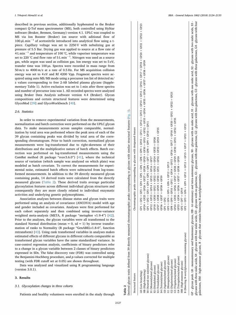
Patients and healthy volunteers were enrolled in the study through Table2
Derived
glycan
traits
define
dfrom
39directly
measuredch
romatog
raph
icpe
aksco
rrespo
ndingto
glycan
swithsimila
rstructural
features
(Fig.1
).
Structural
feature
Chrom
atog
raph
icpe
aksco
rrespo
ndingto
glycan
swithde
sign
ated
feature
LB(Low
-branc
hing
)GP1
+GP2
+GP3
+GP4
+GP5
+GP6
+GP8
+GP9
+GP1
0+
GP1
1+
GP1
2+
GP1
3+
GP1
4+
GP1
5+
GP1
6+
GP1
7+
GP1
8+
GP2
0+
GP2
1+
GP2
2+
GP2
3HB(H
igh-bran
ching)
GP2
4+
GP2
5+
GP2
6+
GP2
7+
GP2
8+
GP2
9+
GP3
0+
GP3
1+
GP3
2+
GP3
3+
GP3
4+
GP3
5+
GP3
6+
GP3
7+
GP3
8+
GP3
9S0
(Neu
tral/n
otsialylated
glycan
s)GP1
+GP2
+GP3
+GP4
+GP5
+GP6
+GP7
+GP8
+GP9
+GP1
0+
GP1
1S1
(Mon
osialylatedglycan
s)GP1
2+
GP1
3+
GP1
4+
GP1
5+
GP1
6+
GP1
7S2
(Disialylatedglycan
s)GP1
8+
GP2
0+
GP2
1+
GP2
2+
GP2
3+
GP2
4+
GP2
5+
GP2
6S3
(Trisialylated
glycan
s)GP2
7+
GP2
8+
GP2
9+
GP3
0+
GP3
1+
GP3
2+
GP3
3+
GP3
4+
GP3
5S4
(Tetrasialylated
glycan
s)GP3
6+
GP3
7+
GP3
8+
GP3
9G0(A
galactosylated
glycan
s)GP1
+GP2
G1(M
onog
alactosylatedglycan
s)GP3
+GP4
+GP5
+GP6
+GP1
2+
GP1
3G2(D
igalactosylatedglycan
s)GP8
+GP9
+GP1
0+
GP1
1+
GP1
4+
GP1
5+
GP1
6+
GP1
7+
GP1
8+
GP2
0+
GP2
1+
GP2
2+
GP2
3G3(Triga
lactosylated
glycan
s)GP2
4+
GP2
5+
GP2
6+
GP2
7+
GP2
8+
GP2
9+
GP3
0+
GP3
1+
GP3
2+
GP3
5G4(Tetraga
lactosylated
glycan
s)GP3
3+
GP3
4+
GP3
6+
GP3
7+
GP3
8+
GP3
9HM
(High-man
nose
glycan
s)GP2
+GP7
+GP1
9B(Bisecting
N-acetylgluco
samineco
ntaining
glycan
s)GP2
+GP3
+GP6
+GP9
+GP1
1+
GP1
2+
GP1
5+
GP1
7+
GP2
1+
GP2
3
GP-glycan
peak
.LB-mon
oanten
nary
andbian
tenn
aryglycan
s,HB-tri-anten
nary
andtetra-an
tenn
aryglycan
s,S0
-glycan
swithno
sialic
acid,S
1-glycan
swithon
esialic
acid,S
2-g
lycans
withtw
osialic
acids,S3
-glycan
swiththreesialic
acids,S4
-glycans
withfour
sialic
acids,G0-g
lycans
withno
galactose,
G1-g
lycans
withon
ega
lactose,
G2-g
lycans
withtw
oga
lactoses,G
3-g
lycans
withthreega
lactoses,G
4-g
lycans
withfour
galactoses,HM
-high
-man
nose
glycan
s,B-glycan
sthat
containbisectingN-acetylgluco
samine.
I. Trbojević-Akmačić et al. BBA - General Subjects 1862 (2018) 2124–2133
2127
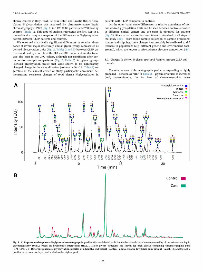
clinical centers in Italy (ITA), Belgium (BEL) and Croatia (CRO). Totalplasma N-glycosylation was analyzed by ultra-performance liquidchromatography (UPLC) (Fig. 1) in 1128 CLBP patients and 760 healthycontrols (Table 1). This type of analysis represents the first step in abiomarker discovery – a snapshot of the differences in N-glycosylationpattern between CLBP patients and controls.
We observed statistically significant differences in relative abun-dance of several major structuraly similar glycan groups represented asderived glycosylation traits (Fig. 2, Tables 2 and 3) between CLBP pa-tients and healthy controls of the ITA and BEL cohorts. A similar trendwas also seen in the CRO cohort, although not significant after cor-rection for multiple comparisons. (Fig. 2, Table 3). All glycan groups(derived glycosylation traits) that were shown to be significantlychanged change in the same direction (column “effect” in Table 3) re-gardless of the clinical center of study participants' enrolment, de-monstrating consistent changes of total plasma N-glycosylation in
patients with CLBP compared to controls.On the other hand, some differences in relative abundance of sev-
eral derived glycosylation traits can be seen between controls enrolledin different clinical centers and the same is observed for patients(Fig. 2). Since extreme care has been taken to standardize all steps ofthe study [30] – from blood sample collection to sample processing,storage and shipping, these changes can probably be attributed to dif-ferences in populations (e.g. different genetic and environment back-ground), which are known to affect plasma glycome composition [44].
3.2. Changes in derived N-glycan structural features between CLBP andcontrols
The relative area of chromatographic peaks corresponding to highlybranched – denoted as “HB” in Table 3 – glycan structures is increased(and, concomitantly, the % Area of chromatographic peaks
Fig. 1. A) Representative plasma N-glycans chromatographic profile. Glycans labeled with 2-aminobenzamide have been separated by ultra-performance liquidchromatography (UPLC) based on hydrophilic interactions (HILIC). Major glycan structures are shown for each glycan containing chromatographic peak(GP1–GP39). B) Different plasma N-glycosylation profiles of a healthy individual (Control) and a chronic low back pain patient (Case). Chromatographicprofiles have been overlayed and scaled to the highest peak.
I. Trbojević-Akmačić et al. BBA - General Subjects 1862 (2018) 2124–2133
2128
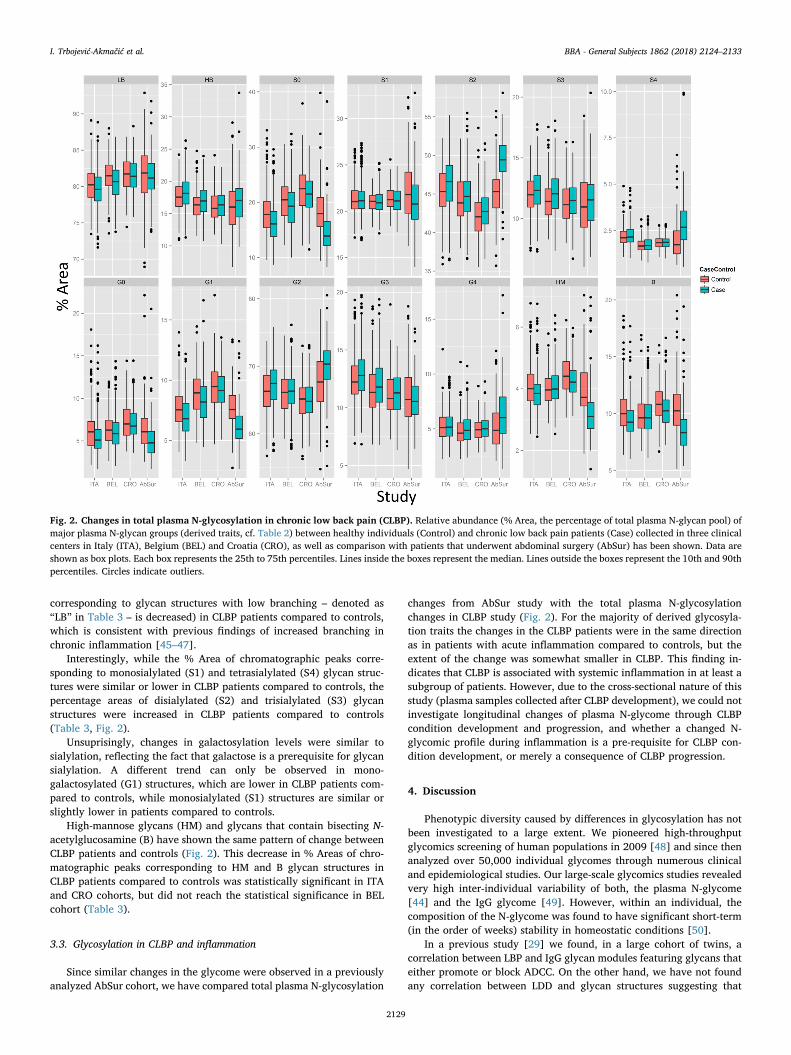
corresponding to glycan structures with low branching – denoted as“LB” in Table 3 – is decreased) in CLBP patients compared to controls,which is consistent with previous findings of increased branching inchronic inflammation [45–47].
Interestingly, while the % Area of chromatographic peaks corre-sponding to monosialylated (S1) and tetrasialylated (S4) glycan struc-tures were similar or lower in CLBP patients compared to controls, thepercentage areas of disialylated (S2) and trisialylated (S3) glycanstructures were increased in CLBP patients compared to controls(Table 3, Fig. 2).
Unsuprisingly, changes in galactosylation levels were similar tosialylation, reflecting the fact that galactose is a prerequisite for glycansialylation. A different trend can only be observed in mono-galactosylated (G1) structures, which are lower in CLBP patients com-pared to controls, while monosialylated (S1) structures are similar orslightly lower in patients compared to controls.
High-mannose glycans (HM) and glycans that contain bisecting N-acetylglucosamine (B) have shown the same pattern of change betweenCLBP patients and controls (Fig. 2). This decrease in % Areas of chro-matographic peaks corresponding to HM and B glycan structures inCLBP patients compared to controls was statistically significant in ITAand CRO cohorts, but did not reach the statistical significance in BELcohort (Table 3).
3.3. Glycosylation in CLBP and inflammation
Since similar changes in the glycome were observed in a previouslyanalyzed AbSur cohort, we have compared total plasma N-glycosylation
changes from AbSur study with the total plasma N-glycosylationchanges in CLBP study (Fig. 2). For the majority of derived glycosyla-tion traits the changes in the CLBP patients were in the same directionas in patients with acute inflammation compared to controls, but theextent of the change was somewhat smaller in CLBP. This finding in-dicates that CLBP is associated with systemic inflammation in at least asubgroup of patients. However, due to the cross-sectional nature of thisstudy (plasma samples collected after CLBP development), we could notinvestigate longitudinal changes of plasma N-glycome through CLBPcondition development and progression, and whether a changed N-glycomic profile during inflammation is a pre-requisite for CLBP con-dition development, or merely a consequence of CLBP progression.
4. Discussion
Phenotypic diversity caused by differences in glycosylation has notbeen investigated to a large extent. We pioneered high-throughputglycomics screening of human populations in 2009 [48] and since thenanalyzed over 50,000 individual glycomes through numerous clinicaland epidemiological studies. Our large-scale glycomics studies revealedvery high inter-individual variability of both, the plasma N-glycome[44] and the IgG glycome [49]. However, within an individual, thecomposition of the N-glycome was found to have significant short-term(in the order of weeks) stability in homeostatic conditions [50].
In a previous study [29] we found, in a large cohort of twins, acorrelation between LBP and IgG glycan modules featuring glycans thateither promote or block ADCC. On the other hand, we have not foundany correlation between LDD and glycan structures suggesting that
Fig. 2. Changes in total plasma N-glycosylation in chronic low back pain (CLBP). Relative abundance (% Area, the percentage of total plasma N-glycan pool) ofmajor plasma N-glycan groups (derived traits, cf. Table 2) between healthy individuals (Control) and chronic low back pain patients (Case) collected in three clinicalcenters in Italy (ITA), Belgium (BEL) and Croatia (CRO), as well as comparison with patients that underwent abdominal surgery (AbSur) has been shown. Data areshown as box plots. Each box represents the 25th to 75th percentiles. Lines inside the boxes represent the median. Lines outside the boxes represent the 10th and 90thpercentiles. Circles indicate outliers.
I. Trbojević-Akmačić et al. BBA - General Subjects 1862 (2018) 2124–2133
2129
inflammation is not connected with this specific subtype of LBP. Al-though in this study we explored one aspect of LBP, complex patho-physiology of this condition still remains poorly understood and bio-marker potential of N-glycome not yet defined. Additionally, a genomewide association study of LBP in over 400,000 individuals revealed onlythree replicable associations between genetic variants (not related ex-plicitly to glycosylation) and LBP, indicating that genetics has verylimited potential for stratification of LBP patients (manuscript in sub-mission).
Therefore, the disease mechanisms underlying the syndrome ofCLBP remain incompletely understood, making patient prognosis andtherapy challenging. Previous studies implicated protein glycosylationas one of the players involved in inflammation [29]. To deepen ourunderstanding of CLBP pathology and potentially improve therapy ef-ficiency on an individual patient basis, we explored changes in totalplasma N-glycosylation in 1888 healthy individuals and CLBP patientsin an international multi-center retrospective clinical study.
We found statistically significant differences in plasma N-glycosy-lation between patients and matching (by age, gender and clinicalcentre) healthy controls consistent with changes in glycosylation pre-viously reported for chronic inflammation – an increase in the level ofhigh-branched glycan structures [45–47]. Increase in N-glycanbranching is a result of N-acetylglucosaminyl (GlcNAc) transferase IV(transfers β1,4-linked GlcNAc to tri-manosyl core creating a third an-tenna) and GlcNAc transferase V (transfers β1,6-linked GlcNAc to thetri-mannosyl core creating a fourth antenna) function [51]. One of theplasma glycoproteins that contributes to majority of tri-antennary andtetra-antennary sialylated (highly branched) glycans is α-1-acid glyco-protein [52], which is an acute phase protein whose concentrationsignificantly increases as a response to inflammatory stimuli. Interest-ingly, it has been shown that the relative abundance of total bi-an-tennary glycans and α1,3-fucosylated bi-, tri- and tetra-antennary α-1-acid glycoprotein glycan structures are increasing in acute and chronicinflammation compared to healthy individuals, while the relativeabundance of total tri-antennary and total tetra-antennary glycans aredecreasing [53]. Fucose bound in an α1,3 linkage to the antenna is partof a sialyl Lewis X (SLex) structure that consists of a terminal α2,3-sialicacid residue linked to galactose that is attached to GlcNAc. Increase inSLex structures is another previously identified signature of chronicinflammation [54].
Increased levels of glycans with bisecting GlcNAc has been pre-viously connected with inflammation, mostly on the level of IgG [55].On the other hand, it is known that presence of bisecting GlcNAc duringglycan synthesis prevents formation of tri-antennary and tetra-an-tennary glycan structures, causing a decrease in glycan branching [51].Since the reative amount of high-branched glycan structures on thelevel of total plasma glycoproteins was increased in CLBP patientscompared to controls, it is not surprising that glycans containing bi-secting GlcNAc were decreased in CLBP.
Although high-mannose glycans are usually not very abundant insecreted proteins relative to other glycan types and are considered to beincomplete products of the N-glycosylation process, its importance viainteractions with mannose receptors on macrophages and dendriticcells as part of the innate immune response is well recognized [56].Additionally it has been shown that high-mannose glycans increaseserum clearence of IgG [57] and can initiate complement pathway bybinding to mannose-binding lectin [58].
Observed increase in disialylated and trisialylated glycan structuresin CLBP patients potentially indicate an increase in relative abundanceof these specific glycan structures or an amount of specific glycopro-teins carrying disialylated and trisialylated glycans, instead of a sys-tematic increase in level of total plasma glycoproteins sialylation. Thesetargeted changes in disialylated and trisialylated glycan structures inCLBP are a novel finding.
To the best of our knowledge, this is a first large clinical study onCLBP patients and healthy individuals providing a total plasma N-Ta
ble3
Differen
cesin
derive
dstructural
features
(TRAITs,de
fine
din
Table2)
ofplasmaproteinN-glycans
(Fig.1
)be
tweench
roniclow
back
pain
casesan
dhe
althyco
ntrolsin
thethreepo
pulation
s-Italy
(ITA
),Be
lgium
(BEL
)an
dCroatia
(CRO),toge
ther
withmeta-an
alysis
ofallthreepo
pulation
s(M
ETA).
TRAIT
ITA
BEL
CRO
MET
Aeff
ect
SEp.va
lq.va
leff
ect
SEp.va
lq.va
leff
ect
SEp.va
lq.va
leff
ect
SEp.va
lq.va
l
LB(Low
-branc
hing
)−
0.22
00.07
22.23
E−03
3.47
E−03
−0.34
00.08
12.81
E−05
3.94
E−04
−0.11
50.12
13.39
E−01
5.05
E−01
−0.24
70.04
95.14
E−07
1.20
E−06
HB(H
igh-bran
ching)
0.22
10.07
22.18
E−03
3.47
E−03
0.28
40.08
14.76
E−04
1.11
E−03
0.09
10.12
14.48
E−01
5.23
E−01
0.22
30.04
96.25
E−06
9.72
E−06
S0(N
eutral)
−0.44
20.07
15.45
E−10
3.44
E−09
−0.31
90.08
31.23
E−04
7.26
E−04
−0.18
60.12
11.23
E−01
3.05
E−01
−0.35
60.04
94.22
E−13
5.91
E−12
S1(M
onosialylated)
0.02
70.07
27.05
E−01
7.05
E−01
0.00
80.08
39.27
E−01
9.27
E−01
−0.16
30.12
01.71
E−01
3.41
E−01
−0.01
20.04
98.03
E−01
8.03
E−01
S2(D
isialylated)
0.34
40.07
11.36
E−06
3.17
E −06
0.16
00.08
45.56
E−02
8.65
E−02
0.19
00.12
11.16
E−01
3.05
E−01
0.25
40.04
92.79
E−07
7.82
E−07
S3(Trisialylated
)0.20
50.07
34.53
E−03
5.76
E−03
0.29
70.08
23.11
E−04
1.09
E−03
0.18
20.12
11.31
E−01
3.05
E−01
0.23
50.05
02.25
E−06
3.93
E−06
S4(Tetrasialylated
)0.11
40.07
11.07
E−01
1.25
E−01
0.04
80.08
35.59
E−01
7.12
E−01
−0.09
60.12
34.31
E−01
5.23
E−01
0.05
70.05
02.52
E−01
2.71
E−01
G0(A
galactosylated
)−
0.39
80.06
21.85
E−10
2.59
E−09
−0.27
10.07
74.03
E−04
1.11
E−03
−0.11
40.11
33.11
E−01
5.05
E−01
−0.31
20.04
41.93
E−12
1.35
E−11
G1(M
onog
alactosylated)
−0.42
30.07
13.18
E−09
1.11
E−08
−0.31
10.08
21.56
E−04
7.26
E−04
−0.10
80.11
93.60
E−01
5.05
E−01
−0.33
00.04
91.58
E−11
7.36
E−11
G2(D
igalactosylated)
0.28
50.06
82.97
E −05
5.94
E−05
0.07
60.08
23.51
E−01
4.91
E−01
0.07
40.11
85.28
E−01
5.69
E−01
0.17
90.04
81.91
E−04
2.43
E−04
G3(Triga
lactosylated
)0.21
20.07
12.71
E−03
3.80
E−03
0.22
80.07
83.54
E−03
7.07
E−03
−0.02
00.12
28.70
E−01
8.70
E−01
0.18
20.04
81.65
E−04
2.30
E−04
G4(Tetraga
lactosylated
)0.07
00.06
93.04
E−01
3.27
E−01
0.16
50.08
03.91
E−02
6.85
E−02
0.22
60.12
16.12
E−02
2.86
E−01
0.12
80.04
87.35
E−03
8.58
E−03
HM
(High-man
nose
glycan
s)−
0.34
50.06
48.51
E−08
2.38
E−07
0.03
10.07
96.99
E−01
8.15
E−01
−0.34
00.11
43.00
E−03
2.10
E−02
−0.21
90.04
61.68
E−06
3.35
E−06
B(Bisecting
N-acetylgluco
samineco
ntaining
glycan
s)−
0.43
30.07
07.37
E−10
3.44
E−09
−0.02
20.08
37.85
E−01
8.45
E−01
−0.36
90.11
81.83
E−03
2.10
E−02
−0.28
10.04
98.20
E−09
2.87
E−08
Effect-be
taco
efficien
testimated
based
onregression
mod
el,SE
-stan
dard
errorof
estimated
beta
coeffi
cien
t,p.va
l-p-va
lue,
q.va
l-ad
justed
p-va
lueusingaFD
Rap
proa
ch.Ea
chTR
AIT
represen
tsasum
ofch
romatog
raph
icpe
aksco
rrespo
ndingto
glycan
swithsimila
rstructural
features.L
B-m
onoa
nten
nary
andbian
tenn
aryglycan
s,HB-tri-anten
nary
andtetra-an
tenn
aryglycan
s,S0
-glycans
withno
sialic
acid,S
1-g
lycans
withon
esialic
acid,S2
-glycan
swithtw
osialic
acids,
S3-glycan
swiththreesialic
acids,
S4-glycan
swithfour
sialic
acids,
G0-glycan
swithno
galactose,
G1-glycan
swithon
ega
lactose,
G2-glycan
swithtw
oga
lactoses,G3-glycan
swiththreega
lactoses,G4-glycan
swithfour
galactoses,H
M-high
-man
nose
glycan
s,B-glycan
sthat
containbisectingN-acetylgluco
samine.
I. Trbojević-Akmačić et al. BBA - General Subjects 1862 (2018) 2124–2133
2130

glycomics perspective and a potential biomarker for this phenotype.The next step would be population stratification according to their in-flammatory response. We are currently evaluating patients with acuteLBP observing if they develop or do not develop chronic pain. With thisprospective study we plan to go a step further and evaluate a biomarkerpotential of glycome in acute systemic inflammation.
Supplementary data to this article can be found online at https://doi.org/10.1016/j.bbagen.2018.07.003.
Author contributions
MM, RR, DP, LK, FMKW, JvZ, MA, and GL designed the researchstudy. AS, MDG, MM, CD, KB and DM acquired samples and researchstudy participants' data. IT-A, MV, AS, JK, JJ, AM, JŠ, JŠ and MNperformed the experiments. FV, LCK, and YSA performed quality con-trol and analyzed the data. IT-A, FV, and GL drafted the manuscript andall authors edited the final version of the manuscript.
Acknowledgements
This research was supported by the European Commission FP7“PainOmics” project (contract No. 602736), as well as funding from theEuropean Structural and Investments Funds for project “CroatianNational Centre of Research Excellence in Personalized Healthcare”(contract No. KK.01.1.1.01.0010). AM and JŠ are part of the“GlyCoCan” project, which is funded from the European Union'sHorizon 2020 research and innovation programme under the MarieSkłodowska-Curie grant agreement No. 676421.
We would like to thank all participants enrolled in this studythrough clinical centers in Italy (PainTherapy Service, FondazioneIRCCS Policlinico San Matteo, Pavia; and Anesthesia, Critical Care andPain Medicine Unit, Division of Surgical Sciences, Department ofMedicine and Surgery, University of Parma, Parma), Belgium(Department of anesthesiology and multidisciplinary pain center,Ziekenhuis Oost-Limburg, Genk, Belgium), and Croatia (St. CatherineSpecialty Hospital, Zabok/Zagreb, Croatia).
Conflict of interest statement
G. Lauc is the founder and owner of Genos Ltd., a private researchorganization that specializes in high-throughput glycomic analysis andhas several patents in this field. I. Trbojević-Akmačić, F. Vučković, M.Vilaj, A. Skelin, J. Krištić, J. Jurić, A. Momčilović, J. Šimunović, J.Štambuk, M. Novokmet are employees of Genos Ltd. Y. S. Aulchenkoand L. C. Karssen are owners of Maatschap PolyOmica, a private or-ganization providing services and doing research and development inthe field of computational and statistical (gen)omics. All other authorsdeclare no conflicts of interest.
References
[1] T. Vos, R.M. Barber, B. Bell, A. Bertozzi-Villa, S. Biryukov, I. Bolliger, F. Charlson,A. Davis, L. Degenhardt, D. Dicker, L. Duan, H. Erskine, V.L. Feigin, A.J. Ferrari, C.Fitzmaurice, T. Fleming, N. Graetz, C. Guinovart, J. Haagsma, G.M. Hansen, S.W.Hanson, K.R. Heuton, H. Higashi, N. Kassebaum, H. Kyu, E. Laurie, X. Liang, K.Lofgren, R. Lozano, M.F. MacIntyre, M. Moradi-Lakeh, M. Naghavi, G. Nguyen, S.Odell, K. Ortblad, D.A. Roberts, G.A. Roth, L. Sandar, P.T. Serina, J.D. Stanaway, C.Steiner, B. Thomas, S.E. Vollset, H. Whiteford, T.M. Wolock, P. Ye, M. Zhou, M.A.Ãvila, G.M. Aasvang, C. Abbafati, A.A. Ozgoren, F. Abd-Allah, M.I.A. Aziz, S.F.Abera, V. Aboyans, J.P. Abraham, B. Abraham, I. Abubakar, L.J. Abu-Raddad, N.M.E. Abu-Rmeileh, T.C. Aburto, T. Achoki, I.N. Ackerman, A. Adelekan, Z. Ademi, A.K.Adou, J.C. Adsuar, J. Arnlov, E.E. Agardh, M.J. Al Khabouri, S.S. Alam, D. Alasfoor,M.I. Albittar, M.A. Alegretti, A. V. Aleman, Z.A. Alemu, R. Alfonso-Cristancho, S.Alhabib, R. Ali, F. Alla, P. Allebeck, P.J. Allen, M.A. AlMazroa, U. Alsharif, E.Alvarez, N. Alvis-Guzman, O. Ameli, H. Amini, W. Ammar, B.O. Anderson, H.R.Anderson, C.A.T. Antonio, P. Anwari, H. Apfel, V.S.A. Arsenijevic, A. Artaman, R.J.Asghar, R. Assadi, L.S. Atkins, C. Atkinson, A. Badawi, M.C. Bahit, T. Bakfalouni, K.Balakrishnan, S. Balalla, A. Banerjee, S.L. Barker-Collo, S. Barquera, L. Barregard, L.H. Barrero, S. Basu, A. Basu, A. Baxter, J. Beardsley, N. Bedi, E. Beghi, T. Bekele, M.L. Bell, C. Benjet, D.A. Bennett, I.M. Bensenor, H. Benzian, E. Bernabe, T.J. Beyene,
N. Bhala, A. Bhalla, Z. Bhutta, K. Bienhoff, B. Bikbov, A. Bin Abdulhak, J.D. Blore, F.M. Blyth, M.A. Bohensky, B.B. Basara, G. Borges, N.M. Bornstein, D. Bose, S.Boufous, R.R. Bourne, L.N. Boyers, M. Brainin, M. Brauer, C.E.G. Brayne, A.Brazinova, N.J.K. Breitborde, H. Brenner, A.D.M. Briggs, P.M. Brooks, J. Brown, T.S.Brugha, R. Buchbinder, G.C. Buckle, G. Bukhman, A.G. Bulloch, M. Burch, R.Burnett, R. Cardenas, N.L. Cabral, I.R. Campos-Nonato, J.C. Campuzano, J.R.Carapetis, D.O. Carpenter, V. Caso, C.A. Castaneda-Orjuela, F. Catala-Lopez, V.K.Chadha, J.C. Chang, H. Chen, W. Chen, P.P. Chiang, O. Chimed-Ochir, R.Chowdhury, H. Christensen, C.A. Christophi, S.S. Chugh, M. Cirillo, M. Coggeshall,A. Cohen, V. Colistro, S.M. Colquhoun, A.G. Contreras, L.T. Cooper, C. Cooper, K.Cooperrider, J. Coresh, M. Cortinovis, M.H. Criqui, J.A. Crump, L. Cuevas-Nasu, R.Dandona, L. Dandona, E. Dansereau, H.G. Dantes, P.I. Dargan, G. Davey, D. V.Davitoiu, A. Dayama, V. De La Cruz-Gongora, S.F. De La Vega, D. De Leo, B. DelPozo-Cruz, R.P. Dellavalle, K. Deribe, S. Derrett, D.C. Des Jarlais, M. Dessalegn, G.A.DeVeber, S.D. Dharmaratne, C. Diaz-Torne, E.L. Ding, K. Dokova, E.R. Dorsey, T.R.Driscoll, H. Duber, A.M. Durrani, K.M. Edmond, R.G. Ellenbogen, M. Endres, S.P.Ermakov, B. Eshrati, A. Esteghamati, K. Estep, S. Fahimi, F. Farzadfar, D.F.J. Fay, D.T. Felson, S.M. Fereshtehnejad, J.G. Fernandes, C.P. Ferri, A. Flaxman, N. Foigt, K.J.Foreman, F.G.R. Fowkes, R.C. Franklin, T. Furst, N.D. Futran, B.J. Gabbe, F.G.Gankpe, F.A. Garcia-Guerra, J.M. Geleijnse, B.D. Gessner, K.B. Gibney, R.F. Gillum,I.A. Ginawi, M. Giroud, G. Giussani, S. Goenka, K. Goginashvili, P. Gona, T.G. DeCosio, R.A. Gosselin, C.C. Gotay, A. Goto, H.N. Gouda, R.L. Guerrant, H.C. Gugnani,D. Gunnell, R. Gupta, R. Gupta, R.A. Gutierrez, N. Hafezi-Nejad, H. Hagan, Y.Halasa, R.R. Hamadeh, H. Hamavid, M. Hammami, G.J. Hankey, Y. Hao, H.L. Harb,J.M. Haro, R. Havmoeller, R.J. Hay, S. Hay, M.T. Hedayati, I.B.H. Pi, P.Heydarpour, M. Hijar, H.W. Hoek, H.J. Hoffman, J.C. Hornberger, H.D. Hosgood,M. Hossain, P.J. Hotez, D.G. Hoy, M. Hsairi, H. Hu, G. Hu, J.J. Huang, C. Huang, L.Huiart, A. Husseini, M. Iannarone, K.M. Iburg, K. Innos, M. Inoue, K.H. Jacobsen, S.K. Jassal, P. Jeemon, P.N. Jensen, V. Jha, G. Jiang, Y. Jiang, J.B. Jonas, J. Joseph, K.Juel, H. Kan, A. Karch, C. Karimkhani, G. Karthikeyan, R. Katz, A. Kaul, N.Kawakami, D.S. Kazi, A.H. Kemp, A.P. Kengne, Y.S. Khader, S.E.A.H. Khalifa, E.A.Khan, G. Khan, Y.H. Khang, I. Khonelidze, C. Kieling, D. Kim, S. Kim, R.W.Kimokoti, Y. Kinfu, J.M. Kinge, B.M. Kissela, M. Kivipelto, L. Knibbs, A.K. Knudsen,Y. Kokubo, S. Kosen, A. Kramer, M. Kravchenko, R. V. Krishnamurthi, S.Krishnaswami, B.K. Defo, B.K. Bicer, E.J. Kuipers, V.S. Kulkarni, K. Kumar, G.A.Kumar, G.F. Kwan, T. Lai, R. Lalloo, H. Lam, Q. Lan, V.C. Lansingh, H. Larson, A.Larsson, A.E.B. Lawrynowicz, J.L. Leasher, J.T. Lee, J. Leigh, R. Leung, M. Levi, B.Li, Y. Li, Y. Li, J. Liang, S. Lim, H.H. Lin, M. Lind, M.P. Lindsay, S.E. Lipshultz, S.Liu, B.K. Lloyd, S.L. Ohno, G. Logroscino, K.J. Looker, A.D. Lopez, N. Lopez-Olmedo, J. Lortet-Tieulent, P.A. Lotufo, N. Low, R.M. Lucas, R. Lunevicius, R.A.Lyons, J. Ma, S. Ma, M.T. MacKay, M. Majdan, R. Malekzadeh, C.C. Mapoma, W.Marcenes, L.M. March, C. Margono, G.B. Marks, M.B. Marzan, J.R. Masci, A.J.Mason-Jones, R.G. Matzopoulos, B.M. Mayosi, T.T. Mazorodze, N.W. McGill, J.J.McGrath, M. McKee, A. McLain, B.J. McMahon, P.A. Meaney, M.M. Mehndiratta, F.Mejia-Rodriguez, W. Mekonnen, Y.A. Melaku, M. Meltzer, Z.A. Memish, G. Mensah,A. Meretoja, F.A. Mhimbira, R. Micha, T.R. Miller, E.J. Mills, P.B. Mitchell, C.N.Mock, T.E. Moffitt, N.M. Ibrahim, K.A. Mohammad, A.H. Mokdad, G.L. Mola, L.Monasta, M. Montico, T.J. Montine, A.R. Moore, A.E. Moran, L. Morawska, R. Mori,J. Moschandreas, W.N. Moturi, M. Moyer, D. Mozaffarian, U.O. Mueller, M.Mukaigawara, M.E. Murdoch, J. Murray, K.S. Murthy, P. Naghavi, Z. Nahas, A.Naheed, K.S. Naidoo, L. Naldi, D. Nand, V. Nangia, K.M.V. Narayan, D. Nash, C.Nejjari, S.P. Neupane, L.M. Newman, C.R. Newton, M. Ng, F.N. Ngalesoni, N.T.Nhung, M.I. Nisar, S. Nolte, O.F. Norheim, R.E. Norman, B. Norrving, L.Nyakarahuka, I.H. Oh, T. Ohkubo, S.B. Omer, J.N. Opio, A. Ortiz, J.D. Pandian, C.I.A. Panelo, C. Papachristou, E.K. Park, C.D. Parry, A.J.P. Caicedo, S.B. Patten, V.K.Paul, B.I. Pavlin, N. Pearce, L.S. Pedraza, C.A. Pellegrini, D.M. Pereira, F.P. Perez-Ruiz, N. Perico, A. Pervaiz, K. Pesudovs, C.B. Peterson, M. Petzold, M.R. Phillips, D.Phillips, B. Phillips, F.B. Piel, D. Plass, D. Poenaru, G. V. Polanczyk, S. Polinder, C.A.Pope, S. Popova, R.G. Poulton, F. Pourmalek, D. Prabhakaran, N.M. Prasad, D. Qato,D.A. Quistberg, A. Rafay, K. Rahimi, V. Rahimi-Movaghar, S.U. Rahman, M. Raju, I.Rakovac, S.M. Rana, H. Razavi, A. Refaat, J. Rehm, G. Remuzzi, S. Resnikoff, A.L.Ribeiro, P.M. Riccio, L. Richardson, J.H. Richardus, A.M. Riederer, M. Robinson, A.Roca, A. Rodriguez, D. Rojas-Rueda, L. Ronfani, D. Rothenbacher, N. Roy, G.M.Ruhago, N. Sabin, R.L. Sacco, K. Ksoreide, S. Saha, R. Sahathevan, M.A. Sahraian, U.Sampson, J.R. Sanabria, L. Sanchez-Riera, I.S. Santos, M. Satpathy, J.E. Saunders,M. Sawhney, M.I. Saylan, P. Scarborough, B. Schoettker, I.J.C. Schneider, D.C.Schwebel, J.G. Scott, S. Seedat, S.G. Sepanlou, B. Serdar, E.E. Servan-Mori, K.Shackelford, A. Shaheen, S. Shahraz, T.S. Levy, S. Shangguan, J. She, S.Sheikhbahaei, D.S. Shepard, P. Shi, K. Shibuya, Y. Shinohara, R. Shiri, K. Shishani, I.Shiue, M.G. Shrime, I.D. Sigfusdottir, D.H. Silberberg, E.P. Simard, S. Sindi, J.A.Singh, L. Singh, V. Skirbekk, K. Sliwa, M. Soljak, S. Soneji, S.S. Soshnikov, P. Speyer,L.A. Sposato, C.T. Sreeramareddy, H. Stoeckl, V.K. Stathopoulou, N. Steckling, M.B.Stein, D.J. Stein, T.J. Steiner, A. Stewart, E. Stork, L.J. Stovner, K. Stroumpoulis, L.Sturua, B.F. Sunguya, M. Swaroop, B.L. Sykes, K.M. Tabb, K. Takahashi, F. Tan, N.Tandon, D. Tanne, M. Tanner, M. Tavakkoli, H.R. Taylor, B.J. Te Ao, A.M.Temesgen, M. Ten Have, E.Y. Tenkorang, A.S. Terkawi, A.M. Theadom, E. Thomas,A.L. Thorne-Lyman, A.G. Thrift, I.M. Tleyjeh, M. Tonelli, F. Topouzis, J.A. Towbin,H. Toyoshima, J. Traebert, B.X. Tran, L. Trasande, M. Trillini, T. Truelsen, U.Trujillo, M. Tsilimbaris, E.M. Tuzcu, K.N. Ukwaja, E.A. Undurraga, S.B. Uzun, W.H.Van Brakel, S. Van De Vijver, R. Van Dingenen, C.H. Van Gool, Y.Y. Varakin, T.J.Vasankari, M.S. Vavilala, L.J. Veerman, G. Velasquez-Melendez, N.Venketasubramanian, L. Vijayakumar, S. Villalpando, F.S. Violante, V. V. Vlassov,S. Waller, M.T. Wallin, X. Wan, L. Wang, J. Wang, Y. Wang, T.S. Warouw, S.Weichenthal, E. Weiderpass, R.G. Weintraub, A. Werdecker, K.R. Wessells, R.Westerman, J.D. Wilkinson, H.C. Williams, T.N. Williams, S.M. Woldeyohannes, C.D.A. Wolfe, J.Q. Wong, H. Wong, A.D. Woolf, J.L. Wright, B. Wurtz, G. Xu, G. Yang,
I. Trbojević-Akmačić et al. BBA - General Subjects 1862 (2018) 2124–2133
2131
Y. Yano, M.A. Yenesew, G.K. Yentur, P. Yip, N. Yonemoto, S.J. Yoon, M. Younis, C.Yu, K.Y. Kim, M.E.S. Zaki, Y. Zhang, Z. Zhao, Y. Zhao, J. Zhu, D. Zonies, J.R. Zunt, J.A. Salomon, C.J.L. Murray, Global, regional, and national incidence, prevalence,and years lived with disability for 301 acute and chronic diseases and injuries in188 countries, 1990–2013: a systematic analysis for the Global Burden of DiseaseStudy 2013, Lancet. 386 (2015) 743–800.
[2] V. Katz, J. Schofferman, J. Reynolds, The sacroiliac joint: a potential cause of painafter lumbar fusion to the sacrum, J. Spinal Disord. Tech. 16 (2003) 96–99.
[3] S.P. Cohen, Sacroiliac joint pain: a comprehensive review of anatomy, diagnosis andtreatment, Anesth. Analg. 101 (2005) 1440–1453.
[4] L. Manchikanti, V. Pampati, B. Fellows, A.G. Baha, The inability of the clinicalpicture to characterize pain from facet joints, Pain Phys. 3 (2000) 158–166.
[5] A. Schwarzer, C. Aprill, R. Derby, J. Fortin, G. Kine, N. Bogduk, Clinical features ofpatients with pain stemming from the lumbar zygapophysial joints. Is the lumbarfacet syndrome a clinical entity? Spine (Phila Pa 1976) 19 (1994) 1132–1137.
[6] L. Manchikanti, V. Pampati, B. Fellows, C.E. Bakhit, The diagnostic validity andtherapeutic value of lumbar facet joint nerve blocks with or without adjuvantagents, Curr. Rev. Pain 4 (2000) 337–344.
[7] S.P. Cohen, C.E. Argoff, E.J. Carragee, Management of low back pain, BMJ 337(2008).
[8] L. Kapural, A. Ng, J. Dalton, E. Mascha, M. Kapural, M. de La Garza, N. Mekhail,Intervertebral disc biacuplasty for the treatment of lumbar discogenic pain: resultsof a six-month follow-up, Pain Med. 9 (2008) 60–67.
[9] A.Y. Wong, J. Karppinen, D. Samartzis, Low back pain in older adults: risk factors,management options and future directions, Scoliosis Spinal Disord. 12 (2017) 14.
[10] E. Fehrmann, S. Kotulla, L. Fischer, T. Kienbacher, K. Tuechler, P. Mair,G. Ebenbichler, B. Paul, The impact of age and gender on the ICF-based assessmentof chronic low back pain, Disabil. Rehabil. 0 (2018) 1–10.
[11] I. Heuch, I. Heuch, K. Hagen, J.-A. Zwart, Physical activity level at work and risk ofchronic low back pain: a follow-up in the Nord-Trøndelag Health Study, PLoS One12 (2018) e0175086.
[12] G. Lippi, C. Dagostino, R. Buonocore, R. Aloe, C. Bonaguri, G. Fanelli, M. Allegri,The serum concentrations of leptin and MCP-1 independently predict low back painduration, Clin. Chem. Lab. Med. 55 (2017) 1368–1374.
[13] M.M. Wertli, R. Eugster, U. Held, J. Steurer, R. Kofmehl, S. Weiser, Catastrophizing -a prognostic factor for outcome in patients with low back pain: a systematic review,Spine J. 14 (2014) 2639–2657.
[14] M. Le Borgne, A.H. Boudoukha, A. Petit, Y. Roquelaure, Chronic low back pain andthe transdiagnostic process: how do cognitive and emotional dysregulations con-tribute to the intensity of risk factors and pain? Scand J Pain 17 (2017) 309–315.
[15] A. Varki, Biological roles of oligosaccharides: all of the theories are correct,Glycobiology 3 (1993) 97–130.
[16] A.J. Hülsmeier, P. Paesold-Burda, T. Hennet, N-glycosylation site occupancy inserum glycoproteins using multiple reaction monitoring liquid chromatography-mass spectrometry, Mol. Cell. Proteomics 6 (2007) 2132–2138.
[17] S.L. King, H.J. Joshi, K.T. Schjoldager, A. Halim, T.D. Madsen, M.H. Dziegiel,A. Woetmann, S.Y. Vakhrushev, H.H. Wandall, Characterizing the O-glycosylationlandscape of human plasma, platelets, and endothelial cells, Blood Adv. 1 (2017)429–442.
[18] K.W. Marek, I.K. Vijay, J.D. Marth, A recessive deletion in the GlcNAc-1-phos-photransferase gene results in peri-implantation embryonic lethality, Glycobiology9 (1999) 1263–1271.
[19] H.H. Freeze, Genetic defects in the human glycome, Nat. Rev. Genet. 7 (2006)537–551.
[20] G. Lauc, M. Pezer, I. Rudan, H. Campbell, Mechanisms of disease: the human N-glycome, Biochim. Biophys. Acta, Gen. Subj. 1860 (2016) 1574–1582.
[21] G.P. Subedi, A.W. Barb, The structural role of antibody N-glycosylation in receptorinteractions, Structure 23 (2015) 1573–1583.
[22] G.P. Subedi, A.W. Barb, The immunoglobulin G1 N-glycan composition affectsbinding to each low affinity fc γ receptor, MAbs 8 (2016) 1512–1524.
[23] M. Novokmet, E. Lukić, F. Vučković, Ž. Đurić, T. Keser, K. Rajšl, D. Remondini,G. Castellani, H. Gašparović, O. Gornik, G. Lauc, Changes in IgG and total plasmaprotein glycomes in acute systemic inflammation, Sci. Rep. 4 (2014) 1–10.
[24] O. Gornik, G. Lauc, Glycosylation of serum proteins in inflammatory diseases, Dis.Markers 25 (2008) 267–278.
[25] R. Goulabchand, T. Vincent, F. Batteux, J.F. Eliaou, P. Guilpain, Impact of auto-antibody glycosylation in autoimmune diseases, Autoimmun. Rev. 13 (2014)742–750.
[26] F. Vučkovïć, J. Krištïć, I. Gudelj, M. Teruel, T. Keser, M. Pezer, M. Pučïć-Bakovïć,J. Štambuk, I. Trbojevïć-Akmačïć, C. Barrios, T. Pavïc, C. Menni, Y. Wang, Y. Zhou,L. Cui, H. Song, Q. Zeng, X. Guo, B.A. Pons-Estel, P. McKeigue, A. Leslie Patrick,O. Gornik, T.D. Spector, M. Harjaček, M. Alarcon-Riquelme, M. Molokhia, W. Wang,G. Lauc, Association of systemic lupus erythematosus with decreased im-munosuppressive potential of the IgG glycome, Arthritis Rheum. 67 (2015)2978–2989.
[27] I. Trbojević Akmačić, N.T. Ventham, E. Theodoratou, F. Vučković, N.A. Kennedy,J. Krištić, E.R. Nimmo, R. Kalla, H. Drummond, J. Štambuk, M.G. Dunlop,M. Novokmet, Y. Aulchenko, O. Gornik, H. Campbell, M. Pučić Baković, J. Satsangi,G. Lauc, Inflammatory bowel disease associates with proinflammatory potential ofthe immunoglobulin G glycome, Inflamm. Bowel Dis. 21 (2015) 1237–1247.
[28] F.H. Routier, E.F. Hounsell, P.M. Rudd, N. Takahashi, A. Bond, F.C. Hay, A. Alavi,J.S. Axford, R. Jefferis, Quantitation of the oligosaccharides of human serum IgGfrom patients with rheumatoid arthritis : a critical evaluation of different methods,J. Immunol. Methods 2013 (1998) 113–130.
[29] M.B. Freidin, T. Keser, I. Gudelj, J. Stambuk, D. Vucenovic, M. Allegri, T. Pavic,M. Simurina, S.M. Fabiane, G. Lauc, F.M.K. Williams, The association between low
back pain and composition of IgG glycome, Sci. Rep. 6 (2016) 26815.[30] C. Dagostino, M. De Gregori, C. Gieger, J. Manz, I. Gudelj, G. Lauc, L. Divizia,
W. Wang, M. Sim, I.K. Pemberton, J. MacDougall, F. Williams, J. Van Zundert,D. Primorac, Y. Aulchenko, L. Kapural, M. Allegri, PainOmics group, validation ofstandard operating procedures in a multicenter retrospective study to identify-omics biomarkers for chronic low back pain, PLoS One 12 (2017) e0176372.
[31] M. Allegri, M. De Gregori, C.E. Minella, C. Klersy, W. Wang, M. Sim, C. Gieger,J. Manz, I.K. Pemberton, J. MacDougall, F.M. Williams, J. Van Zundert, K. Buyse,G. Lauc, I. Gudelj, D. Primorac, A. Skelin, Y.S. Aulchenko, L.C. Karssen, L. Kapural,R. Rauck, G. Fanelli, PainOMICS group, “omics” biomarkers associated with chroniclow back pain: protocol of a retrospective longitudinal study, BMJ Open 6 (2016)e012070.
[32] R. Freynhagen, R. Baron, U. Gockel, T.R. Tölle, pain DETECT : a new screeningquestionnaire to identify neuropathic components in patients with back pain, Curr.Med. Res. Opin. 22 (2006) 1911–1920.
[33] M. De Gregori, L. Diatchenko, P.M. Ingelmo, V. Napolioni, P. Klepstad, I. Belfer,V. Molinaro, G. Garbin, G.N. Ranzani, G. Alberio, M. Normanno, F. Lovisari,M. Somaini, S. Govoni, E. Mura, D. Bugada, T. Niebel, M. Zorzetto, S. De Gregori,M. Molinaro, G. Fanelli, M. Allegri, Human genetic variability contributes topostoperative morphine consumption, J. Pain 17 (2016) 628–636.
[34] I. Gudelj, M. Baciarello, I. Ugrina, M. De Gregori, V. Napolioni, P.M. Ingelmo,D. Bugada, S. De Gregori, L. Đerek, M. Pučić-Baković, M. Novokmet, O. Gornik,G. Saccani Jotti, T. Meschi, G. Lauc, M. Allegri, O. Gornik, T. Pavic, G. Lauc,G. Lauc, A. Vojta, V. Zoldoš, G. Lauc, J. Krištić, V. Zoldoš, R.B. Parekh,M.A. Wolfert, G.-J. Boons, J.N. Arnold, R. Saldova, U.M.A. Hamid, P.M. Rudd,C.A. Reis, H. Osorio, L. Silva, C. Gomes, L. David, S.G. Raja, G.D. Dreyfus,H.L. Rittner, A. Brack, C. Stein, D. Bugada, C.L. Wu, S.N. Raja, M. De Gregori,Z.Y. Ren, I.C. Hwang, A. Knezevic, M. Pucic, F. Vučković, G. Thanabalasingham,M. Novokmet, I.T. Akmačić, V. Vanhooren, J.C. Kalff, A. Cruickshank, W. Fraser,H. Burns, J. Van Damme, A. Shenkin, A. Gonzalez-Quintela, A. Shenkin, J.V. Castell,H. Ohzato, G. Lauc, O. Gornik, B. der LEC, P.F. de Haan, E.C. Havenaar, D.W. Van,M. Sperandio, J.B. Lowe, J. Mitoma, H.E. Miwa, Y. Song, R. Alvarez,R.D. Cummings, P. Stanley, R.S. Tirumalai, J.L. Dage, B.L. Ackermann, H.B. Halsall,T. Imre, D. Kolarich, A. Weber, P.L. Turecek, H.P. Schwarz, F. Altmann, K. Mills,J. Nilsson, H. Hwang, A. Harazono, F. Clerc, Y. Satomi, Y. Shimonishi, T. Takao,Y. Satomi, Y. Shimonishi, T. Hase, T. Takao, N.L. Wilson, B.L. Schulz, N.G. Karlsson,N.H. Packer, J.N. Arnold, K.R. Reiding, D. Blank, D.M. Kuijper, A.M. Deelder,M. Wuhrer, B. Adamczyk, W.B. Struwe, A. Ercan, P.A. Nigrovic, P.M. Rudd,S. Zhang, K. Jiang, C. Sun, H. Lu, Y. Liu, P. Pompach, W. van Dijk, E.C. Havenaar,E.C.B. der Linden, E.C.B. der Linden, E.C. van Ommen, W. van Dijk, E.C. Havenaar,W. JM, B. Fournet, D. Konan, D. Biou, G. Durand, M. De Gregori, I.T. Akmačić,D. Bates, M. Mächler, B. Bolker, S. Walker, Y. Benjamini, Y. Hochberg, Changes intotal plasma and serum N-glycome composition and patient-controlled analgesiaafter major abdominal surgery, Sci. Rep. 6 (2016) 31234.
[35] I. Trbojević Akmačić, I. Ugrina, J. Štambuk, I. Gudelj, F. Vučković, G. Lauc,M. Pučić Baković, High throughput glycomics: optimization of sample preparation,Biochemist 80 (2015) 934–942.
[36] A. Agakova, F. Vučković, L. Klarić, G. Lauc, F. Agakov, Automated integration of aUPLC glycomic profile, in: G. Lauc, M. Wuhrer (Eds.), High-Throughput GlycomicsGlycoproteomics, 1503rd ed., Methods Mol. Biol. Humana Press, New York, 2017,pp. 217–233.
[37] L. Royle, C.M. Radcliffe, R.A. Dwek, P.M. Rudd, Detailed structural analysis of N-glycans released from glycoproteins in SDS-PAGE gel bands using HPLC combinedwith exoglycosidase array digestions, Methods Mol. Biol. 347 (2006) 125–143.
[38] R. Saldova, A. Asadi Shehni, V.D. Haakensen, I. Steinfeld, M. Hilliard, I. Kifer,Å. Helland, Z. Yakhini, A.L. Børresen-Dale, P.M. Rudd, Association of N-glycosy-lation with breast carcinoma and systemic features using high-resolution quanti-tative UPLC, J. Proteome Res. 13 (2014) 2314–2327.
[39] C.A. Cooper, E. Gasteiger, N.H. Packer, GlycoMod - a software tool for determiningglycosylation compositions from mass spectrometric data, Proteomics 1 (2001)340–349.
[40] A. Ceroni, K. Maass, H. Geyer, R. Geyer, A. Dell, S.M. Haslam, GlycoWorkbench: atool for the computer-assisted annotation of mass spectra of glycans, J. ProteomeRes. 7 (2008) 1650–1659.
[41] J.T. Leek, W.E. Johnson, H.S. Parker, A.E. Jaffe, J.D. Storey, The SVA package forremoving batch effects and other unwanted variation in high-throughput experi-ments, Bioinformatics 28 (2012) 882–883.
[42] W. Viechtbauer, Conducting meta-analyses in R with the metafor package, J. Stat.Softw. 36 (2010) 1–48.
[43] Y.S. Aulchenko, S. Ripke, A. Isaacs, C.M. van Duijn, GenABEL: an R library forgenome-wide association analysis, Bioinformatics 23 (2007) 1294–1296.
[44] A. Knežević, O. Polašek, O. Gornik, I. Rudan, H. Campbell, C. Hayward, A. Wright,I. Kolčić, N. O'Donoghue, J. Bones, P.M. Rudd, G. Lauc, Variability, heritability andenvironmental determinants of human plasma N-glycome, J. Proteome Res. 8(2009) 694–701.
[45] J.N. Arnold, R. Saldova, U.M. Abd Hamid, P.M. Rudd, Evaluation of the serum N-linked glycome for the diagnosis of cancer and chronic inflammation, Proteomics 8(2008) 3284–3293.
[46] K. Fassbender, W. Zimmerli, A. Aeschlimann, M. Kellner, W.M. It, Glycosylation ofα1-acid glycoprotein in relation to duration of disease in acute and chronic infec-tion and inflammation, Clinica Chim. Acta. 203 (1991) 315–328.
[47] A. Mackiewicz, K. Mackiewicz, Glycoforms of serum alpha 1-acid glycoprotein asmarkers of inflammation and cancer, Glycoconj. J. 12 (1995) 241–247.
[48] A. Knežević, J. Bones, S.K. Kračun, O. Gornik, P.M. Rudd, G. Lauc, High throughputplasma N-glycome profiling using multiplexed labelling and UPLC with fluores-cence detection, Analyst 136 (2011) 4670.
I. Trbojević-Akmačić et al. BBA - General Subjects 1862 (2018) 2124–2133
2132
[49] M. Pučić, A. Knežević, J. Vidič, B. Adamczyk, M. Novokmet, O. Polašek, O. Gornik,S. Šupraha-Goreta, M.R. Wormald, I. Redžić, H. Campbell, A. Wright, N.D. Hastie,J.F. Wilson, I. Rudan, M. Wuhrer, P.M. Rudd, D. Josić, G. Lauc, High throughputisolation and glycosylation analysis of IgG-variability and heritability of the IgGglycome in three isolated human populations, Mol. Cell. Proteomics 10 (2011)(M111.010090).
[50] O. Gornik, J. Wagner, M. Pučić, A. Knežević, I. Redžić, G. Lauc, Stability of N-glycanprofiles in human plasma, Glycobiology 19 (2009) 1547–1553.
[51] I. Brockhausen, S. Narasimhan, H. Schachter, The biosynthesis of highly branchedN-glycans: studies on the sequential pathway and functional role of N-acet-ylglucosaminyltransferases I, II, III, IV, V and VI, Biochimie 70 (1988) 1521–1533.
[52] F. Clerc, K.R. Reiding, B.C. Jansen, G.S.M. Kammeijer, A. Bondt, M. Wuhrer, Humanplasma protein N-glycosylation, Glycoconj. J. 33 (2016) 309–343.
[53] K. Higai, Y. Aoki, Y. Azuma, K. Matsumoto, Glycosylation of site-specific glycans ofα1-acid glycoprotein and alterations in acute and chronic inflammation, Biochim.Biophys. Acta, Gen. Subj. 1725 (2005) 128–135.
[54] E.C.M. Brinkman-Van Der Linden, P.F. De Haan, E.C. Havenaar, W. Van Dijk,Inflammation-induced expression of sialyl Lewis(x) is not restricted to α1-acidglycoprotein but also occurs to a lesser extent on α1- antichymotrypsin and hap-toglobin, Glycoconj. J. 15 (1998) 177–182.
[55] M. Wuhrer, M.H.J. Selman, L.A. McDonnell, T. Kümpfel, T. Derfuss, M. Khademi,T. Olsson, R. Hohlfeld, E. Meinl, M. Krumbholz, Pro-inflammatory pattern of IgG1Fc glycosylation in multiple sclerosis cerebrospinal fluid, J. Neuroinflammation 12(2015) 1–14.
[56] Y. van Kooyk, G.A. Rabinovich, Protein-glycan interactions in the control of innateand adaptive immune responses, Nat. Immunol. 9 (2008) 593–601.
[57] A.M. Goetze, Y.D. Liu, Z. Zhang, B. Shah, E. Lee, P.V. Bondarenko, G.C. Flynn, High-mannose glycans on the Fc region of therapeutic IgG antibodies increase serumclearance in humans, Glycobiology 21 (2011) 949–959.
[58] R. Malhotra, M.R. Wormald, P.M. Rudd, P.B. Fischer, R.A. Dwek, R.B. Sim,Glycosylation changes of IgG associated with rheumatoid arthritis can activatecomplement via the mannose-binding protein, Nat. Med. 1 (1995) 237–243.
I. Trbojević-Akmačić et al. BBA - General Subjects 1862 (2018) 2124–2133
2133


![[Date] [Participant Name Participant Address1 …Date] [Participant Name Participant Address1 Participant City ST Zip] Dear Participant: RE: Request for Hardship Distribution under](https://img.dokumen.tips/doc/110x75/5b002b357f8b9af1148c48bc/date-participant-name-participant-address1-date-participant-name-participant.jpg)