Embed Size (px)
Citation preview
Bacterial Resistance to â-Lactam Antibiotics: Compelling Opportunism,Compelling Opportunity †
Jed F. Fisher, Samy O. Meroueh, and Shahriar Mobashery*
Department of Chemistry and Biochemistry, University of Notre Dame, Notre Dame, Indiana 46556
Received June 3, 2004
Contents1. Introduction 3952. Penicillin-Binding Proteins 398
2.1. Enzymes of Cell Wall Biosynthesis 3982.2. â-Lactam-Sensing Proteins 402
3. â-Lactamases 4043.1. Overview and Classification 4043.2. Class A â-Lactamases 4063.3. Class C â-Lactamases 4113.4. Class D â-Lactamases 4123.5. Class B Metallo-â-lactamases 413
4. Other Resistance Mechanisms 4164.1. Porin Deletion 4164.2. Transporter Expression 417
5. Envoi 4176. Acknowledgments 4197. Abbreviations 4198. Note Added in Proof 4199. References 419
1. IntroductionThe simplistic image of the bacterium as an iso-
lated, planktonic, self-cloning automaton is refuted.We now recognize bacteria as microorganisms ofenormous diversity and adaptability. They can thriveunder conditions that we regard as extremesin theabsence of oxygen and at high temperatures, tochoose but two examplessand they can adjust withsurprising alacrity to their environment, and to theircircumstance, so as to improve their fitness forsurvival.
The focus of this review is that of bacterial bio-chemical adaptation to a particular circumstance ofprofound concern to the human species: that ofbacterial tolerance and resistance to the â-lactamantibiotics.1 The â-lactam antibiotic family originallywas limited to the penicillin (sulfur-containing pe-nams) and then the cephalosporin (sulfur-containing
cephems) â-lactams but now includes natural andsynthetic monocyclic â-lactams, carbapenems, oxap-enams, carbacephems, and oxacephems (Scheme 1).The â-lactams are one of the three largest antibioticclasses (the others are the macrolides and fluoroqui-nolones).2,3 Mere words cannot properly emphasizethe role that these antibiotics have to the preserva-tion of human health. Nor do words adequatelyemphasize the disquieting reality that at the samemoment we profit from the use of antibiotics, thatthere is cost and that this cost is inexorable bacterialresistance.4-8
Bacterial resistance is not a new phenomenon. Wenow recognize that resistance is the inevitable con-sequence of organisms competing for the same eco-logical niche. Yet it is only during the past 60 yearsthat resistance has been transformed by man (as thedriving evolutionary force)9 from what might bereasonably described as stasissbacteria competingagainst bacteriasto that of a disequilibrium of chemi-cal warfare2,10-14 where bacteria additionally competedirectly with us. Assuredly, this is a competition withuncertain outcome. While the phenomenon of bacte-rial resistance is evolutionarily ancient,15,16 the con-sequence of this (so very recent) warfare is that ofaccelerated dispersion of the mechanisms for resis-tance across the bacterial kingdom, increasing selec-tion for bacteria that have acquired these mecha-nisms, and devaluation of our antibiotic arma-mentarium.
Bacterial resistance mechanisms with respect tothe â-lactam antibiotics are divided between thosethat occur at the level of primary metabolism (alteredand acquired proteins and enzymes) and those thatoccur at the level of secondary metabolism (thebiosynthesis of modified â-lactams that are betterantagonists of the altered proteins). While the real-ization of the outstanding importance of secondarymetabolites as drug templates dates to the momentof their very discovery by man,17,18 the recognitionthat these secondary metabolites occupy a logicalplace in the evolution of bacterial resistance is a morerecent consensus, as discussed in several lucidreviews13,19-23 The parallel medicinal chemistry de-velopment of â-lactam structure is presented byDalhoff and Thomson24 and is not further discussedhere. Our focus is the bacterial response to theselection pressure exerted by the â-lactam antibiotics.Among the changes that are accomplished are alter-ations (mutations) in the molecular target of the
† This review is dedicated to our Ph.D. mentors: To Chris Walsh,on the occasion of his 60th birthday and with appreciation for hisunique ability to inspire and guide self-discovery. To Bill Hase,for his guidance and creating an environment that expectedperfection in every aspect of scientific investigation. To MichaelJohnston, for his creativity, passionate engagement, and single-minded ability to contribute to the many things that he has tackledin life.* To whom correspondence should be addressed. E-mail: [email protected].
395Chem. Rev. 2005, 105, 395−424
10.1021/cr030102i CCC: $53.50 © 2005 American Chemical SocietyPublished on Web 02/09/2005
â-lactams, the (ancient) transformation of theseenzymes into families of â-lactam hydrolytic deacti-vating enzymes (the â-lactamases), the expression ofprotein inhibitors of the â-lactamases, the deletionof porin proteins in the membrane, the acquisitionand activation of efflux exporter proteins, and themodification of the cell wall to minimize â-lactamantibiotic access to their targets. While the alterationin the cell wall biosynthesizing targets and theexpression of â-lactamases may be regarded as theprimary bacterial defensive measures, none of thesedefensive measures is unimportant. Depending onthe bacterium and the particular circumstance of theâ-lactam challenge, the bacterium that devises asuccessful combination of these responses is thebacterium that survives. This review covers the
primary literature of the past 4 years with citationto the preceding literature via the many outstandingconcurrent reviews on this topic.
Notwithstanding the likely familiarity of the readerwith the â-lactam antibiotics as mechanism-basedenzyme inhibitors of cell wall biosynthesis (inhibitingthe â-lactam “binding protein” enzymes) and enzymesubstrates (of the hydrolytic antibiotic-destroyingenzymes, the â-lactamases),16 an overview of thechemistry of the â-lactams is essential context. Thepoint of reference is the eponymous â-lactam (thatis, a 2-azetidinone) four-membered cyclic amide ringof the penicillins and cephalosporins (Scheme 1). Theneighboring carboxylate (on the second ring of thesebicyclic structures) and the acylamino substituentupon the â-lactam immediately (from our contempo-rary perspective) define these structures as confor-mationally constrained tripeptides, having an ex-posed C-terminus and imbued with the capacity toacylate (with opening of the â-lactam) susceptiblenucleophiles. The antibiotic property implies thatthese peptidomimetics mimic an essential peptidemotif possessed by the bacteria and engage thismimicry to the purpose of confounding acylation of acritical enzymatic target. Indeed, this hypothesiscoincides to the guiding â-lactam presumptionstheTipper-Strominger hypothesissthat dates from thediscovery by Tipper and Strominger (and simulta-neously and independently by Wise and Park) thata penicillin-derived entity is irreversibly incorporatedinto the transpeptidase and carboxypeptidase en-zymes of bacterial cell wall biosynthesis.25,26 Amongthe cell wall biosynthetic enzymes an obvious can-didate for such interference is the peptidoglycancross-linking transpeptidase wherein the peptidogly-can D-Ala-D-Ala terminus is cleaved in the serineacylation half-reaction (with loss of the terminalD-Ala) and the cross-link is formed in the deacylationhalf-reaction (by acyl transfer to an amine substitu-ent of the neighboring peptidoglycan strand). Shouldthis enzyme be presented with a substrate mimeticwherein the initial acylation reaction remains en-abled, but the capacity for deacylation is abolished,
Jed F. Fisher is an alumnus of S.U.N.Y. Stony Brook (B.S., under BillFowler) and M.I.T. (Ph.D., under Chris Walsh). Following a postdoctoralstay at Harvard (with Jeremy Knowles), a faculty position at the Universityof Minnesota, and nearly two decades in the pharmaceutical industry (atUpjohn and Pharmacia), he joined the Mobashery group at the Universityof Notre Dame in 2004. His scientific accomplishments include thecollaborative discovery of the utility of coenzyme mimetics to discernflavoenzyme mechanism, the acyl−enzyme pathway for â-lactamasesubstrate hydrolysis and inhibitor inactivation, the autocatalytic pathwayfor anthracycline and mitomycin reductive nucleophilic substitution, thefirst rational design of an inhibitor of protein−protein association, and thevirtue of glycosylation to alter peptidomimetic pharmacodynamics.
Samy Meroueh studied chemistry at Wayne State University, where hereceived his Ph.D. degree in Physical Chemistry, under the supervisionof William L. Hase. He then joined the group of Shahriar Mobashery,where he is currently a Walther Cancer Institute Fellow at the Universityof Notre Dame in South Bend, IN. He is using a variety of tools, includingcomputational, calorimetry, and X-ray crystallography, toward understandingthe catalytic machinery of enzymes that are involved in antimicrobialresistance and the design and discovery of molecules that would targetthose enzymes as potential antimicrobial agents.
Shahriar Mobashery received his undergraduate (1981) and doctoral (1985)degrees from the University of Southern California and University ofChicago, respectively. After postdoctoral research (1986−1988) at Rock-efeller University, he joined the faculty at Wayne State University in 1989.Mobashery and his research group relocated to the University of NotreDame in 2003, where he holds the position of Navari Family Professor ofLife Sciences in the Department of Chemistry and Biochemistry.
396 Chemical Reviews, 2005, Vol. 105, No. 2 Fisher et al.

then the enzyme will fail to complete its catalyticcycle.27,28 The loss of these enzymatic activitiesdisrupts the homeostasis of cell wall integrity, lead-ing (through a poorly understood process that ulti-mately involves activation of cell wall degradativeenzymes, termed autolysins) to lethal cell walldefects.1,29-31 A bacterium unable to maintain theintegrity of its cell wall will be unable to reproduce(a bacteriostatic antibiotic) or survive (a bactericidalantibiotic) wherein the impaired cell wall no longercontains the osmotic pressure of the cytoplasm.32,33
Within this mechanistic perspective is found thetwo important strategies for bacterial acquisition ofresistance to the â-lactam antibiotics. If a cell walltranspeptidase is deceived by a peptidomimetic in theacylating half-reaction, then resistance can be achievedby alteration of the transpeptidase such that acyla-tion by the peptidomimetic does not happen. Second,if a cell wall transpeptidase is unable to completedeacylation of the peptidomimetic, then resistance
may be achieved by alteration of the transpeptidaseso as to enable this reaction. The first answer is thatof mutated (resistant) â-lactam (penicillin) bindingproteins (PBPs). The second answer is that of thetransformation of the PBPs to new, fully capacitatedsoften operational at the substrate diffusional rateconstantshydrolytic (wherein water is the acyl ac-ceptor) enzymes, the â-lactamases. As these alter-ations must be accomplished without loss of bacterialfitness, it may be expected that specific circumstancewill make one the more effective strategy than theother. This has proven to be so. Both contribute.However, whereas the altered PBP is generallyregarded as a relatively recent development in Gram-positive bacteria resistance, the advent of â-lacta-mases is understood as an ancient evolutionary eventin bacteria resistance. With respect to the last 60years, it is far less the evolutionary development ofthese resistant enzymes as it is their broadeneddistribution, which is of immediate concern.
We have posited a relationship between the mech-anisms by which â-lactams serve as antibiotics andthe primary mechanisms by which bacteria acquireresistance to these mechanisms in terms of enzymaticacylation and deacylation half-reactions. Furtherdevelopment of these concepts requires, however, aclearer description of these events as chemical reac-tions. Two reactions are pertinent. Under enzymaticcontrol each comprises an acylation event (whereinan enzyme active site serine is involved for both) anda deacylation event (wherein for the transpeptidasethe incoming nucleophile is usually a lysine, orni-thine, or diaminopimelate amino group and whereinfor the â-lactamase the acyl acceptor is water). Forthe transpeptidase, the net reaction is the cross-linking of the peptidoglycan NAM-pentapeptide, themajor constituent of the cell wall. For the â-lacta-mase, the net reaction is â-lactam hydrolysis to thebiologically harmless penicilloate.
An objective of the two following sectionsstherelationship of the PBPs and â-lactamases to bacte-rial â-lactam resistancesis to communicate at thesimplest possible level the operation of these acyla-tion/deacylation reactions at the protein level. Thisunderstanding remains a challenging task. The firstaspect of this task is the comparative transition-stateenergies for ordinary (acyclic) amide (or peptide)cleavage and â-lactam opening. As Page discussed,34
the historical presumption that â-lactam antibioticability correlates to the release of significant strainenergy upon â-lactam ring opening is overvalued.Rather, the strain energy is modest and such greaterâ-lactam reactivity as does exist among the â-lactamfamily members (for example, the Sweet-Woodwardcorrelation)35 is unlikely to be expressed in the critical(rate-limiting) enzymatic acylation step.36 Moreover,the spatial requirements for general acid catalysisof transient tetrahedral species collapse to the acyl-enzyme are rather different for a peptide comparedto a â-lactam.34 The answers to the critical questionsas to why â-lactams successfully inhibit the PBPs andhave become favorable substrates for the PBP-derived â-lactamases are found in the experimentsthat address these questions: How is the â-lactam
Scheme 1
Bacterial Resistance to â-Lactam Antibiotics Chemical Reviews, 2005, Vol. 105, No. 2 397
recognized as a peptidomimetic by both enzymeclasses? How does the â-lactam exploit for acylationthe PBP catalytic machinery, for which it is notintended? How does the resulting PBP acyl-enzymeresist the catalytic machinery for deacylation (ortransacylation)? How has the â-lactamase acyl-enzyme become fully competent for acyl transfer towater? The extraction of answers to these questionsrequires challenging experimental design. This de-sign would be more straightforward if the mechanis-tic basis for enzyme catalysis was evident fromprotein structure. It is not!34,37-39 Enzyme catalysisis the subtle orchestration of a panoply of electrostaticforces, often significantly influenced by distal (non-active site) changes in protein structure. While wecan visualize atomssthe structures of several PBPsand of numerous â-lactamases are knownswe mustintuit forces and conjecture the transition states thatthey stabilize. The relationship of such conjectures(and transition states) to bacterial â-lactam resis-tance is the objective of the remainder of this review.
2. Penicillin-Binding Proteins
2.1. Enzymes of Cell Wall BiosynthesisTo have survived means to have been opportunis-
tic. Among the survivors, across the eons of time, arethe single-cell microorganisms of the domain bacte-ria. The seminal observation that a crystal violetstain is retained by some bacteria, but not by others,is known now to signify two different exoskeletonconstructs. The positive staining bacteria have asingle multilayered polymericsthat of a cross-linkedpeptidoglycansexoskeleton, while the nonstainingbacteria have a thinner (two and in places threelayers) of a polymeric (and also a cross-linked pep-tidoglycan) exoskeleton, further surrounded by agellike periplasmic layer,32 itself enclosed by a com-plex (outer) membrane bilayer. Despite this substan-tial difference, there are at the functional level andat the molecular level remarkable similarities be-tween the two. The peptidoglycan exoskeleton (termedthe murein sacculus) is durable and elastic, strongenough to contain the osmotic turgor of the living bac-terium yet permitting nutrient access to the porinsand transporter proteins.40 The cell wall componentsssynthesized in the cytoplasm and transported acrossthe cytoplasmic membrane for polymerizationsofboth are remarkably similar.41 For Gram-negativebacteria (and for many Gram-positive ones), in thefinal cell wall assembly step the D-Ala terminus ofthe pentapeptide-functionalized N-acetylglucosamine(termed N-acetylmuramic acid or NAM, and as-sembled with N-acetylglucosamine or NAG, as aNAG-NAM disaccharide repeat) is removed. Theresulting acyl species is then transferred (cross-linked) to an amino group of a neighboring chain,thereby unifying the peptidoglycan sacculus as asingle polymeric macromolecule (Scheme 2).26 Giventhe sophistication of this process (which is alsointimate to the resealing of the sacculus during celldivision), cooperative multienzyme catalysis (thatincludes the “transpeptidase” just described) is im-plicated. The study of this enzyme ensemble (termed
the divisome when as part of cell division) to bacterialshape, integrity, and function is of outstandingscientific importance.26,32,42-45 With regard to antibi-otics (not only the â-lactams, although these are ourfocus), this ensemble is the story of compellingopportunity and compelling opportunism.
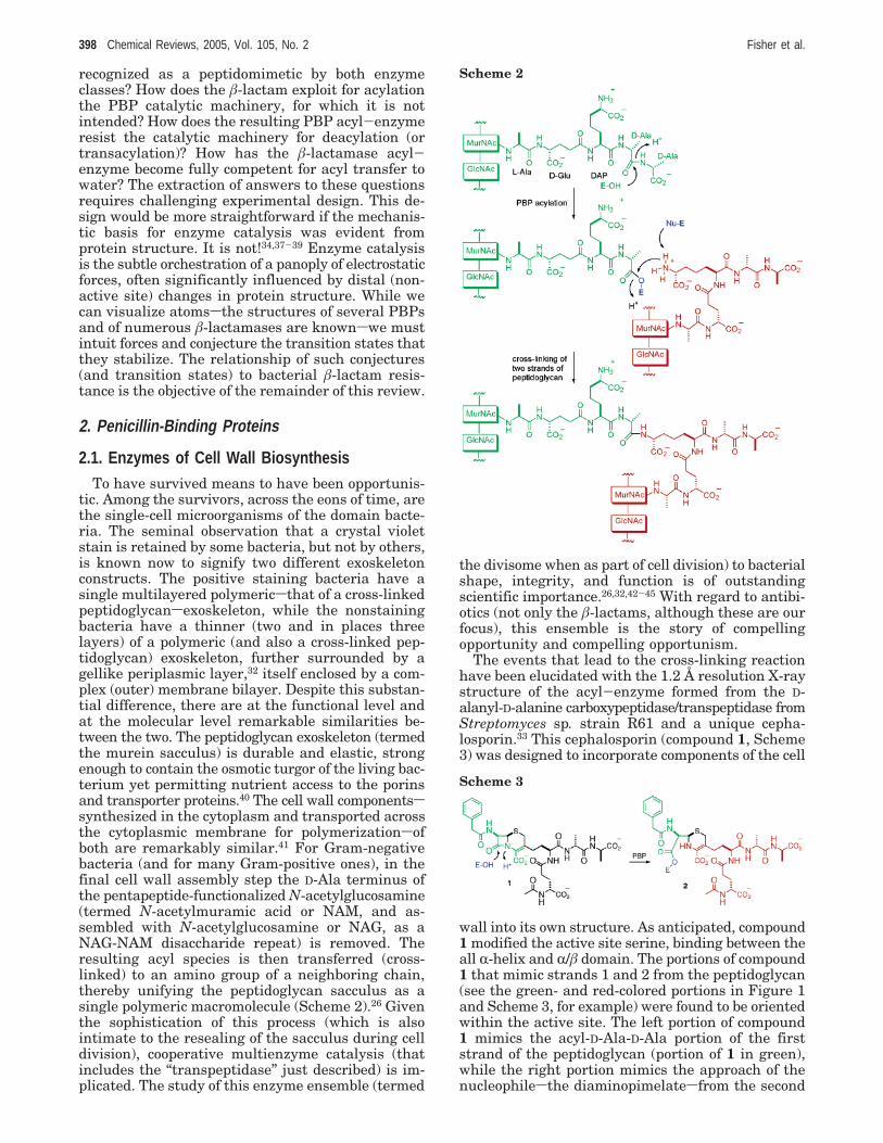
The events that lead to the cross-linking reactionhave been elucidated with the 1.2 Å resolution X-raystructure of the acyl-enzyme formed from the D-alanyl-D-alanine carboxypeptidase/transpeptidase fromStreptomyces sp. strain R61 and a unique cepha-losporin.33 This cephalosporin (compound 1, Scheme3) was designed to incorporate components of the cell
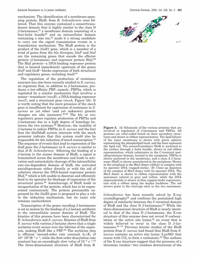
wall into its own structure. As anticipated, compound1 modified the active site serine, binding between theall R-helix and R/â domain. The portions of compound1 that mimic strands 1 and 2 from the peptidoglycan(see the green- and red-colored portions in Figure 1and Scheme 3, for example) were found to be orientedwithin the active site. The left portion of compound1 mimics the acyl-D-Ala-D-Ala portion of the firststrand of the peptidoglycan (portion of 1 in green),while the right portion mimics the approach of thenucleophilesthe diaminopimelatesfrom the second
Scheme 2
Scheme 3
398 Chemical Reviews, 2005, Vol. 105, No. 2 Fisher et al.

strand of the peptidoglycan (red portions in structure2). The acylation is proposed to occur after activationof the active site Ser62 (located at the N-terminus ofan R-helix, which is expected to modulate the serinepKa as proposed by Moews et al.46) by the generalbase Lys65swhich is in direct contact with Ser62 ata distance of 3.0 Å. This mechanism is consistentwith previously proposed mechanisms of the acyla-tion of PBPs, where the universally conserved lysineacts as the general base, abstracting a proton fromserine, followed by the back-donation of the proton(from lysine) to the peptide or â-lactam nitrogen.47
To provide a more detailed picture of the cross-linkingevent, a computational model was constructed fromthe high-resolution structure by extending compound2 to include the full pentapeptide and a NAG-NAMextension. The resulting model was fully solvated and
energy minimized (shown in Figure 1A). The pepti-doglycan strands were found to form a network ofelectrostatic interactions (shown in Figure 1B). Theseinteractions should play important roles in properlypositioning the peptidoglycan strands and for otherimportant events such as deprotonation of diami-nopimelate in the cross-linking event. It is of interestto note that the three-dimensional model differedfrom a previous model48 that used only the apoPBP2x structure with respect to the location of thesaccharide-binding grooves.
The description of behavior as either moral orimmoral is (primarily) a human characteristic. Forother organisms this distinction is irrelevant, and thefocus of their behavior is survival to the point ofreproduction. Among the bacteria (and fungi, forwhich bacteria are a food source) survivalsthat is,preservation within an ecological nichesrequiresexploitation of vulnerability. In addition, the biosyn-thetic enzymes of bacterial cell wall biosynthesis arevulnerable. The basis of their vulnerability (whichis one and the same with that of the bacterium) isthe combination of essentiality and exposure. Theseenzymes are located underneath the very cell wallthat they assemble. For the Gram-negative bacteriathese enzymes are either within the periplasmicspace orsfor the most essential of these enzymesswith active sites exposed to the periplasmic space anda transmembrane domain (with small cytoplasmicanchor) within the cytoplasmic membrane. Hence,bimolecular encounter with an inhibitor of theseenzymes requires only the successful passage ofthe inhibitorsintermingling with solute nutrientssthrough the lipopolysaccharide of the outer mem-brane into contact with the peptidoglycan surface ofthe periplasmic space. While this simple requirementcannot be underestimated (especially insofar asantibacterial design and for resistance development)for the penicillin and cephalosporin â-lactams se-creted by the biosynthesizing bacteria and fungiwithin the niche, the passage and encounter withthese biosynthetic enzymes is straightforward. As-tonishingly, each enzyme of the ensemble is capableof inactivation (via the same mechanism of irrevers-ible acylation) by an appropriately substituted â-lac-tam. The inactivation is facile for susceptible Gram-positive and -negative strains and less so for resistantbacteria. This truly remarkable event is commemo-rated by historical nomenclature: these enzymes arecollectively the penicillin-binding proteins (or PBPs)of the bacteria.
The chemically intriguing aspect of this event isthe recognition by each enzyme regardless of thespecific cell wall biosynthetic role. The inescapableconclusion is a fundamental of homology of structureand of alignment with the active site. However,despite this homology, all â-lactams do not inhibitall PBPs, likely due to subtle differences in the activesite and distal regions in the protein. Regardless ofcell morphology (Gram positive or negative) andregardless of individual specific synthetic function,these enzymes must possess such similarity as toimplicate a mere handful of, if not a single, ancestralprogenitor(s). As the intact â-lactam antibiotic was
Figure 1. (A) Stereoview of the three-dimensional struc-tures of two strands of peptidoglycan bound to the activesite of the D,D-transpeptidase/D,D-carboxypeptidase fromStreptomyces R61 PBP, constructed computationally fromthe 1.2 Å resolution structure for the acyl-enzyme specieswith compound 1 (the extension reaches the NAG-NAMunits on the peptidoglycan). The protein is shown in yellowribbon representation, while the bound computationalmodel representing the two strands of the peptidoglycanare shown in green and red capped-sticks representation.The blue van der Waals surface defines the active site. (B)Schematic representing the peptidoglycan from A, showingthe various hydrogen-bonding interactions and color-codedaccording to the three-dimensional model.
Bacterial Resistance to â-Lactam Antibiotics Chemical Reviews, 2005, Vol. 105, No. 2 399
recognized by this ancestral enzyme as a mimic ofthe peptidic terminus of the peptidoglycan, so toothere is continued recognition of these antibiotics bythe offspring of this enzyme. Once the â-lactamantibiotic is recognized by the PBP, the recognitionculminates in the formation of a stable acyl-enzymespecies. Hydrolysis of the acyl-enzyme ester bondis slow in PBPs, with half-lifes that substantiallyexceed the doubling time of the organism. To ap-preciate the degree of inefficiency of this step inPBPs, comparison of the deacylation rate constantwith that of â-lactamasessresistance enzymes thatare believed to have descended from the PBPssreveals that the rate of deacylation in PBPs is up to6 orders of magnitude slower.49 The irreversibility ofthe deacylation step in PBPs is at the root of theantimicrobial action of â-lactam antibiotics.
The basis for these differences must clearly derivefrom the differences in the targetsPBP and â-lac-tamasessstructures. The first X-ray diffraction struc-tures of two low molecular weight PBPs were solvednearly 25 years ago.50-52 Since then only a handfulof additional structures have been solved, despite thevery large number of PBPs that are now known.Those with solved structures include the low Mr PBPsfrom Streptomyces R6153 and Streptomyces K15 (PDBcode 1SKF),54,55 a zinc-dependent PBP from Strepto-myces albus G (PDB code 1LBU),50 the PBP5 fromEscherichia coli (PDB code 1NZO),56 and the high Mr(and â-lactam resistant) PBP2x (PDB code 1PMD)48
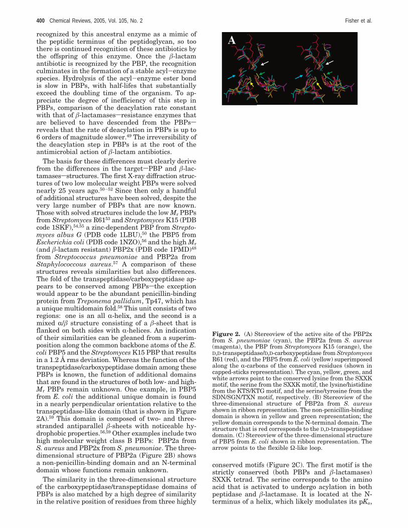
from Streptococcus pneumoniae and PBP2a fromStaphylococcous aureus.57 A comparison of thesestructures reveals similarities but also differences.The fold of the transpeptidase/carboxypeptidase ap-pears to be conserved among PBPssthe exceptionwould appear to be the abundant penicillin-bindingprotein from Treponema pallidum, Tp47, which hasa unique multidomain fold.58 This unit consists of tworegions: one is an all R-helix, and the second is amixed R/â structure consisting of a â-sheet that isflanked on both sides with R-helices. An indicationof their similarities can be gleaned from a superim-position along the common backbone atoms of the E.coli PBP5 and the Streptomyces K15 PBP that resultsin a 1.2 Å rms deviation. Whereas the function of thetranspeptidase/carboxypeptidase domain among thesePBPs is known, the function of additional domainsthat are found in the structures of both low- and high-Mr PBPs remain unknown. One example, in PBP5from E. coli the additional unique domain is foundin a nearly perpendicular orientation relative to thetranspeptidase-like domain (that is shown in Figure2A).59 This domain is composed of two- and three-stranded antiparallel â-sheets with noticeable hy-drophobic properties.56,59 Other examples include twohigh molecular weight class B PBPs: PBP2a fromS. aureus and PBP2x from S. pneumoniae. The three-dimensional structure of PBP2a (Figure 2B) showsa non-penicillin-binding domain and an N-terminaldomain whose functions remain unknown.
The similarity in the three-dimensional structureof the carboxypeptidase/transpeptidase domains ofPBPs is also matched by a high degree of similarityin the relative position of residues from three highly
conserved motifs (Figure 2C). The first motif is thestrictly conserved (both PBPs and â-lactamases)SXXK tetrad. The serine corresponds to the aminoacid that is activated to undergo acylation in bothpeptidase and â-lactamase. It is located at the N-terminus of a helix, which likely modulates its pKa,
Figure 2. (A) Stereoview of the active site of the PBP2xfrom S. pneumoniae (cyan), the PBP2a from S. aureus(magenta), the PBP from Streptomyces K15 (orange), theD,D-transpeptidase/D,D-carboxypeptidase from StreptomycesR61 (red), and the PBP5 from E. coli (yellow) superimposedalong the R-carbons of the conserved residues (shown incapped-sticks representation). The cyan, yellow, green, andwhite arrows point to the conserved lysine from the SXXKmotif, the serine from the SXXK motif, the lysine/histidinefrom the KTS/KTG motif, and the serine/tyrosine from theSDN/SGN/TXN motif, respectively. (B) Stereoview of thethree-dimensional structure of PBP2a from S. aureusshown in ribbon representation. The non-penicillin-bindingdomain is shown in yellow and green representation; theyellow domain corresponds to the N-terminal domain. Thestructure that is red corresponds to the D,D-transpeptidasedomain. (C) Stereoview of the three-dimensional structureof PBP5 from E. coli shown in ribbon representation. Thearrow points to the flexible Ω-like loop.
400 Chemical Reviews, 2005, Vol. 105, No. 2 Fisher et al.

thus facilitating its activation as a nucleophile andnucleofugacity as a leaving group.46 The lysine in thismotif is the general base that activates the serine foracylation.60 The second conserved motif is the (S/Y)-XN tripeptide sequence. This triad is SDN in Strep-tomyces R61, SGN in the Streptomyces K15 D,D-transpeptidase, and YSN in the Streptomyces R61D,D-peptidase. The S/Y residue in this motif is thoughtto be required for back-donation of a proton to thenitrogen atom of the â-lactam ring after formationof the tetrahedral intermediate. From the superim-position of the amino acids within the active sites ofthese PBPs (as shown in Figure 2C) it appears thatthe position of the hydroxyl (whether serine ortyrosine) is conserved (comparing the SDN and YXNmotifs). The third conserved motif in PBPs is a KTSor KTG motif (except in the case of Streptomyces R61where it is an HT(S/G)). In â-lactamases the role ofthis lysine (or histidine) is important in modulatingthe pKa of the universally conserved lysine that istwo residues downstream of the active site serine.
The overall similarity of the transpeptidase/car-boxypeptidase region and the conserved relativepositions of highly conserved residues in PBPs do nottranslate into similar catalytic (or functional) behav-ior. PBP5 of E. coli is a case in point. While mostPBPs have low deacylation rate constants, PBP5 hasan unusually high deacylation rate constant with ahalf-life t1/2 < 10 min with penicillin G; this is to becontrasted to the more typical deacylation rateconstant of 8.3 × 10⁻⁶ s-1 for the acyl-enzyme speciesof PBP2a with the same substrate. What makes thisPBP so unusual? Earlier studies had shown that aG105D mutation reduced the deacylation rate by 30-fold.61 The X-ray structure of this mutant did notreveal the basis for the reduced deacylation59 sincethe mutation does not occur in the active site.However, the X-ray structure of the wild-type en-zyme56 revealed disorder at a loop located near theactive site serine. The position of this loop is remi-niscent of the Ω-loop (vide infra) in class A â-lacta-mases, based on the superimposition of the TEM-1â-lactamase and PBP5.59 The different conformationof the loop adopted by the mutant likely contributesto the deacylation rate difference. This understandingtakes us to the curious event (and a theme of thisreview). What are the molecular events that resultin failed recognition of â-lactam antibiotics by thePBPs? Alteration of the PBPs is a dominant mech-anism of Gram-positive resistance. For this compel-ling reason the focus of Gram-positive PBP biochem-istry has changed from the historical (the enzymesof the nonpathogenic Bacillus subtilis)62 to those ofthe resistant and pathogenic S. pneumoniae and S.aureus. How are the PBPs of these resistant patho-gens different? Two limiting possibilities exist. ThePBPs are the same but exist in increased copynumber or the PBPs are altered by selection ofmutant variants so as to diminish recognition of theâ-lactam without compromise of the peptidoglycanbiosynthetic role. Both processes exist, but it is thelatter that has proven to be the most versatilesandexpandingsmechanism of Gram-positive bacterialresistance.
The expansion of resistant PBPs is a medicalproblem with microbiological (what is the ecologicalcircumstance where resistance is acquired from onebacterium by another), molecular biological (what arethe mutations, and what is the genetic basis for theirtransfer), biochemical (how do these mutations defeatrecognition of the â-lactam as a mechanism-basedPBP inhibitor), and chemical (what will be the designof new generation â-lactam antibiotics effective againstresistant pathogens) manifestations. A decrease inthe spread of antimicrobial drug resistance willrequire societal change and scientific discovery inresponse to each of these manifestations. Of thesefour we will briefly address the microbiological andmolecular biological, and focus on the biochemical.The topic of the chemical is reviewed elsewhere.24
An excellent point of biochemical entry to thereality of S. aureus â-lactam resistance is Pucci andDougherty’s analysis, by saturating penicillin inac-tivation, of the PBP distribution and stoichiometryin susceptible and resistant S. aureus.63 While it islong known that substituent changes made to thepenicillin and cephalosporin periphery influence rela-tive affinity (specificity) among the penicillin-bindingproteins, by judicious substituent choice and highconcentration it is nonetheless possible to saturate(to titrate to the point of complete inactivation) theentire ensemble and so obtain their relative abun-dance (copies per bacterium). Susceptible S. aureuscontains four PBPs, three of which are “high” molec-ular mass enzymes (70-80 kDa) and one of which isa low molecular mass enzyme (46 kDa). The high Mrenzymes are PBP1 (approximately 185 enzymes percell), PBP2 (460 enzymes), and PBP3 (150 enzymes).The low Mr enzyme is PBP4 (285 enzymes per cell).While this is a smaller number of enzymes than isfound in other bacteria species (B. subtilis has 12;E. coli has 16) the critical biosynthetic enzymes (withrespect to â-lactam lethality) for all are the (videinfra) high Mr enzymes, which are usually bifunc-tional (one activity is the â-lactam-sensitive transpep-tidase activity, and the second is a non-â-lactam-sensitive transglycosylase activity). For S. aureus thebifunctional enzyme target of the â-lactams (whereinthe transpeptidase but not the transglycosylaseactivity is inhibited) is the PBP2 enzyme.
What is the PBP composition of the â-lactam-resistant S. aureus? A comparison of the PBP com-position of a resistant strain, by saturating penicillininactivation, shows that the resistant bacteriumcontains the same four PBPs as the susceptiblebacterium (and in the same quantity per bacteriumas the susceptible bacterium) and an additional fifthenzyme (termed PBP2a).63,64 The PBP2a is presentin substantial quantity (approximately 800 copies percell with some variability) and is a “low affinity”enzyme with respect to â-lactam binding and inac-tivation. When resistant S. aureus is challenged bya â-lactam antibiotic, the transpeptidase activity ofits PBP2 enzyme is inactivated but the transpepti-dase activity of its PBP2a enzyme is unaffected. ThePBP2a performs the transpeptidase function of thenow inactivated PBP2, and the bacterium survives.Simply stated, the â-lactam concentration attained
Bacterial Resistance to â-Lactam Antibiotics Chemical Reviews, 2005, Vol. 105, No. 2 401
during chemotherapy is insufficient to inactivate thetranspeptidase activity of this new enzyme. (ThePBP2a contains a second domain that is presumedto possess a catalytic function which is not transg-lycosylase activity. The role of this second PBP2adomain remains unknown.) In the presence of â-lac-tam antibiotics the functioning transglycosylase do-main of the PBP2 (its transpeptidase having beeninactivated) works cooperatively with the activetranspeptidase of the PBP2a to maintain the cell wallintegrity of the resistant S. aureus.65,66 Therefore, thesalient issues to the understanding of Gram-positiveâ-lactam resistance are the circumstance of PBP2aacquisition, the genetic origin of PBP2a, and themolecular alteration(s) within the transpeptidasePBP2a domain that result in a change from high tolow susceptibility to â-lactam inactivation.
The appearance of methicillin resistance soon fol-lowed (within a year) the introduction of methicillinin the clinic.67 The mechanism by which the mecAgene, carried by mobile genetic elements known asthe staphylococcal cassette chromosome mec (SCC-mec),68 was acquired by S. aureus remains unknown.It is suggested that this gene was acquired fromStaphylococcus sciuri (a bacterium found in the gutof animals) which possesses a close mecA genehomologue.69 Upon activation of the mecA gene, thePBP2a protein is expressed. It was shown in the mid-1980s that the presence of PBP2a in S. aureus con-ferred resistance to the clinically used â-lactams,70-72
as evidenced by a 500-fold increase in the MIC(minimal inhibitory concentration) for penicillins.These bacteria came to be known as methicillin-resistant S. aureus (or MRSA). Kinetic characteriza-tion of the reaction of PBP2a with â-lactams providedvaluable mechanistic information and revealed thatthe resistance to â-lactams was not merely due to alarge Kd value (a commonly held belief) but to theacylation rate constant as well.73 The dissociationconstant of the PBP2a-benzylpenicillin complex was13 mM,74 similar to what is found for other (suscep-tible) PBPs. A much clearer difference is the apparentsecond-order rate constant for acylation of the activesite serine. For the reaction of PBP2a with benzyl-penicillin this rate constant is 2-3 orders of magni-tude smaller than those found for other high molec-ular weight PBPs (such as the S. aureus PBP2 andS. pneumoniae PBP2x).74 The structural features ofPBP2a that are responsible for the poor acylationwere elucidated recently with X-ray structures for theapo and acyl-enzyme complex of PBP2a with ben-zyl-penicillin, nitrocefin, and methicillin.57 Compari-son of the apo-PBP and acyl-PBP structures re-vealed noticeable differences. In the acyl-enzymestructure the CR, Câ, and Oγ of Ser403sthe acylatedserinesare 1.1, 1.4, and 1.8 Å away from the sameatoms in the apo-structure, suggesting that theR-helix that holds Ser403 must undergo a conforma-tional change for acylation to occur.57
Structural modifications in PBPs that result inincreased resistance are not confined to S. aureus.The three-dimensional structures of two additionalPBPs have been solved by X-ray crystallography. Oneis PBP2x from S. pneumoniae. PBP2x is a class B
high molecular weight PBP, and its structure wassolved nearly a decade ago.48 The X-ray diffractionstructure of a mutant PBP2x provided the firstglimpse into the structural bases for resistance bymutation of PBPs.75,76 Two drug-resistant PBP2xmutants have been characterized. The first X-raystructure describes the effects of mutations Thr338Alaand Met339Phe, which along with other mutationsalter the acylation efficiency by 20-fold. Both of thesemutations occur close to the active site Ser-337, theresidue that is acylated by â-lactam antibiotics. Theeffect of these mutations is attributed to the disrup-tion of hydrogen-bonding interactions between Thr388and a conserved water molecule. Also, a conforma-tional change that occurs for the â3 strand is at-tributed to the collective effects of the larger Phe339side chain and smaller Ala338 chain. These confor-mational changes enable an alternative conformationfor Ser337 that might be less prone to activation. Themost likely candidate for the base that promotesSer337 acylation is Lys340, whose amine is inhydrogen-bonding contact (3 Å) with the serinehydroxyl. The structure of another mutant of PBP2xhas been recently solved.75 It reveals similar confor-mational changes as the PBP2x above except that inthis case the effects are attributed to the change inpolarity introduced by Gln552Glu and to a narroweractive site.
PBP5, a transpeptidase from Enterococcus faeciumsa bacterium which incidentally does not produceâ-lactamasesshas also been implicated in resistanceto â-lactams through either modification or overex-pression of the enzyme.77,78 The enterococci are lessvirulent than S. aureus and S. pneumoniae but havebecome prominent in the clinic due to their increasedlevels of resistance to a variety of antibiotics.79
PBP5fm has low affinity for â-lactam antibiotics. Tworeasons for this decreased affinity are suggested fromthe structure of this protein.80 A glutamate (Glu622)near the active site may present a steric barrier toâ-lactam binding. This interpretation is consistentwith the reduced affinity for benzylpenicillin thatresults when the equivalent site in PBP2x is changedto glutamate.81 Second, an arginine (Arg464) mayinteract with its neighbors in the conserved loopspanning residues 461-465, resulting in a more rigidcleft that would lead to the reduced affinity ofPBP5fm.80
2.2. â-Lactam-Sensing Proteins
Resistance to antibiotics in Gram-positive andGram-negative bacteria has manifested itself throughvarious mechanisms, including the production ofâ-lactamases or PBPs that are insensitive to theaction of â-lactams, such as the case of PBP2a fromS. aureus. In the mid-1980s Lampen and co-workersobserved that the synthesis of the â-lactamase fromBacillus licheniformis 749/I gradually peaks at 1-1.5h after exposure to a â-lactam and decreases slowlyin the following 1-2 h82 (induction in S. aureus isfar more rapid, complete within 11 min83). Thisexperiment confirmed that â-lactamase production isinducible and implied the presence of a transduction
402 Chemical Reviews, 2005, Vol. 105, No. 2 Fisher et al.
mechanism. The identification of a membrane-span-ning protein, BlaR, from B. licheniformis soon fol-lowed. That this enzyme contained a sensor/trans-duscer domain that is highly similar to the class Dâ-lactamases,84 a membrane domain consisting of afour-helix bundle85 and an intracellular domaincontaining a zinc ion,84 made it a strong candidateto carry out the signal transmission events in atransduction mechanism. The BlaR protein is theproduct of the blaR1 gene, which is a member of atriad of genes from the bla divergon; blaP and blaIare the remaining genes that encode the effectorprotein (â-lactamase) and repressor protein BlaI.86
The BlaI proteinsa DNA-binding repressor proteinthat is located immediately upstream of the genesblaP and blaRsblocks expression of both structuraland regulatory genes, including itself.87
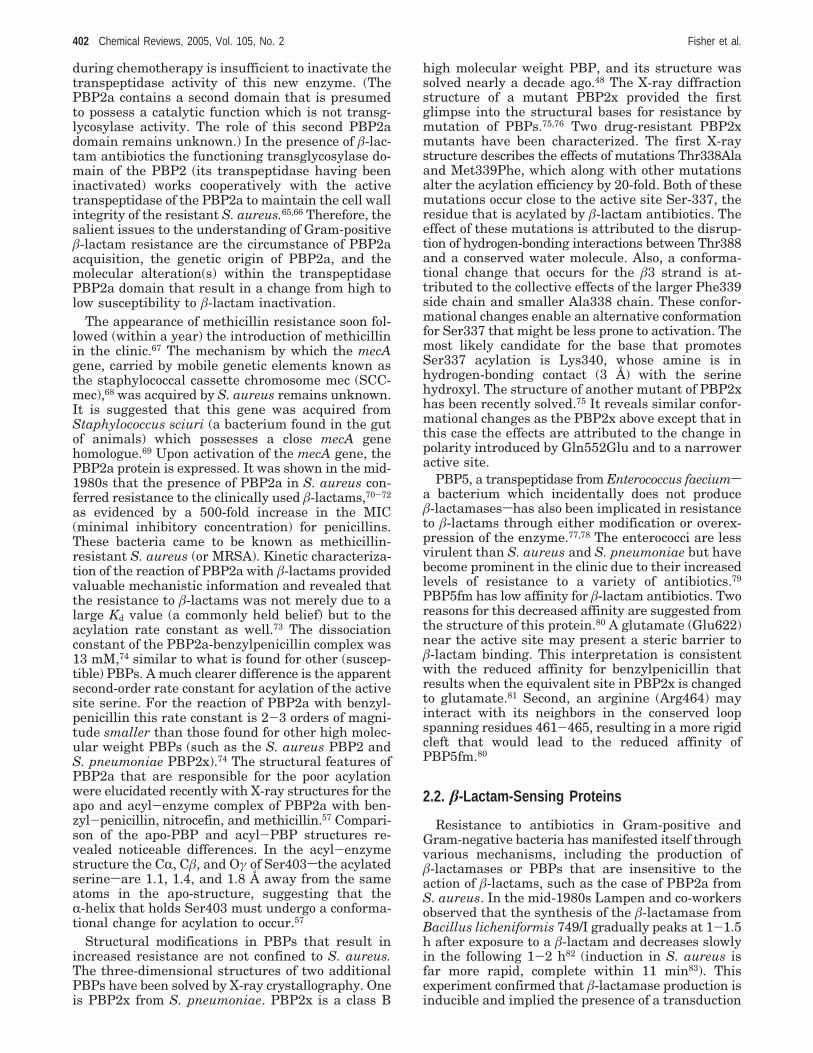
The regulation of the production of resistanceenzymes has also been recently studied in S. aureus,an organism that, in addition to â-lactamases, pro-duces a low-affinity PBP, namely, PBP2a, which isregulated by a similar mechanism that involves asensor-transducer (mecR), a DNA-binding repressor(mecI), and a structural gene (mecA, Figure 3A). Itis worth noting that the mere presence of the mecAgene is insufficient for expression of resistance in S.aureus as yet other (and yet unknown) geneticchanges are also necessary.88,89 The bla or mecregulatory genes regulate production of PBP2a andâ-lactamase due to a high degree of homology be-tween the two systems.90 However, the inability ofâ-lactams to induce PBP2a in S. aureus and the factthat the blaI/blaR system interacts with the mecApromoter indicate that this system could also beresponsible for the induction of mecA transcription.91
The sequence of events that lead to expression of theblaZ gene (for â-lactamase) in S. aureus is similar tothat of B. licheniformis: following the binding of aâ-lactam to the sensor domain of BlaR, a signal istransmitted across the membrane and leads to acti-vation and autocatalytic cleavage of the intracellularzinc-ion-dependent domain of blaR; the activatedmetalloprotease either directly or with the aid ofcofactors cleaves the DNA-bound repressor proteinBlaI,92 which is left unable to dimerize and efficientlybind to its operator for blockage of expression of thestructural genes.90 Autocleavage of BlaR leads toincapacitation of the protein, which has to be regen-erated continuously. The protein presumably ex-pressed by the blaR2 gene is proposed to play a rolein the induction mechanism, but its exact roleremains unelucidated.
Transcription of the genes encoding â-lactamasesis set in motion by the binding of a â-lactam antibioticto the extracellular sensor domain of BlaR. Thekinetics of this process have been characterized forB. licheniformis and S. aureus. Kinetics of BlaR fromS. aureus with various â-lactams shows that a singleacylation event occurs over the lifetime of the organ-ism, making BlaR like a PBP.93 The acylation stepis efficient (second-order rate constant k2/Ks of104-106 M-1 s-1, while the first-order deacylation rateconstant has an exceedingly slow value of 10-5 s-1.93
The three-dimensional structure of BlaR from B.
licheniformis has been recently solved by X-raycrystallography94 and confirms the postulated highdegree of similarity between the C-terminal domainof BlaR and the class D â-lactamases,84 While thethree-dimensional structure of BlaR is nearly identi-cal to that of the class D â-lactamases, the X-raystructure of this enzyme does not reveal N-carboxy-lation at the active site lysine,94 an event that iswidely believed to occur in the class D â-lac-tamases.95-97 Previous kinetic studies of the BlaRprotein from S. aureus had found that BlaR from S.aureus contains an active site lysine (Lys392) thatreacts with CO2 to form a carbamate.93 The authorsof the X-ray structure suggest that the presence of athreonine residuestwo residues downstream of the
Figure 3. (A) Schematic of the various proteins that areinvolved in regulation of â-lactamase and PBP2a. Allproteins are color-coded based on their secondary struc-tures and shown in ribbon representation. The lipid bilayerof the inner membrane is shown with green spheresrepresenting the phospholipid head, and the lines representthe lipid tail. The sensor/transducer BlaR is anchored tothe surface through a helix bundle, shown in red ribbonrepresentation, which culminates in the C-terminal met-alloproteinase domain, located in the cytoplasm. PBP2a isshown anchored to the membrane, and a class A â-lacta-mase (BlaZ) is shown unanchored in the periplasm. Shownin the cytoplasm is the MecI dimer (ribbon) in complex withits operator DNA (capped-sticks). (B) Close-up depictionof the complex of MecI dimer with its operator DNA. TheMecI dimer is shown in ribbon representation with themonomers colored in gray and yellow, while the DNAoligonucleotide is shown in blue capped-sticks representa-tion with a ribbon along the duplex backbone. The redarrows point to the cleavage sites in the two monomers.
Bacterial Resistance to â-Lactam Antibiotics Chemical Reviews, 2005, Vol. 105, No. 2 403

active serine residue where a valine residue wouldbe usually located in class D â-lactamasesis likelythe reason behind the lack of carboxylation of thatlysine residue.94 However, the signature 13C NMRsignal for N-carboxylated lysine in BlaR of S. aureushas been documented in solution.93
More recently, the structures of the acyl-enzymecomplex of BlaR from S. aureus with benzylpenicillinand ceftazidime have been independently solved byX-ray crystallography.98,99 These confirm the simi-larities in the structures of BlaR from S. aureus toBlaR of B. licheniformis and the class D â-lactamases.Interestingly, both of these studies found that BlaRwas not carboxylated in the acyl-enzyme complex,in contrast to the 13C NMR, fluorescence, and mu-tagenesis studies identifying the carboxylated lysinein the resting BlaR protein.93 QM/MM calculationscarried out by Birck et al.99 reveal that upon proto-nation of the carbamate nitrogen in the class Dâ-lactamases a barrierless decarboxylation occurs.The authors postulate that the same N-decarbox-ylation event occurs in BlaR, but unlike the class Dâ-lactamases decarboxylation results in an inactiveenzyme unable to recarboxylate due to a hydrogenbond between Lys392 and Asn439. This conclusionis consistent with the fact that BlaR undergoes asingle acylation event over the lifetime of the organ-ism.
The nature of the signaling event across themembrane that transpires as a result of â-lactambinding to the sensor domain of BlaR is not wellunderstood. Golemi-Kotra and co-workers have shownthat binding of the antibiotic to BlaR of S. aureus isaccompanied by significant conformational changesthat likely have a role in the signal transductionmechanism.93 A recent study of the transductionmechanism in B. licheniformis did not identify aconformational change in the C-terminal domain.100
An alternative mechanism was noted based on aninterdomain conformational change in the membraneprotein consisting of the loss of interaction betweenthe C-terminal domain and an L2 loop of BlaR thatconnects two R-helices of the four-helix bundle. It wasshown that in the absence of the antibiotic the sensordomain and the L2 loop form an interaction. Recentmutations of highly conserved residues in the L2 loopappear to be lending credence to this mechanism, asthe organisms exhibited no â-lactamase activity,since the level of antibiotic remained at similar levelsto that of a â-lactamase-negative control.98 Whereasthe mechanism that leads to the transduction of thesignal appears to be contentious, the event thatfollows is accepted to consist of autocleavage of thezinc-containing intracellular domain,87 which in thecase of S. aureus is followed by inactivation of therepressor proteins MecI/BlaI, while in B. lichenifor-mis this process is thought to occur through anintermediary coactivator.101
The structures of BlaI102 and MecI103,104 have beenrecently solved by NMR and X-ray crystallography,respectively. The BlaI and MecI proteins from S.aureus share 60% identity and 31 to 41% identity toBlaI from B. licheniformis.102 The structure of MecIconsists of a dimer in the shape of a triangle (shown
in Figure 3B).104 Dimerization occurs at the C-termi-nal domain, while the DNA-binding domain is locatedat the N-terminus (see Figure 3B for structure ofMecI-DNA complex). The topology of MecI follows a“winged-helix” architecture103,104 with a helix-turn-helix DNA-binding motif; the second helix of thismotif binds to the major groove of DNA with up to16 hydrogen bonds and salt bridges (see Figure3B).104 The high level of conservation of the residuesthat form contact between the repressor protein andthe operator DNA in S. aureus and B. licheniformissuggests that this complexation is likely similar inBlaI and MecI.104
3. â-Lactamases
3.1. Overview and ClassificationThe â-lactamases predate the antibiotic era. The
evolution of these enzymes is presumed to have takenplace in parallel to the biosynthetic steps leading toâ-lactam antibiotics.105 Indeed, the first â-lactamasewas identified in the early 1940s prior to the firstlarge-scale use of penicillins in Boston 2 yearssubsequent.16 However, extensive clinical use of theseantibioticssâ-lactams comprise approximately 55%of all antibiotics used currentlyshas accelerated theselection process for the emergence of once rare genesfor these antibiotic-resistant enzymes. The rarebacterium that harbored the gene for a â-lactamasewould have had the opportunity to grow unencum-bered once the susceptible organisms in a heteroge-neous population of bacteria were eliminated in thecourse of an antibiotic treatment regimen. In essence,the less fit bacterium is eliminated by the â-lactamchallenge and the resistant organism experiencesunlimited nutritional resources to propagate. Theonce rare gene for the â-lactamase is amplified.
Sharing of genetic materials among microbialpopulations is relatively facile. Genes, often residingon inherently mobile genetic elements such as plas-mids and transposons, are shared not merely mem-bers of a given species of bacteria but also amongunrelated genera. The facility of genetic sharing isunderscored by the observation that some organisms,such as the Streptoccoci, are able to acquire free-standing stretches of nucleic acids (containing entiregenes) directly from the environment. All theseprocesses have contributed by clinical selection to theamplification of once rare antibiotic-resistant genesand their liberal sharing among various bacterialpopulations.
The account given above outlines the plausibleevents that have given rise to the emergence of the“parental” genes for â-lactamases. As a consequenceof the inappropriate use of â-lactam antibiotics forthe past half a century, especially in the community,many variants of the parental enzymes have emerged.This accelerated evolution of the antibiotic-resistantgenes has been abetted by the creative moleculartinkering of medicinal chemists in the past decades,the fruits of which are an ensemble of â-lactamantibiotic structures. The dynamics between thediscovery, the creation of new â-lactam antibiotics,and the clinical responses by microbial population to
404 Chemical Reviews, 2005, Vol. 105, No. 2 Fisher et al.
these developments have been outlined by Bush andMobashery.106 In light of the different properties ofthese various types of â-lactam antibioticssfor ex-ample, antibiotics that target different sets of PBPsor those that have been imparted with resistance tothe action of â-lactamasessthe clinical response bybacteria has been the selection of mutant variantsof â-lactamases that often have broadened the cata-lytic ability of the enzymes. For example, the TEM-1â-lactamase, a plasmid-borne enzyme described firstin 1963, has given rise to 133 variants (as of February2004).15 While this variety does reflect the successfultherapeutic use of â-lactams, it also may be takenas presaging the obsolescence of these versatileantibiotics sometime in the future.
Prompted by a critical biochemical requirement,nature deftly develops catalysts to meet the need.Despite the outstanding stability of the peptide bond(half-life of approximately 500 years when uncata-lyzed for hydrolysis),107,108 multiple classes of pro-teases have evolved to hydrolyze this bond, in lightof the central importance of proteolysis to manybiological processes. The same has been true forâ-lactamases in the face of the life or death optionsto the organisms presented by the antibiotics. Thereare four known classes of bona fide â-lactamases,each of which operates by a distinct reaction mech-anism.15,105,109,110
While a handful of â-lactamases were known in theearly 1970s, the number at the present exceeds 470(communication by Dr. Karen Bush). Two generaltypes of â-lactamases are known: those that requirethe zinc ion for their function, and those that pursuea transient serine acylation/deacylation strategy (ifthe unique â-lactamase activity of the T. pallidumPBP is also found in other organisms, this will yetbe another type of â-lactamase as it does not requirea zinc ion nor does it pursue the covalent catalyticstrategy of the serine enzymes).58,111 The widelyaccepted molecular classification places â-lactamasesinto four classes: three serine-dependent enzymeclasses (classes A, C, and D) and one metal-depend-ent (class B). This classification is not to be confusedwith that of Ghuysen for the PBPs in which the twogroups of low molecular weight and high molecularweight PBPs are divided among classes A, B, and C(for a total of six PBP classes).
Both PBPs and â-lactamases are present in theperiplasmic space of Gram-negative bacteria. InGram-positive organisms (which lack the outer mem-brane) the PBPs are located on the outer surface ofthe cytoplasmic membrane and the â-lactamases areeither excreted or bound to the cytoplasmic mem-brane.112 All â-lactamases are expected to havedivested completely their ability to bind the pepti-doglycan substrate of their ancestral PBPs.14,113 Ifnot, the opportunity to function as a vanguardagainst the incoming antibiotics would be lost (thestructural means to this end was revealed recentlyfor the class C â-lactamases).113 Moreover, thissame conclusion may be intuited from the ability ofmany of the class A and C â-lactamases to act atthe diffusion-limited rate for their preferred sub-strates.114,115
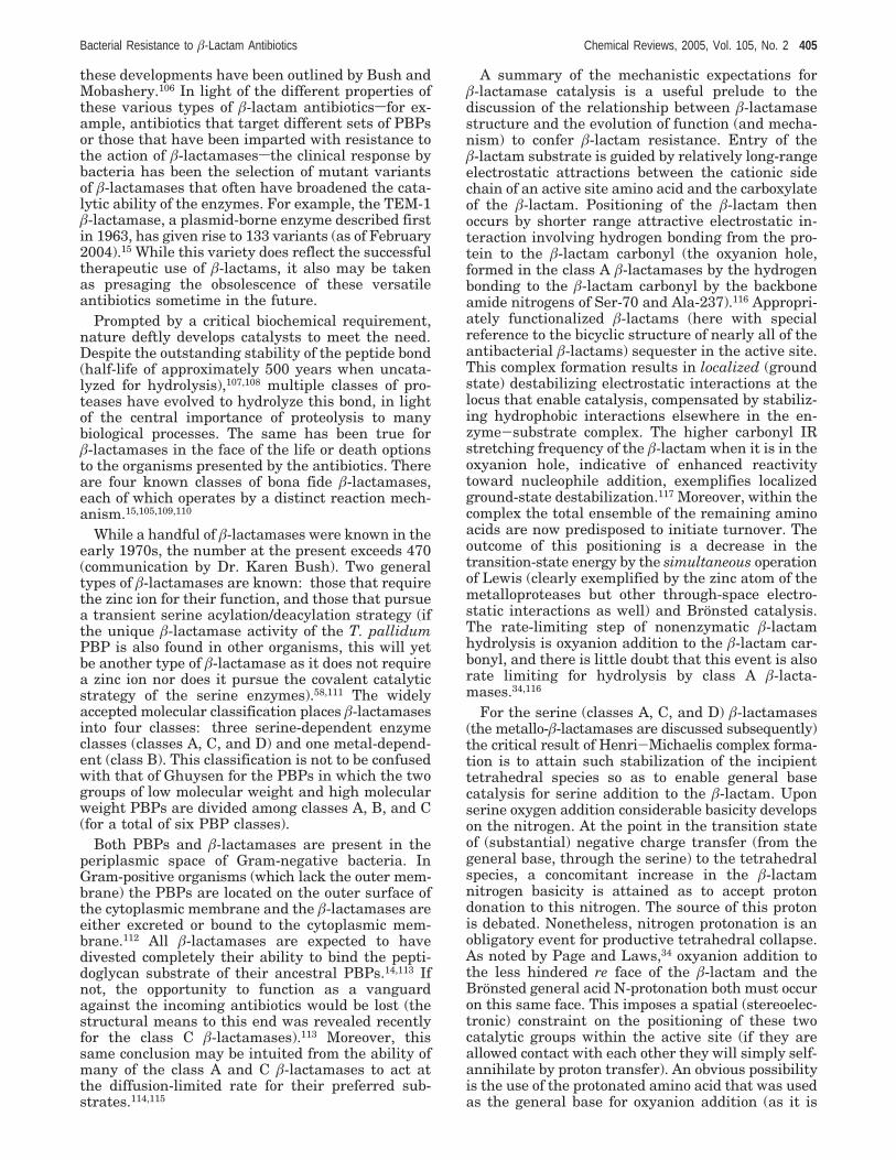
A summary of the mechanistic expectations forâ-lactamase catalysis is a useful prelude to thediscussion of the relationship between â-lactamasestructure and the evolution of function (and mecha-nism) to confer â-lactam resistance. Entry of theâ-lactam substrate is guided by relatively long-rangeelectrostatic attractions between the cationic sidechain of an active site amino acid and the carboxylateof the â-lactam. Positioning of the â-lactam thenoccurs by shorter range attractive electrostatic in-teraction involving hydrogen bonding from the pro-tein to the â-lactam carbonyl (the oxyanion hole,formed in the class A â-lactamases by the hydrogenbonding to the â-lactam carbonyl by the backboneamide nitrogens of Ser-70 and Ala-237).116 Appropri-ately functionalized â-lactams (here with specialreference to the bicyclic structure of nearly all of theantibacterial â-lactams) sequester in the active site.This complex formation results in localized (groundstate) destabilizing electrostatic interactions at thelocus that enable catalysis, compensated by stabiliz-ing hydrophobic interactions elsewhere in the en-zyme-substrate complex. The higher carbonyl IRstretching frequency of the â-lactam when it is in theoxyanion hole, indicative of enhanced reactivitytoward nucleophile addition, exemplifies localizedground-state destabilization.117 Moreover, within thecomplex the total ensemble of the remaining aminoacids are now predisposed to initiate turnover. Theoutcome of this positioning is a decrease in thetransition-state energy by the simultaneous operationof Lewis (clearly exemplified by the zinc atom of themetalloproteases but other through-space electro-static interactions as well) and Bronsted catalysis.The rate-limiting step of nonenzymatic â-lactamhydrolysis is oxyanion addition to the â-lactam car-bonyl, and there is little doubt that this event is alsorate limiting for hydrolysis by class A â-lacta-mases.34,116
For the serine (classes A, C, and D) â-lactamases(the metallo-â-lactamases are discussed subsequently)the critical result of Henri-Michaelis complex forma-tion is to attain such stabilization of the incipienttetrahedral species so as to enable general basecatalysis for serine addition to the â-lactam. Uponserine oxygen addition considerable basicity developson the nitrogen. At the point in the transition stateof (substantial) negative charge transfer (from thegeneral base, through the serine) to the tetrahedralspecies, a concomitant increase in the â-lactamnitrogen basicity is attained as to accept protondonation to this nitrogen. The source of this protonis debated. Nonetheless, nitrogen protonation is anobligatory event for productive tetrahedral collapse.As noted by Page and Laws,34 oxyanion addition tothe less hindered re face of the â-lactam and theBronsted general acid N-protonation both must occuron this same face. This imposes a spatial (stereoelec-tronic) constraint on the positioning of these twocatalytic groups within the active site (if they areallowed contact with each other they will simply self-annihilate by proton transfer). An obvious possibilityis the use of the protonated amino acid that was usedas the general base for oxyanion addition (as it is
Bacterial Resistance to â-Lactam Antibiotics Chemical Reviews, 2005, Vol. 105, No. 2 405
already on this face and makes the self-annihilationissue moot). Hydrolysis of the serine acyl-enzyme,the deacylation step, is energetically less demanding(a more reactive carbonyl and thus having a dimin-ished need for general acid catalysis in tetrahedralcollapse).
Within the active sites of the serine â-lactamasesare, therefore, two ensembles of amino acids. Thefirst is the catalytic ensemble comprised of a mini-mum of five amino acids: the serine, the general basefor the serine, the oxyanion hole (two amino acids),and the cationic recognition site for the carboxylate.These amino acids are expected to be invariant (orvery highly conserved). The second is the recognitionensemble (in one form or another, all of the remain-ing amino acids) that complements the hydrophobicand hydrophilic segments presented by the remain-ing structure of the â-lactam. These recognitionamino acids will be variable and correspond to theamino acids that will mutate under selection pres-sure. For a given â-lactamase and given substratethe complementarity to the rate-limiting transitionstate and to overall recognition of the â-lactam willvary. This variability is, of course, quantified as theunique kcat/Km for the substrate. While this may seemobvious, there is a less appreciated corollary. Therelative individual role played by any member ofthese ensembles during catalysis (measured, say, interms of an amino acid pKa) is therefore substratedependent. Deletion of an essential amino acidswhilean essential tool of mechanistic enzymologysaltersthe energetic landscape of the active site in suchfashion as to make all subsequent interpretationcautionary. The criteria that determine which aminoacids of the recognition ensemble transform underselection pressure are straightforward. The integrityof the catalytic ensemble must be preserved, and theintegrity of the protein as the whole (for example, asmeasured in terms of thermal stability or expression/folding capability) can only be lightly varied.118-121
Likewise, mutations that require only a single nucle-otide change and that preserve common codon usageare more probable than those that do not.120 Thisimplies that certain â-lactamase families are antici-pated to be more “plastic” than others, and indeed,the serine â-lactamases provide such a contrast withthe great phenotypic diversity of the serine class ATEM enzymes as compared to the class A SHVenzymes, which have diversified but without sub-stantial phenotypic evolution.15 Given these boundaryconditions and superlative kinetic and structuraldata for these enzymes, one might presume that theassignment of function within the catalytic ensembleand within the recognition ensemble as these developunder selection pressure would be straightforward.One would be wrong.
3.2. Class A â-LactamasesThis is the largest and best mechanistically char-
acterized serine â-lactamase class. Historically, theseâ-lactamases were described as “penicillinases” astheir ability to catalyze penicillin hydrolysis wasgreater than that for cephalosporins. They havebecome so efficient at their function that they are
diffusion controlled, where the apparent second-orderrate constant kcat/Km has reached the (upper) diffu-sion limit estimated from collision theory. As a result,class A â-lactamases may be described as havingreached catalytic perfection for their preferred sub-strates.114 Variants with much broader substratepreferences are now known, including enzymes im-parting clinical resistance to late generation â-lac-tams.15,109,122-124 The class A â-lactamases are closelyrelated in sequence to low molecular weight classC PBPs such as PBP4 of E. coli, H. influenza, andM. tuberculosis.60 As judged by the comparison ofcrystal structures, the catalytic domain of the largerE. coli PBP5 (low Mr PBP class A) shows highsimilarity as well.56,125 In terms of bacterial resis-tance, three class A â-lactamases subclasses domi-nate: the (historically Gram-negative plasmid pen-icillinase) TEM/SHV, the P. aeruginosa PER/OXA/TOHO cephalosporinases, and the CTX-M (NMC-A)carbapenemase subclasses.126 As of October 2004, 135TEM and 57 SHV â-lactamase variants are known(http://www.lahey.org/studies/webt.htm). While thesequence homology among the three is easily recog-nizable and the fundamental catalytic mechanism foreach is the same, the differences render broad struc-tural and mechanistic generalizationssespecially asthey relate to resistance developmentsunwarranted.
Several class A variants resist inactivation by themechanism-based inhibitors (clavulanate and sul-bactam) used in clinical formulations with otherwiseâ-lactamase-susceptible penicillins. Until very re-cently the occurrence of these inactivation-resistantclass A â-lactamases was limited to the aforemen-tioned TEM subclass, and hence, the term “inhibitor-resistant TEM” (IRT) was coined. However, in lightof the discovery of inhibitor resistance in the SHV-type enzymes, this group is better referred to as“inhibitor-resistant â-lactamases”. Furthermore, newclass A â-lactamases that are active against the morerecent cephalosporins (ceftazidime and cefotaximeand the monobactam aztreonam) and others that areactive against the carbapenems are known col-lectively (also with other class C and D enzymes) as“expanded-spectrum â-lactamases” (ESBL).124
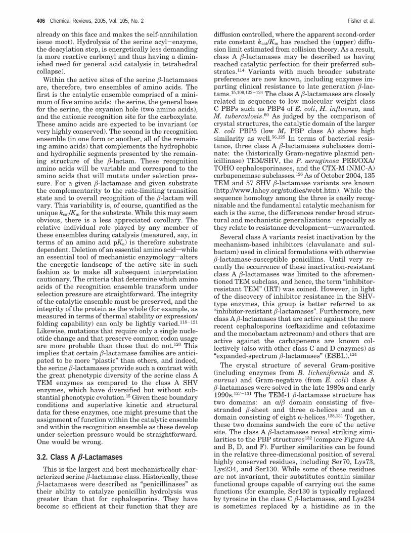
The crystal structure of several Gram-positive(including enzymes from B. licheniformis and S.aureus) and Gram-negative (from E. coli) class Aâ-lactamases were solved in the late 1980s and early1990s.127-131 The TEM-1 â-lactamase structure hastwo domains: an R/â domain consisting of five-stranded â-sheet and three R-helices and an Rdomain consisting of eight R-helices.128,131 Together,these two domains sandwich the core of the activesite. The class A â-lactamases reveal striking simi-larities to the PBP structures132 (compare Figure 4Aand B, D, and F). Further similarities can be foundin the relative three-dimensional position of severalhighly conserved residues, including Ser70, Lys73,Lys234, and Ser130. While some of these residuesare not invariant, their substitutes contain similarfunctional groups capable of carrying out the samefunctions (for example, Ser130 is typically replacedby tyrosine in the class C â-lactamases, and Lys234is sometimes replaced by a histidine as in the
406 Chemical Reviews, 2005, Vol. 105, No. 2 Fisher et al.
structure of the Streptomyces R61 D,D-peptidase/transpeptidase).
The remaining invariant amino acid is Glu166.This glutamate is located on a loop (termed theΩ-loop) that sequesters, by hydrogen bonding, asingle water molecule as a bridge to the Ser70adjacent to the re face of the â-lactam carbonyl. Thisglutamate has been proposed by some to have a rolein the rate-limiting acylation step.133-137 This mech-anism envisions that this glutamate, acting throughthe bridging water as a proton shuttle, activatesSer70 for nucleophilic addition to the â-lactam.136,138
The alternative possibility for the general base, theinvariant Lys73,129,131 must then be electrostaticstabilization (such as to increase the Ser70 acidityand hence nucleophilicity). The evidence in favor ofGlu166 as the serine-activating general base issummarized. A compelling argument in favor of aLewis acid, rather than a Bronsted base, role forLys73 was the 15N NMR determination of its pKa asgreater than 10.139 This determination was followedby Poisson-Boltzmann electrostatic calculations byLamotte-Brasseur et al. supporting the pKa > 10assignment,140,141 although others (using the samecalculation method) estimated a value of 8.142 Morerecently each of three methodssenzyme kinetics, 15NNMR, and free-energy calculations using the ther-
modynamic integrationssupports a pKa value forLys73 of 8.0-8.5.143 Ultrahigh-resolution crystalstructures of the TEM-1133 and SHV-2136 enzymesshow a spatial arrangement of Glu166 and theinvariant water, in the presence of a bound transitionstate mimic, to be consistent with serine activationvia the proton shuttle.133 The TEM-1 structure re-solves a hydrogen atom on Glu166, while the SHV-2structure shows the hydrogen of the Ser70 hydroxylpointing to the conserved water molecule. Followingserine addition the Ser130 hydroxyl is positionedideally to shuttle the proton on Lys73 to the â-lactamnitrogen of the tetrahedral species (Figure 4B),driving ring opening to the acyl-enzyme species. Itis widely accepted that Glu166 is the general basefor hydrolysis of the acyl-enzyme ester. In fact, site-directed mutagenesis of Glu166 (E166A) abolishesdeacylation while impairing (but not abolishing) theacylation process by a factor of 103.127,137
Over the past three decades several strategies haveemerged, in the guise of new â-lactams, to incapaci-tate the class A â-lactamases. The first strategy isexemplified by clavulanate and the penam sulfones(sulbactam and tazobactam), which are poor PBPinactivators but excellent â-lactamase inactivators.144
The key mechanistic event for both is a quite similarfragmentation reaction of the respective serine acyl-
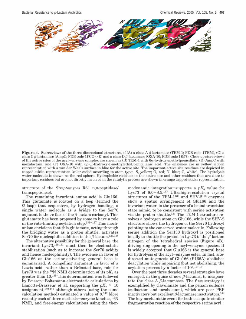
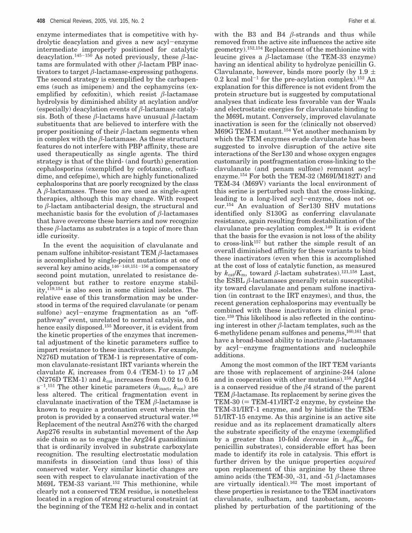
Figure 4. Stereoviews of the three-dimensional structures of (A) a class A â-lactamase (TEM-1; PDB code 1TEM), (C) aclass C â-lactamase (AmpC; PDB code 1FCO), (E) and a class D â-lactamase (OXA-10; PDB code 1K57). Close-up stereoviewsof the active sites of the acyl-enzyme complex are shown as (B) TEM-1 with 6R-hydroxymethylpenicillate, (D) AmpC withmoxalactam, and (F) OXA-10 with 6â-(1-hydroxy-1-methylethyl)penicillanic acid. The enzymes are in yellow ribbonrepresentation with a van der Waals surface in blue for the active site. The important active site residues are depicted incapped-sticks representation (color-coded according to atom type: S, yellow; O, red; N, blue; C, white). The hydrolyticwater molecule is shown as the red sphere. Hydrophobic residues in the active site and other residues that are close toimportant residues but are not directly involved in the catalytic process are shown in orange capped-sticks representation.
Bacterial Resistance to â-Lactam Antibiotics Chemical Reviews, 2005, Vol. 105, No. 2 407
enzyme intermediates that is competitive with hy-drolytic deacylation and gives a new acyl-enzymeintermediate improperly positioned for catalyticdeacylation.145-150 As noted previously, these â-lac-tams are formulated with other â-lactam PBP inac-tivators to target â-lactamase-expressing pathogens.The second strategy is exemplified by the carbapen-ems (such as imipenem) and the cephamycins (ex-emplified by cefoxitin), which resist â-lactamasehydrolysis by diminished ability at acylation and/or(especially) deacylation events of â-lactamase cataly-sis. Both of these â-lactams have unusual â-lactamsubstituents that are believed to interfere with theproper positioning of their â-lactam segments whenin complex with the â-lactamase. As these structuralfeatures do not interfere with PBP affinity, these areused therapeutically as single agents. The thirdstrategy is that of the third- (and fourth) generationcephalosporins (exemplified by cefotaxime, ceftazi-dime, and cefepime), which are highly functionalizedcephalosporins that are poorly recognized by the classA â-lactamases. These too are used as single-agenttherapies, although this may change. With respectto â-lactam antibacterial design, the structural andmechanistic basis for the evolution of â-lactamasesthat have overcome these barriers and now recognizethese â-lactams as substrates is a topic of more thanidle curiosity.
In the event the acquisition of clavulanate andpenam sulfone inhibitor-resistant TEM â-lactamasesis accomplished by single-point mutations at one ofseveral key amino acids,146-148,151-156 a compensatorysecond point mutation, unrelated to resistance de-velopment but rather to restore enzyme stabil-ity,119,154 is also seen in some clinical isolates. Therelative ease of this transformation may be under-stood in terms of the required clavulanate (or penamsulfone) acyl-enzyme fragmentation as an “off-pathway” event, unrelated to normal catalysis, andhence easily disposed.155 Moreover, it is evident fromthe kinetic properties of the enzymes that incremen-tal adjustment of the kinetic parameters suffice toimpart resistance to these inactivators. For example,N276D mutation of TEM-1 is representative of com-mon clavulanate-resistant IRT variants wherein theclavulate Ki increases from 0.4 (TEM-1) to 17 µM(N276D TEM-1) and kcat increases from 0.02 to 0.16s-1.151 The other kinetic parameters (kinact, krec) areless altered. The critical fragmentation event inclavulanate inactivation of the TEM â-lactamase isknown to require a protonation event wherein theproton is provided by a conserved structural water.146
Replacement of the neutral Asn276 with the chargedAsp276 results in substantial movement of the Aspside chain so as to engage the Arg244 guanidiniumthat is ordinarily involved in substrate carboxylaterecognition. The resulting electrostatic modulationmanifests in dissociation (and thus loss) of thisconserved water. Very similar kinetic changes areseen with respect to clavulanate inactivation of theM69L TEM-33 variant.152 This methionine, whileclearly not a conserved TEM residue, is nonethelesslocated in a region of strong structural constraint (atthe beginning of the TEM H2 R-helix and in contact
with the B3 and B4 â-strands and thus whileremoved from the active site influences the active sitegeometry).152,154 Replacement of the methionine withleucine gives a â-lactamase (the TEM-33 enzyme)having an identical ability to hydrolyze penicillin G.Clavulanate, however, binds more poorly (by 1.9 (0.2 kcal mol-1 for the pre-acylation complex).152 Anexplanation for this difference is not evident from theprotein structure but is suggested by computationalanalyses that indicate less favorable van der Waalsand electrostatic energies for clavulanate binding tothe M69L mutant. Conversely, improved clavulanateinactivation is seen for the (clinically not observed)M69G TEM-1 mutant.154 Yet another mechanism bywhich the TEM enzymes evade clavulanate has beensuggested to involve disruption of the active siteinteractions of the Ser130 and whose oxygen engagescustomarily in postfragmentation cross-linking to theclavulanate (and penam sulfone) remnant acyl-enzyme.154 For both the TEM-32 (M69I/M182T) andTEM-34 (M69V) variants the local environment ofthis serine is perturbed such that the cross-linking,leading to a long-lived acyl-enzyme, does not oc-cur.154 An evaluation of Ser130 SHV mutationsidentified only S130G as conferring clavulanateresistance, again resulting from destabilization of theclavulanate pre-acylation complex.149 It is evidentthat the basis for the evasion is not loss of the abilityto cross-link157 but rather the simple result of anoverall diminished affinity for these variants to bindthese inactivators (even when this is accomplishedat the cost of loss of catalytic function, as measuredby kcat/Km, toward â-lactam substrates).121,158 Last,the ESBL â-lactamases generally retain susceptibil-ity toward clavulanate and penam sulfone inactiva-tion (in contrast to the IRT enzymes), and thus, therecent generation cephalosporins may eventually becombined with these inactivators in clinical prac-tice.159 This likelihood is also reflected in the continu-ing interest in other â-lactam templates, such as the6-methylidene penam sulfones and penems,160,161 thathave a broad-based ability to inactivate â-lactamasesby acyl-enzyme fragmentations and nucleophileadditions.
Among the most common of the IRT TEM variantsare those with replacement of arginine-244 (aloneand in cooperation with other mutations).158 Arg244is a conserved residue of the â4 strand of the parentTEM â-lactamase. Its replacement by serine gives theTEM-30 () TEM-41)/IRT-2 enzyme, by cysteine theTEM-31/IRT-1 enzyme, and by histidine the TEM-51/IRT-15 enzyme. As this arginine is an active siteresidue and as its replacement dramatically altersthe substrate specificity of the enzyme (exemplifiedby a greater than 10-fold decrease in kcat/Km forpenicillin substrates), considerable effort has beenmade to identify its role in catalysis. This effort isfurther driven by the unique properties acquiredupon replacement of this arginine by these threeamino acids (the TEM-30, -31, and -51 â-lactamasesare virtually identical).162 The most important ofthese properties is resistance to the TEM inactivatorsclavulanate, sulbactam, and tazobactam, accom-plished by perturbation of the partitioning of the
408 Chemical Reviews, 2005, Vol. 105, No. 2 Fisher et al.

acyl-enzyme between hydrolysis (where there islittle change in normal turnover) and the slowerfragmentation and cross-linking (where this inactiva-tion event is even further suppressed).145,154,163 Twopossible roles for Arg244 in normal substrate turn-over have been proposed; both are consistent withthe alteration in the steady-state kinetics (decreasedkcat/Km). The first possibility is that the arginine sidechain participates, with a highly ordered proximalwater, in â-lactam substrate recognition and activesite orientation via electrostatic pairing with thecarboxylate substituent, which is found in all bicyclicâ-lactam antibiotics.155,164 The likelihood of such aninteraction is well substantiated both by active sitesimulation and by crystallography.118,119,154 The sec-ond proposal is that Arg244 facilitates turnover byassisting, now via electrostatic repulsion, in productdissociation.165 These two possibilities are not mutu-ally exclusive. With respect to resistance, the para-mount question is clearly that of the structuralconsequence of arginine replacement and the cor-relation of that consequence to the mechanism ofclavulanate and penam sulfone TEM â-lactamaseinactivation. The answer, it appears, is the pivotalrole of the proximal (to the arginine guanidinium)water that is lost to the active site when this arginineis replaced.146,154 This water molecule provides thecritical proton catalyst necessary to the fragmenta-tion event of the clavulanate acyl-enzyme. Uponarginine replacement this water molecule is lost andthese inactivators of the parent TEM become ordi-nary substrates for these IRT TEM variants. OtherIRT variants (such as occur at Met69) that retain thisarginine are nonetheless also able to resist theseinactivators by perturbing the second residue, Ser130,critical to this fragmentation (and its sequelae).
With respect to resistance development, a particu-lar objective is the understanding of the relationshipbetween the mutations securing the IRT class Avariants and the mutations (such as the R164S) thatsecure to the class A â-lactamases the ability tohydrolyze late generation cephalosporins (the classA ESBL).166-170 The consensussfor the momentsisthat these two phenotypes are mutually exclusive.For example, the TEM â-lactamase double mutant(R164S, R244S) retains clavulanate resistance but isno longer capable of ceftazidime hydrolysis.166,168 Theobvious possibility identified by this observation thatinactivator/late generation cephalosporin combina-tion therapy might prove clinically advantageous isnow in the process of preliminary evaluation.159
The outstanding features of the carbapenem (e.g.,imipenem) and cephamycin (e.g., cefoxitin) classes ofâ-lactamase inhibitors are the respective 6R-hydroxy-ethyl and 7R-methoxy substitution of these â-lactams.The potential for these substituents to interfere withnucleophile approach to the â-lactam carbonyl isimmediately evident, and this hypothesis is provenespecially with respect to catalytic deacylation.171 Ofthe two, cefoxitin is the more straightforward. It hasa standard cephalosporin scaffold but with an un-usual 7R-methoxy substituent in a position occupiedcustomarily by a hydrogen in the cephalosporins.Thus, cefoxitin engages many of the standard recog-
nition features (albeit abnormally) for class A â-lac-tamase substrates. The critical mechanistic eventoccurs upon serine acylation. In the cefoxitin acyl-enzyme intermediate the 7R-methoxy not only dis-places the catalytic water172,173 but also interfereswith the Asn132 side chain. This side chain iscompelled to move from its customary location (wherewith normal substrates it is engaged in a hydrogenbond with the substrate 7â-amide).173 This movementalters the cefoxitin 7â-side chain in such a way as toinduce further active site distortion, especially of theΩ-loop. The cumulative effect is a remarkably stableacyl-enzyme. Subtler (but no less complex nor lessintriguing) mechanisms operate for the class A car-bapenemases. As the dominant mechanism for car-bapenem resistance in pathogens involves the acqui-sition of class B or metallo-â-lactamases, however,the enzymatic basis for resistance due to expressionof a modified class A â-lactamase has been less wellstudied. Yet there is no doubt that the new mecha-nism coincides, in part, with a stunning structuraltransformation of the TEM peptide, the introductionof a second disulfide bond (Cys69-Cys238). Thepresence of this cystine is intimately related to theacquisition of the carbapenemase activity.174-176 Therole(s) that this insertion has with respect to themechanism has only begun to be understood. Tobegin with, for the E. cloacae NMC-A enzyme anearly 100-fold diminution in the ability to hydrolyzepenicillins but a 100-fold improvement in the abilityto hydrolyze imipenem is seen.177 Crystallographicinspection of a stable acyl-enzyme species showsthat the positioning of the acyl-enzyme is verysimilar to that seen for TEM acyl-enzymes (notablynormal oxyanion hole hydrogen bonding) but impor-tantly a repositioning of Asn132 so as to open theactive site for efficient catalytic delivery of thedeacylation water. That even further structural andmechanistic accommodation may be anticipated issuggested by the structure of the related (70%sequence identity to NMC-A) class A SME-1 carbap-enemase (and which also shows impaired penicillinhydrolysis). In the resting enzyme the presumptiveacylation/deacylation general base Glu166 (on theΩ-loop) hydrogen bonds directly to Ser70 (without awater bridge).175 This suggests that the role of thesecond cystine is to enable an alternative approach(evading the steric barrier of the 6R-hydroxyethylsubstituent) of the serine in the acylation half-reaction.
The remaining aspect of class A â-lactamase evolu-tion as it relates to the acquisition of â-lactamresistance by bacterial pathogens is the ESBL en-zymes.15 Following the clinical appearance of thethird-generation oxyimino-cephalosporins (ceftriax-one, cefotaxime) some two decades ago, resistantbacteria appeared. The basis for the resistance wasthe acquisition and dissemination of class A, C, andD â-lactamases capable (often with a narrow sub-strate spectrum) of hydrolysis of these oxyimino-cephalosporins. Three related class A groups arepertinent: the TEM and SHV ESBL variants initiallyamong the Enterobacteriaceae but increasingly amongthe Pseudomonas;122 the VEB and PER variants
Bacterial Resistance to â-Lactam Antibiotics Chemical Reviews, 2005, Vol. 105, No. 2 409
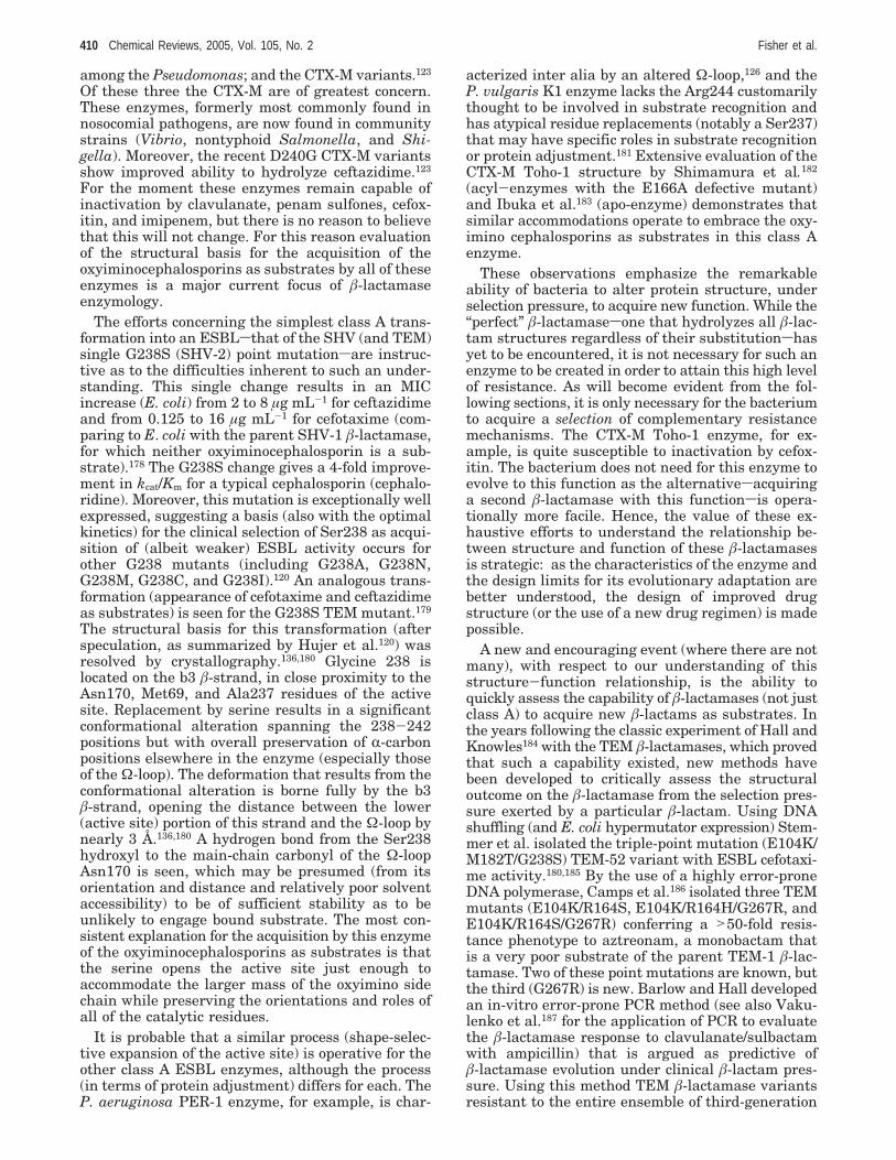
among the Pseudomonas; and the CTX-M variants.123
Of these three the CTX-M are of greatest concern.These enzymes, formerly most commonly found innosocomial pathogens, are now found in communitystrains (Vibrio, nontyphoid Salmonella, and Shi-gella). Moreover, the recent D240G CTX-M variantsshow improved ability to hydrolyze ceftazidime.123
For the moment these enzymes remain capable ofinactivation by clavulanate, penam sulfones, cefox-itin, and imipenem, but there is no reason to believethat this will not change. For this reason evaluationof the structural basis for the acquisition of theoxyiminocephalosporins as substrates by all of theseenzymes is a major current focus of â-lactamaseenzymology.
The efforts concerning the simplest class A trans-formation into an ESBLsthat of the SHV (and TEM)single G238S (SHV-2) point mutationsare instruc-tive as to the difficulties inherent to such an under-standing. This single change results in an MICincrease (E. coli) from 2 to 8 µg mL-1 for ceftazidimeand from 0.125 to 16 µg mL-1 for cefotaxime (com-paring to E. coli with the parent SHV-1 â-lactamase,for which neither oxyiminocephalosporin is a sub-strate).178 The G238S change gives a 4-fold improve-ment in kcat/Km for a typical cephalosporin (cephalo-ridine). Moreover, this mutation is exceptionally wellexpressed, suggesting a basis (also with the optimalkinetics) for the clinical selection of Ser238 as acqui-sition of (albeit weaker) ESBL activity occurs forother G238 mutants (including G238A, G238N,G238M, G238C, and G238I).120 An analogous trans-formation (appearance of cefotaxime and ceftazidimeas substrates) is seen for the G238S TEM mutant.179
The structural basis for this transformation (afterspeculation, as summarized by Hujer et al.120) wasresolved by crystallography.136,180 Glycine 238 islocated on the b3 â-strand, in close proximity to theAsn170, Met69, and Ala237 residues of the activesite. Replacement by serine results in a significantconformational alteration spanning the 238-242positions but with overall preservation of R-carbonpositions elsewhere in the enzyme (especially thoseof the Ω-loop). The deformation that results from theconformational alteration is borne fully by the b3â-strand, opening the distance between the lower(active site) portion of this strand and the Ω-loop bynearly 3 Å.136,180 A hydrogen bond from the Ser238hydroxyl to the main-chain carbonyl of the Ω-loopAsn170 is seen, which may be presumed (from itsorientation and distance and relatively poor solventaccessibility) to be of sufficient stability as to beunlikely to engage bound substrate. The most con-sistent explanation for the acquisition by this enzymeof the oxyiminocephalosporins as substrates is thatthe serine opens the active site just enough toaccommodate the larger mass of the oxyimino sidechain while preserving the orientations and roles ofall of the catalytic residues.
It is probable that a similar process (shape-selec-tive expansion of the active site) is operative for theother class A ESBL enzymes, although the process(in terms of protein adjustment) differs for each. TheP. aeruginosa PER-1 enzyme, for example, is char-
acterized inter alia by an altered Ω-loop,126 and theP. vulgaris K1 enzyme lacks the Arg244 customarilythought to be involved in substrate recognition andhas atypical residue replacements (notably a Ser237)that may have specific roles in substrate recognitionor protein adjustment.181 Extensive evaluation of theCTX-M Toho-1 structure by Shimamura et al.182
(acyl-enzymes with the E166A defective mutant)and Ibuka et al.183 (apo-enzyme) demonstrates thatsimilar accommodations operate to embrace the oxy-imino cephalosporins as substrates in this class Aenzyme.
These observations emphasize the remarkableability of bacteria to alter protein structure, underselection pressure, to acquire new function. While the“perfect” â-lactamasesone that hydrolyzes all â-lac-tam structures regardless of their substitutionshasyet to be encountered, it is not necessary for such anenzyme to be created in order to attain this high levelof resistance. As will become evident from the fol-lowing sections, it is only necessary for the bacteriumto acquire a selection of complementary resistancemechanisms. The CTX-M Toho-1 enzyme, for ex-ample, is quite susceptible to inactivation by cefox-itin. The bacterium does not need for this enzyme toevolve to this function as the alternativesacquiringa second â-lactamase with this functionsis opera-tionally more facile. Hence, the value of these ex-haustive efforts to understand the relationship be-tween structure and function of these â-lactamasesis strategic: as the characteristics of the enzyme andthe design limits for its evolutionary adaptation arebetter understood, the design of improved drugstructure (or the use of a new drug regimen) is madepossible.
A new and encouraging event (where there are notmany), with respect to our understanding of thisstructure-function relationship, is the ability toquickly assess the capability of â-lactamases (not justclass A) to acquire new â-lactams as substrates. Inthe years following the classic experiment of Hall andKnowles184 with the TEM â-lactamases, which provedthat such a capability existed, new methods havebeen developed to critically assess the structuraloutcome on the â-lactamase from the selection pres-sure exerted by a particular â-lactam. Using DNAshuffling (and E. coli hypermutator expression) Stem-mer et al. isolated the triple-point mutation (E104K/M182T/G238S) TEM-52 variant with ESBL cefotaxi-me activity.180,185 By the use of a highly error-proneDNA polymerase, Camps et al.186 isolated three TEMmutants (E104K/R164S, E104K/R164H/G267R, andE104K/R164S/G267R) conferring a >50-fold resis-tance phenotype to aztreonam, a monobactam thatis a very poor substrate of the parent TEM-1 â-lac-tamase. Two of these point mutations are known, butthe third (G267R) is new. Barlow and Hall developedan in-vitro error-prone PCR method (see also Vaku-lenko et al.187 for the application of PCR to evaluatethe â-lactamase response to clavulanate/sulbactamwith ampicillin) that is argued as predictive ofâ-lactamase evolution under clinical â-lactam pres-sure. Using this method TEM â-lactamase variantsresistant to the entire ensemble of third-generation
410 Chemical Reviews, 2005, Vol. 105, No. 2 Fisher et al.
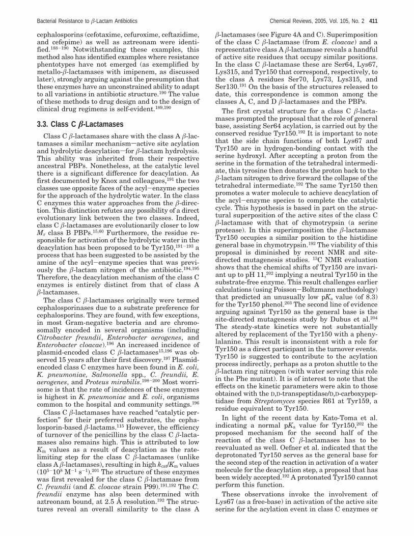
cephalosporins (cefotaxime, cefuroxime, ceftazidime,and cefepime) as well as aztreonam were identi-fied.188-190 Notwithstanding these examples, thismethod also has identified examples where resistancephentotypes have not emerged (as exemplified bymetallo-â-lactamases with imipenem, as discussedlater), strongly arguing against the presumption thatthese enzymes have an unconstrained ability to adaptto all variations in antibiotic structure.190 The valueof these methods to drug design and to the design ofclinical drug regimens is self-evident.189,190
3.3. Class C â-LactamasesClass C â-lactamases share with the class A â-lac-
tamases a similar mechanismsactive site acylationand hydrolytic deacylationsfor â-lactam hydrolysis.This ability was inherited from their respectiveancestral PBPs. Nonetheless, at the catalytic levelthere is a significant difference for deacylation. Asfirst documented by Knox and colleagues,191 the twoclasses use opposite faces of the acyl-enzyme speciesfor the approach of the hydrolytic water. In the classC enzymes this water approaches from the â-direc-tion. This distinction refutes any possibility of a directevolutionary link between the two classes. Indeed,class C â-lactamases are evolutionarily closer to lowMr class B PBPs.15,60 Furthermore, the residue re-sponsible for activation of the hydrolytic water in thedeacylation has been proposed to be Tyr150,191-193 aprocess that has been suggested to be assisted by theamine of the acyl-enzyme species that was previ-ously the â-lactam nitrogen of the antibiotic.194,195
Therefore, the deacylation mechanism of the class Cenzymes is entirely distinct from that of class Aâ-lactamases.
The class C â-lactamases originally were termedcephalosporinases due to a substrate preference forcephalosporins. They are found, with few exceptions,in most Gram-negative bacteria and are chromo-somally encoded in several organisms (includingCitrobacter freundii, Enterobacter aerogenes, andEnterobacter cloacae).196 An increased incidence ofplasmid-encoded class C â-lactamases15,196 was ob-served 15 years after their first discovery.197 Plasmid-encoded class C enzymes have been found in E. coli,K. pneumoniae, Salmonella spp., C. freundii, E.aerogenes, and Proteus mirabilis.198-200 Most worri-some is that the rate of incidences of these enzymesis highest in K. pneumoniae and E. coli, organismscommon to the hospital and community settings.196
Class C â-lactamases have reached “catalytic per-fection” for their preferred substrates, the cepha-losporin-based â-lactams.115 However, the efficiencyof turnover of the penicillins by the class C â-lacta-mases also remains high. This is attributed to lowKm values as a result of deacylation as the rate-limiting step for the class C â-lactamases (unlikeclass A â-lactamases), resulting in high kcat/Km values(105-108 M-1 s-1).201 The structure of these enzymeswas first revealed for the class C â-lactamase fromC. freundii (and E. cloacae strain P99).191,192 The C.freundii enzyme has also been determined withaztreonam bound, at 2.5 Å resolution.192 The struc-tures reveal an overall similarity to the class A
â-lactamases (see Figure 4A and C). Superimpositionof the class C â-lactamase (from E. cloacae) and arepresentative class A â-lactamase reveals a handfulof active site residues that occupy similar positions.In the class C â-lactamase these are Ser64, Lys67,Lys315, and Tyr150 that correspond, respectively, tothe class A residues Ser70, Lys73, Lys315, andSer130.191 On the basis of the structures released todate, this correspondence is common among theclasses A, C, and D â-lactamases and the PBPs.
The first crystal structure for a class C â-lacta-mases prompted the proposal that the role of generalbase, assisting Ser64 acylation, is carried out by theconserved residue Tyr150.192 It is important to notethat the side chain functions of both Lys67 andTyr150 are in hydrogen-bonding contact with theserine hydroxyl. After accepting a proton from theserine in the formation of the tetrahedral intermedi-ate, this tyrosine then donates the proton back to theâ-lactam nitrogen to drive forward the collapse of thetetrahedral intermediate.192 The same Tyr150 thenpromotes a water molecule to achieve deacylation ofthe acyl-enzyme species to complete the catalyticcycle. This hypothesis is based in part on the struc-tural superposition of the active sites of the class Câ-lactamase with that of chymotrypsin (a serineprotease). In this superimposition the â-lactamaseTyr150 occupies a similar position to the histidinegeneral base in chymotrypsin.192 The viability of thisproposal is diminished by recent NMR and site-directed mutagenesis studies. 13C NMR evaluationshows that the chemical shifts of Tyr150 are invari-ant up to pH 11,202 implying a neutral Tyr150 in thesubstrate-free enzyme. This result challenges earliercalculations (using Poisson-Boltzmann methodology)that predicted an unusually low pKa value (of 8.3)for the Tyr150 phenol.203 The second line of evidencearguing against Tyr150 as the general base is thesite-directed mutagenesis study by Dubus et al.204
The steady-state kinetics were not substantiallyaltered by replacement of the Tyr150 with a pheny-lalanine. This result is inconsistent with a role forTyr150 as a direct participant in the turnover events.Tyr150 is suggested to contribute to the acylationprocess indirectly, perhaps as a proton shuttle to theâ-lactam ring nitrogen (with water serving this rolein the Phe mutant). It is of interest to note that theeffects on the kinetic parameters were akin to thoseobtained with the D,D-transpeptidase/D,D-carboxypep-tidase from Streptomyces species R61 at Tyr159, aresidue equivalent to Tyr150.
In light of the recent data by Kato-Toma et al.indicating a normal pKa value for Tyr150,202 theproposed mechanism for the second half of thereaction of the class C â-lactamases has to bereevaluated as well. Oefner et al. indicated that thedeprotonated Tyr150 serves as the general base forthe second step of the reaction in activation of a watermolecule for the deacylation step, a proposal that hasbeen widely accepted.192 A protonated Tyr150 cannotperform this function.
These observations invoke the involvement ofLys67 (as a free-base) in activation of the active siteserine for the acylation event in class C enzymes or
Bacterial Resistance to â-Lactam Antibiotics Chemical Reviews, 2005, Vol. 105, No. 2 411
that it may abstract a proton from Tyr150, which inturn activates the serine. Furthermore, for the deacy-lation step, in the absence of a residue equivalent toGlu166 in class A â-lactamases, crystal structuresfrom E. cloacae P99191 and C. freundii192 argue forthe approach of the hydrolytic water from the â-faceof the â-lactam antibiotic. Bulychev et al. proposethat the nitrogen of the thiazoline (from â-lactamopening) is ideally positioned to promote hydrolyticwater addition to the acyl-enzyme to accomplishdeacylation.193 This would be an example of a sub-strate-assisted catalysis. Two non-â-lactam syntheticmolecules were used to test this concept. Thesecompounds were chemically predisposed to acylatethe active site serine in the E. cloacae class Câ-lactamase, resulting in acyl-enzyme species. Onecompound lacked the amine of the acyl-enzymespecies, whereas the other possessed it. The one withthe amine experienced turnover, whereas the onewithout it served merely as an irreversible inhibitorof the enzyme.194 A recent X-ray crystallographicstructure of an acyl-enzyme species of AmpC â-lac-tamase and moxalactam lends further evidence tothis proposal. The authors note that a water moleculeis organized to take advantage of the ring nitrogenin the hydrolytic step.195
Third-generation cephasloporins have been ef-fectively used as a strategy against class C â-lacta-mases for over a decade. However, class C â-lacta-mases capable of hydrolyzing most third-generationcephalosporins were first isolated in the 1980s205 andyet more recently from a virulent strain of E. cloa-cae.206 These enzymes, termed extended-spectrumâ-lactamases (ESBLs),124 mediate resistance to (ex-tended-spectrum) third-generation cephalosporins(exemplified by ceftazidime, cefotaxime, cefepime,and ceftriaxone) and monobactams (aztreonam) butdo not affect cephamycins (cefoxitin and cefotetan)or carbapenems (Meropenem or imipenem). Thestructural basis for resistance mediated by theseESBLs was revealed by the structure of the E. cloacaeGC1 enzyme, solved by Crichlow et al.207 Theseauthors suggest that conformational flexibility of theexpanded Ω-loop facilitates hydrolysis of third-generation cephalosporins by enabling greater mobil-ity of the acyl moiety. Further evidence implicatingthe Ω-loop modification with resistance to thesethird-generation cephalosporins came from the struc-tures of the AmpC â-lactamase in complex withvarious third-generation cephalosporins.208 A morerecent structure of a phosphonate transition-statemimetic bound to the E. cloacae GC1 and the wild-type C. freundii GN346 class C enzymes affordedfurther insight to the basis for resistance. It wasfound that the designer molecule adopted differentconformations in both enzymes, with the mutatedΩ-loop of the GC1 enzyme able to accommodate thecefotaxime side chain in a different conformation,enabling it to allow the approach of the hydrolyticwater to the acyl-enzyme species.209
3.4. Class D â-LactamasesThe class D â-lactamases are increasingly encoun-
tered among the defensive â-lactamase ensemble of
certain Gram-negative pathogens.15,109,124,210 Theseâ-lactamases were first termed as oxacillinases (andfor this reason are still described as OXA â-lactamasevariants) for their ability to hydrolyze the 5-methyl-3-phenylisoxazole-4-carboxy side chain penicillin class,exemplified by oxacillin and cloxacillin. Over 50 classD OXA variants are now known,211 including variantsthat have dispensed with their oxacillinase activityin the process of acquiring ESBL activity againstcarbapenem and third-generation cephalosporins.212
In the course of these transformations the class Denzymes have expanded from their historical P.aeruginosa niche 213,214 into other Gram-negativepathogens including E. coli,215,216 P. mirabilis,217
Salmonella sp.,218 K. pneumoniae,219,220 and especiallyAcinetobacter baumannii.221-228 While at present theclinical impact of the OXA â-lactamases is associatedwith infections by P. aeruginosa and A. baumannii,the widening Gram-negative distribution providespowerful support for the concern that the clinicalvalue of carbapenems (and third-generation cepha-losporins) may quickly diminish.211,220,222,225,229
These enzymes only recently have become thesubject of detailed structural and mechanistic stud-ies. This is due in some measure to their very recentappearance in clinically relevant pathogens (as dis-tinct from the other three â-lactamase classes) butin greater measure to their heterogeneous properties(not withstanding their interrelatedness) and thehistorical difficulty of their in vitro assay. Theliterature is replete with descriptions of poorly re-producible biphasic (burst-type) progress curves forsome variants, corresponding (in the apparent steady-state) to unusually low kcat/Km values (as exemplifiedby the data of Danel et al.,230 Franceschini et al.,231
Pernot et al.,232 and Heritier et al.226). Among theenzyme properties explored as possible explanationsfor these difficulties were a monomer-dimer equi-librium with possible additional divalent metal de-pendency.230,231,233,234 No credible explanation wasfound. For example, some OXA variants are mono-meric,97 and the dimer Kd in any case (typicallymicromolar) is relevant only to the in-vivo and notin-vitro kinetics, while other variants show littlecapacity for divalent metal binding.224 As a conse-quence of this dilemma, the possibility that othermechanisms (in addition to the OXA â-lactamase)contribute to the â-lactam resistance phenotypehas been discussed.221 Also, while the importance ofporin deletion211,216,219 and peptidoglycan remodeling(in addition to the assembly of mixed class â-lacta-mase ensembles or single â-lactamase hyperexpres-sion) to Gram-negative resistance cannot be under-estimated, the identity of the likely critical andconfounding variable contributing to the in vitroassay variabilitysCO2swas identified by Golemi etal.95,96
The structural basis by which CO2 activates theclass D enzymes for â-lactam hydrolysis was eluci-dated concurrent with several independent crystal-lographic studies of the class D â-lactamases. Thefirst studies on a class D enzymes (the P. aeruginosaOXA-10 â-lactamase) showed that the class D domainfolding was similar to the other two serine â-lacta-
412 Chemical Reviews, 2005, Vol. 105, No. 2 Fisher et al.
mase classes.235,236 However, subsequent crystal-lographic analysis revealed that the active site lysinewas N-carboxylated as a result of addition of thelysine side chain amine (as the free-base amine) toCO2.96,237 The resulting carbamic acid ionizes to givea carbamate functional group in hydrogen-bondingcontact with the active site serine. As the formationof this carbamate is reversible, the earlier reports ofthe absence of lysine carboxylation are explained. Inlight of the high physiological concentration of CO2(low millimolar) this lysine is expected to be fullycarboxylated in vivo.95 Kinetic analysis of a mutantenzyme where this lysine is replaced shows the totalloss of catalytic activity, indicating a direct involve-ment in catalysis.95 The role for this unusual lysine-derived carbamate is general base activation for bothacylation (activation of the serine) and deacylation(activation of water) steps of catalysis.95,237 Moreover,the assignment to this CO2-derived lysine carbamateof this role as general base catalyst is consistent withthe absence of alternative possibilities. Not only doesthe class D Ω-loop not contain a counterpart for theclass A Glu166, but the tyrosine of the conservedparental class D Y144-G145-N146 motif on this loopis replaced by phenylalanine in ESBL (both carbap-enem and cephalosporin) class D variants, obviatingdirect participation of this tyrosine in catalysis (asis more fully discussed elsewhere).97,222,226,238 TheOXA-1 crystal also shows the lysine carbamate97
whereas the OXA-13 enzyme232 does not (almostcertainly an artifact of crystallization). The relation-ship of the CO2 to the complicated kinetics extendsbeyond the relative portion of the lysines that areactivated for catalysis. During catalysis the lysinecarbamate is prone to spontaneous decarboxylationin the middle of the turnover process, thus arrestingcatalysis at the acyl-enzyme stage. This must,however, be regarded as an artifact of in vitro assaysince supplementation of the medium with bicarbon-ate (as a CO2 source) restores the kinetic profile.95 Ithas been argued that the more complicated biphasicturnover profile for these enzymes with some sub-strates is due to a branching mechanism. As theenzyme experiences decarboxylation midcatalysis, itbecomes inactivated (the branching species), pendingthe availability of a CO2 molecule to restore the lysineto its active carboxylated form. The enzyme is thenable to complete the second step of catalysis.95,236
With a few substrates, however, it was shown thatsupplementation of the medium with bicarbonatedoes not simplify the turnover process. For these fewcases a branching mechanism (as might occur by aconformational change at the acyl-enzyme state) hasbeen invoked.
[Dr. Roger Labia, a pioneer of studies of â-lacta-mases, kindly communicated that his early investi-gations of the class D â-lactamases in 1970s werefrustrating because of the complicated and erratickinetic behavior of the enzymes. He opted to abandonstudies of the class D enzymes. He now attributesthe erratic behavior of the enzymes to the seasonalvariations in the quality of the water, which has highcarbonation in the summers and low carbonation inthe winters.]
While there are numerous reports evaluating theâ-lactam substrate profile for the class D OXAenzymes, essentially all of these predate the discov-ery of requirement for in-vitro CO2 (but most unlikelyin vivo) activation. These earlier data may not bereliable. Nonetheless, all these data suggest that thesubstrate profile for these enzymes, individually andas a class, is not broad.211 The value of the class Denzymes to bacteria is the ability of this enzyme toadapt, under selection pressure, to the specific â-lac-tam. For example, while most of the ESBL OXAvariants hydrolyze ceftazidime better than cefepime,the reverse is true for the OXA-30 variant (derivedfrom OXA-1).215 Full discussions of the orientation(and contacts) within the class D active site for(inhibitor) acyl-enzymes232,237 and possible orienta-tions for substrates97 are presented elsewhere.
A final issue is the origin of the class D enzymesin comparison to the two other serine â-lactamaseclasses. All three classes are now encountered as bothchromosomal- and plasmid-borne genes. However,the evolutionary history of each class is distinct.15 Incontrast to the class C AmpC family, where themobilization to plasmids is a modern (antibiotic era)event, plasmid mobilization of the class A TEM andclass D OXA are ancient events.239 The primarydistinction between the class A and C â-lactamasesis the much more rapid and extensive diversificationsat this point in timesof the former within the Gram-negative bacteria. Also of particular interest is thecomplete absence of this diversification within theGram-positive bacteria given the estimate by Halland Barlow that the horizontal transfer of the classC gene from the Gram-negative to the Bacillus Gram-positive bacteria is an ancient event of some 320-575Myr (but is after the divergence of B. subtilis fromS. aureus).15 A homology between the hydrophiliccarboxy domain of the BlaR/MecR â-lactam-signalingreceptors and the class D â-lactamases was notedpreviously.84,240 The recent discovery within B. sub-tilis of a gene encoding an enzyme with weak classD â-lactamase activity, yet also resembling a penicil-lin-binding protein,241 suggests that study of the classD gene within those Gram-positive bacteria thatpossess it may further refine our understanding ofthe structural and functional relationships betweenthe PBP and â-lactamase enzymes.
3.5. Class B Metallo- â-lactamasesThe secondsand in many respects no less fore-
bodingsvision of the â-lactamase future is that ofthe disseminated metallo-â-lactamases (MBLs).242
First observed in 1967 by Kawabata and Abrahamas chromosomal enzymes of the innocuous Gram-positive B. cereus, these enzymes occupy a positionof concern (in terms of breadth of distribution andbreadth of â-lactam catalytic activity) with respectto the inexorable expansion of â-lactam resis-tance.109,229,243,244 These metal-dependent (almost al-ways divalent zinc) â-lactamases have a broad â-lac-tam substrate tolerance that encompasses many ofthe newer generation cephalosporins, carbapenems,and other â-lactamase inhibitory (clavulanate andpenam sulfones) â-lactams important to the treat-
Bacterial Resistance to â-Lactam Antibiotics Chemical Reviews, 2005, Vol. 105, No. 2 413
ment of Gram-negative infection.210,229 As a differentchemical mechanism (compared to the serine â-lac-tamases) is used by the metallo-â-lactamases forâ-lactam hydrolysissnotably, a mechanism withouta covalent enzyme intermediatesan entirely differentstrategy for their inhibition (or inactivation) will berequired should (or is it when) these enzymes expandbeyond their present niche (that of minor, andopportunistic, Gram-negative pathogens). While sev-eral inhibition strategies have been identified, nonehas yielded anything resembling that of a clinicallyeffective inhibitor. Also, although there exist as yetchemical reasons (in terms of kcat/Km these are notoptimized enzymes) and biochemical reasons (zinc asa limiting nutrient) that ultimately may limit the rolethe metallo-â-lactamases will have as a resistancemechanism (vide infra), the proposal discussed byFast et al.,245 Fabiane et al.,246 and Wommer et al.247
that these are young enzymes, only now in theprocess of maturation under evolutionary pressure,is both credible and worrisome.
The metallo-â-lactamases (also termed Amblerclass B and Bush-Jacoby-Medeiros Group 3 â-lac-tamases)105,243 are small enzymes sharing a commonfour-layer RââR motif with a central â-sandwich andtwo R-helices on either side.246,248 This motif, arisingpossibly by a gene duplication event,60,245 is found alsoin other proteins (glyoxalase and certain flavoen-zymes). The motif has an intrinsic metal-binding sitelocated at an edge of the â-sandwich.249,250 For themetallo-â-lactamases this site is occupied by a diva-lent zinc ion having a tetrahedral array of threehistidines and water. The importance of the zinc ionto â-lactam substrate binding is unquestionable.251,252
The role of the zinc in the hydrolytic mechanism,beyond that of Lewis acid catalysis, is less certain.Nonetheless, there is a consensus that the waterligand of the zinc ion is the â-lactam ring-openingnucleophile (via a mechanism likely with parallel tothat of the zinc metalloproteases), but there are nodirect data establishing this role.
Beyond these structural commonalities the met-allo-â-lactamases possess a surprising breadth ofprimary structure that most notably includes thecreation (in some enzymes) of a second zinc bindingsite.243 In these binuclear enzymes the two zincs areproximal (approximately a 3.5 Å separation) withboth participating in the water coordination. Theligand environment of the second zinc ion is verydifferent from that for the first both in array (trigonalbipyramid) and amino acid ligands (variable amongthe binuclear class). The purpose of the second zincion is regarded as catalytic augmentationsthat is,accomplishing an incremental increase in the â-lac-tam substrate kcat/Kmsand not a role of catalyticnecessity.245,247,253-255 Nonetheless, the specific envi-ronment of the zinc ensemble determines the indi-vidual enzyme catalytic behavior toward substratesand the detail of the rate-limiting (highest energy)catalytic step.245,255-259 The relative affinity of theenzyme for the two zinc ions is unequal, and in-vitroenzyme kinetic analysis requires added Zn2+.247,255,260
Wommer et al.247 discussed in detail the role of theâ-lactam substrate in the recruitment of zinc to, and
hence activation of, the â-lactamase. This process isargued as one of physiological necessity wherein theâ-lactamase exists as an apoenzyme and the avail-able zinc is reserved to other enzymes until theâ-lactam antibiotic is encountered. The likelihood ofa fully zinc complemented metallo-â-lactamase (bi-nuclear site fully occupied) in vivo is regarded assmall. It is therefore understandable why the bacte-rium possessing a metallo-â-lactamases is often foundwith a serine â-lactamase as well.261,262 The demon-strable fact that these enzymes are disseminating isproof of evolutionary pressure for resistance develop-ment within human clinical practice.
While the details245,255,263 of the hydrolytic mecha-nism used by these enzymes is beyond the scope ofthis review, the mechanistic fundamental is assur-edly not. This fundamental is delivery of the zinc-coordinated water, possibly as a hydroxide ion (pKa
) 4.9 to 5.6),245,260,264,265 to the â-lactam carbonyl. (Themetalloprotease carboxypeptidase A has a similarcatalytic pKa that is also assigned to a zinc hydroxide.However, the recent 67Zn NMR study by Lipton etal. forcefully argues against this assumption.266 Ac-cordingly, a wholesale mechanistic revision for met-allo-â-lactamase catalysis may be necessary.) Thepresumed involvement of a zinc hydroxide intermedi-ate for the metallo-â-lactamases has stimulatedexquisite studies on the in-vitro mechanism of metal-catalyzed â-lactam solvolysis.34,267-269 These studiessuggest the rate-limiting step to be either metal-coordinated hydroxide addition or (following thehydroxide addition) the collapse of the tetrahedralspecies. The mechanism is dependent on â-lactamstructure in such fashion as to strongly implicatezinc coordination of the â-lactam in the transitionstate.34,252,253,256,270-272 A similar mechanismsthat is,hydrolysis without a covalent enzyme intermediatesis posited for the enzymatic reaction. Solvent kineticisotope effects for the enzymatic reaction implicateadditional transition-state stabilization by protonflight.255,260 The identity of the enzymatic protondonor/acceptor, however, remains a particular focusof mechanistic study. A notable commonality of thestructure-kinetics analysis of the nonenzymatic sol-volysis and the comparative enzymatic structure-kinetics is the particular susceptibility of the car-bapenems to hydrolysis.34,229,269,270
This realitysthe susceptibility toward metallo-â-lactamase hydrolysis of nearly all (vide infra) of theserine â-lactamase inhibitorssis a growing concern.There are three reasons for this. The first is theburgeoning presence of metallo-â-lactamases (firstthe IMP and now the VIM and SMP class B1variants) as mobile plasmid-encoded (and often alsoas integron, or cassette) genes by the enterobacte-ria.273,274 Second, these enzyme variants have aremarkable ability to alter their substrate capability(as can be accomplished by simple-point mutation,even remote to the active site), raising the possibilityof rapid adjustment to encompass new substrates(and to thwart new inhibitors). Last, the metallo-â-lactamase plasmids often encode additional (multi-substrate) resistance mechanisms.
414 Chemical Reviews, 2005, Vol. 105, No. 2 Fisher et al.
The class B1 metallo-â-lactamases are monomeric,binuclear zinc enzymes constituting the largest met-allo-â-lactamase subclass. Within this subclass thedominant subtypes are the IMP and VIM enzymes.While these subtype enzymes are defined by se-quence, substantial diversity is found within each.For example, the recently observed IMP-12 varianthas 89% sequence identity to its closest (IMP-8)relative but includes 10 amino acid changes atpositions that are otherwise invariant among the 11other subtypes.275 The IMP enzymes are originallyAsian (and the VIM enzymes originally European),but it is quite evident that geographical characteriza-tion of both of these enzymes is irrelevant. Firstobserved in 1988, the IMP enzymes have a broadsubstrate (primarily cephalosporins and carbapen-ems, less so penicillins) acceptance, although at thelevel of specific â-lactam structure the kcat/Km varia-tion can be substantial. Replacement of glycine-196(a noncatalytic residue adjacent to His197, a zincligand) in the IMP-3 and IMP-6 variants with serine(as is found in IMP-1) results in a significant kcat/Km
improvement toward certain cephalosporins andtoward imipenem (IMP-1 kcat/Km is 10-50-fold greatercompared to IMP-3).272,276 Nonetheless, a comprehen-sive in vitro substrate evaluation of IMP-6 andmicrobiological evaluation of E. coli possessing theTEM-1/IMP-6 plasmid indicate the IMP-6 to be thebetter enzyme in terms of extended carbapenemaseactivity.277 Whereas Meropenem and imipenem areessentially equivalent (as measured by kcat/Km) IMP-1substrates, the IMP-6 glycine replacement results ina 6-fold improvement in the Meropenem kcat/Km (anda 2-fold loss for imipenem). This improved kcat/Km
contributes to the significant difference in MIC (E.coli with the IMP-1/TEM-1 plasmid, 64 µg mL-1 forMeropenem and 2-8 µg mL-1 for imipenem; without,0.25 µg mL-1).277 Efforts to correlate IMP sequencewith altered substrate acceptance have been madeby Oelschlaeger et al. (comparing G196 IMP-6 toS196 IMP-1)272 and Moali et al. (evaluating the roleof the distal 60-66 loop).271 In the former study afavorable serine-196-lysine-33 interaction improvespacking and rigidifies histidine-197; this rigiditypropagates throughout the active site. A calculatedenzyme-substrate stability index was found to cor-relate well to the experimental kcat/Km. In the latterstudy the loop was confirmed as nonessential forcatalysis but contributed (especially tryptophan-64)to hydrophobic substrate binding. A mutagenesisstudy by Materon and Palzkill of IMP-1 active siteproximal amino acids identified 52% of the IMP-1amino acids as intolerant to substitution (by com-parison, the TEM-1 value is 33%).278 Materon andPalzkill278 suggest that the IMP metallo-â-lactamasesmay have a relatively more limited ability to adjustto “extended spectrum” â-lactam structure comparedto the TEM enzymes (which have, of course, alreadydone so). A relatedsand no less pertinentsquestionis whether the metallo-â-lactamases have the capa-bility to develop their catalytic apparatus to functionat the substrate diffusion limit (that is, at fullcatalytic competence), as is already the case for theclass A and C serine â-lactamases.115 The extant IMP
kinetic data indicate that while certain cephalospor-ins have kcat/Km values that approach the diffusionlimit of (107-108 M-1 s-1), most substrates have lowervalues (typical carbapenem kcat/Km values are ap-proximately 106 M-1 s-1). Hall’s full mutagenesis in-vitro evaluation of the IMP-1 structure, which failedto identify a mutant enzyme more capable of imi-penem hydrolysis, is consistent with one (or both) ofthese possibilities.279 As the diversity of known car-bapenem structure is not nearly that of the penicillinsand cephalosporins, cautious optimism may be en-tertained that newer generation â-lactams poorlycapable of metallo-â-lactamase hydrolysis may yet bemade.
Two additional aspects may temper this conclusion.It is clearly not possible to determine, by evaluationof enzyme sequence or enzyme kinetics, a “direction”for metallo-â-lactamase variant evolution (whichvariant arose from which variant). Hence, the ap-parent evolutionary limitation of IMP-1 with imi-penem has no predictive value with respect to othermetallo-â-lactamase variants. As bluntly stated byHall, “in order to understand the risks posed bymetallo-â-lactamases, it will be necessary to conductsimilar studies on representative members of eachof the three metallo-â-lactamase subfamilies and toinclude all clinically relevant carbapenems”.279 Sec-ond, it is evident that incremental changes in â-lac-tam fitnesssin terms of PBP inactivation and com-petence as a substrate for â-lactamase hydrolysisssuffice to determine whether a bacterium is susceptibleor resistant. An effect need not be dramatic to beimportant.
The VIM B1 subclass is newer (first observed in1997) and biochemically less well studied.280 A VIMsequence homology with IMP is recognizable (ap-proximately 30-40%), and the overall pattern ofâ-lactam substrate recognition by the two enzymesis similar.229 Seven VIM variants are extant.281 In lessthan 7 years the VIM metallo-â-lactamases havetransitioned from chromosomal expression by non-fermenting Gram-negative bacteria (where it con-tributes to high-level antibiotic resistance in P.aeruginosa pathogenic strains)282,283 to transferableplasmid expression in Gram-negative enterobacte-ria.262,273 The presumptive circumstances leading tothis change (evolutionary pressure for plasmid dis-semination is not necessarily carbapenem but rathermultidrug driven) and probable consequence of thischange (likelihood of carbapenem clinical failure)underscore the caution expressed in the previousparagraph.
A consistent observation from the in vitro evalua-tion of the metallo-â-lactamase substrate spectrumis the inability of these enzymes to hydrolyze themonocyclic N-sulfonyl â-lactam antibiotic aztreonam.Consequently, bacteria that have these metallo-â-lactamases can retain aztreonam susceptibility (al-though moderate to substantial increases in theaztreonam MIC values, due to other plasmid-con-ferred resistance mechanisms or to the presence ofaztreonam-capable serine â-lactamases, is common).As the intrinsic reactivity toward solvolysis of theaztreonam â-lactam is identical to that of the penicil-
Bacterial Resistance to â-Lactam Antibiotics Chemical Reviews, 2005, Vol. 105, No. 2 415
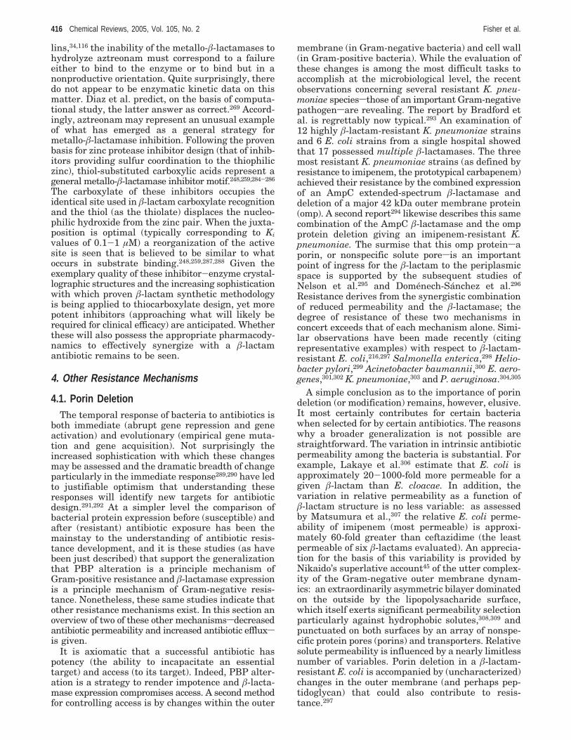
lins,34,116 the inability of the metallo-â-lactamases tohydrolyze aztreonam must correspond to a failureeither to bind to the enzyme or to bind but in anonproductive orientation. Quite surprisingly, theredo not appear to be enzymatic kinetic data on thismatter. Diaz et al. predict, on the basis of computa-tional study, the latter answer as correct.269 Accord-ingly, aztreonam may represent an unusual exampleof what has emerged as a general strategy formetallo-â-lactamase inhibition. Following the provenbasis for zinc protease inhibitor design (that of inhib-itors providing sulfur coordination to the thiophiliczinc), thiol-substituted carboxylic acids represent ageneral metallo-â-lactamase inhibitor motif.248,259,284-286
The carboxylate of these inhibitors occupies theidentical site used in â-lactam carboxylate recognitionand the thiol (as the thiolate) displaces the nucleo-philic hydroxide from the zinc pair. When the juxta-position is optimal (typically corresponding to Kivalues of 0.1-1 µM) a reorganization of the activesite is seen that is believed to be similar to whatoccurs in substrate binding.248,259,287,288 Given theexemplary quality of these inhibitor-enzyme crystal-lographic structures and the increasing sophisticationwith which proven â-lactam synthetic methodologyis being applied to thiocarboxylate design, yet morepotent inhibitors (approaching what will likely berequired for clinical efficacy) are anticipated. Whetherthese will also possess the appropriate pharmacody-namics to effectively synergize with a â-lactamantibiotic remains to be seen.
4. Other Resistance Mechanisms
4.1. Porin DeletionThe temporal response of bacteria to antibiotics is
both immediate (abrupt gene repression and geneactivation) and evolutionary (empirical gene muta-tion and gene acquisition). Not surprisingly theincreased sophistication with which these changesmay be assessed and the dramatic breadth of changeparticularly in the immediate response289,290 have ledto justifiable optimism that understanding theseresponses will identify new targets for antibioticdesign.291,292 At a simpler level the comparison ofbacterial protein expression before (susceptible) andafter (resistant) antibiotic exposure has been themainstay to the understanding of antibiotic resis-tance development, and it is these studies (as havebeen just described) that support the generalizationthat PBP alteration is a principle mechanism ofGram-positive resistance and â-lactamase expressionis a principle mechanism of Gram-negative resis-tance. Nonetheless, these same studies indicate thatother resistance mechanisms exist. In this section anoverview of two of these other mechanismssdecreasedantibiotic permeability and increased antibiotic effluxsis given.
It is axiomatic that a successful antibiotic haspotency (the ability to incapacitate an essentialtarget) and access (to its target). Indeed, PBP alter-ation is a strategy to render impotence and â-lacta-mase expression compromises access. A second methodfor controlling access is by changes within the outer
membrane (in Gram-negative bacteria) and cell wall(in Gram-positive bacteria). While the evaluation ofthese changes is among the most difficult tasks toaccomplish at the microbiological level, the recentobservations concerning several resistant K. pneu-moniae speciessthose of an important Gram-negativepathogensare revealing. The report by Bradford etal. is regrettably now typical.293 An examination of12 highly â-lactam-resistant K. pneumoniae strainsand 6 E. coli strains from a single hospital showedthat 17 possessed multiple â-lactamases. The threemost resistant K. pneumoniae strains (as defined byresistance to imipenem, the prototypical carbapenem)achieved their resistance by the combined expressionof an AmpC extended-spectrum â-lactamase anddeletion of a major 42 kDa outer membrane protein(omp). A second report294 likewise describes this samecombination of the AmpC â-lactamase and the ompprotein deletion giving an imipenem-resistant K.pneumoniae. The surmise that this omp proteinsaporin, or nonspecific solute poresis an importantpoint of ingress for the â-lactam to the periplasmicspace is supported by the subsequent studies ofNelson et al.295 and Domenech-Sanchez et al.296
Resistance derives from the synergistic combinationof reduced permeability and the â-lactamase; thedegree of resistance of these two mechanisms inconcert exceeds that of each mechanism alone. Simi-lar observations have been made recently (citingrepresentative examples) with respect to â-lactam-resistant E. coli,216,297 Salmonella enterica,298 Helio-bacter pylori,299 Acinetobacter baumannii,300 E. aero-genes,301,302 K. pneumoniae,303 and P. aeruginosa.304,305
A simple conclusion as to the importance of porindeletion (or modification) remains, however, elusive.It most certainly contributes for certain bacteriawhen selected for by certain antibiotics. The reasonswhy a broader generalization is not possible arestraightforward. The variation in intrinsic antibioticpermeability among the bacteria is substantial. Forexample, Lakaye et al.306 estimate that E. coli isapproximately 20-1000-fold more permeable for agiven â-lactam than E. cloacae. In addition, thevariation in relative permeability as a function ofâ-lactam structure is no less variable: as assessedby Matsumura et al.,307 the relative E. coli perme-ability of imipenem (most permeable) is approxi-mately 60-fold greater than ceftazidime (the leastpermeable of six â-lactams evaluated). An apprecia-tion for the basis of this variability is provided byNikaido’s superlative account45 of the utter complex-ity of the Gram-negative outer membrane dynam-ics: an extraordinarily asymmetric bilayer dominatedon the outside by the lipopolysacharide surface,which itself exerts significant permeability selectionparticularly against hydrophobic solutes,308,309 andpunctuated on both surfaces by an array of nonspe-cific protein pores (porins) and transporters. Relativesolute permeability is influenced by a nearly limitlessnumber of variables. Porin deletion in a â-lactam-resistant E. coli is accompanied by (uncharacterized)changes in the outer membrane (and perhaps pep-tidoglycan) that could also contribute to resis-tance.297
416 Chemical Reviews, 2005, Vol. 105, No. 2 Fisher et al.
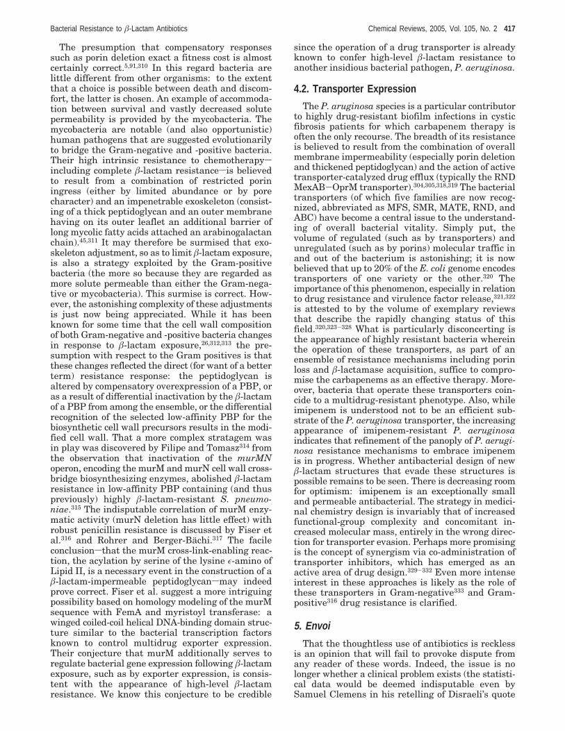
The presumption that compensatory responsessuch as porin deletion exact a fitness cost is almostcertainly correct.5,91,310 In this regard bacteria arelittle different from other organisms: to the extentthat a choice is possible between death and discom-fort, the latter is chosen. An example of accommoda-tion between survival and vastly decreased solutepermeability is provided by the mycobacteria. Themycobacteria are notable (and also opportunistic)human pathogens that are suggested evolutionarilyto bridge the Gram-negative and -positive bacteria.Their high intrinsic resistance to chemotherapysincluding complete â-lactam resistancesis believedto result from a combination of restricted poriningress (either by limited abundance or by porecharacter) and an impenetrable exoskeleton (consist-ing of a thick peptidoglycan and an outer membranehaving on its outer leaflet an additional barrier oflong mycolic fatty acids attached an arabinogalactanchain).45,311 It may therefore be surmised that exo-skeleton adjustment, so as to limit â-lactam exposure,is also a strategy exploited by the Gram-positivebacteria (the more so because they are regarded asmore solute permeable than either the Gram-nega-tive or mycobacteria). This surmise is correct. How-ever, the astonishing complexity of these adjustmentsis just now being appreciated. While it has beenknown for some time that the cell wall compositionof both Gram-negative and -positive bacteria changesin response to â-lactam exposure,26,312,313 the pre-sumption with respect to the Gram positives is thatthese changes reflected the direct (for want of a betterterm) resistance response: the peptidoglycan isaltered by compensatory overexpression of a PBP, oras a result of differential inactivation by the â-lactamof a PBP from among the ensemble, or the differentialrecognition of the selected low-affinity PBP for thebiosynthetic cell wall precursors results in the modi-fied cell wall. That a more complex stratagem wasin play was discovered by Filipe and Tomasz314 fromthe observation that inactivation of the murMNoperon, encoding the murM and murN cell wall cross-bridge biosynthesizing enzymes, abolished â-lactamresistance in low-affinity PBP containing (and thuspreviously) highly â-lactam-resistant S. pneumo-niae.315 The indisputable correlation of murM enzy-matic activity (murN deletion has little effect) withrobust penicillin resistance is discussed by Fiser etal.316 and Rohrer and Berger-Bachi.317 The facileconclusionsthat the murM cross-link-enabling reac-tion, the acylation by serine of the lysine ε-amino ofLipid II, is a necessary event in the construction of aâ-lactam-impermeable peptidoglycansmay indeedprove correct. Fiser et al. suggest a more intriguingpossibility based on homology modeling of the murMsequence with FemA and myristoyl transferase: awinged coiled-coil helical DNA-binding domain struc-ture similar to the bacterial transcription factorsknown to control multidrug exporter expression.Their conjecture that murM additionally serves toregulate bacterial gene expression following â-lactamexposure, such as by exporter expression, is consis-tent with the appearance of high-level â-lactamresistance. We know this conjecture to be credible
since the operation of a drug transporter is alreadyknown to confer high-level â-lactam resistance toanother insidious bacterial pathogen, P. aeruginosa.
4.2. Transporter Expression
The P. aruginosa species is a particular contributorto highly drug-resistant biofilm infections in cysticfibrosis patients for which carbapenem therapy isoften the only recourse. The breadth of its resistanceis believed to result from the combination of overallmembrane impermeability (especially porin deletionand thickened peptidoglycan) and the action of activetransporter-catalyzed drug efflux (typically the RNDMexAB-OprM transporter).304,305,318,319 The bacterialtransporters (of which five families are now recog-nized, abbreviated as MFS, SMR, MATE, RND, andABC) have become a central issue to the understand-ing of overall bacterial vitality. Simply put, thevolume of regulated (such as by transporters) andunregulated (such as by porins) molecular traffic inand out of the bacterium is astonishing; it is nowbelieved that up to 20% of the E. coli genome encodestransporters of one variety or the other.320 Theimportance of this phenomenon, especially in relationto drug resistance and virulence factor release,321,322
is attested to by the volume of exemplary reviewsthat describe the rapidly changing status of thisfield.320,323-328 What is particularly disconcerting isthe appearance of highly resistant bacteria whereinthe operation of these transporters, as part of anensemble of resistance mechanisms including porinloss and â-lactamase acquisition, suffice to compro-mise the carbapenems as an effective therapy. More-over, bacteria that operate these transporters coin-cide to a multidrug-resistant phenotype. Also, whileimipenem is understood not to be an efficient sub-strate of the P. aeruginosa transporter, the increasingappearance of imipenem-resistant P. aeruginosaindicates that refinement of the panoply of P. aerugi-nosa resistance mechanisms to embrace imipenemis in progress. Whether antibacterial design of newâ-lactam structures that evade these structures ispossible remains to be seen. There is decreasing roomfor optimism: imipenem is an exceptionally smalland permeable antibacterial. The strategy in medici-nal chemistry design is invariably that of increasedfunctional-group complexity and concomitant in-creased molecular mass, entirely in the wrong direc-tion for transporter evasion. Perhaps more promisingis the concept of synergism via co-administration oftransporter inhibitors, which has emerged as anactive area of drug design.329-332 Even more intenseinterest in these approaches is likely as the role ofthese transporters in Gram-negative333 and Gram-positive316 drug resistance is clarified.
5. Envoi
That the thoughtless use of antibiotics is recklessis an opinion that will fail to provoke dispute fromany reader of these words. Indeed, the issue is nolonger whether a clinical problem exists (the statisti-cal data would be deemed indisputable even bySamuel Clemens in his retelling of Disraeli’s quote
Bacterial Resistance to â-Lactam Antibiotics Chemical Reviews, 2005, Vol. 105, No. 2 417

about statistics)6,334-338 nor largely what could (orshould) be done to forestall the inevitable crisis butrather how to invest today in the scientific andmedical strategies that will provide preparednesstomorrow. Jefferson’s insightful connection betweenvigilance and liberty is no less appropriate to parlousgeopolitical circumstances as it is the inexorableprogression of resistant human pathogens. Two ques-tions address the relationship of scientific vigilanceto the future role of the â-lactam antibacterials inthe treatment of infections. Do the â-lactams remaina viable template for drug discovery? Do their targetssthe enzymes of cell wall biosynthesissremain aviable target for antibacterial design?
The former question is unquestionably the morecontentious, especially given the precipitous declinein pharma investment in antibacterial discov-ery.2,339-343 The choice of template, always paramountto ultimate success in drug discovery, is yet more sowhen resources are limited. Notwithstanding thehistorical dominance of the â-lactams, which contin-ues to this very day, the trend toward greater massand functional-group complexity in successive â-lac-tam generations strongly suggests that design limitsare being approached. In the past these conceptuallimits have been breached by revelation from Naturesa vastly more imaginative engineer of chemicalstructure than mansbut here as well pharma invest-ment is in decline.344,345 It is evident that the era ofmedicinal chemistry manipulation of the â-lactam,guided by the paradigm of iterative optimization ofMIC values, is coming to an end. This is not to say,however, that future medicinal chemistry effortstoward â-lactam optimization are exercises in futility.Rather, future antibacterial discovery (not just â-lac-tams) will follow the much more complicated processwherein chemical structure is evaluated in terms ofthe interrelationships among bacterial genomics andproteomics relating to several antibacterial tar-gets.290-292,346,347 As has been noted on several occa-sions within this review, bacterial resistance as aphenotype is the result of multiple compensatoryadjustments, many of which are incremental. Anexample of the possible fragility of this accommoda-tion (and as well the depth of our ignorance as tothese compensatory adjustments) is the synergisticand compensatory relationship among the bacterialgenetic background, â-lactamase expression, and themaintenance and expression of plasmid-carried mecAin â-lactam-resistant S. aureus, as discussed byKatayama et al.348 This observation would immedi-ately suggest the beneficial combination of a â-lactamtargeting the PBPs with a â-lactam targeting theâ-lactamase, which has been of course a mainstay ofclinical therapy for some two decades. The conclusionthat this combination therapy would prove successfulis arguably, in retrospect, facile. Whether futureantibacterial therapy of multidrug-resistant micro-organisms will also require complex drug combina-tions (as is already the case for anti-HIV therapy) isnot nearly as obvious. This is, nonetheless, a likelycourse of events. Fortunately, the experimental re-sources needed to accomplish this task are cominginto place. Single-cell microscopy, for example, may
allow one to identify the role of the target in cell wallbiosynthesis as the cell is seen to respond to drugexposure.62,349-352 This response can be correlated toproteomic analysis.289,291,353 The understanding of thestructural basis for â-lactam induction of the bacte-rial SOS response354 and for creation of persisterbacteria populations290,355,356 are but two examples ofhow future â-lactam SAR may be guided.357 Screen-ing to identify drug combinations that synergize withâ-lactams (as is currently being done with effluxpump inhibitors), with respect to target pairings, canbe done on a high-throughput scale. Old â-lactams,with proven safety and performance, can be givencontinued clinical relevance.358 Nonetheless, the like-lihood that future antibacterial chemotherapy will bea multidrug regimen is real.359
The task of identifying and then optimizing mul-tidrug safety and efficacy is daunting. Drug discoveryis already the zenith of the collaboration betweenhuman scientific and engineering ingenuity, and theemerging anticipation that this may now need to bedone on a multifactorial scale is part of the reasonthat the economics for future antimicrobial drugdiscovery are so dismal. While these economics maychange after the crisis (as only a crisis galvanizesconsensus of opinion), the prudent and soberingreminder is that successful drug discovery is em-phatically not instantaneous. Should our vigilance inantibacterial drug discovery falter, the length anddepth of the crisis may be unlike anything modernman has experienced.
There remain, of course, the companion questionsas to what should be done now to preserve â-lactamefficacy for future generations. There is no other anti-bacterial class that can substitute for â-lactam anti-biotics in the foreseeable future, and none are in thepipeline.4,360 These are questions that involve the en-tire breadth of clinical practice, including minimizingglobal environmental antibiotic exposure (the reduc-tion of antibiotics in animal feeds is likely a step inthe right direction), re-appreciating the value of hos-pital quarantine, and re-emphasizing the incredibleimportance of proper hygiene to minimizing infection.At the level of drug therapy, additional possibilitiesare emerging. We now appreciate that the gut is, touse the delectable phrasing of Courvalin and Davies,4a “veritable microbial bordello” with extensive capac-ity for genetic exchange.361,362 Exploratory therapiesthat include â-lactamases (to destroy nonabsorbedâ-lactams, either in situ or post facto) have shownpromise.363-366 The interrelationship of community-and hospital-resistant microorganism reservoirs nowis recognized.367 Judicious use of early generationâ-lactam therapy can mitigate resistance develop-ment against later generation â-lactams.358,368-371
The central importance of the cell wall biosyntheticenzymes as antibacterial targets is irrefutably vali-dated by the â-lactams themselves. Not only arethese enzymes accessible and essential, but also theseare enzymes with a demonstrated commonality forinhibitor (and substrate) recognition at their activesites. The proposal that the â-lactams constitute theonly motif for inhibitor design is not merely unprovenbut largely untested.372 Antibacterial screening is
418 Chemical Reviews, 2005, Vol. 105, No. 2 Fisher et al.
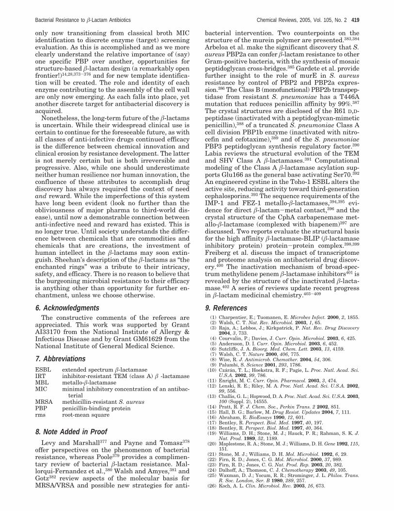
only now transitioning from classical broth MICidentification to discrete enzyme (target) screeningevaluation. As this is accomplished and as we moreclearly understand the relative importance of (say)one specific PBP over another, opportunities forstructure-based â-lactam design (a remarkably openfrontier!)14,28,373-376 and for new template identifica-tion will be created. The role and identity of eachenzyme contributing to the assembly of the cell wallare only now emerging. As each falls into place, yetanother discrete target for antibacterial discovery isacquired.
Nonetheless, the long-term future of the â-lactamsis uncertain. While their widespread clinical use iscertain to continue for the foreseeable future, as withall classes of anti-infective drugs continued efficacyis the difference between chemical innovation andclinical erosion by resistance development. The latteris not merely certain but is both irreversible andprogressive. Also, while one should underestimateneither human resilience nor human innovation, theconfluence of these attributes to accomplish drugdiscovery has always required the context of needand reward. While the imperfections of this systemhave long been evident (look no further than theobliviousness of major pharma to third-world dis-ease), until now a demonstrable connection betweenanti-infective need and reward has existed. This isno longer true. Until society understands the differ-ence between chemicals that are commodities andchemicals that are creations, the investment ofhuman intellect in the â-lactams may soon extin-guish. Sheehan’s description of the â-lactams as “theenchanted rings” was a tribute to their intricacy,safety, and efficacy. There is no reason to believe thatthe burgeoning microbial resistance to their efficacyis anything other than opportunity for further en-chantment, unless we choose otherwise.
6. AcknowledgmentsThe constructive comments of the referees are
appreciated. This work was supported by GrantAI33170 from the National Institute of Allergy &Infectious Disease and by Grant GM61629 from theNational Institute of General Medical Science.
7. AbbreviationsESBL extended spectrum â-lactamaseIRT inhibitor-resistant TEM (class A) â -lactamaseMBL metallo-â-lactamaseMIC minimal inhibitory concentration of an antibac-
terialMRSA methicillin-resistant S. aureusPBP penicillin-binding proteinrms root-mean square
8. Note Added in ProofLevy and Marshall377 and Payne and Tomasz378
offer perspectives on the phenomenon of bacterialresistance, whereas Poole379 provides a complimen-tary review of bacterial â-lactam resistance. Mal-lorqui-Fernandez et al.,380 Walsh and Amyes,381 andGotz382 review aspects of the molecular basis forMRSA/VRSA and possible new strategies for anti-
bacterial intervention. Two counterpoints on thestructure of the murein polymer are presented.383,384
Arbeloa et al. make the significant discovery that S.aureus PBP2a can confer â-lactam resistance to otherGram-positive bacteria, with the synthesis of mosaicpeptidoglycan cross-bridges.385 Gardete et al. providefurther insight to the role of murE in S. aureusresistance by control of PBP2 and PBP2a expres-sion.386 The Class B (monofunctional) PBP2b transpep-tidase from resistant S. pneumoniae has a T446Amutation that reduces penicillin affinity by 99%.387
The crystal structures are disclosed of the R61 D,D-peptidase (inactivated with a peptidoglycan-mimeticpenicillin),388 of a truncated S. pneumoniae Class Acell division PBP1b enzyme (inactivated with nitro-cefin and cefotaxime),389 and of the S. pneumoniaePBP3 peptidoglycan synthesis regulatory factor.390
Labia reviews the structural evolution of the TEMand SHV Class A â-lactamases.391 Computationalmodeling of the Class A â-lactamase acylation sup-ports Glu166 as the general base activating Ser70.392
An engineered cystine in the Toho-1 ESBL alters theactive site, reducing activity toward third-generationcephalosporins.393 The sequence requirements of theIMP-1 and FEZ-1 metallo-â-lactamases,394,395 evi-dence for direct â-lactam-metal contact,396 and thecrystal structure of the CphA carbapenemase met-allo-â-lactamase (complexed with biapenem)397 arediscussed. Two reports evaluate the structural basisfor the high affinity â-lactamase-BLIP (â-lactamaseinhibitory protein) protein-protein complex.398,399
Freiberg et al. discuss the impact of transcriptomeand proteome analysis on antibacterial drug discov-ery.400 The inactivation mechanism of broad-spec-trum methylidene penem â-lactamase inhibitors401 isrevealed by the structure of the inactivated â-lacta-mase.402 A series of reviews update recent progressin â-lactam medicinal chemistry.403-409
9. References(1) Charpentier, E.; Tuomanen, E. Microbes Infect. 2000, 2, 1855.(2) Walsh, C. T. Nat. Rev. Microbiol. 2003, 1, 65.(3) Raja, A.; Lebbos, J.; Kirkpatrick, P. Nat. Rev. Drug Discovery
2004, 3, 733.(4) Courvalin, P.; Davies, J. Curr. Opin. Microbiol. 2003, 6, 425.(5) Andersson, D. I. Curr. Opin. Microbiol. 2003, 6, 452.(6) Sutcliffe, J. A. Bioorg. Med. Chem. Lett. 2003, 13, 4159.(7) Walsh, C. T. Nature 2000, 406, 775.(8) Wise, R. J. Antimicrob. Chemother. 2004, 54, 306.(9) Palumbi, S. Science 2001, 293, 1786.
(10) Czaran, T. L.; Hoekstra, R. F.; Pagie, L. Proc. Natl. Acad. Sci.U.S.A. 2002, 99, 786.
(11) Enright, M. C. Curr. Opin. Pharmacol. 2003, 3, 474.(12) Lenski, R. E.; Riley, M. A. Proc. Natl. Acad. Sci. U.S.A. 2002,
99, 556.(13) Challis, G. L.; Hopwood, D. A. Proc. Natl. Acad. Sci. U.S.A. 2003,
100 (Suppl. 2), 14555.(14) Pratt, R. F. J. Chem. Soc., Perkin Trans. 2 2002, 851.(15) Hall, B. G.; Barlow, M. Drug Resist. Updates 2004, 7, 111.(16) Abraham, E. BioEssays 1990, 12, 601.(17) Bentley, R. Perspect. Biol. Med. 1997, 40, 197.(18) Bentley, R. Perspect. Biol. Med. 1997, 40, 364.(19) Williams, D. H.; Stone, M. J.; Hauck, P. R.; Rahman, S. K. J.
Nat. Prod. 1989, 52, 1189.(20) Maplestone, R. A.; Stone, M. J.; Williams, D. H. Gene 1992, 115,
151.(21) Stone, M. J.; Williams, D. H. Mol. Microbiol. 1992, 6, 29.(22) Firn, R. D.; Jones, C. G. Mol. Microbiol. 2000, 37, 989.(23) Firn, R. D.; Jones, C. G. Nat. Prod. Rep. 2003, 20, 382.(24) Dalhoff, A.; Thomson, C. J. Chemotherapy 2003, 49, 105.(25) Waxman, D. J.; Yocum, R. R.; Strominger, J. L. Philos. Trans.
R. Soc. London, Ser. B 1980, 289, 257.(26) Koch, A. L. Clin. Microbiol. Rev. 2003, 16, 673.
Bacterial Resistance to â-Lactam Antibiotics Chemical Reviews, 2005, Vol. 105, No. 2 419
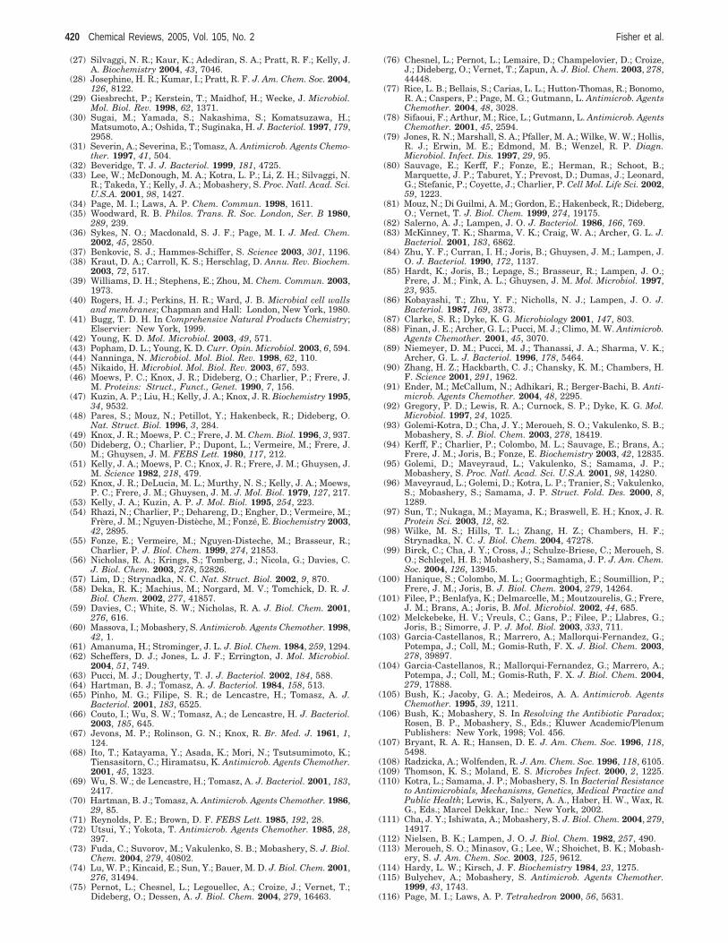
(27) Silvaggi, N. R.; Kaur, K.; Adediran, S. A.; Pratt, R. F.; Kelly, J.A. Biochemistry 2004, 43, 7046.
(28) Josephine, H. R.; Kumar, I.; Pratt, R. F. J. Am. Chem. Soc. 2004,126, 8122.
(29) Giesbrecht, P.; Kerstein, T.; Maidhof, H.; Wecke, J. Microbiol.Mol. Biol. Rev. 1998, 62, 1371.
(30) Sugai, M.; Yamada, S.; Nakashima, S.; Komatsuzawa, H.;Matsumoto, A.; Oshida, T.; Suginaka, H. J. Bacteriol. 1997, 179,2958.
(31) Severin, A.; Severina, E.; Tomasz, A. Antimicrob. Agents Chemo-ther. 1997, 41, 504.
(32) Beveridge, T. J. J. Bacteriol. 1999, 181, 4725.(33) Lee, W.; McDonough, M. A.; Kotra, L. P.; Li, Z. H.; Silvaggi, N.
R.; Takeda, Y.; Kelly, J. A.; Mobashery, S. Proc. Natl. Acad. Sci.U.S.A. 2001, 98, 1427.
(34) Page, M. I.; Laws, A. P. Chem. Commun. 1998, 1611.(35) Woodward, R. B. Philos. Trans. R. Soc. London, Ser. B 1980,
289, 239.(36) Sykes, N. O.; Macdonald, S. J. F.; Page, M. I. J. Med. Chem.
2002, 45, 2850.(37) Benkovic, S. J.; Hammes-Schiffer, S. Science 2003, 301, 1196.(38) Kraut, D. A.; Carroll, K. S.; Herschlag, D. Annu. Rev. Biochem.
2003, 72, 517.(39) Williams, D. H.; Stephens, E.; Zhou, M. Chem. Commun. 2003,
1973.(40) Rogers, H. J.; Perkins, H. R.; Ward, J. B. Microbial cell walls
and membranes; Chapman and Hall: London, New York, 1980.(41) Bugg, T. D. H. In Comprehensive Natural Products Chemistry;
Elservier: New York, 1999.(42) Young, K. D. Mol. Microbiol. 2003, 49, 571.(43) Popham, D. L.; Young, K. D. Curr. Opin. Microbiol. 2003, 6, 594.(44) Nanninga, N. Microbiol. Mol. Biol. Rev. 1998, 62, 110.(45) Nikaido, H. Microbiol. Mol. Biol. Rev. 2003, 67, 593.(46) Moews, P. C.; Knox, J. R.; Dideberg, O.; Charlier, P.; Frere, J.
M. Proteins: Struct., Funct., Genet. 1990, 7, 156.(47) Kuzin, A. P.; Liu, H.; Kelly, J. A.; Knox, J. R. Biochemistry 1995,
34, 9532.(48) Pares, S.; Mouz, N.; Petillot, Y.; Hakenbeck, R.; Dideberg, O.
Nat. Struct. Biol. 1996, 3, 284.(49) Knox, J. R.; Moews, P. C.; Frere, J. M. Chem. Biol. 1996, 3, 937.(50) Dideberg, O.; Charlier, P.; Dupont, L.; Vermeire, M.; Frere, J.
M.; Ghuysen, J. M. FEBS Lett. 1980, 117, 212.(51) Kelly, J. A.; Moews, P. C.; Knox, J. R.; Frere, J. M.; Ghuysen, J.
M. Science 1982, 218, 479.(52) Knox, J. R.; DeLucia, M. L.; Murthy, N. S.; Kelly, J. A.; Moews,
P. C.; Frere, J. M.; Ghuysen, J. M. J. Mol. Biol. 1979, 127, 217.(53) Kelly, J. A.; Kuzin, A. P. J. Mol. Biol. 1995, 254, 223.(54) Rhazi, N.; Charlier, P.; Dehareng, D.; Engher, D.; Vermeire, M.;
Frere, J. M.; Nguyen-Disteche, M.; Fonze, E. Biochemistry 2003,42, 2895.
(55) Fonze, E.; Vermeire, M.; Nguyen-Disteche, M.; Brasseur, R.;Charlier, P. J. Biol. Chem. 1999, 274, 21853.
(56) Nicholas, R. A.; Krings, S.; Tomberg, J.; Nicola, G.; Davies, C.J. Biol. Chem. 2003, 278, 52826.
(57) Lim, D.; Strynadka, N. C. Nat. Struct. Biol. 2002, 9, 870.(58) Deka, R. K.; Machius, M.; Norgard, M. V.; Tomchick, D. R. J.
Biol. Chem. 2002, 277, 41857.(59) Davies, C.; White, S. W.; Nicholas, R. A. J. Biol. Chem. 2001,
276, 616.(60) Massova, I.; Mobashery, S. Antimicrob. Agents Chemother. 1998,
42, 1.(61) Amanuma, H.; Strominger, J. L. J. Biol. Chem. 1984, 259, 1294.(62) Scheffers, D. J.; Jones, L. J. F.; Errington, J. Mol. Microbiol.
2004, 51, 749.(63) Pucci, M. J.; Dougherty, T. J. J. Bacteriol. 2002, 184, 588.(64) Hartman, B. J.; Tomasz, A. J. Bacteriol. 1984, 158, 513.(65) Pinho, M. G.; Filipe, S. R.; de Lencastre, H.; Tomasz, A. J.
Bacteriol. 2001, 183, 6525.(66) Couto, I.; Wu, S. W.; Tomasz, A.; de Lencastre, H. J. Bacteriol.
2003, 185, 645.(67) Jevons, M. P.; Rolinson, G. N.; Knox, R. Br. Med. J. 1961, 1,
124.(68) Ito, T.; Katayama, Y.; Asada, K.; Mori, N.; Tsutsumimoto, K.;
Tiensasitorn, C.; Hiramatsu, K. Antimicrob. Agents Chemother.2001, 45, 1323.
(69) Wu, S. W.; de Lencastre, H.; Tomasz, A. J. Bacteriol. 2001, 183,2417.
(70) Hartman, B. J.; Tomasz, A. Antimicrob. Agents Chemother. 1986,29, 85.
(71) Reynolds, P. E.; Brown, D. F. FEBS Lett. 1985, 192, 28.(72) Utsui, Y.; Yokota, T. Antimicrob. Agents Chemother. 1985, 28,
397.(73) Fuda, C.; Suvorov, M.; Vakulenko, S. B.; Mobashery, S. J. Biol.
Chem. 2004, 279, 40802.(74) Lu, W. P.; Kincaid, E.; Sun, Y.; Bauer, M. D. J. Biol. Chem. 2001,
276, 31494.(75) Pernot, L.; Chesnel, L.; Legouellec, A.; Croize, J.; Vernet, T.;
Dideberg, O.; Dessen, A. J. Biol. Chem. 2004, 279, 16463.
(76) Chesnel, L.; Pernot, L.; Lemaire, D.; Champelovier, D.; Croize,J.; Dideberg, O.; Vernet, T.; Zapun, A. J. Biol. Chem. 2003, 278,44448.
(77) Rice, L. B.; Bellais, S.; Carias, L. L.; Hutton-Thomas, R.; Bonomo,R. A.; Caspers, P.; Page, M. G.; Gutmann, L. Antimicrob. AgentsChemother. 2004, 48, 3028.
(78) Sifaoui, F.; Arthur, M.; Rice, L.; Gutmann, L. Antimicrob. AgentsChemother. 2001, 45, 2594.
(79) Jones, R. N.; Marshall, S. A.; Pfaller, M. A.; Wilke, W. W.; Hollis,R. J.; Erwin, M. E.; Edmond, M. B.; Wenzel, R. P. Diagn.Microbiol. Infect. Dis. 1997, 29, 95.
(80) Sauvage, E.; Kerff, F.; Fonze, E.; Herman, R.; Schoot, B.;Marquette, J. P.; Taburet, Y.; Prevost, D.; Dumas, J.; Leonard,G.; Stefanic, P.; Coyette, J.; Charlier, P. Cell Mol. Life Sci. 2002,59, 1223.
(81) Mouz, N.; Di Guilmi, A. M.; Gordon, E.; Hakenbeck, R.; Dideberg,O.; Vernet, T. J. Biol. Chem. 1999, 274, 19175.
(82) Salerno, A. J.; Lampen, J. O. J. Bacteriol. 1986, 166, 769.(83) McKinney, T. K.; Sharma, V. K.; Craig, W. A.; Archer, G. L. J.
Bacteriol. 2001, 183, 6862.(84) Zhu, Y. F.; Curran, I. H.; Joris, B.; Ghuysen, J. M.; Lampen, J.
O. J. Bacteriol. 1990, 172, 1137.(85) Hardt, K.; Joris, B.; Lepage, S.; Brasseur, R.; Lampen, J. O.;
Frere, J. M.; Fink, A. L.; Ghuysen, J. M. Mol. Microbiol. 1997,23, 935.
(86) Kobayashi, T.; Zhu, Y. F.; Nicholls, N. J.; Lampen, J. O. J.Bacteriol. 1987, 169, 3873.
(87) Clarke, S. R.; Dyke, K. G. Microbiology 2001, 147, 803.(88) Finan, J. E.; Archer, G. L.; Pucci, M. J.; Climo, M. W. Antimicrob.
Agents Chemother. 2001, 45, 3070.(89) Niemeyer, D. M.; Pucci, M. J.; Thanassi, J. A.; Sharma, V. K.;
Archer, G. L. J. Bacteriol. 1996, 178, 5464.(90) Zhang, H. Z.; Hackbarth, C. J.; Chansky, K. M.; Chambers, H.
F. Science 2001, 291, 1962.(91) Ender, M.; McCallum, N.; Adhikari, R.; Berger-Bachi, B. Anti-
microb. Agents Chemother. 2004, 48, 2295.(92) Gregory, P. D.; Lewis, R. A.; Curnock, S. P.; Dyke, K. G. Mol.
Microbiol. 1997, 24, 1025.(93) Golemi-Kotra, D.; Cha, J. Y.; Meroueh, S. O.; Vakulenko, S. B.;
Mobashery, S. J. Biol. Chem. 2003, 278, 18419.(94) Kerff, F.; Charlier, P.; Colombo, M. L.; Sauvage, E.; Brans, A.;
Frere, J. M.; Joris, B.; Fonze, E. Biochemistry 2003, 42, 12835.(95) Golemi, D.; Maveyraud, L.; Vakulenko, S.; Samama, J. P.;
Mobashery, S. Proc. Natl. Acad. Sci. U.S.A. 2001, 98, 14280.(96) Maveyraud, L.; Golemi, D.; Kotra, L. P.; Tranier, S.; Vakulenko,
S.; Mobashery, S.; Samama, J. P. Struct. Fold. Des. 2000, 8,1289.
(97) Sun, T.; Nukaga, M.; Mayama, K.; Braswell, E. H.; Knox, J. R.Protein Sci. 2003, 12, 82.
(98) Wilke, M. S.; Hills, T. L.; Zhang, H. Z.; Chambers, H. F.;Strynadka, N. C. J. Biol. Chem. 2004, 47278.
(99) Birck, C.; Cha, J. Y.; Cross, J.; Schulze-Briese, C.; Meroueh, S.O.; Schlegel, H. B.; Mobashery, S.; Samama, J. P. J. Am. Chem.Soc. 2004, 126, 13945.
(100) Hanique, S.; Colombo, M. L.; Goormaghtigh, E.; Soumillion, P.;Frere, J. M.; Joris, B. J. Biol. Chem. 2004, 279, 14264.
(101) Filee, P.; Benlafya, K.; Delmarcelle, M.; Moutzourelis, G.; Frere,J. M.; Brans, A.; Joris, B. Mol. Microbiol. 2002, 44, 685.
(102) Melckebeke, H. V.; Vreuls, C.; Gans, P.; Filee, P.; Llabres, G.;Joris, B.; Simorre, J. P. J. Mol. Biol. 2003, 333, 711.
(103) Garcia-Castellanos, R.; Marrero, A.; Mallorqui-Fernandez, G.;Potempa, J.; Coll, M.; Gomis-Ruth, F. X. J. Biol. Chem. 2003,278, 39897.
(104) Garcia-Castellanos, R.; Mallorqui-Fernandez, G.; Marrero, A.;Potempa, J.; Coll, M.; Gomis-Ruth, F. X. J. Biol. Chem. 2004,279, 17888.
(105) Bush, K.; Jacoby, G. A.; Medeiros, A. A. Antimicrob. AgentsChemother. 1995, 39, 1211.
(106) Bush, K.; Mobashery, S. In Resolving the Antibiotic Paradox;Rosen, B. P., Mobashery, S., Eds.; Kluwer Academic/PlenumPublishers: New York, 1998; Vol. 456.
(107) Bryant, R. A. R.; Hansen, D. E. J. Am. Chem. Soc. 1996, 118,5498.
(108) Radzicka, A.; Wolfenden, R. J. Am. Chem. Soc. 1996, 118, 6105.(109) Thomson, K. S.; Moland, E. S. Microbes Infect. 2000, 2, 1225.(110) Kotra, L.; Samama, J. P.; Mobashery, S. In Bacterial Resistance
to Antimicrobials, Mechanisms, Genetics, Medical Practice andPublic Health; Lewis, K., Salyers, A. A., Haber, H. W., Wax, R.G., Eds.; Marcel Dekkar, Inc.: New York, 2002.
(111) Cha, J. Y.; Ishiwata, A.; Mobashery, S. J. Biol. Chem. 2004, 279,14917.
(112) Nielsen, B. K.; Lampen, J. O. J. Biol. Chem. 1982, 257, 490.(113) Meroueh, S. O.; Minasov, G.; Lee, W.; Shoichet, B. K.; Mobash-
ery, S. J. Am. Chem. Soc. 2003, 125, 9612.(114) Hardy, L. W.; Kirsch, J. F. Biochemistry 1984, 23, 1275.(115) Bulychev, A.; Mobashery, S. Antimicrob. Agents Chemother.
1999, 43, 1743.(116) Page, M. I.; Laws, A. P. Tetrahedron 2000, 56, 5631.
420 Chemical Reviews, 2005, Vol. 105, No. 2 Fisher et al.
(117) Hokenson, M. J.; Cope, G. A.; Lewis, E. R.; Oberg, K. A.; Fink,A. L. Biochemistry 2000, 39, 6538.
(118) Wang, X.; Minasov, G.; Shoichet, B. K. Proteins: Struct., Funct.,Genet. 2002, 47, 86.
(119) Wang, X.; Minasov, G.; Shoichet, B. K. J. Mol. Biol. 2002, 320,85.
(120) Hujer, A. M.; Hujer, K. M.; Helfand, M. S.; Anderson, V. E.;Bonomo, R. A. Antimicrob. Agents Chemother. 2002, 46, 3971.
(121) Helfand, M. S.; Bethel, C. R.; Hujer, A. M.; Hujer, K. M.;Anderson, V. E.; Bonomo, R. A. J. Biol. Chem. 2003, 278, 52724.
(122) Weldhagen, G. F.; Poirel, L.; Nordmann, P. Antimicrob. AgentsChemother. 2003, 47, 2385.
(123) Bonnet, R. Antimicrob. Agents Chemother. 2004, 48, 1.(124) Bradford, P. A. Clin. Rev. Microbiol. 2001, 14, 933.(125) Davies, C.; White, S. W.; Nicholas, R. A. J. Biol. Chem. 2000,
276, 616.(126) Tranier, S.; Bouthors, A. T.; Maveyraud, L.; Guillet, V.; Souga-
koff, W.; Samama, J. P. J. Biol. Chem. 2000, 275, 28075.(127) Moult, J.; Sawyer, L.; Herzberg, O.; Jones, C. L.; Coulson, A. F.;
Green, D. W.; Harding, M. M.; Ambler, R. P. Biochem. J. 1985,225, 167.
(128) Jelsch, C.; Lenfant, F.; Masson, J. M.; Samama, J. P. FEBS Lett.1992, 299, 135.
(129) Jelsch, C.; Mourey, L.; Masson, J. M.; Samama, J. P. Proteins1993, 16, 364.
(130) Knox, J. R.; Moews, P. C. J. Mol. Biol. 1991, 220, 435.(131) Strynadka, N. C.; Adachi, H.; Jensen, S. E.; Johns, K.; Sielecki,
A.; Betzel, C.; Sutoh, K.; James, M. N. Nature 1992, 359, 700.(132) Tipper, D. J.; Strominger, J. L. Proc. Natl. Acad. Sci. U.S.A.
1965, 54, 1133.(133) Minasov, G.; Wang, X.; Shoichet, B. K. J. Am. Chem. Soc. 2002,
124, 5333.(134) Fujii, Y.; Okimoto, N.; Hata, M.; Narumi, T.; Yasuoka, K.;
Susukita, R.; Suenaga, A.; Futatsugi, N.; Koishi, T.; Furusawa,H.; Kawai, A.; Ebisuzaki, T.; Neya, S.; Hoshino, T. J. Phys.Chem. B 2003, 107, 10274.
(135) Hermann, J. C.; Ridder, L.; Mulholland, A. J.; Holtje, H. D. J.Am. Chem. Soc. 2003, 125, 9590.
(136) Nukaga, M.; Mayama, K.; Hujer, A. M.; Bonomo, R. A.; Knox,J. R. J. Mol. Biol. 2003, 328, 289.
(137) Guillaume, G.; Vanhove, M.; Lamotte-Brasseur, J.; Ledent, P.;Jamin, M.; Joris, B.; Frere, J. M. J. Biol. Chem. 1997, 272, 5438.
(138) Lamotte-Brasseur, J.; Dive, G.; Dideberg, O.; Charlier, P.; Frere,J. M.; Ghuysen, J. M. Biochem. J. 1991, 279, 213.
(139) Damblon, C.; Raquet, X.; Lian, L. Y.; Lamotte-Brasseur, J.;Fonze, E.; Charlier, P.; Roberts, G. C. K.; Frere, J. M. Proc. Natl.Acad. Sci. U.S.A. 1996, 93, 1747.
(140) Lamotte-Brasseur, J.; Lounnas, V.; Raquet, X.; Wade, R. C.Protein Sci. 1999, 8, 404.
(141) Raquet, X.; Lounnas, V.; Lamotte-Brasseur, J.; Frere, J. M.;Wade, R. C. Biophys. J. 1997, 73, 2416.
(142) Swaren, P.; Maveyraud, L.; Guillet, V.; Masson, J. M.; Mourey,L.; Samama, J. P. Structure 1995, 3, 603.
(143) Golemi-Kotra, D.; Meroueh, S. O.; Kim, C.; Vakulenko, S. B.;Bulychev, A.; Stemmler, A. J.; Stemmler, T. L.; Mobashery, S.J. Biol. Chem. 2004, 34665.
(144) Finlay, J.; Miller, L.; Poupard, J. A. J. Antimicrob. Chemother.2003, 52, 18.
(145) Imtiaz, U.; Billings, E. M.; Knox, J. R.; Mobashery, S. Biochem-istry 1994, 33, 5728.
(146) Imtiaz, U.; Billings, E.; Knox, J. R.; Manavathu, E. K.; Lerner,S. A.; Mobashery, S. J. Am. Chem. Soc. 1993, 115, 4435.
(147) Yang, Y.; Janota, K.; Tabei, K.; Huang, N.; Siegel, M. M.; Lin,Y. I.; Rasmussen, B. A.; Shlaes, D. M. J. Biol. Chem. 2000, 275,26674.
(148) Kuzin, A. P.; Nukaga, M.; Nukaga, Y.; Hujer, A.; Bonomo, R.A.; Knox, J. R. Biochemistry 2001, 40, 1861.
(149) Helfand, M. S.; Totir, M. A.; Carey, P.; Hujer, A. M.; Bonomo,R. A.; Carey, P. R. Biochemistry 2003, 42, 13386.
(150) Padayatti, P. S.; Helfand, M. S.; Totir, M. A.; Carey, M. P.; Hujer,A. M.; Carey, P. R.; Bonomo, R. A.; van den Akker, F. Biochem-istry 2004, 43, 843.
(151) Swaren, P.; Golemi, D.; Cabantous, S.; Bulychev, A.; Maveyraud,L.; Mobashery, S.; Samama, J. P. Biochemistry 1999, 38, 9570.
(152) Meroueh, S. O.; Roblin, P.; Golemi, D.; Maveyraud, L.; Vaku-lenko, S. B.; Zhang, Y.; Samama, J. P.; Mobashery, S. J. Am.Chem. Soc. 2002, 124, 9422.
(153) Madec, S.; Blin, C.; Krishnamoorthy, R.; Picard, B.; Chaibi, E.B.; Fouchereau-Peron, M.; Labia, R. FEMS Microbiol. Lett. 2002,211, 13.
(154) Wang, X.; Minasov, G.; Shoichet, B. K. J. Biol. Chem. 2002, 277,24744.
(155) Wang, X.; Minasov, G.; Blazquez, J.; Caselli, E.; Prati, F.;Shoichet, B. K. Biochemistry 2003, 42, 8434.
(156) Doucet, N.; De Wals, P. Y.; Pelletier, J. N. J. Biol. Chem. 2004,279, 46295.
(157) Pagan-Rodriguez, D.; Zhou, X.; Simmons, R.; Bethel, C. R.; Hujer,A. M.; Helfand, M. S.; Jin, Z.; Guo, B.; Anderson, V. E.; Lily,M.; Ng, L. M.; Bonomo, R. A. J. Biol. Chem. 2004, 279, 19494.
(158) Chaibi, E. B.; Sirot, D.; Paul, G.; Labia, R. J. Antimicrob.Chemother. 1999, 43, 447.
(159) Lavigne, J. P.; Bonnet, R.; Michaux-Charachon, S.; Jourdan, J.;Caillon, J.; Sotto, A. J. Antimicrob. Chemother. 2004, 53, 616.
(160) Nukaga, M.; Abe, T.; Venkatesan, A. M.; Mansour, T. S.; Bonomo,R. A.; Knox, J. R. Biochemistry 2003, 42, 13152.
(161) Beharry, Z.; Chen, H.; Gadhachanda, V. R.; Buynak, J. D.;Palzkill, T. Biochem. Biophys. Res. Commun. 2004, 313, 541.
(162) Bret, L.; Chaibi, E. B.; Chanal-Claris, C.; Sirot, D.; Labia, R.;Sirot, J. Antimicrob. Agents Chemother. 1997, 41, 2547.
(163) Delaire, M.; Labia, R.; Samama, J. P.; Masson, J. M. J. Biol.Chem. 1992, 267, 20600.
(164) Zafaralla, G.; Manavathu, E. K.; Lerner, S. A.; Mobashery, S.Biochemistry 1992, 31, 3847.
(165) Delaire, M.; Labia, R.; Samama, J. P.; Masson, J. M. J. Biol.Chem. 1992, 267, 20600.
(166) Schroeder, W. A.; Locke, T. R.; Jensen, S. E. Antimicrob. AgentsChemother. 2002, 46, 3568.
(167) Randegger, C. C.; Hachler, H. J. Antimicrob. Chemother. 2001,47, 547.
(168) Stapleton, P. D.; Shannon, K. P.; French, G. L. Antimicrob.Agents Chemother. 1999, 43, 1881.
(169) Vakulenko, S. B.; Taibi-Tronche, P.; Toth, M.; Massova, I.;Lerner, S. A.; Mobashery, S. J. Biol. Chem. 1999, 274, 23052.
(170) Vakulenko, S.; Golemi, D. Antimicrob. Agents Chemother. 2002,46, 646.
(171) Maveyraud, L.; Mourey, L.; Kotra, L. P.; Pedelacq, J. D.; Guillet,V.; Mobashery, S.; Samama, J. P. J. Am. Chem. Soc. 1998, 120,9748.
(172) Vilanova, B.; Donoso, J.; Frau, J.; Munoz, F. Helv. Chim. Acta1999, 82, 1274.
(173) Fonze, E.; Vanhove, M.; Dive, G.; Sauvage, E.; Frere, J. M.;Charlier, P. Biochemistry 2002, 41, 1877.
(174) Sougakoff, W.; Naas, T.; Nordmann, P.; Collatz, E.; Jarlier, V.Biochim. Biophys. Acta 1999, 1433, 153.
(175) Sougakoff, W.; L’Hermite, G.; Pernot, L.; Naas, T.; Guillet, V.;Nordmann, P.; Jarlier, V.; Delettre, J. Acta Crystallogr., Sect.D 2002, 58, 267.
(176) Majiduddin, F. K.; Palzkill, T. Antimicrob. Agents Chemother.2003, 47, 1062.
(177) Mourey, L.; Miyashita, K.; Swaren, P.; Bulychev, A.; Samama,J. P.; Mobashery, S. J. Am. Chem. Soc. 1998, 120, 9382.
(178) Hujer, A. M.; Hujer, K. M.; Bonomo, R. A. Biochim. Biophys.Acta 2001, 1547, 37.
(179) Cantu, C., III; Palzkill, T. J. Biol. Chem. 1998, 273, 26603.(180) Orencia, M. C.; Yoon, J. S.; Ness, J. E.; Stemmer, W. P. C.;
Stevens, R. C. Nat. Struct. Biol. 2001, 8, 238.(181) Nukaga, K.; Mayama, K.; Crichlow, G. V.; Knox, J. R. J. Mol.
Biol. 2002, 317, 109.(182) Shimamura, T.; Ibuka, A. S.; Fushinobu, S.; Wakagi, T.; Ishiguro,
M.; Ishii, Y.; Matsuzawa, H. J. Biol. Chem. 2002, 277, 46601.(183) Ibuka, A. S.; Ishii, Y.; Galleni, M.; Ishiguro, M.; Yamaguchi, K.;
Frere, J. M.; Matsuzawa, H.; Sakai, H. Biochemistry 2003, 42,10634.
(184) Hall, A.; Knowles, J. R. Nature 1976, 264, 803.(185) Stemmer, W. P. C. J. Mol. Catal. B 2002, 19, 3.(186) Camps, M.; Naukkarinen, J.; Johnson, B. P.; Loeb, L. A. Proc.
Natl. Acad. Sci. U.S.A. 2003, 100, 9727.(187) Vakulenko, S. B.; Geryk, B.; Kotra, L. P.; Mobashery, S.; Lerner,
S. A. Antimicrob. Agents Chemother. 1998, 42, 1542.(188) Barlow, M.; Hall, B. G. Genetics 2003, 163, 1237.(189) Barlow, M.; Hall, B. G. Genetics 2003, 164, 23.(190) Hall, B. G. Nat. Rev. Microbiol. 2004, 2, 430.(191) Lobkovsky, E.; Moews, P. C.; Liu, H.; Zhao, H.; Frere, J. M.;
Knox, J. R. Proc. Natl. Acad. Sci. U.S.A. 1993, 90, 11257.(192) Oefner, C.; D′Arcy, A.; Daly, J. J.; Gubernator, K.; Charnas, R.
L.; Heinze, I.; Hubschwerlen, C.; Winkler, F. K. Nature 1990,343, 284.
(193) Gherman, B. F.; Goldberg, S. D.; Cornish, V. W.; Friesner, R. A.J. Am. Chem. Soc. 2004, 126, 7652.
(194) Bulychev, A.; Massova, I.; Miyashita, K.; Mobashery, S. J. Am.Chem. Soc. 1997, 119, 7619.
(195) Patera, A.; Blaszczak, L. C.; Shoichet, B. K. J. Am. Chem. Soc.2000, 122, 10504.
(196) Rice, L. B.; Bonomo, R. A. Drug Resist. Update 2000, 3, 178.(197) Bauernfeind, A.; Chong, Y.; Schweighart, S. Infection 1989, 17,
316.(198) Bauernfeind, A.; Chong, Y.; Lee, K. Yonsei Med. J. 1998, 39,
520.(199) Livermore, D. M. Clin. Microbiol. Rev. 1995, 8, 557.(200) Philippon, A.; Arlet, G.; Lagrange, P. H. Eur. J. Clin. Microbiol.
Infect. Dis. 1994, 13 (Suppl 1), S17.(201) Galleni, M.; Amicosante, G.; Frere, J. M. Biochem. J. 1988, 255,
123.(202) Kato-Toma, Y.; Iwashita, T.; Masuda, K.; Oyama, Y.; Ishiguro,
M. Biochem. J. 2003, 371, 175.(203) Lamotte-Brasseur, J.; Dubus, A.; Wade, R. C. Proteins 2000, 40,
23.
Bacterial Resistance to â-Lactam Antibiotics Chemical Reviews, 2005, Vol. 105, No. 2 421

(204) Dubus, A.; Normark, S.; Kania, M.; Page, M. G. Biochemistry1994, 33, 8577.
(205) Knothe, H.; Shah, P.; Krcmery, V.; Antal, M.; Mitsuhashi, S.Infection 1983, 11, 315.
(206) Nukaga, M.; Haruta, S.; Tanimoto, K.; Kogure, K.; Taniguchi,K.; Tamaki, M.; Sawai, T. J. Biol. Chem. 1995, 270, 5729.
(207) Crichlow, G. V.; Kuzin, A. P.; Nukaga, M.; Mayama, K.; Sawai,T.; Knox, J. R. Biochemistry 1999, 38, 10256.
(208) Powers, R. A.; Caselli, E.; Focia, P. J.; Prati, F.; Shoichet, B. K.Biochemistry 2001, 40, 9207.
(209) Nukaga, M.; Kumar, S.; Nukaga, K.; Pratt, R. F.; Knox, J. R. J.Biol. Chem. 2004, 279, 9344.
(210) Nordmann, P.; Poirel, L. Clin. Microbiol. Infect. 2002, 8, 321.(211) Heritier, C.; Poirel, L.; Nordmann, P. Antimicrob. Agents
Chemother. 2004, 48, 1670.(212) Bou, G.; Martınez-Beltran, J. Antimicrob. Agents Chemother.
2000, 44, 428.(213) Poirel, L.; Girlich, D.; Naas, T.; Nordmann, P. Antimicrob. Agents
Chemother. 2001, 45, 447.(214) Toleman, M. A.; Rolston, K.; Jones, R. N.; Walsh, T. R. Antimi-
crob. Agents Chemother. 2003, 47, 2859.(215) Dubois, V.; Arpin, C.; Quentin, C.; Texier-Maugein, J.; Poirel,
L.; Nordmann, P. Antimicrob. Agents Chemother. 2003, 47, 2380.(216) Oliver, A.; Weigel, L. M.; Rasheed, J. K.; McGowan, J. E.; Raney,
P.; Tenover, F. C. Antimicrob. Agents Chemother. 2002, 46, 3829.(217) Bonnet, R.; Marchandin, H.; Chanal, C.; Sirot, D.; Labia, R.; De
Champs, C.; Jumas-Bilak, E.; Sirot, D. Antimicrob. AgentsChemother. 2002, 46, 2004.
(218) Orman, B. E.; Pineiro, S. A.; Arduino, S.; Galas, M.; Melano, R.;Caffer, M. I.; Sordelli, D. O.; Centron, D. Antimicrob. AgentsChemother. 2002, 46, 3963.
(219) Melano, R.; Corso, A.; Petroni, A.; Centron, D.; Orman, B.;Pereyra, A.; Moreno, N.; Galas, M. J. Antimicrob. Chemother.2003, 52, 36.
(220) Poirel, L.; Heritier, C.; Nordmann, P. Antimicrob. AgentsChemother. 2004, 48, 348.
(221) Bou, G.; Cervero, G.; Dominguez, M. A.; Quereda, C.; Martinez-Beltran, J. J. Clin. Microbiol. 2000, 38, 3299.
(222) Afzal-Shah, M.; Woodford, N.; Livemore, D. M. Antimicrob.Agents Chemother. 2001, 45, 583.
(223) Danes, C.; Navia, M. N.; Ruiz, J.; Marco, F.; Jurado, A.; Jimenezde Anta, M. T.; Vila, J. J. Antimicrob. Chemother. 2002, 50, 261.
(224) Lopez-Otsoa, F.; Gallego, L.; Towner, K. J.; Tysall, L.; Woodford,N.; Livermore, D. M. J. Clin. Microbiol. 2002, 40, 4741.
(225) Dalla-Costa, L. M.; Coelho, J. M.; Souza, H. A. P.; Castro, M. E.S.; Stier, C. J. N.; Bragagnolo, K. L.; Rea-Neto, A.; Penteado-Filho, S. R.; Livermore, D. M.; Woodford, N. J. Clin. Microbiol.2003, 41, 3403.
(226) Heritier, C.; Poirel, L.; Aubert, D.; Nordmann, P. Antimicrob.Agents Chemother. 2003, 47, 268.
(227) Mushtaq, S.; Ge, Y.; Livermore, D. Antimicrob. Agents Chemoth-er. 2004, 48, 1313.
(228) Zarrilli, R.; Crispino, M.; Bagattini, M.; Barretta, E.; Di Popolo,A.; Triassi, A. M.; Villari, P. J. Clin. Microbiol. 2004, 42, 946.
(229) Livermore, D. M.; Woodford, N. Curr. Opin. Microbiol. 2000, 3,489.
(230) Danel, F.; Frere, J. M.; Livemore, D. M. Biochim. Biophys. Acta2001, 1546, 132.
(231) Franceschini, N.; Segatore, B.; Perilli, M.; Vessilier, S.; Franchi-no, L.; Amicosante, G. J. Antimicrob. Chemother. 2002, 49, 395.
(232) Pernot, L.; Frenois, F.; Rybkine, T.; L’Hermite, G.; Petrella, S.;Delettre, J.; Jarlier, V.; Collatz, E.; Sougakoff, W. J. Mol. Biol.2001, 310, 859.
(233) Danel, F.; Paetzel, M.; Strynadka, N. C. J.; Page, M. G. P.Biochemistry 2001, 40, 9412.
(234) Franceschini, N.; Boschi, L.; Pollini, S.; Herman, R.; Perilli, M.;Galleni, M.; Frere, J. M.; Amicosante, G.; Rossolini, G. M.Antimicrob. Agents Chemother. 2001, 45, 3509.
(235) Paetzel, M.; Danel, F.; de Castro, L.; Mosimann, S. C.; Page, M.G. P.; Strynadka, N. C. J. Nat. Struct. Biol. 2000, 7, 918.
(236) Golemi, D.; Maveyraud, L.; Vakulenko, S.; Tranier, S.; Ishiwata,A.; Kotra, L. P.; Samama, J. P.; Mobashery, S. J. Am. Chem.Soc. 2000, 122, 6132.
(237) Maveyraud, L.; Golemi-Kotra, D.; Ishiwata, A.; Meroueh, O.;Mobashery, S.; Samama, J. P. J. Am. Chem. Soc. 2002, 124,2461.
(238) Donald, H. M.; Scaife, W.; Amyes, S. G. B.; Young, H. K.Antimicrob. Agents Chemother. 2000, 44, 196.
(239) Barlow, M.; Hall, B. G. J. Mol. Evol. 2002, 55, 314.(240) Zhu, Y.; Englebert, S.; Joris, B.; Ghuysen, J. M.; Kobayashi, T.;
Lampen, J. O. J. Bacteriol. 1992, 174, 6171.(241) Colombo, M. L.; Hanique, S.; Baurin, S. L.; Bauvois, C.; De
Vriendt, K.; Van Beeumen, J. J.; Frere, J. M.; Joris, B. Antimi-crob. Agents Chemother. 2004, 48, 484.
(242) Garau, G.; Garcia-Saez, I.; Bebrone, C.; Anne, C.; Mercuri, P.;Galleni, M.; Frere, J. M.; Dideberg, O. Antimicrob. AgentsChemother. 2004, 48, 2347.
(243) Galleni, M.; Lamotte-Brasseur, J.; Rossolini, G. M.; Spencer, J.;Dideberg, O.; Frere, J. M. Antimicrob. Agents. Chemother. 2001,45, 660.
(244) Gniadkowski, M. Clin. Microbiol. Infect. 2001, 7, 597.(245) Fast, W.; Wang, Z.; Benkovic, S. J. Biochemistry 2001, 40, 1640.(246) Fabiane, S. M.; Sohi, M. K.; Wan, T.; Payne, D. J.; Bateson, J.
H.; Mitchell, T.; Sutton, B. J. Biochemistry 1998, 37, 12404.(247) Wommer, S.; Rival, S.; Hein, U.; Galleni, M.; Frere, J. M.;
Franceschini, N.; Amicosante, G.; Rasmussen, B.; Bauer, R.;Adolph, H. W. J. Biol. Chem. 2002, 277, 24142.
(248) Concha, N. O.; Janson, C. A.; Rowling, P.; Pearson, S.; Cheever,C. A.; Clarke, B. P.; Lewis, C.; Galleni, M.; Frere, J. M.; Payne,D. J.; Bateson, J. H.; Abdel-Meguid, S. S. Biochemistry 2000,39, 4288.
(249) Gomes, C. M.; Frazao, C.; Xavier, A. V.; Legall, J.; Teixeira, M.Protein Sci. 2002, 11, 707.
(250) Schilling, O.; Wenzel, N.; Naylor, M.; Vogel, A.; Crowder, M. W.;Makaroff, C.; Meyer-Klaucke, W. Biochemistry 2003, 42, 11777.
(251) Spencer, J.; Clarke, A. R.; Walsh, T. R. J. Biol. Chem. 2001, 276,33638.
(252) Rasia, R. M.; Vila, A. J. J. Biol. Chem. 2004, 279, 26046.(253) Ullah, J. H.; Walsh, T. R.; Taylor, I. A.; Emery, D. C.; Verma,
C. S.; Gamblin, S. J.; Spencer, J. J. Mol. Biol. 1998, 284, 125.(254) Rasia, R. M.; Ceolın, M.; Vila, A. J. Protein Sci. 2003, 12, 1538.(255) Garrity, J. D.; Carenbauer, A. L.; Herron, L. R.; Crowder, M.
W. J. Biol. Chem. 2004, 279, 920.(256) Dal Peraro, M.; Vila, A. J.; Carloni, P. Proteins: Struct., Funct.,
Bioinform. 2004, 54, 412.(257) Dal Peraro, M.; Vila, A. J.; Carloni, P. Inorg. Chem. 2003, 42,
4245.(258) Garceia-Saez, I.; Mercuri, P. S.; Papamicael, C.; Kahn, R.; Frere,
J. M.; Galleni, M.; Rossolini, G. M.; Dideberg, O. J. Mol. Biol.2003, 325, 651.
(259) Garceia-Saez, I.; Hopkins, J.; Papamicael, C.; Franceschini, N.;Amicosante, G.; Rossolini, G. M.; Galleni, M.; Frere, J. M.;Dideberg, O. J. Biol. Chem. 2003, 278, 23868.
(260) Bounaga, S.; Laws, A. P.; Galleni, M.; Page, M. I. Biochem. J.1998, 331, 703.
(261) Bush, K. Clin. Infect. Dis. 2001, 32, 1085.(262) Miriagou, V.; Tzelepi, E.; Gianneli, D.; Tzouvelekis, L. S.
Antimicrob. Agents Chemother. 2003, 47, 395.(263) Yanchak, M. P.; Taylor, R. A.; Crowder, M. W. Biochemistry
2000, 39, 11330.(264) Krauss, M.; Gilson, H. S. R.; Gresh, N. J. Phys. Chem. B 2001,
105, 8040.(265) Rasia, R. M.; Vila, A. J. Biochemistry 2002, 41, 1853.(266) Lipton, A. S.; Heck, R. W.; Ellis, P. D. J. Am. Chem. Soc. 2004,
126, 4735.(267) Montoya-Pelaez, P. J.; Brown, R. S. Inorg. Chem. 2002, 41, 309.(268) Montoya-Pelaez, P. J.; Gibson, G. T. T.; Neverov, A. A.; Brown,
R. S. Inorg. Chem. 2003, 42, 8624.(269) Dıaz, N.; Sordo, T. L.; Suarez, D.; Mendez, R.; Martın-Villacorta,
J. New J. Chem. 2004, 28, 15.(270) Diaz, N.; Suarez, D.; Merz, K. M., Jr. J. Am. Chem. Soc. 2001,
123, 9867.(271) Moali, C.; Anne, C.; Lamotte-Brasseur, J.; Groslambert, S.;
Devreese, B.; Van Beeumen, J.; Galleni, M.; Frere, J. M. Chem.Biol. 2003, 10, 319.
(272) Oelschlaeger, P.; Schmid, R. D.; Pleiss, J. Biochemistry 2003,42, 8945.
(273) Luzzaro, F.; Docquier, J. D.; Colinon, C.; Endimiani, A.; Lom-bardi, G.; Amicosante, G.; Rossolini, G.; Toniolol, A. Antimicrob.Agents Chemother. 2004, 48, 648.
(274) Weldhagen, G. F. Int. J. Antimicrob. Agents 2004, 23, 556.(275) Docquier, J. D.; Riccio, M. L.; Mugnaioli, C.; Luzzaro, F.;
Endimiani, A.; Toniolo, A.; Amicosante, G.; Rossolini, G. M.Antimicrob. Agents Chemother. 2003, 47, 1522.
(276) Iyobe, S.; Kusadokoro, H.; Ozaki, J.; Matsumura, N.; Minami,S.; Haruata, S.; Sawai, T.; O’Hara, K. Antimicrob. AgentsChemother. 2000, 44, 2023.
(277) Yano, H.; Kuga, A.; Okamoto, R.; Kitasato, H.; Kobayashi, T.;Inoue, M. Antimicrob. Agents Chemother. 2001, 45, 1343.
(278) Materon, I. C.; Palzkill, T. Protein Sci. 2001, 10, 2556.(279) Hall, B. G. Antimicrob. Agents Chemother. 2004, 48, 1032.(280) Docquier, J. D.; Lamotte-Brasseur, J.; Galleni, M.; Amicosante,
G.; Frere, J. M.; Rossolini, G. M. J. Antimicrob. Chemother. 2003,51, 257.
(281) Toleman, M. A.; Rolston, K.; Jones, R. N.; Walsh, T. R. Antimi-crob. Agents Chemother. 2004, 48, 329.
(282) Cornaglia, G.; Mazzariol, A.; Lauretti, L.; Rossolini, M.; Fontana,R. Clin. Infect. Dis. 2000, 31, 1119.
(283) Yatsuyanagi, J.; Saito, S.; Harata, S.; Suzuki, N.; Ito, Y.; Amano,K.; Enomoto, K. Antimicrob. Agents Chemother. 2004, 48, 626.
(284) Siemann, S.; Clarke, A. J.; Viswanatha, T.; Dmitrienko, G. I.Biochemistry 2003, 42, 1673.
(285) Tsang, W. Y.; Dhanda, A.; Schofield, C. J.; Frere, J. M.; Galleni,M.; Page, M. I. Bioorg. Med. Chem. Lett. 2004, 2004, 1737.
422 Chemical Reviews, 2005, Vol. 105, No. 2 Fisher et al.

(286) Buynak, J. D.; Chen, H.; Vogeti, L.; Gadhachanda, V. R.;Buchanan, C. A.; Palzkill, T.; Shaw, R. W.; Spencer, J.; Walsh,T. R. Bioorg. Med. Chem. Lett. 2004, 14, 1299.
(287) Heinz, U.; Bauer, R.; Wommer, S.; Meyer-Klaucke, W.; Papam-ichaels, C.; Bateson, J.; Adolph, H. W. J. Biol. Chem. 2003, 278,20659.
(288) Mollard, C.; Moali, C.; Papamicael, C.; Damblon, C.; Vessilier,S.; Amicosante, G.; Schofield, C. J.; Galleni, N.; Frere, J. M.;Roberts, G. C. K. J. Biol. Chem. 2001, 276, 45015.
(289) Goh, E. B.; Yim, G.; Tsui, W.; McClure, J.; Surette, M. G.; Davies,J. Proc. Natl. Acad. Sci. U.S.A. 2002, 99, 17025.
(290) Kaldalu, N.; Mei, R.; Lewis, K. Antimicrob. Agents Chemother.2004, 48, 890.
(291) Bandow, J. E.; Brotz, H.; Leichert, L. I. O.; Labischinski, H.;Hecker, M. Antimicrob. Agents Chemother. 2003, 47, 948.
(292) Mills, S. D. J. Antimicrob. Chemother. 2003, 51, 749.(293) Bradford, P. A.; Urban, C.; Mariano, N.; Projan, S. J.; Rahal, J.
J.; Bush, K. Antimicrob. Agents Chemother. 1997, 41, 563.(294) Cao, V. T. B.; Arlet, G.; Ericsson, B. M.; Tammelin, A.; Courvalin,
P.; Lambert, T. J. Antimicrob. Chemother. 2000, 46, 895.(295) Nelson, E. C.; Segal, H.; Elisha, B. G. J. Antimicrob. Chemother.
2003, 52, 899.(296) Domenech-Sanchez, A.; Martınez-Martınez, L.; Hernandez-Alles,
S.; del Carmen Conejo, M.; Pascual, AÄ .; Tomas, J. M.; Albertı,S.; Benedı, V. J. Antimicrob. Agents Chemother. 2003, 48, 3332.
(297) Martinez, M. B.; Flickinger, M.; Higgins, L.; Krick, T.; Nelses-tuen, G. L. Biochemistry 2001, 40, 11965.
(298) Armand-Lefvre, L.; Leflon-Guibout, V.; Bredin, J.; Barguelli, F.;Amor, A.; Pages, J. M.; Nicolas-Chanoine, M. H. Antimicrob.Agents Chemother. 2003, 47, 1165.
(299) Kwon, D. H.; Dore, M. P.; Kim, J. J.; Kato, M.; Lee, M.; Wu, J.Y.; Graham, D. Y. Antimicrob. Agents Chemother. 2003, 47, 2169.
(300) Fernandez-Cuencal, F.; Martınez-Martınez, L.; Conejo, M. C.;Ayala, J. A.; Perea, E. J.; Pascual, A. J. Antimicrob. Chemother.2003, 51, 565.
(301) Gayet, S.; Chollet, R.; Molle, G.; Pages, J. M.; Chevalier, J.Antimicrob. Agents Chemother. 2003, 47, 1555.
(302) Bornet, C.; Saint, N.; Fetnaci, L.; Dupont, M.; Davin-Regli, A.;Bollet, C.; Pages, J. M. Antimicrob. Agents Chemother. 2004, 48,2153.
(303) Jacoby, G. A.; Mills, D. M.; Chow, N. Antimicrob. AgentsChemother. 2004, 48, 3203.
(304) Okamoto, K.; Gotoh, N.; Nishino, T. Antimicrob. Agents Chemoth-er. 2001, 45, 1964.
(305) Pai, H.; Kim, J. W.; Kim, J.; Lee, J.; Choe, K. W.; Gotoh, N.Antimicrob. Agents Chemother. 2001, 45, 480.
(306) Lakaye, B.; Dubus, A.; Joris, B.; Frere, J. M. Antimicrob. AgentsChemother. 2002, 46, 2901.
(307) Matsumura, N.; Minami, S.; Watanabe, Y.; Iyobe, S.; Mitsuhashi,S. Antimicrob. Agents Chemother. 1999, 43, 2084.
(308) Kotra, L. P.; Golemi, D.; Amro, N. A.; Liu, G. Y.; Mobashery, S.J. Am. Chem. Soc. 1999, 121, 8707.
(309) Snyder, D. S.; McIntosh, T. J. Biochemistry 2000, 39, 11777.(310) Gillespie, S. H.; McHugh, T. D. Trends Microbiol. 1997, 5, 337.(311) Faller, M.; Niederweis, M.; Schulz, G. E. Science 2004, 303, 1189.(312) Antignac, A.; Boneca, I. G.; Rousselle, J. C.; Namane, A.; Carlier,
J. P.; Vazquez, J. A.; Fox, A.; Alonso, J. M.; Taha, M. K. J. Biol.Chem. 2003, 278, 31529.
(313) Antignac, A.; Rousselle, J. C.; Namane, A.; Labigne, A.; Taha,M. K.; Boneca, I. G. J. Biol. Chem. 2003, 278, 31521.
(314) Filipe, S. R.; Tomasz, A. Proc. Natl. Acad. Sci. U.S.A. 2000, 99,4891.
(315) Smith, A. M.; Klugman, K. P. Antimicrob. Agents Chemother.2001, 45, 2393.
(316) Fiser, A.; Filipe, S. R.; Tomasz, A. Trends Microbiol. 2003, 11,547.
(317) Rohrer, S.; Berger-Bachi, B. Antimicrob. Agents Chemother.2003, 47, 837.
(318) Okamoto, K.; Gotoh, N.; Nishino, T. Antimicrob. Agents Chemoth-er. 2002, 46, 2696.
(319) Middlemiss, J. K.; Poole, K. J. Bacteriol. 2004, 186, 1258.(320) Van Bambekel, F.; Glupczynski, Y.; Plesiat, P.; Pechere, J. C.;
Tulkens, P. M. J. Antimicrob. Chemother. 2003, 51, 1055.(321) Burns, D. L. Curr. Opin. Microbiol. 2003, 6, 29.(322) Cascales, E.; Christie, P. J. Nat. Rev. Microbiol. 2003, 1, 137.(323) Borges-Walmsley, M. I.; McKeegan, K. S.; Walmsley, A. R.
Biochem. J. 2003, 376, 313.(324) Grkovic, S.; Brown, M. H.; Skurray, R. A. Microbiol. Mol. Biol.
2002, 66, 671.(325) McKeegan, K. S.; Borges-Walmsley, M. I.; Walmsley, A. R.
Trends Microbiol. 2003, 11, 21.(326) Murakami, S.; Yamaguchi, A. Curr. Opin. Struct. Biol. 2003,
13, 443.(327) Paulsen, I. T. Curr. Opin. Microbiol. 2003, 6, 446.(328) Schmacher, M. A.; Brennan, R. G. Mol. Microbiol. 2002, 45, 885.(329) Chevalier, J.; Bredin, J.; Mahamoud, A.; Mallea, M.; Barbe, J.;
Pages, J. M. Antimicrob. Agents Chemother. 2004, 48, 1043.(330) Nakayama, K.; Ishida, Y.; Ohtsuka, M.; Kawato, H.; Yoshida,
K.; Yokomizo, Y.; Hosono, S.; Ohta, T.; Hoshino, K.; Ishida, H.;
Renau, T. E.; Leger, R.; Zhang, J. Z.; Lee, V. J.; Watkins, W. J.Bioorg. Med. Chem. Lett. 2003, 13, 4201.
(331) Mallea, M.; Mahamoud, A.; Chevalier, J.; Alibert-Franco, S.;Brouant, P.; Barbe, J.; Pages, J. M. Biochem. J. 2003, 376, 801.
(332) Renau, T.; Leger, R.; Filonova, L.; Flamme, E.; Wang, M.; Yen,R.; Madsen, D.; Griffith, D.; Chamberland, S.; Dudley, M.; Lee,V.; Lomovskaya, O.; Watkins, W.; Ohta, T.; Nakayama, K.;Ishida, Y. Bioorg. Med. Chem. Lett. 2003, 13, 2755.
(333) Zgurskaya, H.; Nikaido, H. Mol. Microbiol. 2000, 37, 219.(334) McGeer, A.; Low, D. E. Nat. Med. 2003, 9, 390.(335) McCormick, A. W.; Whitney, C. G.; Farley, M. M.; Lynfield, R.;
Harrison, L. H.; Bennett, N. M.; Schaffner, W.; Reingold, A.;Hadler, J.; Cieslak, P.; Samore, M. H.; Lipsitch, M. Nat. Med.2003, 9, 424.
(336) Service, R. F. Science 2004, 303, 1798.(337) Sheldon, T. Nat. Med. 2004, 10, 6.(338) Oliveira, D. C.; Tomasz, A.; de Lencastre, H. Lancet Infect. Dis.
2002, 2, 180.(339) Projan, S. J. Curr. Opin. Microbiol. 2003, 6, 427.(340) Projan, S. J. Curr. Opin. Pharmacol. 2003, 3, 457.(341) Shlaes, D. M. Curr. Opin. Pharmacol. 2003, 3, 470.(342) Projan, S. J. Curr. Opin. Pharmacol. 2002, 2, 513.(343) Livermore, D. Nat. Rev. Microbiol. 2004, 2, 73.(344) Eisner, T. Proc. Natl. Acad. Sci. U.S.A. 2003, 100, 14517.(345) Meinwald, J.; Eisner, T. Helv. Chim. Acta 2003, 86, 3633.(346) Coates, A.; Hu, Y.; Bax, R.; Page, C. Nat. Rev. Drug Discovery
2002, 1, 895.(347) Hutter, B.; Schaab, C.; Albrecht, S.; Borgmann, M.; Brunner,
N. A.; Freiberg, C.; Ziegelbauer, K.; Rock, C. O.; Ivanov, I.;Loferer, H. Antimicrob. Agents Chemother. 2004, 48, 2838.
(348) Katayama, Y.; Zhang, H. Z.; Hong, D.; Chambers, H. F. J.Bacteriol. 2003, 185, 5465.
(349) Weiss, D. S.; Chen, J. C.; Ghigo, J. M.; Boyd, D.; Beckwith, J. J.Bacteriol. 1999, 181, 508.
(350) Zhao, G.; Meier, T. I.; Kahl, S. D.; Gee, K. R.; Blaszczak, L. C.Antimicrob. Agents Chemother. 1999, 43, 1124.
(351) Kyriacou, S. V.; Brownlow, W. J.; Xu, X. H. N. Biochemistry 2004,43, 140.
(352) Steel, C.; Wan, Q.; Xu, X. H. N. Biochemistry 2004, 43, 175.(353) Campbell, D. A.; Szardenings, A. K. Curr. Opin. Chem. Biol.
2003, 7, 296.(354) Miller, C.; Thomsen, L. E.; Gaggero, C.; Mosseri, R.; Ingmer,
H.; Cohen, S. N. Science 2004, 305, 1629.(355) Spoering, A. L.; Lewis, K. J. Bacteriol. 2001, 183, 6746.(356) Balaban, N. Q.; Merrin, J.; Chait, R.; Kowalik, L.; Leibler, S.
Science 2004, 305, 1622.(357) Hastings, P. J.; Rosenberg, S. M.; Slack, A. Trends Microbiol.
2004, 12, 401.(358) Rice, L. B. Curr. Opin. Pharmacol. 2003, 3, 459.(359) Rand, K. H.; Houck, H. J. Antimicrob. Agents Chemother. 2004,
48, 2871.(360) Barrett, C. T.; Barrett, J. F. Curr. Opin. Biotechnol. 2003, 14,
621.(361) Gustafsson, I.; Sjolund, M.; Torell, E.; Johannesson, M.; Eng-
strandl, L.; Cars, O.; Andersson, D. I. J. Antimicrob. Chemother.2003, 52, 645.
(362) Salyers, A. A.; Gupta, A.; Wang, Y. Trends Microbiol. 2004, 12,412.
(363) Kummerer, K.; Henniger, A. Clin. Microbiol. Infect. 2003, 9,1203.
(364) Harmoinen, J.; Mentula, S.; Heikkila, M.; van der Rest, M.;Rajala-Schultz, P. J.; Donskey, C. J.; Frias, R.; Koski, P.;Wickstrand, N.; Jousimies-Somer, H.; Westermarck, E.; Linde-vall, K. Antimicrob. Agents Chemother. 2004, 48, 75.
(365) Harmoinen, J.; Vaali, K.; Koski, P.; Syrjanen, K.; Laitinen, O.;Lindevall, K.; Westermarck, E. J. Antimicrob. Chemother. 2003,51, 361.
(366) Stiefel, U.; Pultz, N. J.; Harmoinen, J.; Koski, P.; Lindevall, K.;Helfand, M. S.; Donskey, C. J. J. Infect. Dis. 2003, 188, 1605.
(367) Cooper, B. S.; Medley, G. F.; Stone, S. P.; Kibbler, C. C.; Cookson,B. D.; Roberts, J. A.; Duckworth, G.; Lai, R.; Ebrahim, S. Proc.Natl. Acad. Sci. U.S.A. 2004, 101, 10223.
(368) Livermore, D. M.; Dudley, M. N. Curr. Opin. Microbiol. 2000,3, 487.
(369) Monroe, S.; Polk, R. Curr. Opin. Microbiol. 2000, 3, 496.(370) Bantar, C.; Vesco, E.; Heft, C.; Salamone, F.; Krayeski, M.;
Gomez, H.; Coassolo, M. A.; Fiorillo, A.; Franco, D.; Arango, C.;Duret, F.; Oliva, M. E. Antimicrob. Agents Chemother. 2004, 48,392.
(371) Livemore, D. M.; Brown, D. F. J.; Quinn, J. P.; Carmeli, Y.;Paterson, D. L.; Yu, V. L. Clin. Microbiol. Infect. 2004, 10, 84.
(372) Chandrakala, B.; Shandil, R. K.; Mehra, U.; Ravishankar, S.;Kaur, P.; Usha, V.; Joe, B.; deSousa, S. M. Antimicrob. AgentsChemother. 2004, 48, 30.
(373) Tondi, D.; Powers, R. A.; Caselli, E.; Negri, M. C.; Blazquez, J.;Costi, M. P.; Shoichet, B. K. Chem. Biol. 2001, 8, 593.
(374) Powers, R. A.; Shoichet, B. K. J. Med. Chem. 2002, 45, 3222.(375) Powers, R. A.; Morandi, F.; Shoichet, B. K. Structure 2002, 10,
1013.
Bacterial Resistance to â-Lactam Antibiotics Chemical Reviews, 2005, Vol. 105, No. 2 423
(376) Trehan, I.; Morandi, F.; Blaszczak, L. C.; Shoichet, B. K. Chem.Biol. 2002, 9, 971.
(377) Levy, S. B.; Marshall, B. Nat. Med. Suppl. 2004, 10, S122.(378) Payne, D.; Tomasz, A. Curr. Opin. Microbiol. 2004, 7, 435.(379) Poole, K. Cell. Mol. Life Sci. 2004, 61, 2200.(380) Mallorqui-Fernandez, G.; Marrero, A.; Garcia-Pique, S.; Garcia-
Castellanos, R.; Gomis-Ruth, F. X. FEMS Microbiol. Lett. 2004,235, 1.
(381) Walsh, F. M.; Amyes, S. G. B. Curr. Opin. Microbiol. 2004, 7,439.
(382) Gotz, F. Curr. Opin. Microbiol. 2004, 7, 477.(383) Dmitriev, B. A.; Toukach, F. V.; Holst, O.; Rietschel, E. T.; Ehlers,
S. J. Bacteriol. 2004, 186, 7141.(384) Vollmer, W.; Holtje, J. V. J. Bacteriol. 2004, 186, 5978.(385) Arbeloa, A.; Hugonnet, J.-E.; Sentilhes, A.-C.; Josseaume, N.;
Dubost, L.; Monsempes, C.; Blanot, D.; Brouard, J.-P.; Arthur,M. J. Biol. Chem. 2004, 279, 41546.
(386) Gardete, S.; Ludovice, A. M.; Sobral, R. G.; Filipe, S. R.; DeLencastre, H.; Tomasz, A. J. Bacteriol. 2004, 186, 1705.
(387) Pagliero, E.; Chesnel, L.; Hopkins, J.; Croize, J.; Dideberg, O.;Vernet, T.; Di Guilmi, A. M. Antimicrob. Agents Chemother.2004, 48, 1848.
(388) Silvaggi, N. R.; Josephine, H. R.; Kuzin, A. P.; Nagarajan, R.;Pratt, R. F.; Kelly, J. A. J. Mol. Biol. 2005, 345, 251.
(389) Macheboeuf, P.; Di Guilmi, A. M.; Job, V.; Vernet, T.; Dideberg,O.; Dessen, A. Proc. Natl. Acad. Sci. U.S.A. 2005, 102, 577.
(390) Morlot, C.; Pernot, L.; Le Gouellec, A.; Di Guilmi, A. M.; Vernet,T.; Dideberg, O.; Dessen, A. J. Biol. Chem. 2005, in press.
(391) Labia, R. Curr. Med. Chem. Anti-Infect. Agents 2004, 3, 251.(392) Ke, Y.-Y.; Lin, T.-H. Biophys. Chem. 2005, 114, 103.(393) Shimizu-Ibuka, A.; Matsuzawa, H.; Sakai, H. Biochemistry 2004,
43, 15737.(394) Materon, I. C.; Beharry, Z.; Huang, W.; Perez, C.; Palzkill, T. J.
Mol. Biol. 2004, 344, 653.
(395) Mercuri, P. S.; Garcia-Saez, I.; De Vriendt, K.; Thamm, I.;Devreese, B.; Van Beeumen, J.; Dideberg, O.; Rossolini, G. M.;Frere, J.-M.; Galleni, M. J. Biol. Chem. 2004, 279, 33630.
(396) Garrity, J. D.; Bennett, B.; Crowder, M. W. Biochemistry 2005,44, 1078.
(397) Garau, G.; Bebrone, C.; Anne, C.; Galleni, M.; Frere, J.-M.;Dideberg, O. J. Mol. Biol. 2005, 345, 785.
(398) Zhang, Z.; Palzkill, T. J. Biol. Chem. 2004, 279, 42860.(399) Reichmann, D.; Rahat, O.; Albeck, S.; Meged, R.; Dym, O.;
Schreiber, G. Proc. Natl. Acad. Sci. U.S.A. 2005, 102, 57.(400) Freiberg, C.; Brotz-Oesterhelt, H.; Labischinski, H. Curr. Opin.
Microbiol. 2004, 7, 451.(401) Venkatesan, A. M.; Agarwal, A.; Abe, T.; Ushirogochi, H.;
Yamamura, I.; Kumagai, T.; Petersen, P. J.; Weiss, W. J.; Lenoy,E.; Yang, Y.; Schlaes, D. M.; Ryan, J. L.; Mansour, T. S. Biorg.Med. Chem. 2004, 12, 5807.
(402) Venkatesan, A. M.; Gu, Y.; Dos Santos, O.; Abe, T.; Agarwal,A.; Yang, Y.; Petersen, P. J.; Weiss, W. J.; Mansour, T. S.;Nukaga, M.; Hujer, A. M.; Bonomo, R. A.; Knox, J. R. J. Med.Chem. 2004, 47, 6556.
(403) Bush, K.; Macielag, M.; Weidner-Wells, M. Curr. Opin. Microbiol.2004, 7, 466.
(404) Buynak, J. D. Curr. Med. Chem. 2004, 11, 1951.(405) Georgopapadakou, N., H. Exp. Opin. Invest. Drugs 2004, 13,
1307.(406) Long, T. E. IDrugs 2003, 6, 351.(407) Singh, G. S. Mini-Rev. Med. Chem. 2004, 4, 69.(408) Singh, G. S. Mini-Rev. Med. Chem. 2004, 4, 93.(409) Thomson, C. J.; Power, E.; Ruebsamen-Waigmann, H.; La-
bischinski, H. Curr. Opin. Microbiol. 2004, 7, 445.
CR030102I
424 Chemical Reviews, 2005, Vol. 105, No. 2 Fisher et al.