Embed Size (px)
Citation preview

B7x/B7-H4 modulates the adaptive immune response andameliorates renal injury in antibody-mediated nephritis
R. D. Pawar,*† B. Goilav,‡ Y. Xia,*†
L. Herlitz,§ J. Doerner,† S. Chalmers,†
K. Ghosh,† X. Zang† andC. Putterman*†
*The Divisions of Rheumatology, †Department of
Microbiology & Immunology, ‡Pediatric
Nephrology, Albert Einstein College of Medicine,
and §The Department of Pathology, Columbia
University Medical Center, NY, USA
Summary
Kidney disease is one of the leading causes of death in patients with lupusand other autoimmune diseases affecting the kidney, and is associated withdeposition of antibodies as well as infiltration of T lymphocytes andmacrophages, which are responsible for initiation and/or exacerbation ofinflammation and tissue injury. Current treatment options have relativelylimited efficacy; therefore, novel targets need to be explored. Theco-inhibitory molecule, B7x, a new member of the B7 family expressed pre-dominantly by non-lymphoid tissues, has been shown to inhibit the prolif-eration, activation and functional responses of CD4 and CD8 T cells. In thisstudy, we found that B7x was expressed by intrinsic renal cells, and wasup-regulated upon stimulation with inflammatory triggers. After passiveadministration of antibodies against glomerular antigens, B7x−/− mice devel-oped severe renal injury accompanied by a robust adaptive immune responseand kidney up-regulation of inflammatory mediators, as well as local infil-tration of T cells and macrophages. Furthermore, macrophages in the spleenof B7x−/− mice were polarized to an inflammatory phenotype. Finally, treat-ment with B7x-immunoglobulin (Ig) in this nephritis model decreasedkidney damage and reduced local inflammation. We propose that B7x canmodulate kidney damage in autoimmune diseases including lupus nephritisand anti-glomerular basement membrane disease. Thus, B7x mimetics maybe a novel therapeutic option for treatment of immune-mediated kidneydisease.
Keywords: B7x, co-inhibition, co-stimulation, nephritis, T cells
Accepted for publication 7 September 2014
Correspondence: C. Putterman, Division of
Rheumatology, Albert Einstein College of
Medicine, F701N, 1300 Morris Park Avenue,
Bronx, NY 10461, USA.
E-mail: [email protected]
Introduction
Activated T cells, in concert with dendritic cells (DC),macrophages and B cells, play a significant role in systemicautoimmunity and in disease pathology [1–4]. For example,kidney involvement in systemic lupus erythmatosus (SLE)(lupus nephritis, LN) is associated with significant infiltra-tion of immune cells and increased levels of inflammatorymediators in the kidneys, which can lead ultimately tosevere tissue injury and damage [1–4].
The B7 family of co-stimulatory and co-inhibitory mol-ecules plays an important role in T cell activation or inhibi-tion. Predominant expression of B7 family members isobserved in antigen-presenting cells (APC) and in lym-phoid tissues. B7-1 (CD80), B7-2 (CD86) and inducible Tcell co-stimulator ligand (ICOSL) (B7-h) are co-stimulatorsof T cell responses, whereas programmed death ligand 1
(PDL1) (B7-H1), PDL2 (B7-DC), B7-H3 and humanendogenous retrovirus subfamily H (HERV-H) LTR-associating protein 2 (HHLA2) [5] inhibit T cell prolifera-tion and activation as well as cytokine release [6,7]. T cellsexpress CD28, cytotoxic T lymphocyte antigen-4 (CTLA-4),PD-1 and ICOS which bind to B7 members present onAPC, leading to activating or inhibitory signals [6–8].
In order to keep the number of T cells in check andfurther limit their activation, proliferation, expansion andfunctional responses, several therapeutic approaches target-ing T cell-mediated pathways have been considered for thetreatment of diseases involving overactive T cell responses.CTLA-4-immunoglobulin (Ig) is one such approach whichis used for patients with LN as well as in rheumatoid arthri-tis and multiple sclerosis [9–12]. CTLA-4-Ig targets andinhibits the interaction of B7-1 and B7-2 with CD28 onactivated T cells. Nevertheless, available treatment options
bs_bs_banner
Clinical and Experimental Immunology ORIGINAL ARTICLE doi:10.1111/cei.12452
329© 2014 British Society for Immunology, Clinical and Experimental Immunology, 179: 329–343
for blocking co-stimulatory pathways are not effective inmany patients; hence, new treatment options need to beexplored and evaluated.
B7 family members are expressed by both haematopoieticand non-haematopoietic tissues as well as tumour cells. Onenew member of the B7 family is B7x (also known as B7-H4,B7S1), which is a negative regulator of T cell proliferation,activation and release of interleukin (IL)-2 and interferon(IFN)-γ [13]. B7x binds to an unknown receptor on T cellswhich is distinct from CD28, CTLA4, ICOS and PD-1·[6]B7x is expressed predominantly by non-lymphoid (lung,liver, pancreas, testes, ovary, placenta, small intestine, skel-etal muscle), rather than lymphoid tissues (spleen andthymus) [7]. B7x is also detectable on activated T cells, Bcells, DC, monocytes and macrophages [7]. B7x is the onlyglycosyl phosphatidylinositol-linked cell membrane proteinin the B7 family, and there is 87% amino acid identitybetween human and mouse B7x [7].
To characterize the role of B7x in immunological disor-ders, several animal models have been used. B7x was foundto restrict immune responses in murine models of autoim-mune diabetes, experimental autoimmune encephalitis(EAE) and rheumatoid arthritis [13–20]. However, a possi-ble role in kidney diseases has not been explored previously.
Nephrotoxic serum nephritis is a model of acute kidneyinjury which is widely used to evaluate mechanisms ofkidney injury mediated by pathogenic antibodies, includingLN and anti-glomerular basement membrane disease [21–23]. In this model, passively transferred nephrotoxic anti-bodies deposit in kidneys, activating complement and otherinflammatory cascades, which leads to recruitment ofimmune cells into the kidneys, which further aggravatesrenal injury [21,22,24–26].
In this study, we found that B7x was expressed by kidneyresident cells, and could be induced further with inflamma-tory stimuli. We further found that B7x−/− mice developedheightened adaptive immune responses and were more sus-ceptible to antibody-mediated nephritis, and exhibited a sig-nificant increase in T cell, macrophage and neutrophilinfiltration in kidneys compared to the wild-type mice. B7x−/−
mice also exhibited polarization of splenic macrophages intoan inflammatory M1 phenotype. Therapeutic administrationof B7x-Ig in this model attenuated kidney injury and reducedthe expression of inflammatory mediators in kidneys, anddecreased splenic CD4 and CD8 T cells and inflammatorymacrophages. These findings suggest that B7x-Ig or similarmimetics of B7x have potential as a novel treatment optionfor immune mediated nephritis.
Materials and methods
Mice
Nine to ten 10-week-old C57BL/6 (B6) female mice werepurchased from Jackson Laboratories (Bar Harbor, ME,
USA). Mice were housed four to five per cage in the animalfacility of the Albert Einstein College of Medicine (Bronx,NY, USA) and acclimatized in the facility for 1–2 weeksprior to experiments. B7x−/− mice were bred at the EinsteinInstitute for Animal Studies. Generation of B7x−/− mice onthe B6 background has been described previously [15]. Allanimal study protocols were approved by the InstitutionalAnimal Care and Use Committee of the Albert EinsteinCollege of Medicine.
Induction of nephrotoxic serum nephritis
Nephrotoxic serum nephritis was induced as described pre-viously [21,22]. Briefly, mouse glomeruli were isolated fromfemale BALB/c mice and sonicated, and this antigenicpreparation was used for immunization of rabbits [27].Rabbit serum collected after sufficient antibody titres wereachieved was used as nephrotoxic serum (NTS). Wild-typeB6 and B7x−/− mice were primed intraperitoneally (i.p.) with250 μg of rabbit IgG (Southern Biotech, Birmingham, AL,USA) emulsified with complete Freund’s adjuvant (CFA)(Sigma-Aldrich, St Louis, MO, USA) on day 0. On day 5,mice received an intravenous injection of either nephro-toxic serum or phosphate-buffered saline (PBS). Blood andurine were collected at baseline (day 0) and at day 12. Micewere killed on day 12, and kidney tissues were harvested foranalysis by histopathology, immunohistochemistry, immu-nofluorescence and mRNA expression levels. The spleenswere obtained for analysis of macrophages, T cells and Bcells by flow cytometry.
In-vivo treatment with B7x-Ig
In independent experiments, B6 mice were challenged withNTS as above and treated with B7x-Ig or control Ig (humanIgG; BioXcell, West Lebanon, NH, USA) at a dose of 200 μgin 200 μl PBS i.p. per injection on days 6, 9 and 12. An addi-tional control group received the same volume of PBS i.p.following the schedule above. Serum and urine were col-lected and analysed as above, and the mice were killed atday 12. Kidney tissues were harvested for analysis by histo-pathology, immunofluorescence and mRNA expressionlevels, and the spleens were collected for analysis ofmacrophages, T cells and B cells by flow cytometry.
Enzyme-linked immunosorbent assay (ELISA) forserum IgG
Serum total and class-specific IgG antibodies were meas-ured at day 12. In brief, 96-well microplates were coatedwith goat anti-mouse IgG, IgG1, IgG2b or IgG3 (SouthernBiotech) at 1 μg/ml overnight at 4°C. The plates werewashed and blocked with blocking buffer [2% bovineserum albumin (BSA) in PBS], followed by washing andincubation with serum samples or standards for 2 h at
R. D. Pawar et al.
330 © 2014 British Society for Immunology, Clinical and Experimental Immunology, 179: 329–343

37°C. Alkaline–phosphatase-conjugated anti-mouse IgG orclass-specific antibodies (Southern Biotech) were used forthe detection of bound antibodies, followed by the additionof phosphatase substrate (Sigma-Aldrich) for colour devel-opment, which was read at 405 nm.
Histological analysis
The histology of kidneys from PBS- and NTS-challenged B6and B7x−/− mice was evaluated by an experienced renalpathologist who was blinded to the mouse strains and treat-ment groups. Kidney sections were stained withhaematoxylin and eosin (H&E) and periodic acid Schiff(PAS), and evaluated for mesangial proliferation,endocapillary proliferation, PAS+ deposits andtubulointerstitial lesions, as described previously [21,28].Between 50 and 100 randomly chosen glomeruli were scoredfor each mouse. A score from 0 to 4 was given for eachparameter above, based on the percentage of glomeruli dis-playing a particular lesion (0 = not observed, 1 = 1–25%,2 = 26–50%, 3 = 51–75%, 4 > 75%). Tubulointerstitiallesions, including interstitial inflammation, tubulointerstitialfibrosis, and acute tubular injury were also scored based onthe percentage of cortical tissue displaying the pathologicallesion, using the 0–4 scale detailed above. Images were cap-tured using light microscopy (Olympus BX41) at ×600magnification.
Immunohistochemical staining
IgG deposition in kidneys was analysed by immunofluores-cence. Kidney sections were deparaffinized by a series ofincubations in xylene and decreasing ethanol concentra-tions (100, 90, 80 and 70%), followed by extensive rinsingwith water and PBS. The sections were then blocked sepa-rately with biotin and streptavidin blocking reagents(Vector Laboratories, Burlingame, CA, USA) for 15 mineach, and washed between each blocking step with Tris-buffered saline containing 0·05% Tween-20 (TBST). Slideswere then blocked with PBS containing 2% BSA and 5%normal horse serum (Vector Laboratories) for 30 min, andwashed three times. The sections were incubated withbiotinylated anti-mouse IgG (Vector Laboratories) for 1 hat room temperature (RT). Sections were washed as aboveand then incubated with streptavidin-Alexa488 (JacksonImmunoResearch, West Grove, PA, USA) for 20 min in thedark, and washed again. Slides were covered with mountingmedium, and the images captured using fluorescencemicroscopy (Zeiss Axiscope) at ×400 magnification.
For analysis of neutrophils, T cells and macrophages, thesections were deparaffinized as described above and antigenretrieval was performed using citrate buffer (pH 6) at 95°Cfor 20 min and washed with distilled water and PBS. Thesections were incubated with peroxide block (Dako,Carpinteria, CA, USA), washed three times in TBST and
blocked as above. Separate staining with primary antibod-ies, including rat anti-mouse Ly6G antibody (AbD Serotec,Raleigh, NC, USA), goat anti-human CD3 antibody(eBioscience, San Diego, CA, USA) and rat anti-mouseCD68 antibody (eBioscience), was performed for 2 h at RT.The sections were washed as above and incubated withbiotin-conjugated goat anti-rat IgG or goat anti-human IgGfor 1 h at RT, followed by streptavidin–horseradishperoxidase (HRP) (Thermo Scientific, Waltham, MA, USA)for 20 min. The 3′,3′-diaminobenzidine (DAB) substrate(Dako) was used for colour development for 1–2 min.Slides were rinsed with water for 5 min, followed bycounterstaining with Mayer’s haematoxylin (Sigma-Aldrich) for another 2 min. Slides were rinsed in water, air-dried and mounted with Permount (Fisher, Pittsburgh, PA,USA). Images were captured using light microscopy (ZeissAxiscope) at ×40 magnification.
Real-time polymerase chain reaction (PCR)
RNA was extracted from kidney tissue using the RNeasyminikit (Qiagen, Valencia, CA, USA). For cDNA synthesis,reverse transcription was performed using the SuperScriptII system (Invitrogen, San Diego, CA, USA). Real-time PCRwas performed in duplicate or triplicate using the SYBRgreen PCR mastermix (Power SyBr; Applied Biosystems,Warrington, UK) and the ABI PRISM 7900HT SequenceDetection System (Applied Biosystems), and analysed usingthe 2∧ΔCt method as described previously [22].Glyceraldehyde 3-phosphate dehydrogenase (GAPDH) wasused as a housekeeping gene.
In-vitro stimulation of podocytes, endothelial cells andtubular cells
Podocytes [29], glomerular endothelial cells [30] andtubular cells [31] from established cell lines were used forthe stimulation experiments. In brief, cells in Dulbecco’smodified Eagle’s medium (DMEM) or RPMI-1640 medium(Thermo Scientific) containing 10% fetal bovine serum(FBS) and 1% penicillin and streptomycin (PS) were platedat a density of 105/per well in six-well plates. Upon reaching70% confluence, cells were switched to serum-free mediaand stimulated with either lipopolysaccharide (LPS)(Invivogen, San Diego, CA, USA) at 5 μg/ml or a combina-tion of tumour necrosis factor (TNF)-α and IFN-γ at5 ng/ml each (eBioscience) for 6, 12 and 24 h. RNA was iso-lated using the RNeasy minikit (Qiagen), and real-time PCRfor B7x was performed as above. Fold changes in mRNAlevels were expressed as a ratio to basal mRNA levels (justprior to stimulation).
For analysis of B7x expression on tubular cells andpodocytes by flow cytometry, cells were stimulated for 24 h,harvested and incubated for 10 min in Fc block (anti-mouse CD16/32). Phycoerythrin (PE)-conjugated anti-
B7x in antibody mediated nephritis
331© 2014 British Society for Immunology, Clinical and Experimental Immunology, 179: 329–343

mouse B7x antibody (eBioscience) diluted 1:100 in 2% FBS/PBS was added for 30 min, followed by two washes withFBS/PBS. The cells were then analysed by flow cytometry.
Western blot
Podocytes were stimulated for 3, 6 and 12 h, as describedabove, and the cells harvested using lysis buffer[radioimmunoprecipitation assay (RIPA); Cell Signaling,Danvers, MA, USA] containing a protease inhibitor cocktail(Thermo Scientific). The lysate was centrifuged, and thesupernatant was collected and loaded onto a 4–15% TGXgel (Bio-Rad, Hercules, CA, USA) in Tris–glycine–sodiumdodecyl sulphate (SDS) buffer for electrophoresis. Thetransfer was performed using Tris–glycine buffer containingmethanol at 4°C for 2 h on a polyvinylidene difluoride(PVDF) membrane (Immobilon, Millipore, Billerica, MA,USA). The membrane was blocked with 5% non-fat drymilk in Tris-buffered saline containing 0·05% Tween-20(TBST) for 1 h, followed by five washes with TBST. Themembrane was incubated overnight with biotin-conjugatedgoat anti-mouse B7x (R&D Systems, Minneapolis, MN,USA) diluted in blocking buffer at 4°C. The membrane wasthen washed five times with TBST, incubated withstreptavidin–HRP diluted in blocking buffer for 20 min andincubated with enhanced chemiluminescent (ECL) sub-strate (Pierce, Rockford, IL, USA) for 2–5 min. The densityof the bands was calculated using ImageJ software (NationalInstitutes of Health, Bethesda, MD, USA). Relative densitywas calculated as a ratio to the density of the respectiveband for β-actin. HRP-conjugated rabbit anti-mouseβ-actin antibody (Cell Signaling) was used for detection ofβ-actin.
Flow cytometry
Splenocytes were prepared from B6 and B7x−/− mice post-challenge, as described previously [32]. In brief, spleenswere chopped into fine pieces in ice-cold RPMI-1640(Invitrogen) medium containing 2% heat-inactivated FBS(Invitrogen) and 1% penicillin–streptomycin (PS), and thenpassed through 70-μm filters by gentle mincing. The cellsuspension was then washed twice with buffer [PBS con-taining BSA (0·5%) and ethylenediamine tetraacetic acid(EDTA) (0·5 mM)], and treated with a red blood cell (RBC)lysis solution containing 0·14 M NH4CL, KHCO3 andEDTA, pH7 for 5–10 min at RT. The cells were washed threetimes with buffer, and incubated with Fc block (anti-mouseCD16/32) for 10 min.
Three staining panels were chosen for separate staining ofB cells, T cells and macrophages: (a) CD3, CD4, CD8 andCD25 for T cells; (c) CD19 and MHCII for B cells; and (c)CD11c, CD11b, F480 and Ly6C for macrophages. Antibod-ies against surface markers were incubated with the cells for30 min followed by two washes with buffer. The cells were
resuspended in buffer, and analysed by flow cytometryusing LSRII (BD Biosciences, San Jose, CA, USA) andFlowJo software (Tree Star, Inc., Ashland, OR, USA). A totalof 50 000 events were recorded per sample. The antibodiesused were PE-CD19, fluorescein isothiocyanate (FITC)-major histocompatibility complex (MHCII), PE-CD3,PE-cyanin (Cy)7-CD4, APC-Cy7-CD8, FITC-CD25,PE-Cy7-CD11c (BD Biosciences), APC-CD11b (Biolegend,San Diego, CA) and peridinin chlorophyll (PerCP-F480)and PE-Ly6C (eBioscience).
Generation of B7x-Ig
A fusion protein of murine B7x and the Fc part of humanIgG was constructed as described previously [5,33], fol-lowed by expression of the fused protein in Drosophila cells.Cells were grown in Schneider’s Drosophila medium(Invitrogen) containing 10% FBS (ultra-low IgG), 1% PSand hygromycin for 7 days, washed with PBS and switchedto serum-free media (Express Five; Invitrogen) with 1% PS,hygromycin and 0·05 mM copper sulphate for 7 days forexpression of B7x-Ig. The secreted protein was purifiedfrom the supernatant using Protein G Hi-trap columns (GEHealthcare, Pittsburgh, PA, USA), dialyzed extensively andsterile-filtered.
Statistical analysis
Differences between groups were compared by a two-tailed unpaired t-test unless otherwise indicated. Non-parametric data were analysed using Mann–Whitney’sU-test. Graphing and statistical analyses were performedusing GraphPad Prism (version 4), with P-values ≤ 0·05considered significant.
Results
B7x is up-regulated in renal cells and kidney tissuefollowing inflammatory stimuli
B7x has been reported to be expressed by non-lymphoidorgans; therefore, we analysed its expression in several celltypes present in kidney, including tubular cells, podocytesand glomerular endothelial cells. We found that all these celltypes expressed low levels of B7x mRNA under basal condi-tions; however, B7x was significantly induced after stimula-tion in a time-dependent manner (Fig. 1a). In tubular cells,B7x mRNA induction was significant at 6, 12 and 24 h post-stimulation with LPS, whereas with TNF-α + IFN-γ stimu-lation the increase was significant only at 24 h. Inpodocytes, the induction of B7x was significant at 6 h post-stimulation with LPS and TNF-α + IFN-γ. In glomerularendothelial cells, significant increases were observed at 6, 12and 24 h with LPS stimulation, whereas with TNF-α + IFN-γ stimulation the only significant increase wasobserved at 6 h.
R. D. Pawar et al.
332 © 2014 British Society for Immunology, Clinical and Experimental Immunology, 179: 329–343

To evaluate the protein expression of B7x on residentkidney cells, we performed additional analyses which sup-ported the above findings. Using Western blot, we foundthat B7x expression in podocytes exists in two different
isoforms, 100 kDa and 75 kDa, as described previously intumours [34], and is up-regulated after stimulation withLPS or TNF-α + IFN-γ (data not shown). Furthermore,using flow cytometry, we found that B7x was induced in
10·0
7·5
5·0
2·5
Fol
d ch
ange
ver
sus
0 h
2·0
MediumLPSTNF-α+IFN-γ
TNF-α+IFN-γ
MediumLPS
TNF-α+ IF
N-γ
1·0
06
*
*
*
*
*
*
*
*
*
*
**
12Hours post-stimulation Hours post-stimulation Hours post-stimulation
Tubular cells
(b)
(a)
50
Med
ium LPS
%
% B7x+
B7x expression
40
1·0K
Cou
nt
800
600
400
200
0
101 102 103
30
20
10
0
Podocytes Glomerular endothelial cells
24 6 12 24 6 12 24 6 12 24 6 12 24 6 12 24 6 12 24 6 12 24 6 12 24
Fig. 1. B7x is up-regulated by inflammatory stimuli. (a) Tubular cells, podocytes and glomerular endothelial cells were stimulated with media alone,
lipopolysaccharide (LPS) or tumour necrosis factor (TNF)-α + interferon (IFN)-γ (in triplicate) for 6, 12 and 24 h, and analysed for B7x mRNA
levels by real-time polymerase chain reaction (PCR). Fold change in B7x mRNA with each treatment was compared with media alone at each
time-point. Data are representative of two independent experiments, and is expressed as mean ± standard error of the mean (s.e.m.). *P ≤ 0·05, by
unpaired t-test. (b) Tubular cells were stimulated with media alone, LPS or TNF-α + IFN-γ for 24 h, and B7x expression analysed by flow cytometry.
A total of 10 000 events were recorded per sample. Data in the left panel display the mean ± s.e.m. of three independent experiments. *P ≤ 0·05, by
unpaired t-test. A representative histogram is shown in the right panel.
B7x in antibody mediated nephritis
333© 2014 British Society for Immunology, Clinical and Experimental Immunology, 179: 329–343

tubular cells post-stimulation with LPS or TNF-α + IFN-γ,confirming the presence of surface B7x on these cells(Fig. 1b). A similar, but much attenuated B7x induction wasobserved on podocytes but not glomerular endothelial cellsfollowing stimulation (data not shown). Endotoxin concen-trations in the media were undetectable or negligible(<0·01 EU/ml), and addition of polymyxin B did not alterB7x expression. Furthermore, the effect of LPS on B7xexpression was decreased significantly with polymyxin Bpreincubation, confirming that this effect was specific (datanot shown).
B7x deficiency leads to an enhanced humoralimmune response
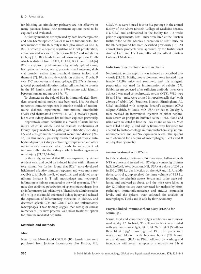
To evaluate the immune response following the NTS chal-lenge, we analysed the levels of total IgG and IgG subclassesin the serum of B6 and B7x−/− mice. Seven days after theserum transfer, B7x−/− mice displayed elevated levels of totalIgG (P = 0·02), IgG2b (P = 0·05) and IgG1 (P = 0·06)(Fig. 2a–c) compared to B6 mice. Additionally, levels ofanti-rabbit IgG were also found to be significantly higher inB7x−/− mice compared to B6 mice (P = 0·04) post-NTS chal-lenge (Fig. 2d). Interestingly, basal levels of serum IgG inB7x−/− mice were also higher than the B6 mice (P = 0·01)(Fig. 2a).
B7x-deficient mice are more susceptible to renaldamage and inflammatory cell infiltration
Histological analysis of kidneys revealed that both B6 andB7x−/− mice displayed marked renal damage followingpassive antibody transfer, with endocapillary hyper-cellularity, mesangial proliferation, PAS+ deposits andtubular lesions (Fig. 2e). However, the most severe indicatorof glomerular damage, crescent formation, was more pro-nounced in B7x−/− mice compared to B6 mice (Fig. 2e).Moreover, the number of infiltrating neutrophils was higherin kidneys of B7x−/− mice compared to B6 mice (Fig. 2f).Kidneys of B6 and B7x−/− mice at baseline (naive) or controlchallenged with PBS did not show any significant pathology(data not shown).
In nephrotoxic serum nephritis, recruitment and infiltra-tion of immune cells into the kidneys occurs due to theinflammatory cascades initiated after the deposition of anti-bodies directed against glomerular antigens, followed by
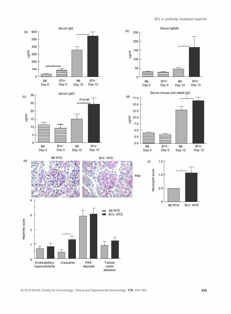
up-regulation of inflammatory mediators at the site ofinjury and complement activation [21,22]. Using immuno-histochemical analysis, we found that CD3+ T cells andCD68+ macrophages were increased significantly in the glo-meruli and interstitium of B7x−/− mice compared to B6 mice(Fig. 3a,b). Furthermore, glomerular IgG deposition wasmore pronounced in B7x−/− mice (Fig. 3c). Finally, B7x wassignificantly up-regulated in the kidneys of NTS-challengedB6 mice (Fig. 3d). Consistent with the relative over-expression patterns of B7x induced in vitro by inflamma-tory stimuli in resident kidney cells (Fig. 1), in-vivo B7xexpression was observed primarily in the tubules in thismodel (Fig. 3d).
Up-regulated levels of inflammatory mediators arefound in B7x-deficient kidneys following NTS challenge
Inflammatory cytokines and chemokines as well as otherrenal injury markers are induced in the kidney post-NTSchallenge [21,22]. Therefore, we analysed the levels ofinflammatory mediators in the kidney relevant to thismodel, pre- and post-NTS challenge. When normalizedseparately to the baseline levels in each strain, we found thatlevels of CXCL13 (P < 0·05), IL-23 (P < 0·0001), IFN regula-tory factor 5 (IRF5) (P < 0·01) and ICOSL (P < 0·01) wereup-regulated markedly in B7x−/− mice compared to B6 micefollowing induction of nephritis (Fig. 3e). CCL2, CXCL2,CCR5 and TGF-β (Fig. 3e), as well as ICAM-1, regulatedupon activation normal T cell expressed and secreted(RANTES) and the pro-apoptotic genes BCL-like protein 4(BAX) and apoptotic protease activating factor 1 (APAF-1)were at similar levels in both strains post-challenge (datanot shown).
Inflammatory macrophages are increased in B7x−/− mice
Splenic macrophages which express CD11b and CD11c canbe separated into two subsets (CD11b+CD11chi orCD11b+CD11clow), which can be subdivided further based onthe levels of F480 (F480hi or F480low) and Ly6C (Ly6chi orLy6clow) expression. At baseline, there were no differences inany macrophage subset between B6 and B7x−/− mice (data notshown). Analysis of macrophages subpopulations post-NTSchallenge revealed that the B7x−/− mice displayed higher rela-tive (%; Fig. 4a) and absolute (9517 ± 616 versus 7148 ± 706,P < 0·05) levels of macrophages which express low levels of
▶
Fig. 2. B7x−/− mice develop a robust humoral immune response, pronounced renal pathology and neutrophil infiltration post-nephrotoxic serum
(NTS) challenge. (a–d) Immunoglobulin (Ig)G, IgG2b, IgG1 and anti-rabbit IgG levels in serum of B6 and B7x−/− mice pre- (day 0) and
post-challenge (day 12) with NTS. Data are representative of two independent experiments, and are expressed as mean ± standard error of the mean
(s.e.m.), n = 9–10 per group. *P ≤ 0·05, by unpaired t-test. (e) Kidney histopathology was analysed by periodic acid Schiff (PAS) and haematoxylin
and eosin (H&E) staining, and images captured at ×600 magnification. A representative image from each strain is shown. Data are expressed as
mean ± s.e.m., n = 9–10 per group. *P ≤ 0·05, by Mann–Whitney U-test. (f) Immunohistochemical analysis of infiltration of Ly6G+ neutrophils. Data
are expressed as mean ± s.e.m., n = 6–7 randomly selected mice per group. *P ≤ 0·05, by Mann–Whitney U-test.
R. D. Pawar et al.
334 © 2014 British Society for Immunology, Clinical and Experimental Immunology, 179: 329–343
Serum lgG600(a)
μg/m
l
500
400
300
200
100
0B6
Day 0B7x-/-
Day 0B6
Day 12B7x-/-
Day 12
Serum lgG2b250(b)
μg/m
l
200
100
150
50
0B6
Day 0B7x-/-
Day 0B6
Day 12B7x-/-
Day 12
Serum lgG1
P=0·06(c)
μg/m
l
30
25
20
15
10
5
0B6
Day 0B7x-/-
Day 0B6
Day 12B7x-/-
Day 12
Serum mouse anti-rabbit lgG
17·5(d)
(e) (f)
μg/m
l
15·0
12·5
7·5
10·0
5·0
2·5
0·0
1·5
4
3
2
1
0
1·0
0·5
0
B6Day 0
B6 NTS
B6 NTS
PAS
Endocapillaryhypercellularity
Crescents PAS+
depositsTubularcasts/
dilatation
B6 NTS
Neu
trop
hil s
core
Nep
hriti
s sc
ore
B7x-/-
Day 0
B7x-/- NTS
B7x-/- NTS
B7x-/- NTS
B6Day 12
B7x-/-
Day 12
*
*
*
*
*
*
B7x in antibody mediated nephritis
335© 2014 British Society for Immunology, Clinical and Experimental Immunology, 179: 329–343
Interstitium Interstitium
20 μm
20 μm
20 μm20 μm
20 μm20 μm
B6 NTS
Glomeruli
1·2(a)
(c)
(d)
(e)
(b)
0·8
3
2·5
2·0
1·5
1·0
0·5
0
2
1
0
3
50
40
30
Nor
mal
ized
mR
NA
leve
ls
20
10
3
2
1
0
2
1
0
0·4
0B7x-/- NTS B6 NTS B7x-/- NTS B6 NTS
Glomeruli
B7x-/- NTS
B6 NTS
B6 NTS
B7x-/- NTS
B6 NTS B7x-/- NTS B6 NTS B7x-/- NTS
B6 NTS B7x-/- NTS
B6 PBS
B6 PBS
B6 NTS
B6 NTS
B7x-/- NTS
B6 NTS B7x-/- NTS
T c
ell s
core
Mac
roph
age
scor
e
IgG
dep
ositi
on s
core
Sco
re
MacrophagesT cells**
***
*
*
*
*
*
**
** **
CCL2 CXCL2 CXCL13 CCR5 IL-23 IRF5 ICOSL TGF-β
R. D. Pawar et al.
336 © 2014 British Society for Immunology, Clinical and Experimental Immunology, 179: 329–343
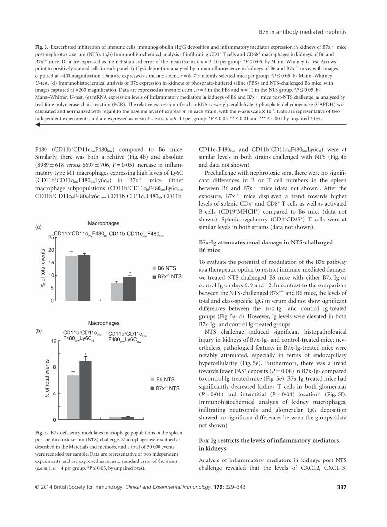
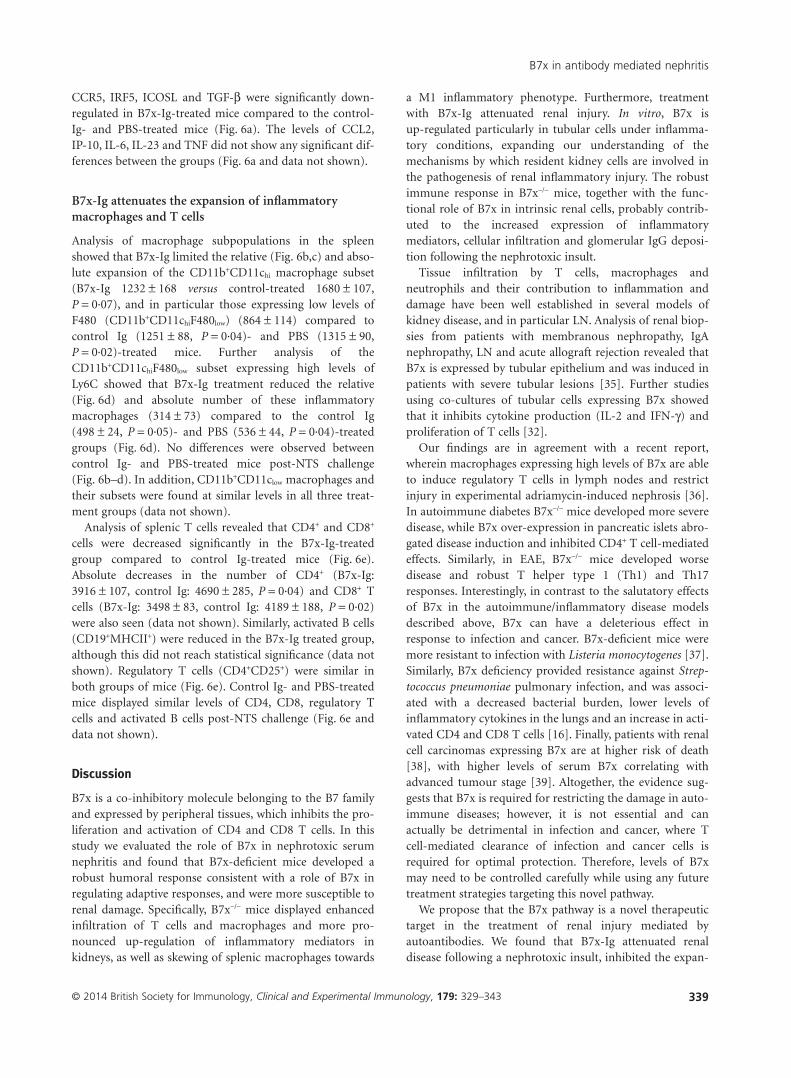
F480 (CD11b+CD11clowF480low) compared to B6 mice.Similarly, there was both a relative (Fig. 4b) and absolute(8989 ± 618 versus 6697 ± 706, P = 0·05) increase in inflam-matory type M1 macrophages expressing high levels of Ly6C(CD11b+CD11clowF480lowLy6chi) in B7x−/− mice. Othermacrophage subpopulations (CD11b+CD11chiF480lowLy6clow,CD11b+CD11chiF480hiLy6clow, CD11b+CD11chiF480hi, CD11b+
CD11chiF480low and CD11b+CD11chiF480lowLy6chi) were atsimilar levels in both strains challenged with NTS (Fig. 4band data not shown).
Prechallenge with nephrotoxic sera, there were no signifi-cant differences in B or T cell numbers in the spleenbetween B6 and B7x−/− mice (data not shown). After theexposure, B7x−/− mice displayed a trend towards higherlevels of splenic CD4+ and CD8+ T cells as well as activatedB cells (CD19+MHCII+) compared to B6 mice (data notshown). Splenic regulatory (CD4+CD25+) T cells were atsimilar levels in both strains (data not shown).
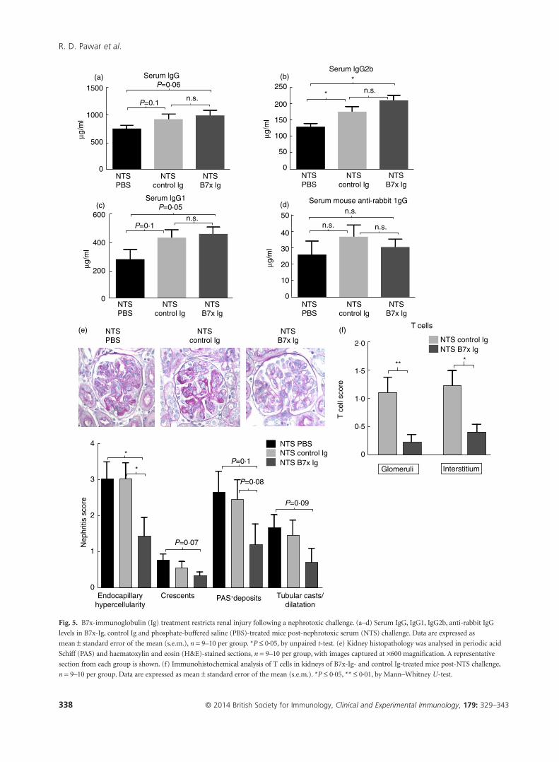
B7x-Ig attenuates renal damage in NTS-challengedB6 mice
To evaluate the potential of modulation of the B7x pathwayas a therapeutic option to restrict immune-mediated damage,we treated NTS-challenged B6 mice with either B7x-Ig orcontrol Ig on days 6, 9 and 12. In contrast to the comparisonbetween the NTS-challenged B7x−/− and B6 mice, the levels oftotal and class-specific IgG in serum did not show significantdifferences between the B7x-Ig- and control Ig-treatedgroups (Fig. 5a–d). However, Ig levels were elevated in bothB7x-Ig- and control Ig-treated groups.
NTS challenge induced significant histopathologicalinjury in kidneys of B7x-Ig- and control-treated mice; nev-ertheless, pathological features in B7x-Ig-treated mice werenotably attenuated, especially in terms of endocapillaryhypercellularity (Fig. 5e). Furthermore, there was a trendtowards fewer PAS+ deposits (P = 0·08) in B7x-Ig- comparedto control Ig-treated mice (Fig. 5e). B7x-Ig-treated mice hadsignificantly decreased kidney T cells in both glomerular(P = 0·01) and interstitial (P = 0·04) locations (Fig. 5f).Immunohistochemical analysis of kidney macrophages,infiltrating neutrophils and glomerular IgG depositionshowed no significant differences between the groups (datanot shown).
B7x-Ig restricts the levels of inflammatory mediatorsin kidneys
Analysis of inflammatory mediators in kidneys post-NTSchallenge revealed that the levels of CXCL2, CXCL13,
Fig. 3. Exacerbated infiltration of immune cells, immunoglobulin (Ig)G deposition and inflammatory mediator expression in kidneys of B7x−/− mice
post-nephrotoxic serum (NTS). (a,b) Immunohistochemical analysis of infiltrating CD3+ T cells and CD68+ macrophages in kidneys of B6 and
B7x−/− mice. Data are expressed as mean ± standard error of the mean (s.e.m.), n = 9–10 per group. *P ≤ 0·05, by Mann–Whitney U-test. Arrows
point to positively stained cells in each panel. (c) IgG deposition analysed by immunofluorescence in kidneys of B6 and B7x−/− mice, with images
captured at ×400 magnification. Data are expressed as mean ± s.e.m., n = 6–7 randomly selected mice per group. *P ≤ 0·05, by Mann–Whitney
U-test. (d) Immunohistochemical analysis of B7x expression in kidneys of phosphate-buffered saline (PBS) and NTS-challenged B6 mice, with
images captured at ×200 magnification. Data are expressed as mean ± s.e.m., n = 8 in the PBS and n = 11 in the NTS group. *P ≤ 0·05, by
Mann–Whitney U-test. (e) mRNA expression levels of inflammatory mediators in kidneys of B6 and B7x−/− mice post-NTS challenge, as analysed by
real-time polymerase chain reaction (PCR). The relative expression of each mRNA versus glyceraldehyde 3-phosphate dehydrogenase (GAPDH) was
calculated and normalized with regard to the baseline level of expression in each strain, with the y-axis scale × 10−3. Data are representative of two
independent experiments, and are expressed as mean ± s.e.m., n = 9–10 per group. *P ≤ 0·05, ** ≤ 0·01 and *** ≤ 0·001 by unpaired t-test.◀
(a)
(b)
*
* B7x-/- NTS
B7x-/- NTS
B6 NTS
B6 NTS
Macrophages
CD11b+CD11clow F480lowLy6Chi
CD11b+CD11clowF480lowCD11b+CD11clowF480hi
CD11b+CD11clowF480lowLy6Clow
Macrophages
25
20
15
10 5 0
% o
f tot
al e
vent
s
12
8
4
0
% o
f tot
al e
vent
s
Fig. 4. B7x deficiency modulates macrophage populations in the spleen
post-nephrotoxic serum (NTS) challenge. Macrophages were stained as
described in the Materials and methods, and a total of 50 000 events
were recorded per sample. Data are representative of two independent
experiments, and are expressed as mean ± standard error of the mean
(s.e.m.), n = 4 per group. *P ≤ 0·05, by unpaired t-test.
B7x in antibody mediated nephritis
337© 2014 British Society for Immunology, Clinical and Experimental Immunology, 179: 329–343
μg/m
l(a)
1500
1000
500
0
Serum lgGP=0·06
P=0.1n.s.
n.s.
n.s.
n.s.n.s.
n.s.
NTSPBS
NTScontrol lg
NTSB7x lg
μg/m
l
(c)600
400
200
0
Serum lgG1P=0·05
P=0·1
P=0·1
P=0·07
P=0·08
P=0·09
NTSPBS
NTScontrol lg
NTSB7x lg
NTSPBS
NTScontrol lg
NTSB7x lg
μg/m
l
(b)250
150
200
100
50
0
Serum lgG2b*
* *
*4
3
2
1
0
*
*
*
NTSPBS
NTScontrol lg
NTSB7x lg
μg/m
l
(d)
(f)(e)
50
30
40
20
10
0
Serum mouse anti-rabbit 1gG
NTSPBS
NTS PBS
NTScontrol lg
NTSB7x lg
NTS control lg
T cells
NTS B7x lg
NTS control lgNTS B7x lg
Endocapillaryhypercellularity
Crescents PAS+deposits Tubular casts/dilatation
Nep
hriti
s sc
ore
InterstitiumGlomeruli
T c
ell s
core
2·0
1·5
1·0
0·5
0
Fig. 5. B7x-immunoglobulin (Ig) treatment restricts renal injury following a nephrotoxic challenge. (a–d) Serum IgG, IgG1, IgG2b, anti-rabbit IgG
levels in B7x-Ig, control Ig and phosphate-buffered saline (PBS)-treated mice post-nephrotoxic serum (NTS) challenge. Data are expressed as
mean ± standard error of the mean (s.e.m.), n = 9–10 per group. *P ≤ 0·05, by unpaired t-test. (e) Kidney histopathology was analysed in periodic acid
Schiff (PAS) and haematoxylin and eosin (H&E)-stained sections, n = 9–10 per group, with images captured at ×600 magnification. A representative
section from each group is shown. (f) Immunohistochemical analysis of T cells in kidneys of B7x-Ig- and control Ig-treated mice post-NTS challenge,
n = 9–10 per group. Data are expressed as mean ± standard error of the mean (s.e.m.). *P ≤ 0·05, ** ≤ 0·01, by Mann–Whitney U-test.
R. D. Pawar et al.
338 © 2014 British Society for Immunology, Clinical and Experimental Immunology, 179: 329–343
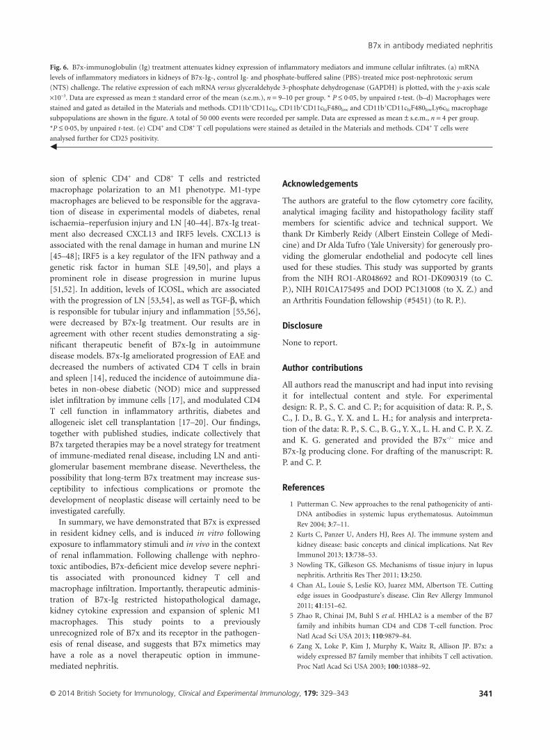
CCR5, IRF5, ICOSL and TGF-β were significantly down-regulated in B7x-Ig-treated mice compared to the control-Ig- and PBS-treated mice (Fig. 6a). The levels of CCL2,IP-10, IL-6, IL-23 and TNF did not show any significant dif-ferences between the groups (Fig. 6a and data not shown).
B7x-Ig attenuates the expansion of inflammatorymacrophages and T cells
Analysis of macrophage subpopulations in the spleenshowed that B7x-Ig limited the relative (Fig. 6b,c) and abso-lute expansion of the CD11b+CD11chi macrophage subset(B7x-Ig 1232 ± 168 versus control-treated 1680 ± 107,P = 0·07), and in particular those expressing low levels ofF480 (CD11b+CD11chiF480low) (864 ± 114) compared tocontrol Ig (1251 ± 88, P = 0·04)- and PBS (1315 ± 90,P = 0·02)-treated mice. Further analysis of theCD11b+CD11chiF480low subset expressing high levels ofLy6C showed that B7x-Ig treatment reduced the relative(Fig. 6d) and absolute number of these inflammatorymacrophages (314 ± 73) compared to the control Ig(498 ± 24, P = 0·05)- and PBS (536 ± 44, P = 0·04)-treatedgroups (Fig. 6d). No differences were observed betweencontrol Ig- and PBS-treated mice post-NTS challenge(Fig. 6b–d). In addition, CD11b+CD11clow macrophages andtheir subsets were found at similar levels in all three treat-ment groups (data not shown).
Analysis of splenic T cells revealed that CD4+ and CD8+
cells were decreased significantly in the B7x-Ig-treatedgroup compared to control Ig-treated mice (Fig. 6e).Absolute decreases in the number of CD4+ (B7x-Ig:3916 ± 107, control Ig: 4690 ± 285, P = 0·04) and CD8+ Tcells (B7x-Ig: 3498 ± 83, control Ig: 4189 ± 188, P = 0·02)were also seen (data not shown). Similarly, activated B cells(CD19+MHCII+) were reduced in the B7x-Ig treated group,although this did not reach statistical significance (data notshown). Regulatory T cells (CD4+CD25+) were similar inboth groups of mice (Fig. 6e). Control Ig- and PBS-treatedmice displayed similar levels of CD4, CD8, regulatory Tcells and activated B cells post-NTS challenge (Fig. 6e anddata not shown).
Discussion
B7x is a co-inhibitory molecule belonging to the B7 familyand expressed by peripheral tissues, which inhibits the pro-liferation and activation of CD4 and CD8 T cells. In thisstudy we evaluated the role of B7x in nephrotoxic serumnephritis and found that B7x-deficient mice developed arobust humoral response consistent with a role of B7x inregulating adaptive responses, and were more susceptible torenal damage. Specifically, B7x−/− mice displayed enhancedinfiltration of T cells and macrophages and more pro-nounced up-regulation of inflammatory mediators inkidneys, as well as skewing of splenic macrophages towards
a M1 inflammatory phenotype. Furthermore, treatmentwith B7x-Ig attenuated renal injury. In vitro, B7x isup-regulated particularly in tubular cells under inflamma-tory conditions, expanding our understanding of themechanisms by which resident kidney cells are involved inthe pathogenesis of renal inflammatory injury. The robustimmune response in B7x−/− mice, together with the func-tional role of B7x in intrinsic renal cells, probably contrib-uted to the increased expression of inflammatorymediators, cellular infiltration and glomerular IgG deposi-tion following the nephrotoxic insult.
Tissue infiltration by T cells, macrophages andneutrophils and their contribution to inflammation anddamage have been well established in several models ofkidney disease, and in particular LN. Analysis of renal biop-sies from patients with membranous nephropathy, IgAnephropathy, LN and acute allograft rejection revealed thatB7x is expressed by tubular epithelium and was induced inpatients with severe tubular lesions [35]. Further studiesusing co-cultures of tubular cells expressing B7x showedthat it inhibits cytokine production (IL-2 and IFN-γ) andproliferation of T cells [32].
Our findings are in agreement with a recent report,wherein macrophages expressing high levels of B7x are ableto induce regulatory T cells in lymph nodes and restrictinjury in experimental adriamycin-induced nephrosis [36].In autoimmune diabetes B7x−/− mice developed more severedisease, while B7x over-expression in pancreatic islets abro-gated disease induction and inhibited CD4+ T cell-mediatedeffects. Similarly, in EAE, B7x−/− mice developed worsedisease and robust T helper type 1 (Th1) and Th17responses. Interestingly, in contrast to the salutatory effectsof B7x in the autoimmune/inflammatory disease modelsdescribed above, B7x can have a deleterious effect inresponse to infection and cancer. B7x-deficient mice weremore resistant to infection with Listeria monocytogenes [37].Similarly, B7x deficiency provided resistance against Strep-tococcus pneumoniae pulmonary infection, and was associ-ated with a decreased bacterial burden, lower levels ofinflammatory cytokines in the lungs and an increase in acti-vated CD4 and CD8 T cells [16]. Finally, patients with renalcell carcinomas expressing B7x are at higher risk of death[38], with higher levels of serum B7x correlating withadvanced tumour stage [39]. Altogether, the evidence sug-gests that B7x is required for restricting the damage in auto-immune diseases; however, it is not essential and canactually be detrimental in infection and cancer, where Tcell-mediated clearance of infection and cancer cells isrequired for optimal protection. Therefore, levels of B7xmay need to be controlled carefully while using any futuretreatment strategies targeting this novel pathway.
We propose that the B7x pathway is a novel therapeutictarget in the treatment of renal injury mediated byautoantibodies. We found that B7x-Ig attenuated renaldisease following a nephrotoxic insult, inhibited the expan-
B7x in antibody mediated nephritis
339© 2014 British Society for Immunology, Clinical and Experimental Immunology, 179: 329–343

(b) (c)
% o
f tot
al e
vent
s%
of t
otal
eve
nts
% o
f tot
al e
vent
s%
of t
otal
eve
nts
4
5
3
2
P=0·07
NTSPBS
NTScontrol lg
NTSB7x lg
(d)
0·15
0·20
0·10
0·05
0·00
P=0·1
NTSPBS
NTScontrol lg
NTSB7x lg
4
2
3
0
*
*
*
*
NTSPBS
NTScontrol lg
NTSB7x lg
(e)10
6
8
4
2
0
T cellsCD4+
CD4+CD25+
CD8+
NTSPBS
NTScontrol lg
NTSB7x lg
(a) 12
10
8
6
4
3
2
1
0
*
*
*
**
*
*
*
*
*
*
*
*
CCL2 CXCL2 CXCL13 CCR5 IL-23 IRF5 ICOSL TGF-β
P=0·07
NTS PBSNTS control lgNTS B7x lg
Macrophages Macrophages
Rel
ativ
e ex
pres
sion
CD11b+CD11chiF480low
MacrophagesCD11b+CD11chiF480lowLy6Chi
CD11b+CD11Chi
R. D. Pawar et al.
340 © 2014 British Society for Immunology, Clinical and Experimental Immunology, 179: 329–343
sion of splenic CD4+ and CD8+ T cells and restrictedmacrophage polarization to an M1 phenotype. M1-typemacrophages are believed to be responsible for the aggrava-tion of disease in experimental models of diabetes, renalischaemia–reperfusion injury and LN [40–44]. B7x-Ig treat-ment also decreased CXCL13 and IRF5 levels. CXCL13 isassociated with the renal damage in human and murine LN[45–48]; IRF5 is a key regulator of the IFN pathway and agenetic risk factor in human SLE [49,50], and plays aprominent role in disease progression in murine lupus[51,52]. In addition, levels of ICOSL, which are associatedwith the progression of LN [53,54], as well as TGF-β, whichis responsible for tubular injury and inflammation [55,56],were decreased by B7x-Ig treatment. Our results are inagreement with other recent studies demonstrating a sig-nificant therapeutic benefit of B7x-Ig in autoimmunedisease models. B7x-Ig ameliorated progression of EAE anddecreased the numbers of activated CD4 T cells in brainand spleen [14], reduced the incidence of autoimmune dia-betes in non-obese diabetic (NOD) mice and suppressedislet infiltration by immune cells [17], and modulated CD4T cell function in inflammatory arthritis, diabetes andallogeneic islet cell transplantation [17–20]. Our findings,together with published studies, indicate collectively thatB7x targeted therapies may be a novel strategy for treatmentof immune-mediated renal disease, including LN and anti-glomerular basement membrane disease. Nevertheless, thepossibility that long-term B7x treatment may increase sus-ceptibility to infectious complications or promote thedevelopment of neoplastic disease will certainly need to beinvestigated carefully.
In summary, we have demonstrated that B7x is expressedin resident kidney cells, and is induced in vitro followingexposure to inflammatory stimuli and in vivo in the contextof renal inflammation. Following challenge with nephro-toxic antibodies, B7x-deficient mice develop severe nephri-tis associated with pronounced kidney T cell andmacrophage infiltration. Importantly, therapeutic adminis-tration of B7x-Ig restricted histopathological damage,kidney cytokine expression and expansion of splenic M1macrophages. This study points to a previouslyunrecognized role of B7x and its receptor in the pathogen-esis of renal disease, and suggests that B7x mimetics mayhave a role as a novel therapeutic option in immune-mediated nephritis.
Acknowledgements
The authors are grateful to the flow cytometry core facility,analytical imaging facility and histopathology facility staffmembers for scientific advice and technical support. Wethank Dr Kimberly Reidy (Albert Einstein College of Medi-cine) and Dr Alda Tufro (Yale University) for generously pro-viding the glomerular endothelial and podocyte cell linesused for these studies. This study was supported by grantsfrom the NIH RO1-AR048692 and RO1-DK090319 (to C.P.), NIH R01CA175495 and DOD PC131008 (to X. Z.) andan Arthritis Foundation fellowship (#5451) (to R. P.).
Disclosure
None to report.
Author contributions
All authors read the manuscript and had input into revisingit for intellectual content and style. For experimentaldesign: R. P., S. C. and C. P.; for acquisition of data: R. P., S.C., J. D., B. G., Y. X. and L. H.; for analysis and interpreta-tion of the data: R. P., S. C., B. G., Y. X., L. H. and C. P. X. Z.and K. G. generated and provided the B7x−/− mice andB7x-Ig producing clone. For drafting of the manuscript: R.P. and C. P.
References
1 Putterman C. New approaches to the renal pathogenicity of anti-
DNA antibodies in systemic lupus erythematosus. Autoimmun
Rev 2004; 3:7–11.
2 Kurts C, Panzer U, Anders HJ, Rees AJ. The immune system and
kidney disease: basic concepts and clinical implications. Nat Rev
Immunol 2013; 13:738–53.
3 Nowling TK, Gilkeson GS. Mechanisms of tissue injury in lupus
nephritis. Arthritis Res Ther 2011; 13:250.
4 Chan AL, Louie S, Leslie KO, Juarez MM, Albertson TE. Cutting
edge issues in Goodpasture’s disease. Clin Rev Allergy Immunol
2011; 41:151–62.
5 Zhao R, Chinai JM, Buhl S et al. HHLA2 is a member of the B7
family and inhibits human CD4 and CD8 T-cell function. Proc
Natl Acad Sci USA 2013; 110:9879–84.
6 Zang X, Loke P, Kim J, Murphy K, Waitz R, Allison JP. B7x: a
widely expressed B7 family member that inhibits T cell activation.
Proc Natl Acad Sci USA 2003; 100:10388–92.
Fig. 6. B7x-immunoglobulin (Ig) treatment attenuates kidney expression of inflammatory mediators and immune cellular infiltrates. (a) mRNA
levels of inflammatory mediators in kidneys of B7x-Ig-, control Ig- and phosphate-buffered saline (PBS)-treated mice post-nephrotoxic serum
(NTS) challenge. The relative expression of each mRNA versus glyceraldehyde 3-phosphate dehydrogenase (GAPDH) is plotted, with the y-axis scale
×10−3. Data are expressed as mean ± standard error of the mean (s.e.m.), n = 9–10 per group. * P ≤ 0·05, by unpaired t-test. (b–d) Macrophages were
stained and gated as detailed in the Materials and methods. CD11b+CD11chi, CD11b+CD11chiF480low and CD11b+CD11chiF480lowLy6chi macrophage
subpopulations are shown in the figure. A total of 50 000 events were recorded per sample. Data are expressed as mean ± s.e.m., n = 4 per group.
*P ≤ 0·05, by unpaired t-test. (e) CD4+ and CD8+ T cell populations were stained as detailed in the Materials and methods. CD4+ T cells were
analysed further for CD25 positivity.◀
B7x in antibody mediated nephritis
341© 2014 British Society for Immunology, Clinical and Experimental Immunology, 179: 329–343

7 Greenwald RJ, Freeman GJ, Sharpe AH. The B7 family revisited.
Annu Rev Immunol 2005; 23:515–48.
8 Zang X, Allison JP. The B7 family and cancer therapy:
costimulation and coinhibition. Clin Cancer Res 2007; 13:5271–
9.
9 Furie R, Nicholls K, Cheng TT et al. Efficacy and safety of
abatacept in lupus nephritis: a twelve-month, randomized,
double-blind study. Arthritis Rheumatol 2014; 66:379–89.
10 Wofsy D, Hillson JL, Diamond B. Abatacept for lupus nephritis:
alternative definitions of complete response support conflicting
conclusions. Arthritis Rheum 2012; 64:3660–5.
11 Genovese MC, Tena CP, Covarrubias A et al. Subcutaneous
abatacept for the treatment of rheumatoid arthritis: longterm data
from the ACQUIRE trial. J Rheumatol 2014; 41:629–39.
12 Viglietta V, Bourcier K, Buckle GJ et al. CTLA4Ig treatment in
patients with multiple sclerosis: an open-label, phase 1 clinical
trial. Neurology 2008; 71:917–24.
13 Lee JS, Scandiuzzi L, Ray A et al. B7x in the periphery abrogates
pancreas-specific damage mediated by self-reactive CD8 T cells. J
Immunol 2012; 189:4165–74.
14 Podojil JR, Liu LN, Marshall SA et al. B7-H4Ig inhibits mouse and
human T-cell function and treats EAE via IL-10/Treg-dependent
mechanisms. J Autoimmun 2013; 44:71–81.
15 Wei J, Loke P, Zang X, Allison JP. Tissue-specific expression of
B7x protects from CD4 T cell-mediated autoimmunity. J Exp Med
2011; 208:1683–94.
16 Hofmeyer KA, Scandiuzzi L, Ghosh K, Pirofski LA, Zang X.
Tissue-expressed B7x affects the immune response to and
outcome of lethal pulmonary infection. J Immunol 2012;
189:3054–63.
17 Wang X, Hao J, Metzger DL et al. Early treatment of NOD mice
with B7-H4 reduces the incidence of autoimmune diabetes. Dia-
betes 2011; 60:3246–55.
18 Wang X, Hao J, Metzger DL et al. B7-H4 induces donor-specific
tolerance in mouse islet allografts. Cell Transplant 2012; 21:99–
111.
19 Azuma T, Zhu G, Xu H et al. Potential role of decoy B7-H4 in the
pathogenesis of rheumatoid arthritis: a mouse model informed by
clinical data. PLOS Med 2009; 6:e1000166.
20 Lee IF, Wang X, Hao J et al. B7-H4.Ig inhibits the development of
type 1 diabetes by regulating Th17 cells in NOD mice. Cell
Immunol 2013; 282:1–8.
21 Xia Y, Campbell SR, Broder A et al. Inhibition of the TWEAK/
Fn14 pathway attenuates renal disease in nephrotoxic serum
nephritis. Clin Immunol 2012; 145:108–21.
22 Pawar RD, Pitashny M, Gindea S et al. Neutrophil gelatinase-
associated lipocalin is instrumental in the pathogenesis of
antibody-mediated nephritis in mice. Arthritis Rheum 2012;
64:1620–31.
23 Xia Y, Pawar RD, Nakouzi AS et al. The constant region contrib-
utes to the antigenic specificity and renal pathogenicity of murine
anti-DNA antibodies. J Autoimmun 2012; 39:398–411.
24 Qing X, Pitashny M, Thomas DB, Barrat FJ, Hogarth MP,
Putterman C. Pathogenic anti-DNA antibodies modulate gene
expression in mesangial cells: involvement of HMGB1 in anti-
DNA antibody-induced renal injury. Immunol Lett 2008; 121:61–
73.
25 Qing X, Zavadil J, Crosby MB et al. Nephritogenic anti-DNA anti-
bodies regulate gene expression in MRL/lpr mouse glomerular
mesangial cells. Arthritis Rheum 2006; 54:2198–210.
26 Fujii T, Hamano Y, Ueda S et al. Predominant role of
FcgammaRIII in the induction of accelerated nephrotoxic glo-
merulonephritis. Kidney Int 2003; 64:1406–16.
27 Salant DJ, Cybulsky AV. Experimental glomerulonephritis.
Methods Enzymol 1988; 162:421–61.
28 Herlitz LC, Bomback AS, Markowitz GS et al. Pathology after
eculizumab in dense deposit disease and C3 GN. J Am Soc
Nephrol 2012; 23:1229–37.
29 Guan F, Villegas G, Teichman J, Mundel P, Tufro A. Autocrine
class 3 semaphorin system regulates slit diaphragm proteins and
podocyte survival. Kidney Int 2006; 69:1564–9.
30 Reidy KJ, Villegas G, Teichman J et al. Semaphorin3a regulates
endothelial cell number and podocyte differentiation during
glomerular development. Development 2009; 136:3979–89.
31 Deocharan B, Qing X, Lichauco J, Putterman C. Alpha-actinin is a
cross-reactive renal target for pathogenic anti-DNA antibodies. J
Immunol 2002; 168:3072–8.
32 Pawar RD, Castrezana-Lopez L, Allam R et al. Bacterial
lipopeptide triggers massive albuminuria in murine lupus nephri-
tis by activating Toll-like receptor 2 at the glomerular filtration
barrier. Immunology 2009; 128:e206–21.
33 Abadi YM, Jeon H, Ohaegbulam KC et al. Host b7x promotes pul-
monary metastasis of breast cancer. J Immunol 2013; 190:3806–
14.
34 Salceda S, Tang T, Kmet M et al. The immunomodulatory protein
B7-H4 is overexpressed in breast and ovarian cancers and pro-
motes epithelial cell transformation. Exp Cell Res 2005; 306:128–
41.
35 Chen Y, Yang C, Xie Z et al. Expression of the novel
co-stimulatory molecule B7-H4 by renal tubular epithelial cells.
Kidney Int 2006; 70:2092–9.
36 Cao Q, Wang Y, Zheng D et al. IL-10/TGF-beta-modified
macrophages induce regulatory T cells and protect against
adriamycin nephrosis. J Am Soc Nephrol 2010; 21:933–42.
37 Zhu G, Augustine MM, Azuma T et al. B7-H4-deficient mice
display augmented neutrophil-mediated innate immunity. Blood
2009; 113:1759–67.
38 Krambeck AE, Thompson RH, Dong H et al. B7-H4 expression in
renal cell carcinoma and tumor vasculature: associations with
cancer progression and survival. Proc Natl Acad Sci USA 2006;
103:10391–6.
39 Thompson RH, Zang X, Lohse CM et al. Serum-soluble B7x is
elevated in renal cell carcinoma patients and is associated with
advanced stage. Cancer Res 2008; 68:6054–8.
40 Cucak H, Grunnet LG, Rosendahl A. Accumulation of
M1-like macrophages in type 2 diabetic islets is followed by a sys-
temic shift in macrophage polarization. J Leukoc Biol 2014;
95:149–60.
41 Nieuwenhuizen L, Schutgens RE, Coeleveld K et al. Hemarthrosis
in hemophilic mice results in alterations in M1-M2 monocyte/
macrophage polarization. Thromb Res 2014; 133:390–5.
42 Bignon A, Gaudin F, Hemon P et al. CCR1 inhibition ameliorates
the progression of lupus nephritis in NZB/W mice. J Immunol
2014; 192:886–96.
43 Ranganathan PV, Jayakumar C, Ramesh G. Netrin-1-treated
macrophages protect the kidney against ischemia-reperfusion
injury and suppress inflammation by inducing M2 polarization.
Am J Physiol Renal Physiol 2013; 304:F948–57.
44 Orme J, Mohan C. Macrophage subpopulations in systemic lupus
erythematosus. Discov Med 2012; 13:151–8.
R. D. Pawar et al.
342 © 2014 British Society for Immunology, Clinical and Experimental Immunology, 179: 329–343

45 Ezzat M, El-Gammasy T, Shaheen K, Shokr E. Elevated produc-
tion of serum B-cell-attracting chemokine-1 (BCA-1/CXCL13) is
correlated with childhood-onset lupus disease activity, severity,
and renal involvement. Lupus 2011; 20:845–54.
46 Lee HT, Shiao YM, Wu TH et al. Serum BLC/CXCL13 concentra-
tions and renal expression of CXCL13/CXCR5 in patients with
systemic lupus erythematosus and lupus nephritis. J Rheumatol
2010; 37:45–52.
47 Schiffer L, Kumpers P, Davalos-Misslitz AM et al. B-cell-attracting
chemokine CXCL13 as a marker of disease activity and renal
involvement in systemic lupus erythematosus (SLE). Nephrol Dial
Transplant 2009; 24:3708–12.
48 Moreth K, Brodbeck R, Babelova A et al. The proteoglycan
biglycan regulates expression of the B cell chemoattractant
CXCL13 and aggravates murine lupus nephritis. J Clin Invest
2010; 120:4251–72.
49 Niewold TB, Kelly JA, Kariuki SN et al. IRF5 haplotypes demon-
strate diverse serological associations which predict serum inter-
feron alpha activity and explain the majority of the genetic
association with systemic lupus erythematosus. Ann Rheum Dis
2012; 71:463–8.
50 Cherian TS, Kariuki SN, Franek BS, Buyon JP, Clancy RM,
Niewold TB. Brief Report: IRF5 systemic lupus erythematosus risk
haplotype is associated with asymptomatic serologic autoimmun-
ity and progression to clinical autoimmunity in mothers of chil-
dren with neonatal lupus. Arthritis Rheum 2012; 64:3383–7.
51 Richez C, Yasuda K, Bonegio RG et al. IFN regulatory factor 5 is
required for disease development in the FcgammaRIIB−/−Yaa and
FcgammaRIIB−/− mouse models of systemic lupus erythematosus.
J Immunol 2010; 184:796–806.
52 Tada Y, Kondo S, Aoki S et al. Interferon regulatory factor 5 is
critical for the development of lupus in MRL/lpr mice. Arthritis
Rheum 2011; 63:738–48.
53 Ding H, Wu X, Wu J et al. Delivering PD-1 inhibitory signal con-
comitant with blocking ICOS co-stimulation suppresses lupus-
like syndrome in autoimmune BXSB mice. Clin Immunol 2006;
118:258–67.
54 Iwai H, Abe M, Hirose S et al. Involvement of inducible
costimulator-B7 homologous protein costimulatory pathway in
murine lupus nephritis. J Immunol 2003; 171:2848–54.
55 Gentle ME, Shi S, Daehn I et al. Epithelial cell TGFbeta signaling
induces acute tubular injury and interstitial inflammation. J Am
Soc Nephrol 2013; 24:787–99.
56 Iyoda M, Shibata T, Wada Y et al. Long- and short-term treatment
with imatinib attenuates the development of chronic kidney
disease in experimental anti-glomerular basement membrane
nephritis. Nephrol Dial Transplant 2013; 28:576–84.
B7x in antibody mediated nephritis
343© 2014 British Society for Immunology, Clinical and Experimental Immunology, 179: 329–343