Embed Size (px)
Citation preview
Pathology of Birds – An Overview
H. L. ShivaprasadCalifornia Animal Health and Food Safety Laboratory System, Fresno Branch
School of Veterinary Medicine, University of California, Davis2789 South Orange Avenue. Fresno, CA 93725
Tele: 559-498-7740, Fax: 559-485-8097E-mail: [email protected]
Contents
1. Introduction..........................................................................................................................2
2. Taxonomy............................................................................................................................3
3. Anatomy..............................................................................................................................5
4. Inflammation........................................................................................................................7
5. Coagulation..........................................................................................................................8
6. Complement.........................................................................................................................8
7. Bacterial Diseases................................................................................................................8
8. Fungal diseases..................................................................................................................15
9. Viral diseases.....................................................................................................................16
10. Parasitic diseases...............................................................................................................28
11. Toxicosis............................................................................................................................35
12. Metabolic diseases.............................................................................................................38
13. Diseases of Malnutrition....................................................................................................43
14. Neoplasia...........................................................................................................................44
15. References:........................................................................................................................46
Presented at C.L. Davis Foundation Conference on Gross Morbid Anatomy of Animals, AFIP, Washington D. C., April 8-12, 2002. HLS (Last revised 3/12/2003)

Birds: Of all the higher forms of life the birds are the most beautiful, most musical, most admired, most watched and most defended. Roger Tory Peterson (1974)
1. Introduction
Similar to other animals, birds are susceptible to a variety of diseases.
Knowledge about the type of birds, their anatomy and how they are
managed does help one understand the type and kind of diseases
different birds are susceptible to. Because some types of birds are raised
for egg production or meat, such as commercial poultry, infectious
diseases can spread rapidly among birds raised in a confined space.
Poultry can also be raised in small numbers as backyard flocks. Often
poultry raised in such conditions are more often exposed to natural
elements and are often not vaccinated; poor nutrition and lax biosecurity
can lead to frequent viral, bacterial, parasitic and nutritional diseases. Pet
and exotic birds such as psittacines and passerines are raised in small or
large aviaries, sold in pet shops and kept as pets. These birds have their
own unique diseases that can be influenced by management. There are
also free flying birds that face ever shrinking habitats and ecosystems
that are undergoing changes. Therefore, diseases in such birds are
greatly influenced by environmental factors. Numerous birds raised or
kept in captivity as in zoos have a different and unique environment.
Regardless of whatever management practices are used, genetics and
nutrition also play a significant role in the initiation and outcome of a
disease.
This handout on the pathology of birds is an outline and is by no means
complete. There are a large number of miscellaneous diseases or
pathological conditions that have not been included in this outline.
2

Those who are interested in learning more are advised to review the
references provided at the end of this outline.
2. Taxonomy
There are over 22,000 subspecies and 9300 species of birds classified in 166 families, 27 orders
in the class Aves. Order Passeriformes contains the largest number with 5243 species of birds,
and the Order Struthioniformes contains only one species (ostrich).
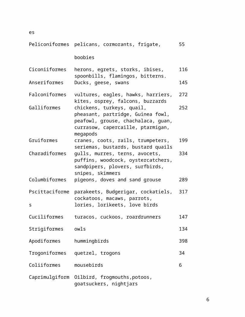
The number of species in each order, along with representative species, is listed in the following
table. The list starts with orders believed to be the most primitive and continuing to the most
advanced.
Order Species # Species
Struthioniformes ostrich 1
Rheiformes rheas 2
Casuariiformes emus, cassowary 4
Apterygiformes kiwis 3
Tinamiformes tinamous 40
Sphenisciformes penguins 17
Gaviiformes loons 4
Podicipediformes grebes 18
Procellariformes albatrosses, fulmars 86
Peliconiformes pelicans, cormorants, frigate, boobies 55
Ciconiiformes herons, egrets, storks, ibises, spoonbills, flamingos, bitterns.
116
Anseriformes Ducks, geese, swans 145
3
Falconiformes vultures, eagles, hawks, harriers, kites, osprey, falcons, buzzards
272
Galliformes chickens, turkeys, quail, pheasant, partridge, Guinea fowl, peafowl, grouse, chachalaca, guan, currasow, capercaille, ptarmigan, megapods
252
Gruiformes cranes, coots, rails, trumpeters, seriemas, bustards, bustard quails
199
Charadiformes gulls, murres, terns, avocets, puffins, woodcock, oystercatchers, sandpipers, plovers, surfbirds, snipes, skimmers
334
Columbiformes pigeons, doves and sand grouse 289
Pscittaciformes parakeets, Budgerigar, cockatiels, cockatoos, macaws, parrots, lories, lorikeets, love birds
317
Cuciliformes turacos, cuckoos, roardrunners 147
Strigiformes owls 134
Apodiformes hummingbirds 398
Trogoniformes quetzel, trogons 34
Coliiformes mousebirds 6
Caprimulgiformes Oilbird, frogmouths,potoos, goatsuckers, nightjars
Coraciformes toucans, barbets, woodpeckers, jacamars 379
Piciformes kingfishers, hornbills, hoopoes, rollers 199
Passeriformes Perching birds; finches, canaries, crows, ravens, magpies, jays, American robins, mockingbirds, bulbuls, sparrows, starlings, mynahs, blackbirds, cardinals, chickadees, thrushes, tits, grosbeaks, buntings, tanagers, orioles, birds of paradise, larks, wrens, waxwings, waxbills, weavers, swallows, manakins, martins, antbirds, shrikes, etc., etc.
5243
4

3. Anatomy
Birds have a close evolutionary relationship with reptiles especially crocodiles. Their unique
anatomy has helped the birds to adapt well to the environment. Even though there are numerous
different species of birds, their anatomy shows a greater uniformity of structure than many single
orders of fishes, amphibians, reptiles and mammals (King and McLelland, 1984). A few salient
features on the anatomy of birds are given below.
Birds skin lack glands except for the uropygeal gland or preen gland present at the base of the
tail. This gland is not found in all birds. Various appendages such as comb, wattles, ear lobes,
snood, caruncles, beard, spurs, claws, scales are present in a variety of birds.
Birds have pneumonic bones and air sacs extend in to the proximal humerus, vertebrae and
pelvic girdle. Laying hens form medullary bone in response to estrogen, which serves as a source
of calcium during laying.
Trachea has complete rings and syrinx is present. Syrinx is large in male waterfowl. Lungs are
attached to the body wall. From trachea the primary bronchi passes through the lungs giving rise
to many secondary bronchi before entering the abdominal air sacs. The secondary bronchi give
rise to tertiary or parabronchioles and from there to atria and air capillaries where air exchange
takes place. Lungs are of ‘paleopulmo’ (unidirectional movement of air) and ‘neopulmo’ (bi-
directional) types. Cervical, clavicular (paired), anterior and posterior thoracic (paired) and
abdominal (paired) air sacs are present.
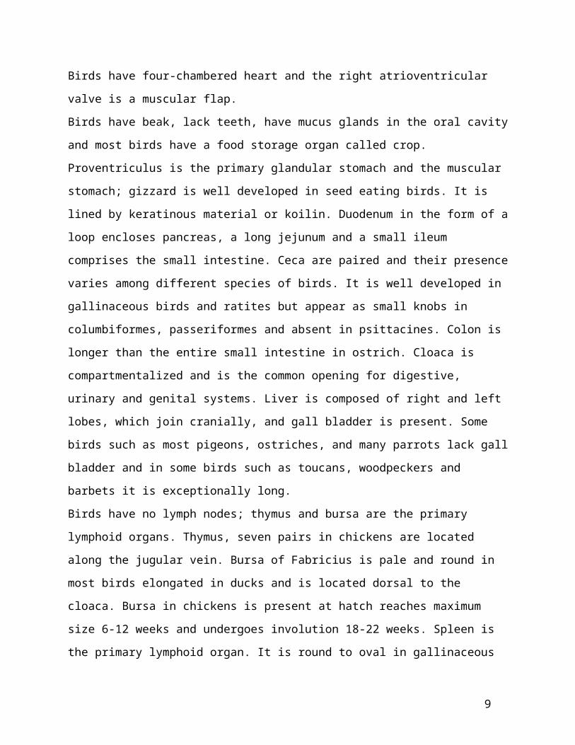
Birds have four-chambered heart and the right atrioventricular valve is a muscular flap.
Birds have beak, lack teeth, have mucus glands in the oral cavity and most birds have a food
storage organ called crop. Proventriculus is the primary glandular stomach and the muscular
stomach; gizzard is well developed in seed eating birds. It is lined by keratinous material or
koilin. Duodenum in the form of a loop encloses pancreas, a long jejunum and a small ileum
comprises the small intestine. Ceca are paired and their presence varies among different species
of birds. It is well developed in gallinaceous birds and ratites but appear as small knobs in
5
columbiformes, passeriformes and absent in psittacines. Colon is longer than the entire small
intestine in ostrich. Cloaca is compartmentalized and is the common opening for digestive,
urinary and genital systems. Liver is composed of right and left lobes, which join cranially, and
gall bladder is present. Some birds such as most pigeons, ostriches, and many parrots lack gall
bladder and in some birds such as toucans, woodpeckers and barbets it is exceptionally long.
Birds have no lymph nodes; thymus and bursa are the primary lymphoid organs. Thymus, seven
pairs in chickens are located along the jugular vein. Bursa of Fabricius is pale and round in most
birds elongated in ducks and is located dorsal to the cloaca. Bursa in chickens is present at hatch
reaches maximum size 6-12 weeks and undergoes involution 18-22 weeks. Spleen is the primary
lymphoid organ. It is round to oval in gallinaceous birds, ducks and psittacines but elongated in
charadiiformes and passeriformes. Ceca have well developed lymphoid tissue, ‘cecal tonsils’ at
the junction of ceca and colon. Cecal tonsils occur as annular bands in waterfowl. Duodenum has
well developed Payer’s patches. Paired lymph node-like structures present in duck and goose,
one at the thoracic inlet near the thyroids and the second pair at the lumbar region. Lymphatics
are present but not well developed.
Kidneys are lobulated, lack renal pelvis and have both mammalian and reptilian nephrons. Renal
portal circulation present, venous blood from the legs, pelvic region and hindgut is carried to the
kidneys. Avian excretory system is uricotelic; uric acid is the end product. Urinary bladder is
absent.
Most avian species have only left ovary and oviduct. Oviduct has five parts, infundibulum,
magnum (albumen secreting), isthmus (shell membranes), uterus (shell formation) and vagina.
Testes are paired and located at the cranial poles of the kidneys. Testes can enlarge greatly
during breeding season. Seminal vesicle, prostate, bulbourethral glands are absent. Ratites,
ducks, geese, swan have intromittent (protruding) phallus.
Thyroids are oval fleshy organs located in association with the common carotid artery at the
thoracic inlet. Parathyroids and ultimobronchial bodies (have C cells) are separate and are
located posterior to the thyroids. Adrenals are located at the anterior poles of the kidneys.
Adrenals lack a distinct cortex (pale staining cells with vacuoles in cytoplasm) and medulla
(basophilic cells) but are ‘scrambled’. Pituitary is present in sella turcica posterior to the optic
chiasm. There is no intermediate lobe in the pituitary. Most of the islets are located in the splenic
lobe of the pancreas.
6

Brain lacks sulci and gyri but cerebellum has folia. Optic lobes are well developed and
prominent. Spinal cord has ‘glycogen body’ in the rhomboid sinus of lumbar segment.
Eyes have pecten, scleral ossicles, single and multiple fovea. Extra orbital nasal or salt glands are
present in the nasal septum, well developed in marine birds.
Ears lack external pinna and have a well-developed sound conducting
structure in the middle ear called columella.
4. Inflammation
Reaction is rapid in birds, 36 hours
Leakage of fibrin and fibrinogen common in early exudate
Intense granulomatous reaction (12 hours)– Coagulum of eosinophilic debris, degranulating heterophils, macrophages and giant cells
Macrophages, heterophils and thrombocytes are active phagocytes
Pus is caseous but liquefaction can occur
Birds respond with granulomatous inflammation to many insults
Acute inflammatory reaction in birds involve edema, congestion and vascular changes mediated by basophils and mast cells – 1-3 hours: basophils, heterophils and monocytes – 2-6 hours: basophils degranulate and die– 6-12 hours: lymphocytes, monocytes, macrophages– 12-36 hours: lymphocytes, macrophages, giant cells
Acute reaction peak by 12 hours (when giant cells appear) – 36-72 hours: regeneration and repair
Fibroblasts, secondary lymphoid follicles, plasma cellsChronic reaction with caseation, macrophages, giant cells, granuloma formation
Cells involved in inflammation– Heterophils: have lance-shaped granules, lack myeloperoxidase and alkaline
phosphatase, have -glucuronidase and acid phosphatase Very phagocytic Granules tend to round up in tissues, difficult to identify
– Eosinophils: have spherical granules Function is not known, delayed type IV hypersensitivity? Associated with eosinophilic enteritis in turkeys due to ascarid
Basophils: contain histamine, involved in acute inflammationThrombocytes: small round to oval cells with clear cytoplasm and small round nucleus (looks like small lymphocyte), phagocyticMonocytes: precursors to cells of MPS, phagocytic, can fuse to form multinucleated giant cells
7

Make monokines; IL-1, IL-2 (IL-15?), IL-6, IL-8, IL-18, TNF, G-CSF, gamma interferon
Lymphocytes: various morphologies involved in subacute inflammation including plasma cells
5. Coagulation
Extrinsic system active and efficient but intrinsic system relatively weak Tissue thromboplastin (III) and platelets play important role Has a vitamin K dependent factor similar to mammalian factor IX
– Plasma thromboplastin and Hageman factor may be lacking – Low levels or absence of factors V and VII
Fibrinogen (I), prothrombin (II), antihemophiliac factor (VIII) and Stuart factor (X) are present
Clot retraction is very slow in birds– Has thrombolytic mechanism and t-PA activation for fibrinolysis
6. Complement
Not well understood Classical complement (CCP) and alternate complement pathways (ACP) are present A few components have been identified such as C1, C3, B
– may lack C2 and C4. Factor B in chickens may play a dual role in ACP and substituting for C2 in CCP Differences in C components may exist among avian species
7. Bacterial Diseases
Disease caused by E. coli, Salmonella, Chlamydophila, Clostridia, Mycobacteria, Mycoplasma, Bordetella, Haemophilus, Pasteurella, Erysipelas, Yersinia pseudotuberculosis, Riemerella anatipestifer, Ornithobacterium rhinotracheale, Staphylococcus, Streptococcus, Pseudomonas and other miscellaneous bacteria
a. Colibacillosis
Disease of great economic significance in poultry
Any one of the syndromes in poultry caused by E. coli– Colisepticemia, air sac disease (CRD), peritonitis, coligranuloma, salpingitis,
omphalitis/yolk sac infection, cellulitis, osteomyelitis/synovitis, swollen head syndrome and panophthalmitis
Enteritis with AEEC, eae gene present – Ceca most commonly involved– Common in turkeys, others; chickens, pigeons, quail, partridges, pheasants, ducks,
ostriches, psittacines, passerines, etc.
Septicemia and enteritis in other birds?
8
b. Salmonellosis
Large group of acute, subacute or chronic diseases caused by one or more members of bacterial genus Salmonella– Pullorum Disease in poultry, S. pullorum– Fowl Typhoid in poultry, S. gallinarum– Paratyphoid in poultry, ducks, pigeons, wild birds, psittacines, passerines, etc.– 10-20 species: S. typhimurium, S. enteritidis, S. heidelberg, S. anatum, S derby, S.
bredeney, etc.
Arizonosis in turkey poults, S. arizonae– Also in chicks, ducklings, psittacines, passerines, etc.
Lesions – Pullorum/Typhoid
In chicks: Septicemic lesions of omphalitis, hepatitis, peritonitis, necrotic typhlitis, pericarditis, splenitis, pneumonia, synovitis, nephritis, ophthalmitis, etc.
Pale yellow nodules in myocardium (histiocytes), intestine and gizzard in
chronic cases
In adults: oophoritis, salpingitis, peritonitis, orchitis– Paratyphoid
S. typhimurium most important In different species of birds: similar to acute septicemic lesions of pullorum and
typhoid, In pigeons: brain, bone, and gonads often involved S. enteritidis can cause septicemic lesions in chicks
Important in egg-associated food poisoning– Arizonosis, in turkey poults
Septicemic lesions, omphalitis, typhlitis, meningitis, ophthalmitis
c. Chlamydiosis
Naturally occurring contagious systemic disease of various species of birds
Chlamydophila psittaci, six serotypes (A, B, C, D, E and F)
Diagnosed in 139 avian species, 15 orders and 30 families
Psittacines, 25% of the reported host species, others; pigeons, passerines, wild and feral birds, rheas, turkeys, pheasants, etc. – Chickens relatively resistant
Clinical signs : vary greatly, species and age of bird, and strain of Chlamydia– Respiratory signs, oculonasal discharge, diarrhea often greenish colored, swelling above
eye (turkeys), conjunctivitis (pigeons), etc.
Lesions : airsacculitis, pericarditis, pneumonia, hepatitis, splenitis, enteritis, conjunctivitis, nasal adenitis (turkeys), synovitis, encephalitis, nephritis, etc.
– Can also infect endothelial cells
9

Chlamydia pneumoniae has been associated with atherosclerosis in humans
Chlamydia-taxonomy (Everett, Bush and Andersen. Int. J Syst. Bact. 49: 415-440,1999)Order: Chlamydiales
Family: ChlamydiaceaeGenus: Chlamydophila
Species: C. psittaciC. pneumoniaeC. pecorumC. felisC. caviaeC. abortus
Genus: ChlamydiaSpecies: C. trachomatis
C. suisC. muridarum
Chlamydophila psittaci– Obligate nonmotile, coccoid intracellular bacteria– Depends on host cells for ATP metabolites– Multiply within membrane-bound inclusions, in the cytoplasm of host cells– Have a non-synchronous multimorphic developmental cycle:
Spore-like, non-vegetative elementary body (EB), uniformly spherical particle of 300nm diameter
Ingestion by host cell, fusion of bacterial endosome with host lysosomes? EB undergoes conversion to metabolically active reticulate body (RB), 800-1200nm RB replicate by binary fission, within a membrane bound vacuole, the chlamydial
inclusion Intermediate bodies (IB) can also be seen Mature to infectious EB’s which infect other cells by lysis of host cells or by
extrusion of chlamydial inclusion
d. Clostridial diseases
C. perfringens (type A most common) - necrotic enteritis in poultry, ratites, psittacines, etc.
C. colinum - ulcerative enteritis in poultry, especially in quail (‘quail disease’), toucans, ratites, etc.
C. difficile - entero/typhlocolitis in ratites (ostrich)
C. sordelli - enterocolitis in ratites (ostrich), omphalitis in baby chickens
C. chauvoei - enteritis in ostriches
Liver may have foci of necrosis and inflammation with the above clostridial diseases
C. septicum - gangrenous dermatitis in poultry, especially chickens (C. perfringens can also cause)
C. botulinum - limberneck in poultry
e. Mycobacteriosis
10

Chronic progressive disease of a variety of species of birds with unthriftiness, loss of weight, diarrhea, etc.– M. avium - wide host spectrum, poultry, pigeons, raptors, ratites, wild birds, psittacines ,
passerines, etc. – M. genavense – most common mycobacteria of psittacines and probably passerines– M. tuberculosis - psittacines, others?– M. bovis – pigeons, psittacine, others?
Lesions – Poultry, pigeons, raptors, ratites; grossly pale yellow or grey nodules in liver, spleen,
intestine, bone marrow, lung, heart, etc. micro: caseous necrosis surrounded by multinucleated giant cells and fibrosis
– Psittacines, passerines, touracos; grossly pale mottling or diffuse enlargement of liver, spleen, intestine, lung, heart, eyelid, skin, etc. micro: diffuse or focal infiltration of foamy macrophages with myriads of acid fast
bacilli in the cytoplasm, necrosis is unusual
f. Mycoplasmosis
Important economic diseases of poultry caused by– M. gallisepticum– M. synoviae– M. meleagridis– M. iowae
14-20 or more Mycoplasma sp. are known– Isolated from chickens, turkeys, pigeons, raptors, ratites, wild birds, psittacines,
passerines, etc.– Pathogenic significance?– M. imitans can cause conjunctivitis and sinusitis in red-legged partridges– M. sturni has been associated with conjunctivitis in a European starling, Blue Jays and
Mockingbirds
M. gallisepticum (MG) – Disease called chronic respiratory disease (CRD) in chickens and infectious sinusitis in turkeys. Other birds susceptible include, quail, pheasants, partridges, peafowl, finches, etc.
– Egg transmitted – Respiratory disease with swollen infraorbital sinus, tracheitis, airsacculitis, conjunctivitis,
etc.– Primarily of lymphocytic inflammation – MG can cause decreased egg production in layers– Some strains of MG can cause neurological signs in turkeys due to vasculitis in the brain – MG has caused conjunctivitis in wild finches in the East and Midwest regions of the US.
M. synoviae (MS) - in chickens, turkeys, geese, quail, partridge, ducks, etc. – Egg transmitted – Subclinical infection of respiratory disease, sinusitis, tracheitis, air sacculitis,
conjunctivitis– It can cause severe synovitis, ulceration
11

– Lymphocytic inflammation, proliferation of synovial cells– Some strains of MS can cause neurological signs in turkeys, (chickens?) due to vasculitis
in the brain – Disseminated vasculitis in synovium, eye, kidney, skeletal muscle, heart, lung, etc., can
be seen in turkeys– Has been associated with amyloid arthropathy in brown-egg laying type chickens
M. melagridis - affects turkeys– Egg transmitted – Airsacculitis in day-old poults– Decreased hatchability, swelling of hock joint, bowing of tarsometatorsus (TS-65
syndrome), deformation of cervical vertebrae (wry neck)
M. iowae - affects turkeys– Egg transmitted – Causes decreased hatchability and embryo mortality
g. Turkey Coryza (Bordetellosis)
Caused by Bordetella avium– Upper respiratory tract infection primarily of young turkey poults; swollen sinus,
collapsed trachea, watery eyes Tracheitis: deciliation, squamous metaplasia, and lymphoplasmacytic inflammation
Decrease of GAGS (?) in the cartilage, effect of toxin?
B. avium can be a significant pathogen in young broiler chickens, ratites, passerines and psittacines
h. Infectious Coryza
Disease primarily of young chickens caused by Haemophilus paragallinarum– Pheasants and guinea fowl are also susceptible
Upper respiratory tract infection; swollen sinus (sinusitis/rhinitis), facial edema, conjunctivitis
Occasionally tracheitis, bronchitis and airsacculitis
i. Fowl Cholera
Also called avian cholera, pasteurellosis
Septicemic disease of birds with high mortality and morbidity
Etiology: P. multocida
Most common in turkeys, chickens, wild waterfowl
Other birds such as geese, quail, pheasants, raptors, psittacines, passerines, zoo birds, etc., are susceptible
Lesions – Acute: petechiae on viscera, consolidated lungs (common in turkeys), enlarged liver with
foci of necrosis, pericarditis, airsacculitis, cellulitis, endocarditis, etc. Mucoid enteritis in waterfowl Peritonitis and oophoritis in breeders
12

– Chronic: swollen wattles, synovitis, otitis, osteomyelitis of cranial bones, sinusitis, conjunctivitis, etc. Esophageal abscesses in raptors
j. Erysipelas
Acute septicemic disease of primarily turkeys
Etiology: Erysipelothrix rhusiopathiae
Chickens, emus, pheasants, ducks, grebes, geese, chukars, raptors, psittacines, zoo birds, etc.
Lesions – Acute cases: hemorrhages over epicardium, abdominal fat, skin, muscle, congested and
enlarged spleen, foci of hepatic necrosis – Chronic cases: vegetative endocarditis and arthritis
k. Pseudotuberculosis
Chronic contagious disease of psittacines, canaries, finches, doves, poultry, raptors, wild birds, etc.
Yersinia pseudotuberculosis
Birds often found dead, loss of weight, digestive and respiratory signs can be seen
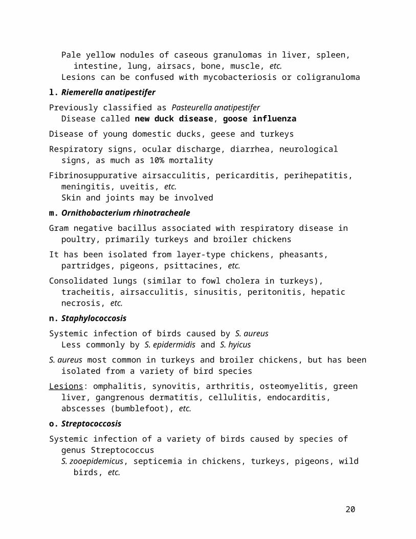
Lesions – Pale yellow nodules of caseous granulomas in liver, spleen, intestine, lung, airsacs, bone,
muscle, etc.– Lesions can be confused with mycobacteriosis or coligranuloma
l. Riemerella anatipestifer
Previously classified as Pasteurella anatipestifer – Disease called new duck disease, goose influenza
Disease of young domestic ducks, geese and turkeys
Respiratory signs, ocular discharge, diarrhea, neurological signs, as much as 10% mortality
Fibrinosuppurative airsacculitis, pericarditis, perihepatitis, meningitis, uveitis, etc.– Skin and joints may be involved
m. Ornithobacterium rhinotracheale
Gram negative bacillus associated with respiratory disease in poultry, primarily turkeys and broiler chickens
It has been isolated from layer-type chickens, pheasants, partridges, pigeons, psittacines, etc.
Consolidated lungs (similar to fowl cholera in turkeys), tracheitis, airsacculitis, sinusitis, peritonitis, hepatic necrosis, etc.
n. Staphylococcosis
Systemic infection of birds caused by S. aureus – Less commonly by S. epidermidis and S. hyicus
13

S. aureus most common in turkeys and broiler chickens, but has been isolated from a variety of bird species
Lesions : omphalitis, synovitis, arthritis, osteomyelitis, green liver, gangrenous dermatitis, cellulitis, endocarditis, abscesses (bumblefoot), etc.
o. Streptococcosis
Systemic infection of a variety of birds caused by species of genus Streptococcus– S. zooepidemicus, septicemia in chickens, turkeys, pigeons, wild birds, etc.
Valvular endocarditis with secondary infarcts in heart, liver, spleen, etc. Others: osteomyelitis, arthritis, tenosynovitis, salpingitis
– S. bovis, septicemia in turkeys, pigeons, etc.– S. faecium, septicemia in ducklings, goslings, chickens, etc.– Enterococcus hirae, encephalomalacia with vascular thrombosis and meningitis in broiler
chicks– Enterococcus faecalis, associated with amyloid arthropathy in chickens (brown-egg
layers)
p. Miscellaneous bacteria
Pseudomonas aeruginosa, Klebsiella pneumoniae - can cause localized or systemic infection in poultry and other birds
Bacillus anthracis - been reported in ostriches as a cause of septicemia
Listeria monocytogenes - can cause septicemia, myocarditis and encephalitis in chickens
Campylobacter jejuni - been associated with enteritis and hepatic necrosis in ostriches
Lawsonia intracellulare - associated with enteritis in ratites
Eubacterium tortuosum - granulomas in liver and spleen, broiler chickens and turkeys
Clostridium piliformis (Tyzzer’s disease) - hepatic necrosis in psittacines
Spirochetosis:– Borrelia anserina, septicemia in poultry and canaries– Borrelia related to B. hirmsii associated with hepatitis, splenitis and encephalitis in
Northern Spotted Owl– Serpulina hyodysenteriae associated with typhlitis in rheas, in poultry?– Serpulina piloscholi in ceca of pheasants, disease?– Spirochete-like in ceca of turkeys, disease?
8. Fungal diseases
a. Aspergillosis
One of the most common fungal diseases of poultry, water fowl, psittacines, passerines, ratites, raptors, zoo birds (penguins), etc.
Aspergillus fumigatus and A. flavus most common – Others: A. niger, A. terreus, A. glaucus, etc.
14

Respiratory signs (brooder pneumonia in poultry), unthrifty, diarrhea, neurological signs, ocular involvement, etc.
Lesions – Pale yellow nodules in lungs, air sac, syrinx, sinus, liver, brain, cloudy cornea, etc.– White plaques with fuzzy green or grey or blue material (conidiophores-fruity bodies) on
air sacs, pleura, etc.– Fibrinosuppurative or granulomatous pneumonia, airsacculitis, syringitis, sinusitis,
encephalitis, ophthalmitis, vasculitis (aortic rupture), hepatitis, osteomyelitis, pericarditis, etc.
b. Candidiasis
Common mycosis of the upper digestive tract – Also called thrush, crop mycosis, moniliasis– Young birds most susceptible
Candida albicans most common etiology
Poultry, psittacines, passerines, ratites, raptors, pigeons, water fowl, etc.
Oral cavity, esophagus and crop involved with white proliferative plaques– Proventriculus, gizzard, intestine less often involved– Systemic and ocular candidiasis have been described
c. Zygomycosis
In ostriches, psittacines, water fowl, canaries involving proventriculus and gizzard and air sacs in a pigeon– Mucor sp, Absidia sp. and Rhizopus sp. isolated– necrotizing lesions with granulomatous reaction
d. Favus (avian ringworm)
Due to Microsporum gallinae, dermatophyte of chickens with white powdery material on head, face and eyelids
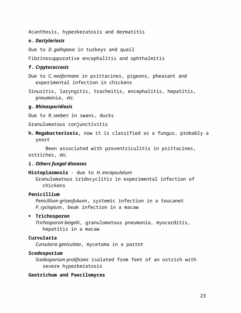
Acanthosis, hyperkeratosis and dermatitis
e. Dactylariosis
Due to D. gallopava in turkeys and quail
Fibrinosuppurative encephalitis and ophthalmitis
f. Crpytococcosis
Due to C. neoformans in psittacines, pigeons, pheasant and experimental infection in chickens
Sinusitis, laryngitis, tracheitis, encephalitis, hepatitis, pneumonia, etc.
g. Rhinosporidiosis
Due to R. seeberi in swans, ducks
Granulomatous conjunctivitis
h. Megabacteriosis, now it is classified as a fungus, probably a yeast
15

Been associated with proventriculitis in psittacines, ostriches, etc.
i. Others fungal diseases
Histoplasmosis - due to H. encapsulatum – Granulomatous iridocyclitis in experimental infection of chickens
Penicillium– Penicillium griseofulvum, systemic infection in a toucanet– P. cyclopium, beak infection in a macaw
Trichosporon – Trichosporon beigelii, granulomatous pneumonia, myocarditis, hepatitis in a macaw
Curvularia – Curvularia geniculata, mycetoma in a parrot
Scedosporium – Scedosporium prolificans isolated from feet of an ostrich with severe hyperkeratosis
Geotrichum and Paecilomyces – Geotrichum candidum and Paecilomyces variota have been isolated with disease
9. Viral diseases
Diseases caused by herpesvirus, retrovirus, coronavirus, paramyxovirus, orthomyxovirus, picornavirus, poxvirus, birnavirus, parvovirus, adenovirus, reovirus, enterovirus, circovirus, papovavirus, arbovirus, bunyavirus and other miscellaneous viruses
a. Marek’s Disease
One of the most common and well studied diseases of young chickens– Quail and turkeys are also susceptible – Etiology : cell-associated herpesvirus
Pathogenesis : virus replicates in feather follicle epithelium, infection through respiratory route, viremia infection of B cells cytolysis infection of activated T cells cytolysis immunosuppression infection of other organs like nerves (paralysis) & blindness latency transformation of T cells (CD4) lymphoma
Lesions – Gross: bursal and thymic atrophy, swollen peripheral nerves, enlarged organs with pale
white tumors in liver, spleen, kidney, lung, proventriculus, intestine, heart, gonads, thymus, irregular/gray iris, prominent feather follicles, etc.
– Microscopic: lymphoid necrosis and depletion in bursa, and thymus, neuritis, encephalomyelitis, pleomorphic lymphocytic lymphoma in various organs Intranuclear inclusion bodies in feather epithelium Atherosclerosis can be produced with MD virus
b. Leukosis/Sarcoma Group
Genus, ALV- related viruses of family Retrovirus– Six subgroups; A, B, C and D (exogenous viruses), E (endogenous) & J (recombinant)
A, B and J are common in the field, C and D are rare
16

– Various oncogenes have been identified (see table 1)
They can produce a variety of neoplasms in chickens
Influenced by strain of virus, dose, route of inoculation, age of host, genotype and sex of host
Neoplasms: sarcoma’s (fibro, osteochondro, myxo, histio, lympho, hemangio), meningioma, mesothelioma, erythroblastosis, myeloblastosis, nephroblastoma, granulosa cell tumor, hepatocellular carcinoma, glioma, (osteopetrosis), etc.
Lymphoid Leukosis– Disease of semimature and mature chickens – Etiology : retrovirus of leukosis/sarcoma group
Exogenous viruses, subgroups A, B, C and D– Transmission
Horizontal, transient viremia, immunity, LL rare Egg transmission, chronic viremia, immune tolerance, LL common
– B cell lymphoma in various organs, bursa of Fabricius, liver, spleen, kidney, gonads, etc.
– Osteopetrosis– thickening of long bones
Effect of virus on osteoblasts
Myelocytomatosis– Neoplastic disease primarily of broiler breeders and broilers– Etiology: retrovirus, subgroup J (leukosis/sarcoma group)– Lesions: liver, spleen, kidney, sternum, etc., with nodules made up immature
granulocytes– Hemangiosarcoma, histiocytoma, myxoma, carcinoma in liver, fibrosarcoma, lymphoma,
ganglioneuroma, renal tumors, etc., have also been associated with subgroup J virus
Reticuloendotheliosis– Includes runting syndrome, chronic lymphoma and acute reticulum cell sarcoma – Primarily in chickens and turkeys but neoplasia associated with REV has been observed
in quail, ducks, pheasants, geese and peafowl– Etiology : retrovirus of REV group, distinctly different from leukosis/sarcoma group,
more closely related to murine leukemia virus– Other viruses in REV group include chick syncytial virus, duck infectious anemia virus,
spleen necrosis virus and others– Lesions
In runting syndrome: thymic and bursal atrophy, neuritis, lymphoma (similar to Marek’s disease)
In chronic lymphoma: bursal and visceral lymphoma (similar to Lymphoid Leukosis)
In acute reticulum cell sarcoma: enlarged liver, spleen, kidney, heart, gonads, etc.
c. Infectious laryngotracheitis
Acute viral respiratory disease of primarily chickens– Pheasants and peafowl are also susceptible
Etiology : herpesvirus
17

Lesions : oculonasal discharge, trachea with hemorrhage and/or fibrinous exudate– Conjunctivitis, tracheitis and sinusitis, syncytia formation and intranuclear inclusion
bodies
d. Infectious Bronchitis
Highly contagious viral respiratory disease of young chickens– Drop in egg production and egg quality in layers
Etiology : coronavirus, many serotypes, and great antigenic variation among strains of virus
Lesions : catarrhal tracheitis, conjunctivitis, bronchitis, and airsacculitis – Fibrinosuppurative inflammation in cases complicated with E. coli– Interstitial nephritis with nephrotropic strains
e. Avian Paramyxoviruses
Based on antigenic relatedness (HI test) avian parmyxoviruses are classified into nine groups
Groups Primary Host Other HostsPMV-1/Newcastle Disease Virus
numerous many
PMV-2/Yukaipa Passerines, turkeys Chickens, psittacines, railPMV-3/Turkeys Turkeys None (chickens ?)PMV-3/Psittacines Psittacine PasserinesPMV-4/Duck Ducks Geese, railsPMV-5/Budgerigar Budgerigar nonePMV-6/Duck Ducks, geese TurkeysPMV-7/Dove Pigeons, dove nonePMV-8/Goose Ducks, geese nonePMV-9/Duck Ducks none
Newcastle Disease – Acute viral disease of chickens, turkeys, pigeons, doves, pheasants, ratites, psittacines,
cormorants, etc. 236 species of birds comprising 27 orders
– Etiology : avian paramyxovirus - 1, isolates vary greatly in pathogenicity to chickens Lentogenic: mild or inapparent infection in chickens Mesogenic: cause disease and mortality in young chickens Velogenic (viscerotropic and neurotropic): lethal infection of chickens of all ages New OIE classification based on chick intra-cerebral pathogenicity index (ICPI)
ICPI of > 0.7 is Newcastle disease virus (formerly Velogenic and mesogenic) ICPI of < 0.7 is Avian Paramyxovirus –1 (formerly Lentogenic)
– Clinical signs Vary with strain, respiratory, digestive, ocular, neurological, sudden death In mature chickens, egg production and quality problems (mesogenic strain)
– Lesions
18

In pigeons: enteritis, pancreatitis, nephritis, encephalitis, respiratory system rarely involved
In chickens; tracheitis, pneumonia, enteritis, conjunctivitis, encephalitis, myocarditis, lymphoid necrosis
Velogenic; hemorrhages in conjunctiva, trachea, oral cavity, esophagus, proventriculus, ceca, rectum
Disseminated vasculitis, lymphoid necrosis and depletion, mucosal necrosis and ulceration
Inclusions are rare but both intranuclear and intracytoplasmic inclusions have been described
In a recent case, discrete eosinophilic intranuclear and intracytoplasmic inclusions in conjunctiva, esophagus, lung, brain, adrenal ganglia of a pheasant and in the brain of a chicken with NDV
Eosinophilic intranuclear inclusions in hepatocytes in doves associated with lentogenic type of NDV
Other Avian Paramyxoviruses
– PMV - 2 (Yucaipa): Respiratory disease in young turkeys and drop in egg production in layers, chickens
are susceptible– PMV - 3, two strains, turkey and psittacine
Turkey: egg production drop in turkeys Psittacine; neurological and digestive problems in psittacines and passerines
Encephalitis with intranuclear and intracytoplasmic inclusion bodies in neurons and glial cells
Myocarditis, pancreatitis with intranuclear inclusions – PMV -5 (Kunitachi):
Enteritis and mortality in budgerigars and lorikeets
f. Avian pneumovirus– Cause of Turkey Rhino Tracheitis (TRT) of turkeys, Swollen Head Syndrome (SHS)
of chickens, highly contagious respiratory diseases– Etiology : pneumovirus (Paramyxoviridae)– Lesions : swollen sinuses, sinusitis, tracheitis, rhinitis, conjunctivitis
g. Avian Influenza
Acute viral disease of poultry; turkeys and chickens and psittacines, passerines, ratites, etc. – It has been isolated from many species of birds – Waterfowl may serve as reservoirs
Etiology: type A influenza virus of family Orthomyxoviridae– Numerous subtypes based on surface antigens, hemagglutinin (13) and neuraminidase (9)
Viruses of H5 (H5N2) and H7 (H7N1) subtypes are considered pathogenic, H1N1 (swine flu) in turkeys
H4N8, H4N6, H3N8 in exotic birds
19
H5N1 in chickens and humans, Hong Kong, 1997
Clinical signs: vary greatly, respiratory, digestive, ocular, neurological, sudden death, etc.– Drop in egg production in layers
Lesions: vary greatly in pathogenicity– Mildly pathogenic: catarrhal tracheitis, sinusitis, airsacculitis, conjunctivitis, pneumonia,
peritonitis, oopharitis, salpingitis, etc.– Highly pathogenic: hemorrhagic lesions in skin of face, comb & shanks and GI tract,
interstitial pneumonia & nephritis, encephalitis, conjunctivitis, myocarditis, adrenalitis, pancreatitis, myositis, lymphoid necrosis, vasculitis and thrombosis, etc.
h. Avian Encephalomyelitis
Viral disease of young (1-3 weeks) chickens, turkeys, pheasants and coturnix quail – Neurological signs (epidemic tremor)– Drop in egg production in layers– Egg transmitted – Etiology : Picornavirus (distantly related to hepatitis A virus, genus:Hepatovirus)
Lesions: neuronal swelling, chromatolysis, lymphocytic perivascular cuffing, gliosis in brain and lymphocytic foci in muscular layer of proventriculus and gizzard, pancreatitis
– A few survivors can develop cataracts
i. Avian Pox
Slow spreading viral disease of chickens, turkeys, quail, pigeons, canaries, raptors, psittacines, ostrich, peacock, waterfowl, etc.
– 60 species of wild birds
Etiology: poxvirus of genus Avipoxvirus, many strains– Fowl pox, turkey pox, pigeon pox, canary pox, quail, mynah, psittacine, junco, sparrow,
starling, etc.
Signs: cutaneous, respiratory, digestive, ocular– Septicemic form in canaries, 70 - 90% mortality
Lesions– Gross:
Dry pox or cutaneous form: proliferative skin lesions on face, eyelids, beak, feet, legs, vent, etc.
Wet pox or diphtheritic form: yellow raised plaques in sinus, trachea, oral cavity esophagus/crop, conjunctiva, etc.
– Micro: Proliferation of epithelial cells, ballooning degeneration with eosinophilic
intracytoplasmic inclusion bodies (pathognomonic) Desquamative pneumonia in canaries Some avipoxviruses are oncogenic, wart-like growth
j. Infectious Bursal disease
Acute viral disease of young chickens (1-6 weeks) and secondary immunosuppression
20

Turkeys and ducks, subclinical infection
Etiology : birnavirus
Lesions : enlarged and edematous bursa of Fabricius some times with hemorrhages and atrophy in later stages, hemorrhages in skeletal muscle, thymic atrophy with virulent IBD
– Lymphoid necrosis and depletion
– Secondary infections with inclusion body hepatitis, gangrenous dermatitis, bursal cryptosporidiosis, etc.
k. Chicken Infectious Anemia Viral disease of young chickens characterized by aplastic anemia and immunosuppression
Chicks 1-3 weeks of age most susceptible
Vertically transmitted
Etiology : a circovirus, genus Gyrovirus, family Circoviridae
Hematology : anemia, hematocrit less than 27% (N 35%), leukopenia, thrombocytopenia
– Due to cytotoxic effect of virus on bone marrow precursor cells
Lesions : pale bone marrow, severe thymic atrophy, atrophy of bursa, hemorrhages in skeletal muscles
Lymphoid necrosis and depletion, bone marrow hypoplasia
– Gangrenous dermatitis, colibacillosis, aspergillosis, viral infection, etc.
Eosinophilic (red) intranuclear inclusions in mononuclear inflammatory cells (macrophages?) of thymus, spleen, bone marrow, bursa, lung, etc. in some cases
– True nature of these inclusions is not known
l. Duck Viral Enteritis
Acute viral disease of primarily adult ducks, geese and swans characterized by high mortality
Etiology: herpesvirus
Lesions: hemorrhages on heart, liver, gizzard, fibrinonecrotic lesions in esophagus, rectum, cloaca, bursa, annular band of hemorrhage and necrosis in intestine, ceca, and thymic atrophy – Necrosis, inflammation and intranuclear inclusions in liver, intestine, thymus, gland of
Harder, conjunctiva, etc.– Esophagitis and bursal necrosis with intranuclear and intracytoplasmic inclusions in
mucosal cells
m. Duck virus hepatitis
Peracute viral infection of ducklings (< 5 weeks) characterized by high mortality
Etiology : – DVH - 1, enterovirus– DVH - 2, astrovirus – DVH - 3, enterovirus (unrelated to DVH1)
21

Lesions : petechiae or ecchymotic hemorrhages and necrosis in liver, minimal inflammation
n. Parvovirus Infection
Goose parvovirus (Derzsy’ disease): highly contagious disease of young geese and Muscovy ducks
– Serofibrinous pericarditis and perihepatitis
– Myocarditis with intranuclear inclusions
Muscovy duck parvovirus– Serologically related to goose parvovirus– Causes locomotor problems with high mortality in 1-3 weeks-old ducks, loss of weight ,
pale leg muscles, serofibrinous pericarditis and perihepatitis Myositis, myocarditis, encephalomyelitis, neuritis, etc.
– Ascites, round hearts in ducks recovered from infection
o. Avian Adenoviruses
Three groups:– Group I - quail bronchitis, inclusion body hepatitis and hydropericardium syndrome in
chickens, also disease in turkeys, pigeons, psittacines, raptors, etc.– Group II - hemorrhagic enteritis virus of turkeys (HEV), marble spleen disease of
pheasants (MSD) and splenomegaly of chickens– Group III - egg drop syndrome of chickens with no apparent lesions but caused
tracheitis and bronchitis in goslings
Quail bronchitis– High mortality in young bobwhite quail associated with bronchopneumonia, tracheitis,
hepatitis, pancreatitis, bursal necrosis, intranuclear inclusions
Inclusion body hepatitis of chickens, also in turkeys, guinea fowl, pigeons, psittacines, etc.– In chickens usually secondary to immunosuppression caused by IBDV, CIAV– Liver enlarged and mottled red/pale, foci of necrosis, inflammation and intranuclear
inclusion bodies, also pancreatitis– Similar lesions seen in turkey poults, guinea fowl, pigeons – Hepatitis, enteritis, bronchitis, pancreatitis, nephritis, encephalitis, etc., associated with
intranuclear inclusions in psittacines
Hemorrhagic enteritis virus (HEV) of turkeys and Marble spleen disease (MSD) of pheasants– Caused by group II adenovirus– Guinea fowl, psittacines, (partridge) susceptible– Young turkeys (4-12 weeks) and pheasants (3-8 months)– With HEV, intestinal hemorrhage and enlarged mottled white spleen and
immunosuppression Intranuclear inclusions in mononuclear cells of spleen and intestine, renal epithelial
cells in HEV – In pheasants mottled white enlarged spleen, MPS cell hyperplasia, intranuclear inclusions – Splenomegaly in chickens
p. Poult Enteritis
22
Disease of young turkeys, multiple etiologies
Viruses include coronavirus (blue comb disease), enterovirus, rotavirus, astrovirus, etc.
Diarrhea, loss of weight, small intestine and ceca distended with watery or frothy contents
Mortality 1 - 55%, caseous exudate in bursa with coronavirus infection
Increased cellularity of lamina propria, necrosis of cells in lamina propria and enterocytes, villus atrophy, lymphoid necrosis in thymus and bursa (virus?)
Eneteritis associated with rotavirus and enterovirus have been described in young pheasants, quail, chukars, etc.
q. Herpesviruses (Marek’s disease, see page 16)
Psittacine Herpesvirus– Probably a diverse group of viruses which infect a variety of psittacines– Three diseases are known
PACHECO’S DISEASE Acute viral disease of a variety of psittacines (common in 1980’s in US) Lesions :
enlarged liver occasionally with petechiae, enlarged spleen, fluid filled intestine, diphteritic membrane in oral cavity, esophagus, etc.
liver necrosis with or without inflammation, enteritis, stomatitis, esophagitis, pancreatitis, conjunctivitis, splenic and bursal necrosis, nephritis with intranuclear inclusion bodies
syncytia formation with inclusions in liver AMAZON TRACHEITIS, disease characterized by tracheitis, bronchitis, rhinitis,
laryngitis with syncytia formation and intranuclear inclusions Virus has some cross reactivity with ILT virus of chickens
BUDGERIGAR HERPESVIRUS, rare disease associated with decreased hatchability and “feather duster” plumage
Miscellaneous Herpesviruses
– GOOSE HERPERVIURS
Lesions like in Duck viral enteritis with intranuclear inclusion bodies and high mortality in goslings
– PIGEON HERPESVIRUS Common in young squabs characterized by hepatitis, pancreatitis, esophagitis
associated with intranuclear inclusions Conjunctivitis, enteritis, myocarditis, encephalitis, laryngitis, splenitis, etc. can also
be seen Raptors and budgies susceptible
– FINCH HERPESVIRUS (cytomegalovirus-like) Disease of primarily Gouldian finches characterized by high mortality, conjunctivitis,
tracheitis, bronchitis, associated with cytomegalic cells and intranuclear inclusions– OTHERS HERPES VIRUSES OF OWLS, FALCONS, EAGLES, CRANES, etc.
Hepatitis associated with intranuclear inclusion bodies
r. Psittacine Beak and Feather Disease
23

Viral disease of many species of psittacines characterized by chronic feather and beak dystrophy – Acute immunosuppression and sudden death in young birds due to secondary bacterial
septicemia and fungal infections
Etiology : Psittacine circovirus, genus circovirus, family Circoviridae
Clinical signs : dystrophic feathers first noticed of the powder down, progress to contour feathers, followed by primary, secondary tail and crest feathers, almost symmetrical
– Dystrophy of the beak
Lesions – Gross: abnormal and loss of feathers, sloughing of claws, beak necrosis, necrosis of oral
mucosa, liver, bursa, thymus, etc.– Microscopic: pterylitis and pulpitis associated with botryoid inclusions in macrophages,
also in bursa, bone marrow, thymus, beak, claws, liver, pancreas, thyroid, testes, etc. Intranuclear inclusions in feather epithelium, intestine, esophagus, hepatocytes
Pigeons and doves – Etilogy: Pigeon circovirus, genus circovirus– feather dystrophy, exudate in bursa (due to bacterial infection), pterylitis, bursal
lymphoid depletion and intracytoplasmic circovirus inclusions in macrophages of bursa of Fabricius, spleen, thymus, cecal tonsil, etc.
– secondary bacterial, parasitic, fungal and other concurrent viral infections common Canaries and Finches
– Etiology: Canary circovirus, genus circovirus, family Circoviridae– Feather dystrophy and characteristic circovirus inclusions in bursa of Fabricius in finches
Gulls and Geese are also susceptible to circovirus– Circovirus inclusions in the bursa of Fabricius
s. Papovavirus
Two genera are known to cause disease in psittacines and passerines– Papillomavirus– Polyomavirus
Papillomavirus – It has been associated with cutaneous papillomas in wild finches (Fringilla) and an
African Grey Parrot – No virus has been associated with papillomas of cloaca, conjunctiva, tongue, larynx, oral
cavity, crop/esophagus, etc., in psittacines
Polyomavirus
Three groups, A, B and C– Causes BUDGERIGAR FLEDGLING DISEASE (BFD)
One of the most common diseases of psittacines (disease of 90’s?) and passerines Antibodies to BFDV been detected in chickens, but chickens are resistant to
infection Etiology : polyomavirus, different strains such as psittacine, passerine, etc., may exist
Variety of psittacines, finches, canaries, seed crackers and blue bills are susceptible
24

Young psittacines are highly susceptible with very high mortality (30 - 100%), also adults
Feather dystrophy in budgerigars, acute death, digestive, neurological, respiratory signs, etc.
Lesions Gross: variation among psittacines and also passerines
In most of psittacines feather dystrophy, hemorrhages in skin, subcutis, skeletal muscle, heart, intestine, liver enlarged and mottled red or with white foci, splenomegaly, pale kidneys, ascites, lung congestion, pale carcass, etc.
In passerines, liver enlarged and mottled white, serosal or subserosal hemorrhage of intestine, pale myocardium, etc.
Microscopic: hemorrhages in various organs, necrosis in spleen, bursa, thymus and bone marrow, midzonal or random necrosis in liver, myocarditis, enteritis, nephritis, membranous glomerulopathy, pancreatitis, conjunctivitis, encephalomyelitis, ganglionitis (spinal), etc. Bluish karyomegalic inclusions in various tissues; epidermis, feather follicle
epithelium, esophagus, kidney, macro/lympho of spleen, bursa, thymus, bone marrow, liver, etc., hepatocytes, myocytes, endothelial cells, glial cells, Purkinje cells, etc.
Buzzards and Falcons are also susceptible– Goose hemorrhagic polyomavirus:
causes high mortality in 4-10 week old geese, 100 % mortality in one week-old geese subcutaneous edema, ascites, hemorrhagic enteritis, nephritis and lymphoid necrosis
in bursa of Fabricius
t. Proventricular Dilation Disease (PDD)
A common chronic disease of psittacines– Characterized by dilation of proventriculus, anorexia, regurgitation, passing of undigested
seeds in feces, diarrhea, neurological signs, loss of weight, etc. Also been seen in a perigrine falcon, Canada geese, etc.
Etiology : not known, presumed to be a virus
Lesions – Gross: dilated thin proventriculus in 70% of cases, distended duodenum, etc.– Microscopic: lymphoplasmacytic ganglioneuritis of splanchnic nerves of
crop/esophagus, proventriculus, gizzard, intestine, adrenalitis, myocarditis, neuritis, encephalomyelitis, choroiditis, etc. (see table 2)
u. Miscellaneous Viral Diseases
Turkey viral hepatitis– Disease of young turkeys– Etiology : Picorna-like virus– Liver and pancreas with foci of necrosis and inflammation
Viral arthritis– Disease primarily of young chickens and turkeys
25

– Etiology : reovirus– Joints enlarged with fluid, proliferative synovitis
Avian nephritis– Highly contagious disease of chickens– Etiology : Astrovirus – Nephritis and secondary visceral urate deposition
Hepadna virus (hepatitis B virus)– Common in ducks, but no significant clinical disease or lesions– Also in swans and geese, no associated clinical disease
Hepatitis E virus– Has been associated with hepatitis-splenomegaly syndrome in mature chickens– Disease is called big liver and spleen disease in Australia– Drop in egg production and increased mortality– Periportal hepatitis, vasculitis, necrosis and hemorrhage in liver. Spleen with increased
number of MPS cells with amyloidosis frequently
Louping ill virus– Red grouse are susceptible
Arboviruses
– WEST NILE VIRUS
Etiology: Flavivirus
First appeared in people in the US in NY city in 1999, flu-like symptoms, 7/61 died
Over 14 orders and over 170 species of birds are susceptible but most severe in crows, blue jays, geese, herons, raptors (owls, hawks), etc.
others; pigeons, kestrels, gulls, storks, geese, magpie, bald eagles, ducks, flamingos, lorikeets, macaw, cockatiel, cockatoo, parakeet, etc.
Chickens and turkeys are resistant to infection
clinical signs vary from sudden death to depression, weight loss, ataxia, tremors, opisthotonus, impaired vision, etc.
gross: hemorrhages in brain, pale areas in the myocardium, enlarged spleen, nephritis, hemorrhage and necrosis in the intestine
histo: primarily nonsuppurative encephalitis and myocarditis. Others include necrosis in spleen, liver, pancreas, and enteritis, nephritis, adrenalitis and pulmonary hemorrhage.
Virus widespread in many tissues but kidneys and brain are good for virus isolation
– EASTERN EQUINE ENCEPHALOMYELITIS VIRUS
Causes neurological signs and encephalitis in pheasants, partridges, finches and turkeys
26
In young turkeys and chickens, myocarditis, lymphoid necrosis in bursa and thymus Hemorrhagic enteritis and splenic necrosis in emus
– WESTERN EQUINE ENCEPHALOMYELITIS VIRUS
Encephalitis, myocarditis, hemorrhagic leiomyositis of intestine associated with vasculitis in emus
Encephalitis in pigeon and neurological signs in turkeys – HIGHLAND J VIRUS:
Encephalitis and myocarditis in partridges and young turkeys Associated with precipitous drop in egg production in turkeys
– ISRAEL TURKEY MENINGOENCEPHALITIS VIRUS
Encephalitis and myocardial necrosis in 10-12 week-old turkeys– BUNYA VIRUS (Turlock-like):
Associated with encephalomyelitis and myocarditis in an ostrich chick
10.Parasitic diseases
Protozoa: coccidia, histomonas, cryptosporidia, sarcocystis, toxoplasma, atoxoplasma, amoeba, microsporidia, trichomonas, leucocytozoon, malaria, haemoproteus, giardia, cochlosoma, spironucleus (Hexamita), balantidium, trypanosomes, hemosporozoa, besnoitia, chilomastix, caryospora, etc.
Nematodes: Ascarids, Capillaria, Syngamus, Tetrameres, Heterakis, Baylisascaris, etc.
Cestodes: Raillietina, Davainea, Hymenolepis, etc.
Trematodes: Prosthogonimus, Schistosomes, etc.
Arthropods: mites, fleas, lice, etc.
Protozoa
A. COCCIDIOSIS
Common disease of many species of birds caused by species of genera primarily Eimeria and Isospora and are quite host specific
Chickens: disease of universal importance – Eimeria tenella (ceca), E. acervulina (upper small int.), E. maxima and E. necatrix (mid
small intestine)– Hemorrhagic, mucoid, necrotic, proliferative enteritis– Numerous coccidia in different stages of development
Turkeys: common, lesions less severe than in chickens– E. adenoides (ceca), E. meleagrimitis (mid small intestine)– Mucoid enteritis, sometimes hemorrhagic and necrotic enteritis
Geese: E. truncata occurs in kidney– Nephritis and urate deposits – E. anseris causes enteritis
Ducks: renal coccidia due to E. boschadis, etc.
Quail, partridges, and pheasants: various species of Eimeria causes enteritis
27

– In quail coccidiosis is commonly associated with ulcerative enteritis caused by Clostridum colinum
Pigeons: E. labbeana causes enteritis
Psittacines: – Species of Eimeria, E. dunsingi and Isospora can cause enteritis in budgerigars, lories,
parakeets, parrots, etc.
Passerines: – Finches: Isospora lacazei has been associated with enteritis
Cranes: – E. gruis and E. reichenowi causes granulomatous enteritis, hepatitis, splenitis,
pneumonia, myocarditis, etc. in whooping and sandhill cranes
B. HISTOMONIASIS
Also called black head, a common protozoal disease of turkeys and partridges– Also in chickens, peafowl, quail, pheasants, rhea, etc.
Etiology : Histomonas meleagridis – Cecal worm, Heterakis gallinarum and earth worms act as accessory hosts
Lesions : saucer shaped depressions or white foci in liver and fibrinonecrotic mucosa and thickened wall of ceca– Granulomatous hepatitis and typhlitis associated with spherical protozoa, 8 - 21 um in
diameter– Nephritis, bursitis (bursa of Fabricius), pneumonia, proventriculitis, pneumonia,
peritonitis secondary to perforating typhlitis can be seen in turkeys
C. CRYPTOSPORIDIOSIS
Common protozoa of various species of birds– Chickens, turkeys, quail, ratites, ducks, pheasants, peafowl, psittacines, passerines, etc.
C. baileyi, C. meleagridis and Cryptosporidium spp in Finches and probably others
Infect various body systems– Cloaca, bursa of Fabricius and trachea most common– Nasal cavity, sinus, bronchus, air sac, conjunctiva– Proventriculus, intestine, ducts of pancreas, salivary and esophageal glands and bile duct– Ureters, collecting tubules of kidney
Inflammation and hyperplasia of epithelium
D. SARCOCYSTOSIS
Systemic protozoal disease of psittacines caused by S. falcatula– Opossum is the definitive host, cowbirds and grackles are intermediate hosts– Old World psittacines highly susceptible– Young new world psittacines, canaries, finches, pigeons are susceptible– Gallinaceous birds and anseriformes are resistant
Sarcocystis with encephalitis has been described in a golden eagle, capercailles and chickens
28

S. riley causes sarcocystosis in skeletal muscle (rice breast) of ducks, an innocuous incidental finding
Lesions – Pulmonary edema, congestion, liver may be enlarged and mottled white, splenomegaly– Lymphoplasmacytic interstitial pneumonia, edema and schizonts in capillaries– Myocarditis, hepatitis, splenitis, nephritis, encephalomyelitis, neuritis, myositis, uveitis,
etc., associated with schizonts– Mature cysts in the heart and skeletal muscle
Generally no reaction to cysts in muscles
E. TOXOPLASMOSIS
Sporadic disease of various species of birds– It has been described in passerines (canaries), chickens, psittacines, pigeons, ducks,
penguin, Japanese quail, chukar partridges, etc.– Turkeys, pheasants, Bob White quail, owls, house sparrows resistant
Etiology : T. gondii
Lesions : encephalomyelitis, ophthalmitis, pneumonia, myocarditis, hepatitis, splenitis, neuritis, myositis, enteritis, adrenalitis, etc. associated with zoites and cysts – Optic nerve; may be enlarged and yellow with necrotizing and granulomatous neuritis in
chickens
F. ATOXOPLASMOSIS
Common coccidian infection of canaries and finches, (previously called Lankesterella)– Also in mynah’s, sparrows, grosbeaks, thrush, cowbirds, raptors and many birds of order
Passeriformes
Etiology : A. serini (Isospora serini), A. adiei and probably others
Lesions – Liver enlarged with white foci, splenomegaly– Enteritis, hepatitis, myocarditis with arteritis, splenitis, myositis, dermatitis, pneumonia,
etc.– Schizonts in cytoplasm of macrophages
Lankesterella-like coccidia associated with pneumonia in a Northern Cardinal
G. MICROSPORIDIOSIS
It can be a significant disease of psittacines
It has been reported in a variety of lovebirds, budgerigars, parrots, cockatiels, etc.– Also reported in ostrich and Hummingbirds– Encephalitozoon hellum, others?
Lesions: granulomatous nephritis, necrosis and inflammation in liver, enteritis, pneumonia, conjunctivitis, etc. associated with Gram-positive organisms
H. TRICHOMONIASIS
A common infection of pigeons and raptors– Also in canaries, finches, doves, psittacines, ducks, poultry, wild birds, etc.
29

Etiology : T. gallinae Tetratrichomonas anatis, Tetratrichomonas sp. in ducksTetratrichomonas gallinarum in mocking bird
Lesions – Granulomatous stomatitis, pharyngitis, esophagitis, ingluvitis, and enteritis– Hepatitis, pericarditis, airsacculitis, tracheitis, pneumonia, meningoencephalitis– Sinusitis, rhinitis, episcleritis– Salpingitis in ducks
I. LEUCOCYTOZOONOSIS
Disease of anseriformes, turkeys, raptors, wild birds and columbiformes– Black flies (Simuliidae) are vectors
Etiology : L. simondi in anseriformes, L. smithi in turkeys, L. marchouxi in columbiformes, L. toddi in falconiformes, L ziemanni in owls– Infect both white and red blood cells
Lesions – Disseminated characteristic megaloschizonts in endothelial cells of liver, spleen, heart,
brain, eye with hepatitis, myocarditis, encephalitis, etc.
L. caulleryi, which causes severe disease in young chickens in south and eastern Asia, is classified as Akiba caulleryi– Vectors are biting midges of genus Culicoides– Lesions are similar to ducks
Leucocytozoon-like infection has been described in budgerigars, parakeets, Nankeen kestrels and a buzzard – Myositis, myocarditis, encephalitis and hemorrhages in skin, pericardium, pancreas,
hepatosplenomegaly, etc.– Granulomatous meningoencephalomyelitis, pectinitis and proliferative areteritis
associated with endothelial parasitic cysts in Nankeen Kestrels
J. AVIAN MALARIA
Hemoprotozoal infection of canaries, penguins and raptors caused by species of Plasmodium– Ducks, pigeons, chickens, grouse, pheasants, sparrows, blackbirds, robin, canary, tern,
etc. are susceptible– Three species of Culicoides are vectors
Etiology : P. relictum, P. elongatum, P. circumflexum, etc.– Infect red blood cells and cells of MPS
Lesions : anemia, interstitial pneumonia, hepatitis, splenitis, encephalitis, etc.– Brown-black pigment (malaria pigment) of RBC’s in MPS cells
K. HAEMOPROTEUS
Hemoprotozoa of some significance primarily in columbiformes– Common in raptors, water fowl, passerines, etc.– Also seen in psittacines
Etiology : H. columbae in pigeons and doves
30

– Biting flies of hippoboscids and Culicoides are vectors– Infects red blood cells and endothelial cells
Lesions : anemia, hepatitis, splenitis, myositis, pneumonia, etc.– Schizonts and megaloschizonts can be seen
L. GIARDIA, SPIRONUCLEUS (HEXAMITA)
Giardia psittaci cause of enteric disease in budgerigars – Others: cockatiels, lovebirds, parrots, conures, many wild birds, herons, egrets, sparrows,
etc.
Hexamita meleagridis an enteric protozoa of turkeys – Others: pheasants, pigeons, quail, partridge, ostrich, peafowl, etc.
Lesions – Emaciation, fluid filled intestine, catarrhal enteritis
M. OTHER ENTERIC PROTOZOA
Cochlosoma anatis, a flagellate enteric protozoon of small intestine is associated with catarrhal enteritis in turkeys
Entamoeba gallinarum common in large intestine of turkeys, significance?
Others: Balantidium spp. in ostriches, Wenyonella philiplevinei in ducks, Blastocystis sp. in pheasants, etc.
N. MISCELLANEOUS PROTOZOA
Hemosporozoa of undetermined taxonomy – Disseminated myositis associated with cysts and high mortality in BW quail in California– Also hepatitis, splenitis, myocarditis, nephritis, etc. associated with cysts,
encephalomyelitis– Probably caused by Heaemoproteus lophortyx?
Large protozoan cysts of undetermined spp. associated with hepatitis, splenitis, pneumonia in canaries, conures, love birds, oropendola, etc.
Amoeba of undetermined spp. associated with meningoencephalitis in a cockatiel
Besnotia: Cyst stages of Besnoitia-like protozoa causes arteritis and endarteritis in shore birds like knots with severe mortality.
Caryopsora: coccida with raptor-mouse cycle. Cysts found in the intestine, and other organs. Disease?
Chilomastix: cyst forming flagellate seen in the intestine of chickens, turkeys, ducks, geese and bustards. Disease?
Babesia shortii: causes anemia in Kestrels
Others: trypanosomes, aegyptianella, etc.
Nematodes
A. ASCARIASIS
Common intestinal parasitism of many birds
31
– Chickens, turkeys, pigeons, partridges, raptors, psittacines (Australian parakeets), passerines, etc.
Etiology : Ascaridia galli (chicken), A. dissimilis (turkey), A. columbae, A. hermaphrodita, species of Porrocaecum and Contracaecum, etc.
Lesions : loss of weight, intussusception, mild enteritis if in large numbers– Eosinophilic enteritis in turkeys– Granulomas in livers due to larval migration– Bile duct hyperplasia and pericholangitis associated with larvae in bile ducts in parakeets
B. CAPILLARIASIS
Significant disease primarily of upper digestive tract in many species of birds– Quail, pheasants, partridges, guinea fowl, turkeys, pigeons, chickens, raptors, ducks,
pscittacines, etc.
Etiology : C. contorta, C. annulata, etc.
Lesions : hyperplastic mucosa, fibrinonecrotic esophagitis, ingluvitis, etc.
C. obsignata, C. caudinflata may cause enteritis in galliformes and columbiformes
C. CEREBROSPINAL NEMATODIASIS
Common condition in a variety of birds
Chickens, emus, ostriches, psittacines, raptors, quail, partridges, wild birds, etc.
Etiology : Baylisascaris procyonis (raccoon) and occasionally B. columnaris (skunk)
Lesions : non-suppurative encephalomyelitis
Larvae of Chandlerella quiscali, a filarid nematode of grackle has been associated with encephalitis in emu
D. SYNGAMUS
Common tracheal worm (gape worm) of pheasants– Others: turkeys, geese, quail, peafowl, chickens
Etiology : Syngamus trachea
Lesions : granulomatous tracheitis and occasionally bronchitis
Cyathostoma bronchialis can cause bronchitis and pneumonia in geese– Also been associated with tracheitis in emus
E. NEMATODES OF PROVENTRICULUS AND GIZZARD
Dispharynx nasuta, Cyrnea colini, Tetrameres americana, Cheilospirura hamulosa and species of Acuaria, Synhimantis, Habronemia, Amidostomum, Hadjelia, etc.– Occur in pigeons, chickens, quail, ducks, turkeys, pheasants, psittacines, passerines, etc.– Lesions : proventriculitis and ventriculitis– Geopetitia aspiculata (Habronemia) causes severe proventriculitis in passeriformes and
others
Libyostrongylus douglassii causes severe proventriculitis in ostriches
32
Eustrogylides ignotus causes severe proventriculitis and peritonitis in fish eating birds such as herons, egrets and mergansers.
F. MISCELLANEOUS NEMATODES
Heterakhis isolonche: fibrous and granulomatous typhlitis in pheasants
Trichostrongylus tenuis: enteritis in young grouse and geese
Oxispirura mansoni: conjunctivitis in galliformes
Thelazia sp. have been associated with conjunctivitis in psittacines
Pelecitus calamiformis(filarid): tenosynovitis associated with adult nematodes in parrots
Microfilariae are common in psittacines, especially cockatoos, not pathogenic
Microfilariae associated with severe pneumonia in magpies
Serratospiculoides amaculata, parasite of air sacs and airways in raptors. Associated with air saccultitis, peritonitis and necrotizing myelitis in a Prairie Falcon
Serratospiculum sp. nine species known in raptors, some associated with airsacculitis and pneumonia
Paronchocerca ciconarum (filarid): myocardial degeneration with adult nematodes in a Marabou Stork
Cardiofilaria - heart worm in psittacines
Cestodes
Common intestinal tapeworms of many species of birds– Chickens, turkeys, ducks, geese, pigeons, psittacines, passerines, wild birds, etc.
Etiology and lesions : – Davainea proglottina (smallest); enteritis in chickens– Raillietina echinobothridia: granulomatous enteritis in chickens– Species of Raillietina, Hymenolepis, Amoebotaenia, etc. are common in psittacines and
finches Pathogenic in large numbers, obstruction, enteritis, etc.
Trematodes
Schistosomiasis: common in waterfowl– Dendritobilharzia sp.– Medial hypertrophy of vessels in the intestinal wall– Rarely hepatitis, encephalitis, nephritis, enteritis– Encephalitis common in swans
Dicroceliidae: been associated with dilated bile ducts and severe cholangiohepatitis in cockatoos and an amazon parrot
33

Gigantobilharzia sp. associated with hemorrhagic ulcerative colitis and cloacitis in a nanday conure
Sphaeridiotrema globulus: causes severe ulcerative hemorrhagic enteritis in swans and cygnet and chickens (experimental)
Philophthalmus gralli: associated with conjunctivitis in ostriches
Collyriclum faba cause cysts in the skin of poultry, wild birds including robins
Prosthogonimus sp. oviduct fluke of poultry
Tanaisia bragai in collecting tubules of kidney in poultry, pigeons, etc.
Cathaemasia hians in the upper esophagus of storks
Athesmia heterolecithodes causes severe hepatitis in guinea fowls
Arthropods
MITES
Most common ectoparasites of a variety of species of birds– Chickens, canaries, finches, psittacines, pigeons, turkeys, pheasants, wild birds, etc.
Etiology (see table 3): species of Dermanyssus, Onithonyssus, Knemidocoptes, Sternostoma, etc.
Lesions – Anemia– Hyperkeratosis, acanthosis, epidermitis, dermatitis– Blockage, granulomatous tracheitis, airsacculitis, pneumonia – Cysts in skin, poor feather growth, loss of feathers, etc.
11.Toxicosis
a. Mycotoxins
Generally ducklings, turkey poults and pheasants are more susceptible
Aflatoxins (B1, B2, G1and G2): B1 most toxic, liver has congestion, necrosis, fatty change, karyomegaly, numerous mitotic figures, bile duct hyperplasia, fibrosis, etc.– Immunosuppression, myocardial, kidney degeneration, impaired blood coagulation, etc.– Turkey X disease due to aflatoxin B1 + cyclopiazonic acid– Model for hepatocarcinogenesis
Citrinin, Ochratoxin, Oosporein: renal tubular necrosis and visceral urate deposition– In addition vacuoles, bile duct hyperplasia, etc. in liver associated with citrinin and
ochratoxin– Ochratoxin can cause glycogen storage disease in chickens– Ochratoxin is embryocidal at low levels
Trichothecenes (T2, DAS, Vomitoxin = DON): radiomimetic effect, oral, proventricular and gizzard erosions, lymphoid necrosis and depletion, hepatic necrosis, etc.
34

Fumonisins: poultry are relatively resistant, hepatic necrosis, biliary hyperplasia, widening of growth plates, myodegeneration, etc.
Moniliformin: Cardiomegaly in broilers, myocardial degeneration, hepatic necrosis
Fusarochromanone: tibial dyschondroplasia
Cyclopiozonic acid: skeletal muscle, heart, liver, GI degeneration
Zearalenone: mycotoxin with estrogen activity – Cystic oviduct with inflammation, cysts in vent, etc.
Ergotism: beak and toe necrosis
b. Heavy metals
Lead: one of the most commonly reported toxic compounds of waterfowl and companion birds– Lesions : anemia, hemorrhagic enteritis, myocardial degeneration, hyalinosis of cerebral
vessels with hemorrhage, nephrosis, demyelination of nerves, etc.– Acid fast intranuclear inclusions in renal tubular epithelial cells
Zinc: common in companion birds, waterfowl– Lesions : gizzard erosion, pancreatic acinar necrosis, enteritis, nephrosis
Iron: seen in a variety of birds– Lesions : hepatic necrosis, hemolysis of RBC
c. Ionophore toxicity
Ionophores widely used as anticoccidials in poultry – Toxicity most common in turkeys and chickens– Also described in quail, guinea fowl, etc.
Monensin, Lasalocid, Salinomycin, Narasin– Various compounds interact and influence toxicity
Lesions : degeneration of skeletal muscles (pectoral muscles not affected) and occasionally heart – In addition demyelination and axonal degeneration of peripheral nerves and spinal cord
have been associated with lasalocid toxicity
d. Selenium, salt, calcium
Selenium: hepatopathy, alopecia of the scalp and dorsal cervical midline, broken or lost digit nails, necrosis of the tip of the beak in ducks
– pulmonary congestion and edema in an ostrich chick
Salt: causes right heart hypertrophy, dilation and ascites in turkey poults and broiler chicks and occasionally encephalomalacia – Water high in salinity causes conjunctivitis, myocardial degeneration, hyperemia of
brain, hyperplasia or atrophy of salt glands, cataracts (reversible if not severe) in wild waterfowl
– Cystic testes in poultry
Calcium: nephrosis in young poultry
35

e. Vitamins
Vitamin D: common in psittacines– Soft tissue mineralization, proventriculus, heart, air sacs, etc.
Vitamin A: conjunctivitis and rickets in chickens– Hepatic degeneration, Ito-cell hyperplasia?
f. Gases; PTFE, Ammonia, CO
Polytetrafluoroethylene (PTFE): Teflon coated pans when overheated release toxic gases – Seen in companion birds, also in free flying birds– In poultry from PTFE-coated light bulbs– Pulmonary congestion and edema
Carbon monoxide: pulmonary congestion and edema, bright red colored blood
Ammonia: conjunctivitis, corneal erosions and ulcers and sinusitis and tracheitis in poultry
Oxygen: in budgerigars, edema and interstitial pneumonia
g. Rodenticides
Diphacinone, Brodifoucum, Bromodiolone: blood in abdominal cavity, hemorrhage in liver
Zinc phosphide: hydropericardium, pulmonary edema, congestion and hemorrhage in liver, congestion in kidney
h. Antibiotics
Gentamicin, Amikacin - nephrosis
Sulfa drugs - disseminated hemorrhages, nephrosis
Tetracyclines - nephrosis
Furazolidone - round heart, hepatocellular vacuoles
Polymyxin E-1. in ostriches, congestion of meningeal vessels, vacuolization of the plexus of Auerbach, edema in the heart and intestinal serosa
i. Plants
Avocado and oleander: myocardial degeneration
Oak: nephrosis
Green acorns (pyrogallol): gastroenteritis, congestion and edema of organs
Coffee senna: degeneration of muscles
Gossypol: hepatic necrosis, biliary hyperplasia, perivascular inflammation
Spring Parsley (Cymopterus watsonii), celery: photosensitization, dermatitis
Blue green algae (Microcystin): Hepatic necrosis
j. Others
Organo-phosphates - acute and delayed neurotoxicity– Acute, inhibition of acetyl cholinesterase, no lesions
36

– Delayed, axonal degeneration in peripheral nerves and spinal cord, adult chicken is the (test) animal model, young birds are resistant
Nitrate, Nitrite: Brown mucus membranes, methemoglobinuria
Arsenilic acid, nitro-phenyl-arsenic acid: demyelination of peripheral nerves
Sodium Sesquicarbonate: nephrosis, visceral gout, fluid in intestine
Quaternary Ammonium: erosions and ulcers in upper GI tract
Domoic acid (marine neurotoxin):– In pelicans and cormorants– Hemorrhage and necrosis in skeletal muscles
Avian vacuolar myelinopathy– Cause not known but toxicity is suspected– In bald eagles, coots and others– Spongy degeneration of the white matter of CNS but most prominent in optic lobes and
swollen axons in spinal cord. Optic nerve also affected.
12.Metabolic diseases
a. Goiter
In male mature buff Cochin chickens (Dr. D. Webb, IL)
Severely enlarged cystic thyroid glands with follicles lined by flattened epithelial cells
Genetics suspected
b. Hemochromatosis
Abnormal accumulation of iron in tissues associated with tissue disturbance such as cirrhosis
Most common in mynahs, toucans, crows, starlings, ducks, fruit doves, horn bills, psittacines, etc.
Hepatic degeneration, fibrosis, myocardial degeneration, ascites, etc.
Genetics, nutrition, hemorrhagic syndromes, starvation, etc.
c. Diabetes mellitus
It has been described in psittacines, toco toucans and a red-tailed hawk
Glucagon is the major glucose - regulating hormone in granivorous birds
Carnivorous birds are similar to mammals
Lesions – Hyperplasia of islet cells– Enlarged islets with eosinophilic cytoplasm in toco toucans– Islet cell tumor
d. Amyloidosis
Most common in ducks and finches
37

Also in other water fowl, game birds, turkeys, psittacines, ratites, canaries, flamingoes, touracos, pigeons, doves, etc.
Commonly associated with chronic infections
Genetics may play a role in its occurrence in commercial ducks, as young as 3-4 week-old ducklings may have severe amyloidosis in the spleen with out apparent co-infections.
Accumulation of amorphous eosinophilic material in various tissues
Liver, spleen, intestine, adrenal, kidney, synovium, heart, pancreas, thyroid, skin, brain, lung, etc.
Lesions – Gross: ascites (water belly) in commercial ducks, firm enlarged and waxy liver
sometimes with nodules, enlarged mottled white spleen, pale or brownish kidneys, etc.– Microscopic: interstitial accumulation of amyloid in liver with hepatocellular atrophy in
severe cases, kidney, adrenal, thyroid, pancreas, spleen, in the lamina propria of intestine and proventriculus, in subepithelium of synovium, bronchus, in myocardium, brain, skin, vessels, etc. Severe hepatic capsular fibrosis and nodules made up of hyperplastic hepatocytes
Amyloid arthropathy in brown egg laying-type chickenso Cause: Enterococcus faecalis, Mycoplasma synoviae and otherso Lesions: Orange colored exudate in the synovium
Amyloid associated with Hepatitis-splenomegaly syndrome in mature broiler and laying-type
chickens
o Cause: Hepatitis E viruso Lesions: enlarged mottled pale spleen and enlarged and hemorrhagic liver. Amyloid
primarily in the spleen
e. Gout (visceral and articular)
Uric acid is the end product of protein and purine metabolism (uricotelic) in birds, where as in mammals’ urea is the end product (ureotelic)
Birds lack the enzyme carbamyl phosphate synthetase to dispose of ammonia and the enzyme uricase to decarboxylate uric acid to allantoin
Gout is a metabolic condition where abnormal accumulation of white chalky or white semifluid-like urates in soft tissues of various organs in the body
Gout occurs as two distinct syndromes, visceral and articular gout
These two syndromes differ in age of onset, frequency, sex predilection, gross and microscopic lesions, pathogenesis and causes
Great deal of confusion exists between the two syndromes because urate deposition takes place in joints in visceral gout also
Term “visceral gout” should be replaced with the term “visceral urate deposits”
See table 4 to help clarify differences between the two syndromes
38

f. Hepatic Lipidosis
Common in some psittacines, budgerigars, amazons, rose breasted cockatoos, shell parakeets, etc.
Diet, environment, toxicities, hormones, genetics (shell parakeets) influence lipidosis
Lesions : obesity, hepatic lipidosis, and lipidosis in liver and other organs in shell parakeets
g. Hemorrhagic fatty liver syndrome (chickens)
Condition of obese layer-type chickens
Fat in abdominal cavity, fatty livers, hemorrhagic or ruptured livers
h. Atherosclerosis
Very common in psittacines especially amazons
Others: ducks, geese, chickens, turkeys, ostriches, flamingoes, herons, vultures, falcons, pigeons, etc.
Etiology : diet, environment, genetics, virus, age, etc.– Marek’s disease virus (herpesvirus) can induce atherosclerosis in chickens– White Carneaux pigeon used as model
Lesions – Thickened walls or plaques in brachiocephalic, carotid, abdominal aorta, etc., coronary
arteries rarely involved– Thickening of intima and media with lipid laden cells
i. Mucopolysaccharidosis type IIIB (emu)
Also called Sanfilippo B syndrome
First description in any animal
Deficiency of N-acetyl--D-glucosaminidase
Probably inherited as a autosomal recessive trait
Signs and lesions – Sudden death, neurological signs – Ruptured liver or subcutaneous hemorrhages– Accumulation of membrane bound substance in neurons of nervous tissue and visceral
organsj. Storage disease in Costa’s Hummingbirds Neurologic signs; flying backwards, swaying, head tilt, flying in circles, head tremors Vacuoles in swollen neurons, cells of bone marrow, spleen, gland of Harder, uropygeal gland
and inner nuclear layer of retina
k. Inherited Muscular dystrophy in chicken Seen in New Hampshire breed and similar dystrophy-like myopathy has been described in
Cornish breed Clin. signs: unable to elevate wings beyond horizontal plane and unable to right themselves
when placed on their backs
39

Lesions: grossly enlargement of pectoral muscles initially and later atrophy, microscopically variation in fiber size, increased nuclei, vacuoles, necrosis, fragmentation and replacement by adipose tissue
l. Myotonic dystrophy in mutant (LWC strain) Japaneese Quails As early as 28 days of age generalized myotonia, muscle stiffness, muscle weakness Lesions: ring fibers, sarcoplasmic masses, internal migration of sarcolemmal nuclei, variation
in fiber size, replacement by fat cells and lenticular cataracts
m. Type II Glycogen storage disease in Japaneese Quail Acid maltase deficiency
n. Aortic aneurysm/rupture
Most common in male turkeys but also seen in ostriches and emus– Most common in the abdominal aorta at the origin of the celiac artery– Longitudinal slit in the posterior abdominal aorta or rarely at the origin of aorta– Medial degeneration and loss of elastic fibers, subintimal thickening, displacement of
elastic lamina, increased amount of collagen, necrosis, rupture– Genetics, hypertension, low copper, vasa vasorum defect?
Coronary artery aneurysm (turkeys)– In male turkeys, 15 -16 weeks-old, 1.5-3.5% mortality– Hemopericardium, hemorrhage across base of heart, subintimal thickening, medial
degeneration of coronary artery, rupture– Genetics, hypertension, low copper, increased body weights?
o. Round heart disease (turkeys)
Also called spontaneous cardiomyopathy
Common condition in young commercial turkeys
Dilated ventricles, chronic passive congestion of liver– Cause is not known, genetics?
p. Ascites syndrome (chickens)
Common condition in broiler chickens
Right heart hypertrophy, dilation, passive congestion of liver and ascites
Rapid growth coupled with insufficient pulmonary capillary capacity aggravates pulmonary hypertension leading to right heart failure
q. Tibial dyschondroplasia (TD)
Common condition in broiler chickens and turkeys and ducks
Abnormal masses of cartilage below the growth plate primarily in the proximal tibiotarsus but occurs also in the tarsometatarsus– Presence of prehypertrophic cartilage with no vascular channels– Etiology : multifactorial, nutrition, genetics, mycotoxin, etc.
40

Similar picture as TD can be seen grossly in the long bones of ratites but it is not TD nor it is pathologic
– In ratites it is normal embryonic cartilage which gets resorbed by 6-8 weeks of age
r. Deep pectoral myopathy
Seen in well muscled broilers and turkeys
Green discoloration of primarily supracoracoideus muscle due to ischemic necrosis
s. Perirenal hemorrhage
Seen in rapidly growing turkeys, 8-14 weeks of age
Hemorrhage over portion or entire kidney
Underlying vascular problems? Probably due to small ruptures or tears in the posterior abdominal aorta and its branches?
t. Xanthomatosis
Common in psittacines and occasionally in chickens
Yellow subcutaneous swelling or nodules in the body cavity
Giant cells, lymphocytes, macrophages with cholesterol clefts
u. Auto immune thyroiditis (chicken)
Occurs spontaneously in obese strain of chicken
Used as model to study Hashimoto’s thyroiditis
Birds have long and silky feathers, large amount of abdominal fat
Thyroids become small gradually as the bird matures and atrophies
Lymphocytic infiltration with few macrophages and lymphoid nodules, loss of architecture, thyroid gland
Antibodies to thyroglobulin
Hyperlipidemia, low T3 and T4 and IgA deficiency
13.Diseases of Malnutrition
a. Xerophthalmia
In non-commercial poultry, psittacines, etc.
Etiology: vitamin A deficiency
Lesions : pustule-like nodules in upper GI tract, exudate in conjunctiva, nasal cavity, nephrosis, opaque dry cornea, hyperkeratosis of plantar surfaces– Microscopic: squamous metaplasia of epithelium, hyperkeratosis
b. Polyneuritis
Rarely seen in poultry
Etiology: vitamin B1 (thiamin) deficiency
41
Lesions: prominent peripheral nerves with edema and infiltration of mononuclear cells
Malacia of the brain in fish eating birds
c. Rickets/osteomalacia
Rickets in younger birds and osteomalacia in older birds – In poultry, ratites, psittacines, zoo birds, etc.– Etiology : deficiency of calcium, phosphorus and vitamin D3 or an imbalance in calcium
to phosphorus ration (normal 2:1)– Lesions : soft and pliable beak, claws and keel, beading of ribs (‘rickety rosary’), enlarged
epiphysis, fractures of long bones, enlarged parathyroid glands Microscopic:
Calcium deficiency - lengthened Proliferating Prehypertrophied Zone and short Degenerating Hypertrophied Zone (DHZ), hyperplasia of parathyroid glands
Phosphorus deficiency/calcium excess: lengthened DHZ Vitamin D3 deficiency - similar to calcium deficiency
d. Encephalomalacia
In young chickens, turkeys, pheasants, geese, ducks, etc.
– Can also be seen in older birds
Etiology : vitamin E deficiency
Lesions : soft and enlarged cerebellum, petechiae, hemorrhages especially in turkey poults– Microscopic: malacia, hemorrhage, vascular thrombosis, demyelination in brain – Occasionally spinal cord involved, poliomyelomalacia
Yellow fat disease in wild birds: herons, etc.– Fat is brownish yellow, steatitis
e. Muscular dystrophy/exudative diathesis
In chickens, quail, turkeys, pelicans, ducks, etc.
Etiology : vitamin E/Selenium deficiency
Lesions – Subcutaneous green-tinged fluid – Pale streaks in skeletal muscles, gizzard and heart (ducks and pelican)
Degeneration of skeletal muscle, heart, gizzard, pancreatic acinar necrosis
f. Curled-toe paralysis
Seen in chicks, turkey poults, ducklings
Etiology : vitamin B2 (riboflavin deficiency)
Lesions : swelling of peripheral nerves, axon and myelin degeneration, Schwann cell proliferation, mononuclear cell infiltration, etc.
g. Pyridoxine (B6) deficiency
Perosis, neurological signs in chicks and ducks and reduced egg production and hatchability in laying chickens and turkeys
42

Microcytic hypochromic anemia in ducks
h. Perosis/slipped tendon (chicks, poults)
Etiology : manganese and choline deficiency
Lesions : deformity of hocks, enlargement of condyle, bowing of tarsometatarsus, slipping of gastrocnemius tendon– Deficiency of pantothenic acid, biotin, folic acid, niacin can cause similar lesions– In addition dermatitis can be seen in birds deficient in biotin and pantothenic acid and
poor feathering and anemia in niacin deficiency
i. Goiter
Common in psittacines especially in budgerigars
Also seen in pheasants, geese, chickens, etc.
Etiology : Iodine deficiency
Lesions : enlarged thyroid glands, some times cystic
Micro: severe hyperplasia of follicle epithelium with papillary projections, pale or lack of colloid, hemorrhage and severe effacement of the gland in extreme cases
14.Neoplasia
Most common in chickens and psittacines especially budgerigars among birds Birds in the order Passeriformes have the lowest incidence In chickens most are caused by viruses such as retroviruses and herpesvirus (see
Leucosis/Sarcoma group and Marek’s Disease) Etiology is not known in other species of birds Incidence in budgerigars range from 16 to 24 % Some of the common tumors in psittacines include renal tumors, fibrosarcoma, lymphoma,
papilloma, tumors of gonads, gastric carcinoma, leiomyosarcoma, pituitary adenoma, etc.
43
15.References:
1. Barnes, J. E. Avian Pathology, Class notes. College of Veterinary Medicine, North Carolina State University, Raleigh, NC.
2. Lowenstine, L. J and others. Comparative Avian Anatomy and Pathology, Class notes. School of Veterinary Medicine, University of California, Davis, CA.
3.Avian histopathology, 1996, Ed. C. Riddell, 2nd Ed, AAAP pub. Kennett Square, PA
4. Color atlas of avian histopathology, 1996, C.J. Randall and R.L. Reece, Mosby-Wolfe
5. Diseases of poultry, 1997, Ed. B. W. Calnek, et al., Iowa State University Press, Ames, Iowa
6. Color atlas of Diseases and Disorders of the Domestic Fowl, 1991, C.J. Randall, Iowa State University Press, Ames, Iowa
7. Avian Medicine: Principles and application, 1994, Ed. B. W. Ritchie, G.J. Harrison and L.R.Harrison, Wingers publishing, Lakeworth, FL
8. Pathology of zoo animals, 1983, L. A. Griner, Zoological society of San Diego, CA
9. The comparative Pathology of zoo animals, 1980, Ed. R. J. Montali and G. Migaki, Smithsonian Institution Press, Washington, DC.
10. Field manual of Wildlife Diseases: General field procedures and diseases of migratory birds, 2001, Ed. M. Friend, US dept. Interior, Fish and Wildlife Service pub, Washington, DC.
44

11. Diseases of Cage and Aviary birds, 1996, W. J. Rosskopf and R.W. Woerpel, Williams and Wilkins, PA
12. Diseases of Wild waterfowl, 1981, G.A. Wobeser, Plenum Press, NY
Journals: Avian Pathology, Avian Diseases, Veterinary Pathology, J Comparative Pathology, Veterinary Record, JAVMA, AJVR, JAMS, Poultry Sci., etc.
45

Table 1: Oncogenes originally identified through their presence in transforming retroviruses
Oncogene Protein Source of virus Tumour
abl Kinase Mouse, cat Pre-B-cell leukaemia
akt Kinase Mouse T-cell lymphoma
crk Kinase activator Chicken Sarcoma
erb-A TH-R Chicken Erythroleukaemia
erb-B EGF-R Chicken Erythroleukaemia
ets TF Chicken Myeloid leukaemia
fes/fps Kinase Chicken/cat o. Sarcoma
fgr Kinase Cat Sarcoma
fms Kinase Cat Sarcoma
fos TF Mouse Osteosarcoma
jun TF Chicken Fibrosarcoma
kit Kinase Cat Sarcoma
mil/raf Kinase Chicken/mouse Sarcoma
mos Kinase Mouse Sarcoma
myb TF Chicken Myeloid leukaemia
myc TF Chicken Myelocytoma, lymphoma, carcinoma
H-ras G-protein Rat Sarcoma
K-ras G-protein Rat Sarcoma
rel TF Turkey Reticuloendotheliosis
ros Kinase Chicken Sarcoma
sea Kinase Chicken Sarcoma, leukaemia
sis PDGF Monkey Sarcoma
ski TF Chicken Carcinoma
src Kinase Chicken Sarcoma
yes Kinase Chicken SarcomaAbbreviations: EGF-R, epidermal growth factor; PDGF, platelet-derived growth factor; TH-R, thyroid hormone receptor; TF, nuclear transcription factor. Chicken genes are shown in bold.
Ref: R. A. Weiss. The oncologist´s debt to the chicken. Avian Path. 27:S8-S15, 1998.
46

Table 2. Relative frequency of microscopic lesions in various organs of psittaciformes diagnosed of having PDD
Organ # Examined # With Lesions %
Prov/Gizzard 112 112 100
Adrenal 56 50 89.3
Intestine 89 77 86.5
Heart 87 69 79.3
Brain/spinal cord 66 52 78.8
Esophagus/crop 61 44 72.1
Peripheral nerves 28 20 71.4
Eye 12 8 66.7
Skin 24 6 25.0
Pancreas 64 11 17.2
Thyroid 31 3 9.6
Liver/lung/spleen/kidney 102 0* 0.0
* Incidental findings not included.
Ref: Shivaprasad et al., Pathology of proventricular dilation syndrome, Vet Path 34:500, 1997.
47
Table 3. Mites in BirdsName Common Name Species of birds affected
Dermanyssus gallinae* Red mite (Roost mite, Poultry mite)
Chickens, turkeys, pigeons, canaries, wild birds
Ornithonyssus sylviarum* Northern Fowl mite Chicken, turkeys, wild birds
O. bursa Tropical Fowl mite Poultry, pigeons, Mynah
Knemidocoptes mutans* Scaly-leg and scaly-face mites Poultry
K. pilae* Scaly-leg and scaly-face mites Psittacines
Procnemidokoptes janssensi Scaly-leg and scaly-face mites Love birds
K. jamaicensis Scaly leg mite Canaries, finches, other passerines
Knemidocoptes gallineae Depluming mite Chickens, pigeons, pheasants
K. laevis Depluming mite Pigeons
Neocnemidocoptes gallinae Depluming mite Pheasants and others
Epidermoptes bilobatus Skin mite Chickens
Myialges nudus Skin mite Grey-cheeked parakeets
Harpyrynchus sp. Skin and feathers Passerines
Syringophilus hipectinatus Feather and quill mites Poultry, wild birds
S. columbae Feather and quill mites Pigeons
(Dermoglyphus sp., Analges sp., Mengninia sp., Freyana sp)
Feather and quill mites Chickens, turkeys
Paraglopsis sp. Feather and quill mites Psittacines, finches
Sternostoma tracheacolum* (Neonyssus, Rhinonyssus)
Respiratory tract mite (trachea, lung, air sac)
Passerines (canaries, finches), psittacines, poultry, pigeon
Cytodites nudus Air sac mite (bronchi, lungs, air sac)
Poultry, pheasants, pigeons, canaries, etc.
Laminosioptes cysticola Cyst mite (skin, subcutis, muscle, abdominal viscera and lungs)
Chicken, turkeys, pigeons, pheasants geese
Hypopial mites Areolar subcutaneous, connective tissue
Pigeons
Family Trombiculidae (Neoschonagastia americana)
Chiggers Poultry (Southern US), turkeys, wild birds, chickens
* Most common
48

Table 4: Differences between Visceral Gout and Articular Gout in Birds
Visceral gout (Visceral urate deposition) Articular gout
1. Onset: It is usually an acute condition but can be chronic.
It is usually a chronic disease.
2. Frequency: It is very common. It is rare or sporadic.
3. Age: 1 day and above. 4-5 months and above. However, immature genetically susceptible chickens may be induced by high protein levels in the diet.
4. Sex: Both males and females are susceptible. Mostly males.5. Gross lesions
Kidney: Kidneys are almost always involved and they look grossly abnormal with deposition of white, chalky precipitates.
Kidneys are normal grossly. Kidneys may become abnormal with white urate deposits if the bird gets dehydrated.
Soft tissues: Visceral organs like liver, myocardium, spleen or serosal surfaces like pleura, pericardium, air sacs, mesentery, etc. are commonly involved.
Soft tissues other than synovium are rarely involved, however, comb, wattles, and trachea have been observed to be involved.
Joints: Soft tissues around the joints may or may not be involved. Surfaces of muscles, synovial sheaths of tendons and joints are involved in severe cases.
Soft tissues around the joints are always involved, especially feet. Other joints of the legs, wing, spine, and mandible are also commonly involved.
6. Microscopic lesions:
Generally no inflammatory reaction in synovium or visceral surfaces. Kidney has inflammatory reaction around tophus.
Granulomatous inflammation in synovium and other tissues.
7. Pathogenesis: It is generally due to failure of urate excretion (renal failure).
It is probably due to a metabolic defect in the secretion of urates by the kidney tubules.
8. Causes: 1. Dehydration.2. Nephrotoxicity: calcium, mycotoxins, (ochratoxins, oosporein, aflatoxins, etc.), certain antibiotics, heavy metals (lead), ethylene glycol, ethoxyquin etc.3. Infectious agents: nephrotropic IBV and avian nephritis virus (chickens), polyomavirus, PMV-1 (pigeons), Salmonella sp., Yersinia sp., Chlamydia psittaci, Eimeria truncata, microsporidia, cryptosporidia, Aspergillus sp., etc.4. Vitamin A deficiency5. Urolithiasis6. Neoplasia (lymphoma, primary renal tumors)8. Immune mediated glomerulonephritis9. Anomalies10. Others?
a. Genetics.b. High protein in the diet.c. Others?
Ref: Shivaprasad, H. L. An overview of anatomy, physiology and pathology of urinary system in birds, AAV Proceedings, pp. 201-205, 1998
49