Embed Size (px)
Citation preview
1
Automated Ultrasound System for Breast Imaging
Mallika Keralapura, Ph.D. Jiayu Chen, Ph.D. July 24, 2014
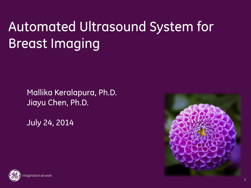
• Mammography has limited effectiveness in women with dense breasts
– Approximately 40% of American women have dense breasts
Having dense breasts increases cancer risk by
a factor of 4-6x1
Unique women, innovative tests
2
1. Boyd, et al, NEJM Jan 2007
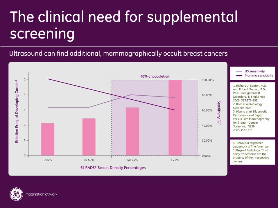
1. Richard J. Santen, M.D., and Robert Mansel, M.D., Ph.D.: Benign Breast Disorders. N Engl J Med 2005; 353:275-285 2. Kolb et al Radiology October 2002 3. Pisano et al. Diagnostic Performance of Digital versus Film Mammography for Breast –Cancer Screening. NEJM 2005;353:1773
BI-RADS is a registered trademark of The American College of Radiology. Third party trademarks are the property of their respective owners.
Ultrasound can find additional, mammographically occult breast cancers
The clinical need for supplemental screening
US sensitivity Mammo sensitivity
4
3
Texas
5
4
New York
California Missouri
Pennsylvania 1
Connecticut
2006 – Insurance Coverage Law
2009 – Density-Inform Law
Florida
Ohio
Virginia
New Hampshire
Delaware
New Jersey
Indiana
Maryland
Kentucky
Nevada
Oregon
Illinois
2009 – Insurance Coverage Law
2013 - Density-Inform Law-POOR
So. Carolina Tennessee
Utah 2 Colorado
Breast Density and Mammography
Reporting Act, October, 2011,
Rosa DeLauro (CT)
Steve Israel (NY)
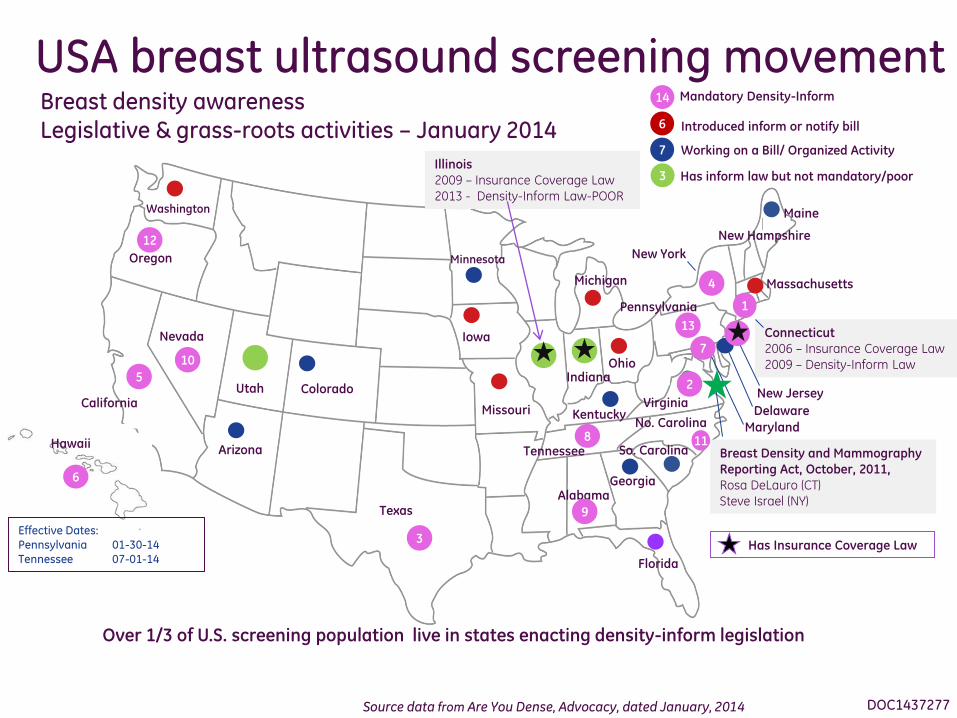
Breast density awareness Legislative & grass-roots activities – January 2014
USA breast ultrasound screening movement
Source data from Are You Dense, Advocacy, dated January, 2014
Iowa
6
Hawaii
Over 1/3 of U.S. screening population live in states enacting density-inform legislation
No. Carolina
Georgia
Alabama
Washington
Michigan
Effective Dates: Pennsylvania 01-30-14 Tennessee 07-01-14
Maine
Massachusetts
7
8
9
12
10
11
14 Mandatory Density-Inform
Introduced inform or notify bill
Working on a Bill/ Organized Activity
6
7
3 Has inform law but not mandatory/poor
Arizona
Minnesota
13
Has Insurance Coverage Law
DOC1437277
5
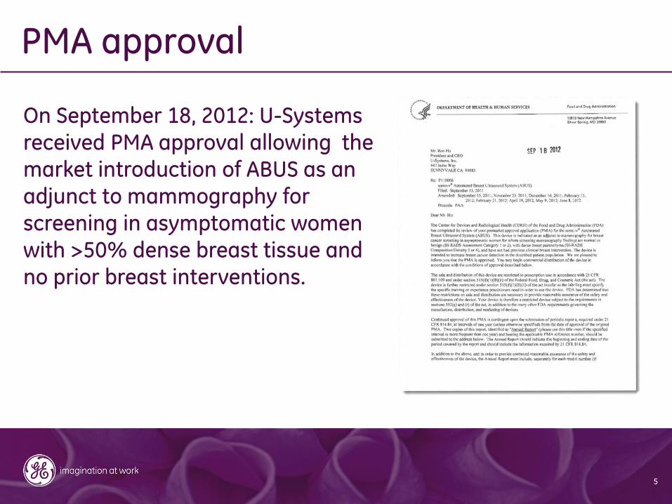
On September 18, 2012: U-Systems received PMA approval allowing the
market introduction of ABUS as an adjunct to mammography for screening in asymptomatic women with >50% dense breast tissue and no prior breast interventions.
PMA approval
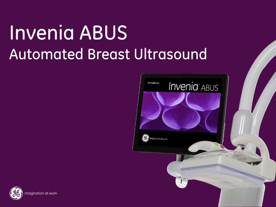
Invenia ABUS Automated Breast Ultrasound
7
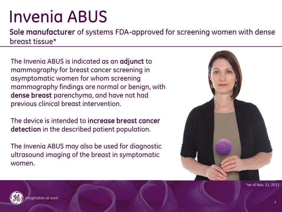
Invenia ABUS Sole manufacturer of systems FDA-approved for screening women with dense breast tissue*
The Invenia ABUS is indicated as an adjunct to
mammography for breast cancer screening in asymptomatic women for whom screening mammography findings are normal or benign, with dense breast parenchyma, and have not had previous clinical breast intervention. The device is intended to increase breast cancer detection in the described patient population.
The Invenia ABUS may also be used for diagnostic ultrasound imaging of the breast in symptomatic women.
*as of Nov. 11, 2013
8
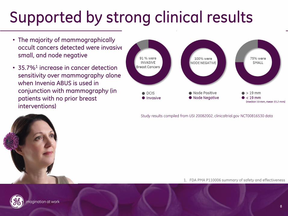
• The majority of mammographically
occult cancers detected were invasive,
small, and node negative
• 35.7%1 increase in cancer detection sensitivity over mammography alone
when Invenia ABUS is used in
conjunction with mammography (in
patients with no prior breast
interventions)
Supported by strong clinical results
1. FDA PMA P110006 summary of safety and effectiveness
Study results compiled from USI 20082002, clinicaltrial.gov NCT00816530 data
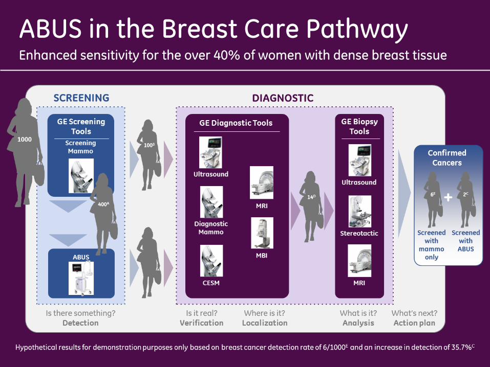
Enhanced sensitivity for the over 40% of women with dense breast tissue
ABUS in the Breast Care Pathway
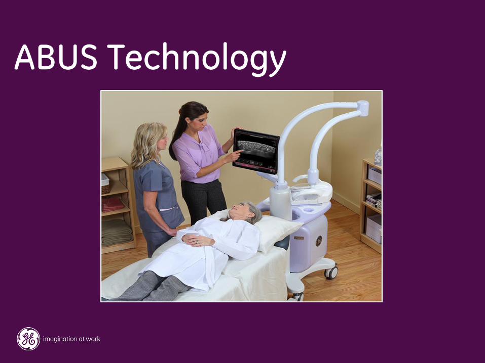
ABUS Technology
11
Basic Technical Requirements for Screening U/S
Caregiver’s perspective
• Automated image acquisition to minimize the operator dependency
• Standardized procedure for reproducibility and workflow efficiency
• High image quality and good tissue coverage for clinical confidence
• Ergonomic machine human interface
Patient’s perspective
• A quick and comfort procedure (~15 min room time)
• No radiation and contrast
• Low cost procedure for patient
12
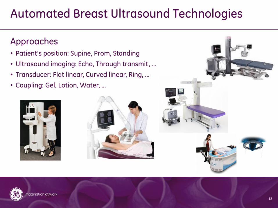
Automated Breast Ultrasound Technologies Approaches
• Patient’s position: Supine, Prom, Standing
• Ultrasound imaging: Echo, Through transmit, …
• Transducer: Flat linear, Curved linear, Ring, …
• Coupling: Gel, Lotion, Water, …
13
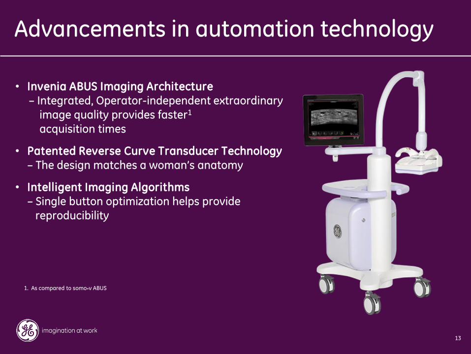
• Invenia ABUS Imaging Architecture – Integrated, Operator-independent extraordinary
image quality provides faster1 acquisition times
• Patented Reverse Curve Transducer Technology – The design matches a woman’s anatomy
• Intelligent Imaging Algorithms – Single button optimization helps provide reproducibility
Advancements in automation technology
1. As compared to somo•v ABUS
14
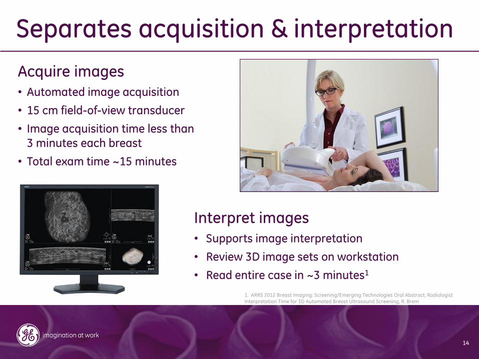
Acquire images
• Automated image acquisition
• 15 cm field-of-view transducer
• Image acquisition time less than 3 minutes each breast
• Total exam time ~15 minutes
Interpret images
• Supports image interpretation
• Review 3D image sets on workstation
• Read entire case in ~3 minutes1
Separates acquisition & interpretation
1. ARRS 2012 Breast Imaging: Screening/Emerging Technologies Oral Abstract; Radiologist Interpretation Time for 3D Automated Breast Ultrasound Screening, R. Brem
15
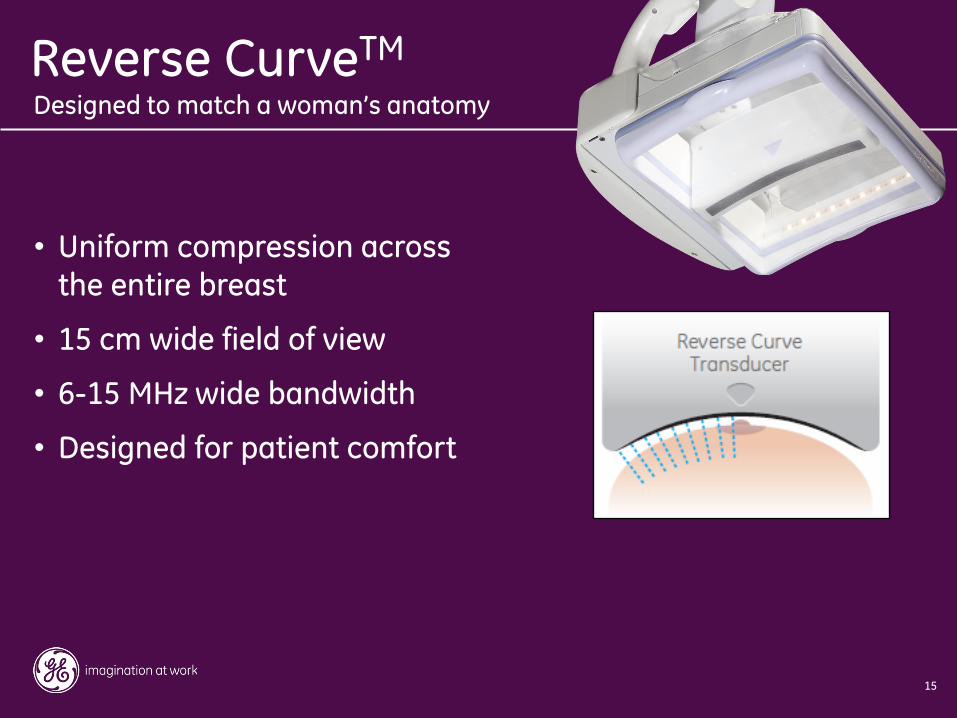
• Uniform compression across the entire breast
• 15 cm wide field of view
• 6-15 MHz wide bandwidth
• Designed for patient comfort
Designed to match a woman’s anatomy
Reverse CurveTM
16
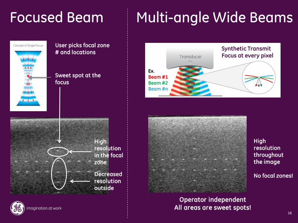
Multi-angle Wide Beams Focused Beam
High resolution in the focal zone
Decreased resolution outside
High resolution throughout the image No focal zones!
User picks focal zone # and locations
Sweet spot at the focus
Operator independent
All areas are sweet spots!
Synthetic Transmit Focus at every pixel
Ex. Beam #1 Beam #2 Beam #n
17
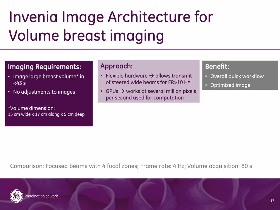
Invenia Image Architecture for Volume breast imaging
Imaging Requirements:
• Image large breast volume* in <45 s
• No adjustments to images
*Volume dimension: 15 cm wide x 17 cm along x 5 cm deep
Approach:
• Flexible hardware allows transmit of steered wide beams for FR>10 Hz
• GPUs works at several million pixels per second used for computation
Benefit:
• Overall quick workflow
• Optimized image
Comparison: Focused beams with 4 focal zones; Frame rate: 4 Hz; Volume acquisition: 80 s
18
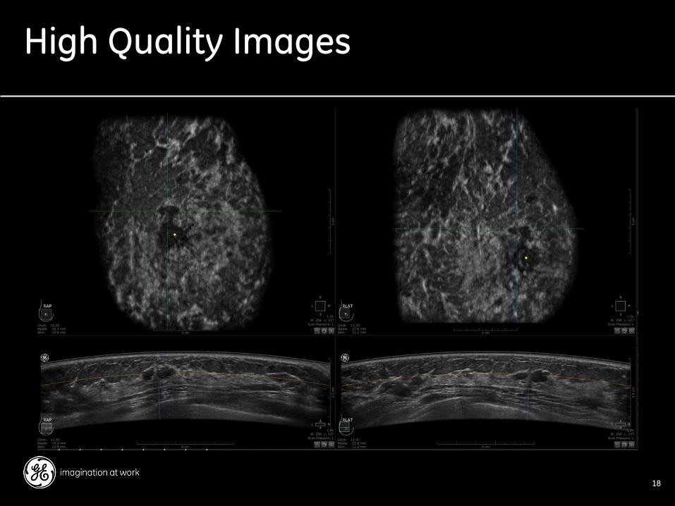
High Quality Images
19
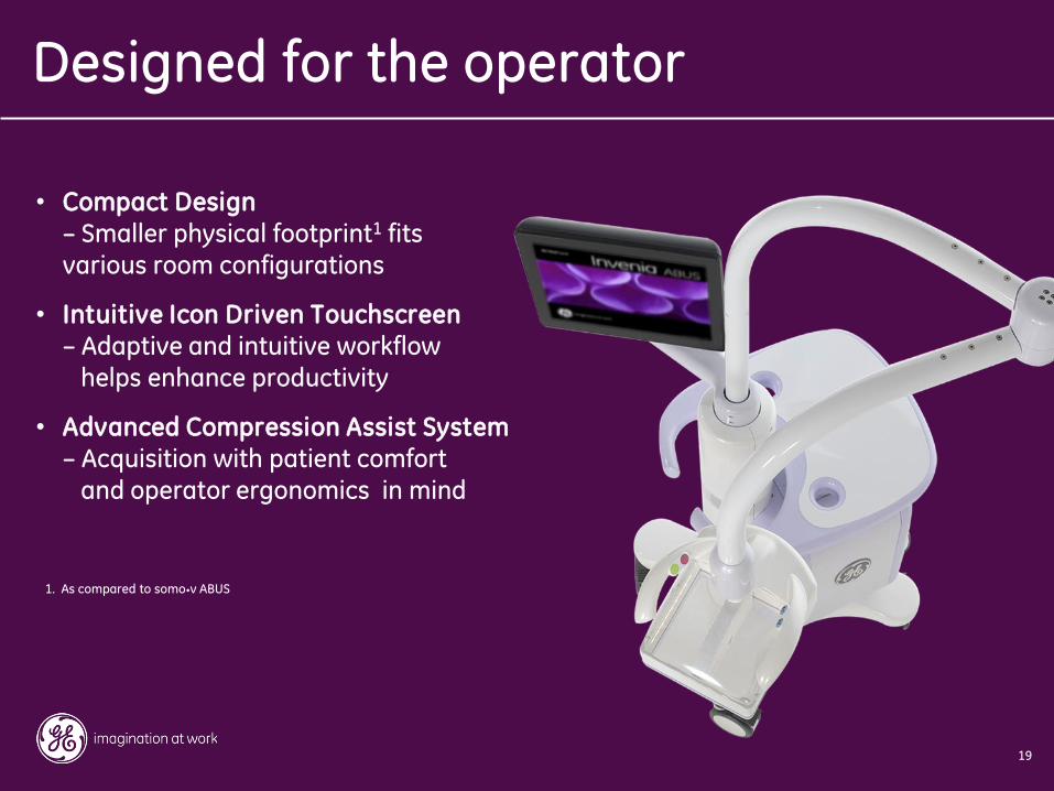
• Compact Design – Smaller physical footprint1 fits various room configurations
• Intuitive Icon Driven Touchscreen
– Adaptive and intuitive workflow helps enhance productivity
• Advanced Compression Assist System – Acquisition with patient comfort and operator ergonomics in mind
Designed for the operator
1. As compared to somo•v ABUS
20
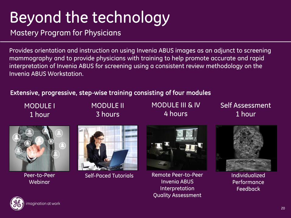
Peer-to-Peer Webinar
MODULE I
1 hour
Self-Paced Tutorials
MODULE II 3 hours
Remote Peer-to-Peer Invenia ABUS Interpretation
Quality Assessment
MODULE III & IV 4 hours
Individualized Performance
Feedback
Self Assessment 1 hour
Provides orientation and instruction on using Invenia ABUS images as an adjunct to screening
mammography and to provide physicians with training to help promote accurate and rapid
interpretation of Invenia ABUS for screening using a consistent review methodology on the
Invenia ABUS Workstation.
Extensive, progressive, step‐wise training consisting of four modules
Mastery Program for Physicians
Beyond the technology

21
Thank You
DOC1458880