Embed Size (px)
Citation preview
Author(s): Gerald Abrams, M.D., 2009
License: Unless otherwise noted, this material is made available under the terms of the Creative Commons Attribution-Non-commercial-Share Alike 3.0 License: http://creativecommons.org/licenses/by-nc-sa/3.0/
We have reviewed this material in accordance with U.S. Copyright Law and have tried to maximize your ability to use, share, and adapt it. The citation key on the following slide provides information about how you may share and adapt this material.
Copyright holders of content included in this material should contact [email protected] with any questions, corrections, or clarification regarding the use of content.
For more information about how to cite these materials visit http://open.umich.edu/education/about/terms-of-use.
Any medical information in this material is intended to inform and educate and is not a tool for self-diagnosis or a replacement for medical evaluation, advice, diagnosis or treatment by a healthcare professional. Please speak to your physician if you have questions about your medical condition.
Viewer discretion is advised: Some medical content is graphic and may not be suitable for all viewers.
Citation Key for more information see: http://open.umich.edu/wiki/CitationPolicy
Use + Share + Adapt
Make Your Own Assessment
Creative Commons – Attribution License
Creative Commons – Attribution Share Alike License
Creative Commons – Attribution Noncommercial License
Creative Commons – Attribution Noncommercial Share Alike License
GNU – Free Documentation License
Creative Commons – Zero Waiver
Public Domain – Ineligible: Works that are ineligible for copyright protection in the U.S. (USC 17 § 102(b)) *laws in your jurisdiction may differ
Public Domain – Expired: Works that are no longer protected due to an expired copyright term.
Public Domain – Government: Works that are produced by the U.S. Government. (USC 17 § 105)
Public Domain – Self Dedicated: Works that a copyright holder has dedicated to the public domain.
Fair Use: Use of works that is determined to be Fair consistent with the U.S. Copyright Act. (USC 17 § 107) *laws in your jurisdiction may differ
Our determination DOES NOT mean that all uses of this 3rd-party content are Fair Uses and we DO NOT guarantee that your use of the content is Fair.
To use this content you should do your own independent analysis to determine whether or not your use will be Fair.
{ Content the copyright holder, author, or law permits you to use, share and adapt. }
{ Content Open.Michigan believes can be used, shared, and adapted because it is ineligible for copyright. }
{ Content Open.Michigan has used under a Fair Use determination. }
Circulatory Derangements I
M1 – Cardiovascular/Respiratory Sequence
Gerald Abrams, MD
Fall 2008
Circulatory Derangements
• I -Congestion, Edema • II -Hemorrhage • III-Thrombosis • IV-Embolism, Ischemia, Infarction • V –Atherosclerosis, Ischemic Ht. Disease
CONGESTION Too much blood in an area
Within the cardiovascular system (unlike hemorrhage)
Vessels engorged with blood
ACTIVE CONGESTION (hyperemia) Increased flow into the area -erythema-
PASSIVE CONGESTION Decreased outflow from area -cyanosis-
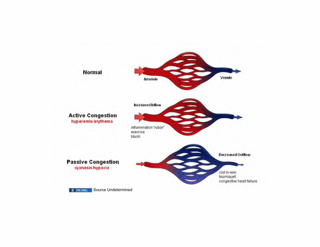
Source Undetermined
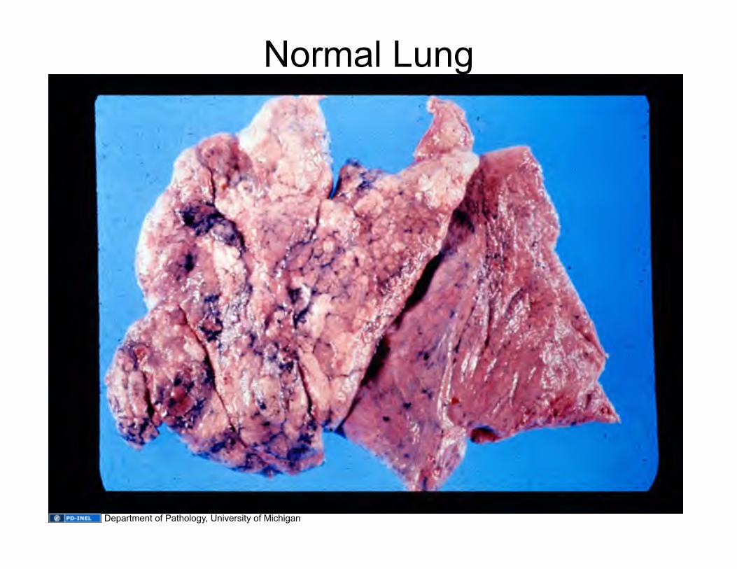
Normal Lung
Department of Pathology, University of Michigan
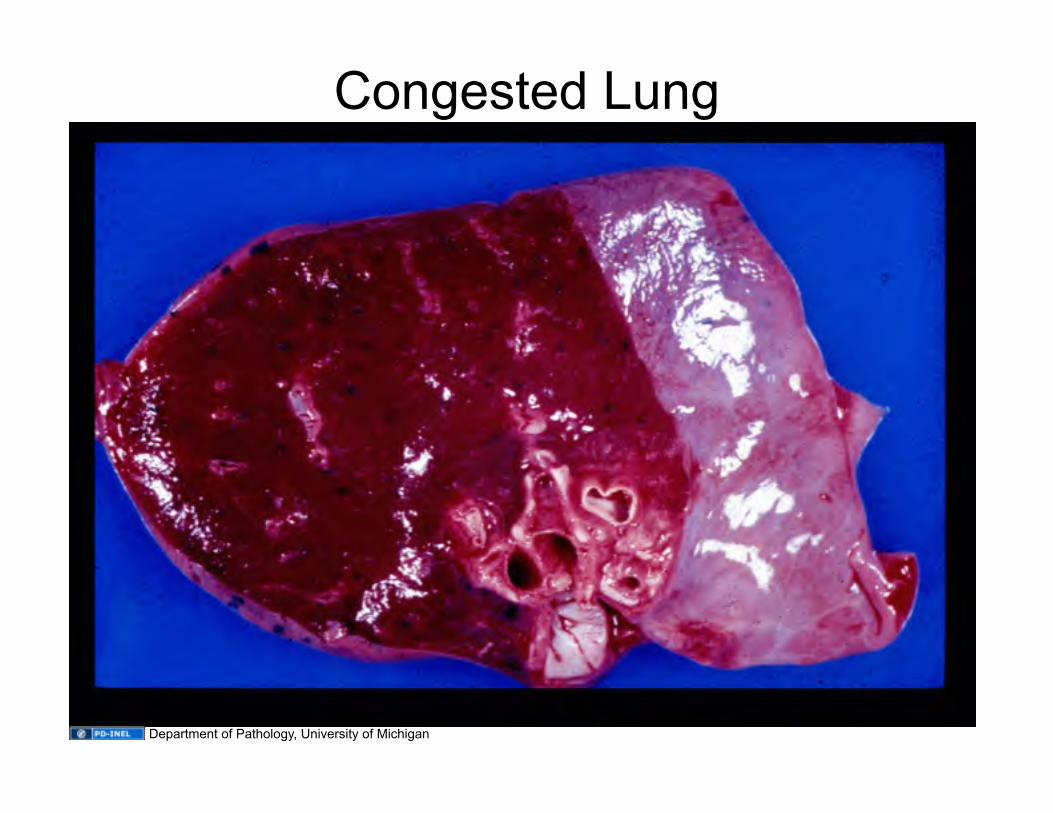
Congested Lung
Department of Pathology, University of Michigan
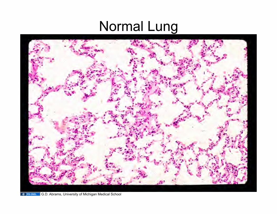
Normal Lung
G.D. Abrams, University of Michigan Medical School

Congested Lung
G.D. Abrams, University of Michigan Medical School
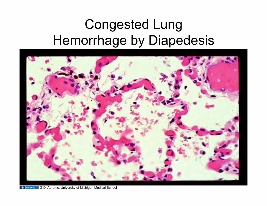
Congested Lung Hemorrhage by Diapedesis
G.D. Abrams, University of Michigan Medical School
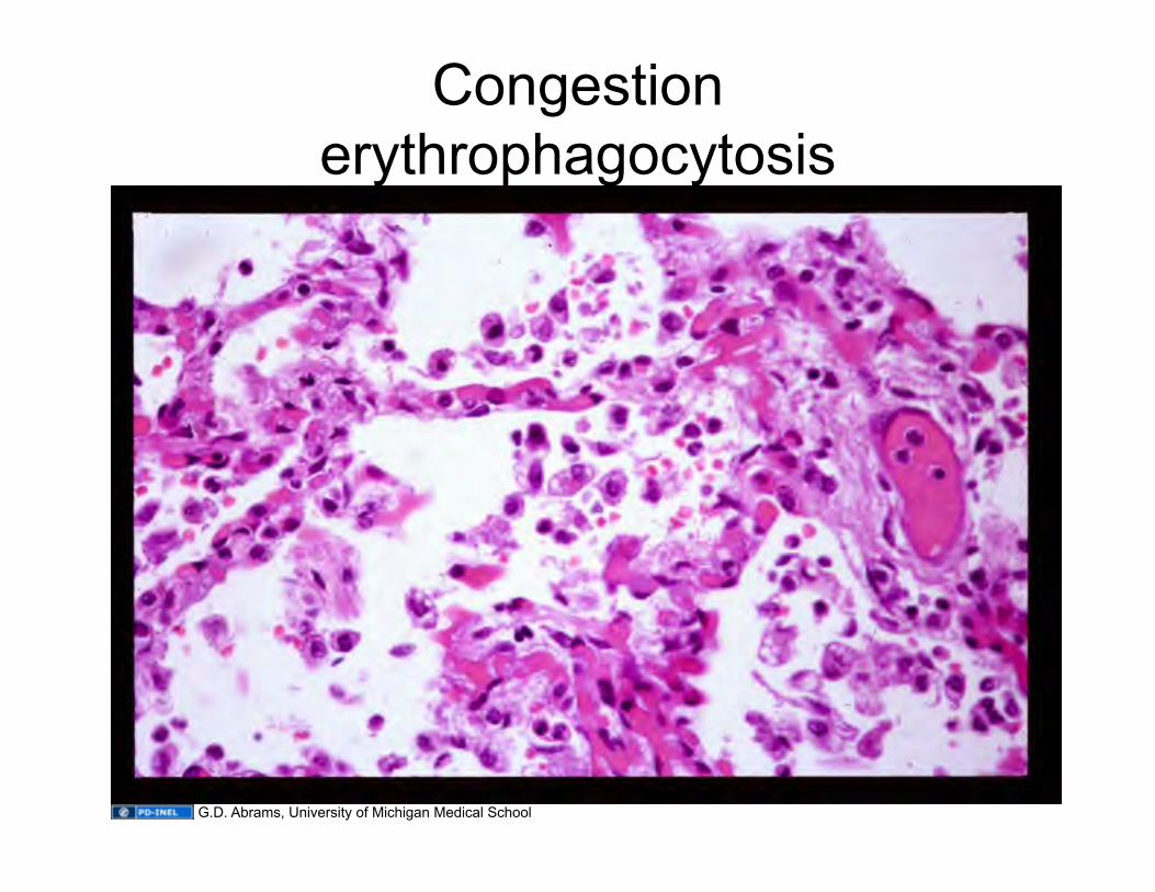
Congestion erythrophagocytosis
G.D. Abrams, University of Michigan Medical School
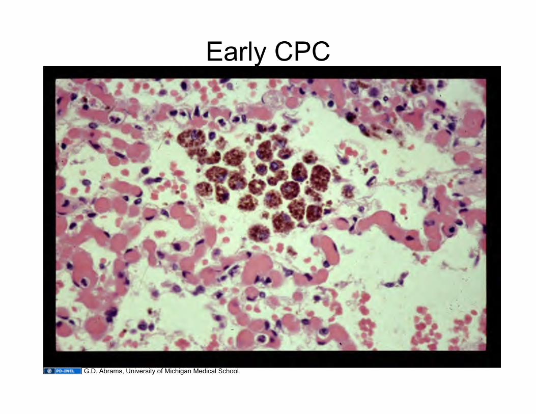
Early CPC
G.D. Abrams, University of Michigan Medical School
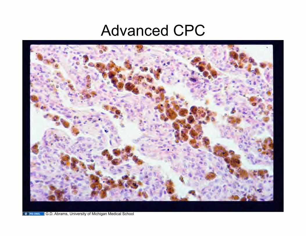
Advanced CPC
G.D. Abrams, University of Michigan Medical School
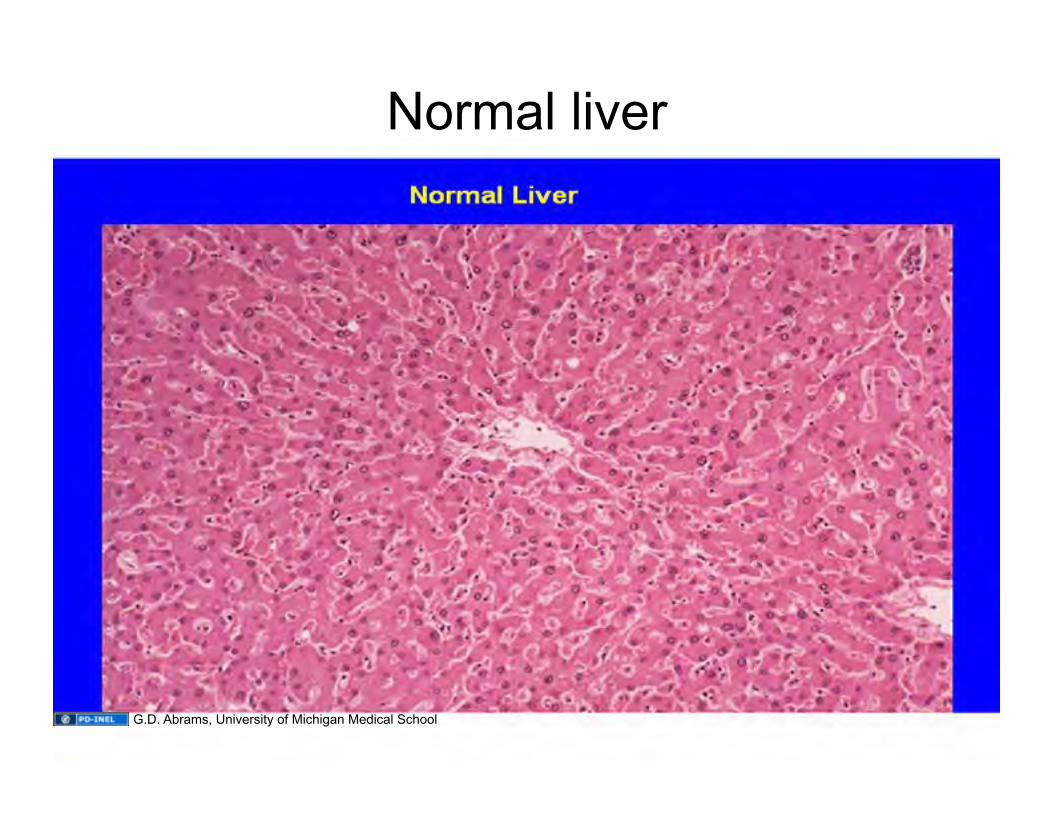
Normal liver
G.D. Abrams, University of Michigan Medical School
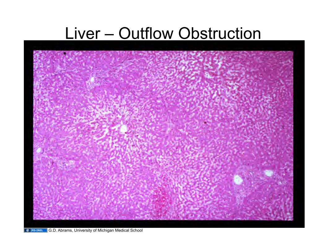
Liver – Outflow Obstruction
G.D. Abrams, University of Michigan Medical School
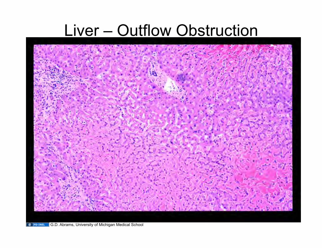
Liver – Outflow Obstruction
G.D. Abrams, University of Michigan Medical School
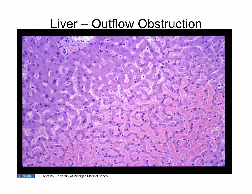
Liver – Outflow Obstruction
G.D. Abrams, University of Michigan Medical School
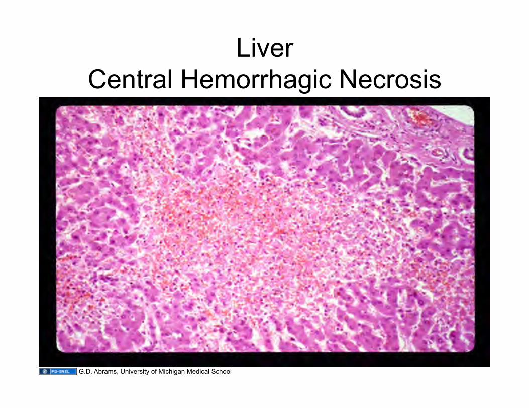
Liver Central Hemorrhagic Necrosis
G.D. Abrams, University of Michigan Medical School
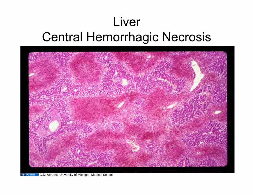
Liver Central Hemorrhagic Necrosis
G.D. Abrams, University of Michigan Medical School
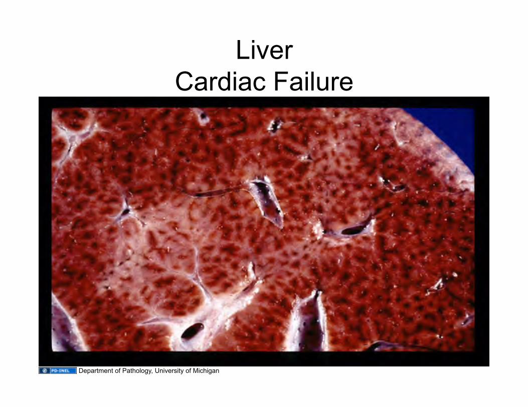
Liver Cardiac Failure
Department of Pathology, University of Michigan
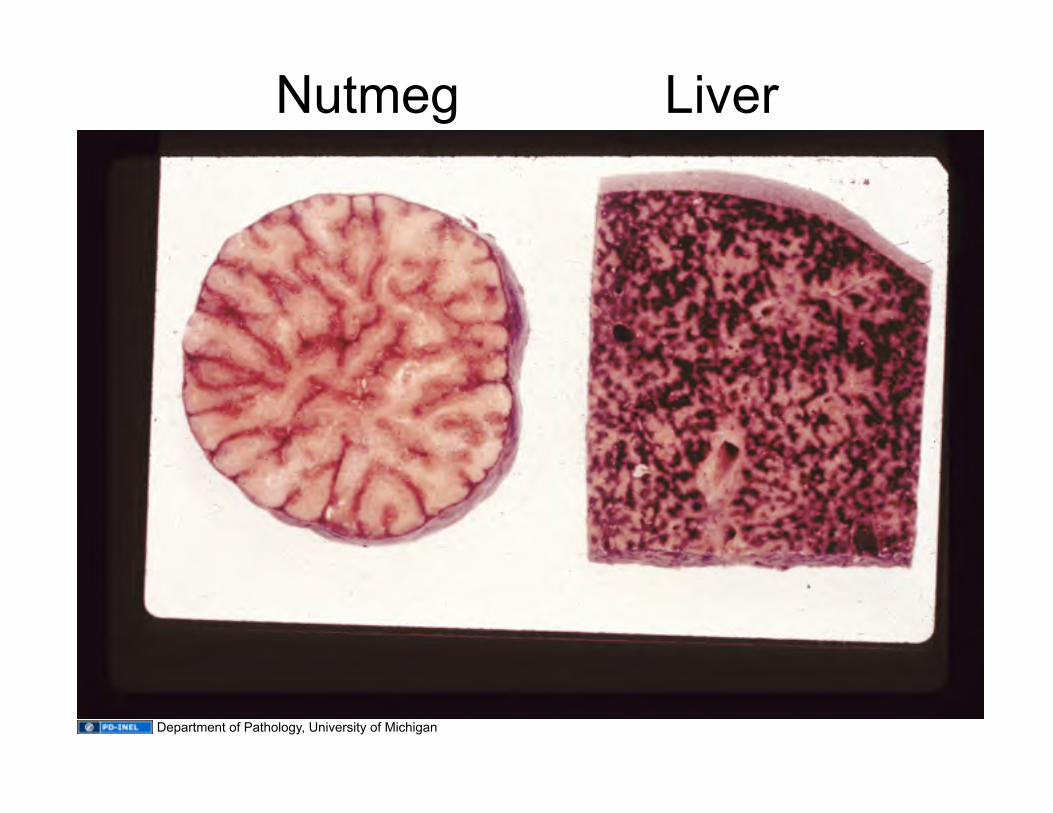
Nutmeg Liver
Department of Pathology, University of Michigan
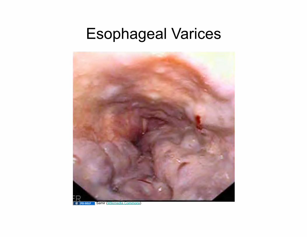
Esophageal Varices
Samir (Wikimedia Commons)
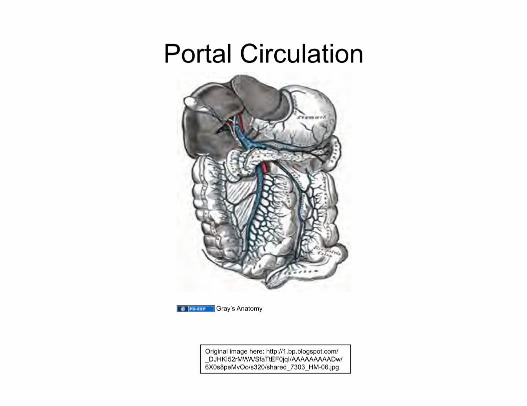
Portal Circulation
Original image here: http://1.bp.blogspot.com/_DJHKI52rMWA/SfaTtEF0jqI/AAAAAAAAADw/6X0s8peMvOo/s320/shared_7303_HM-06.jpg
Gray’s Anatomy
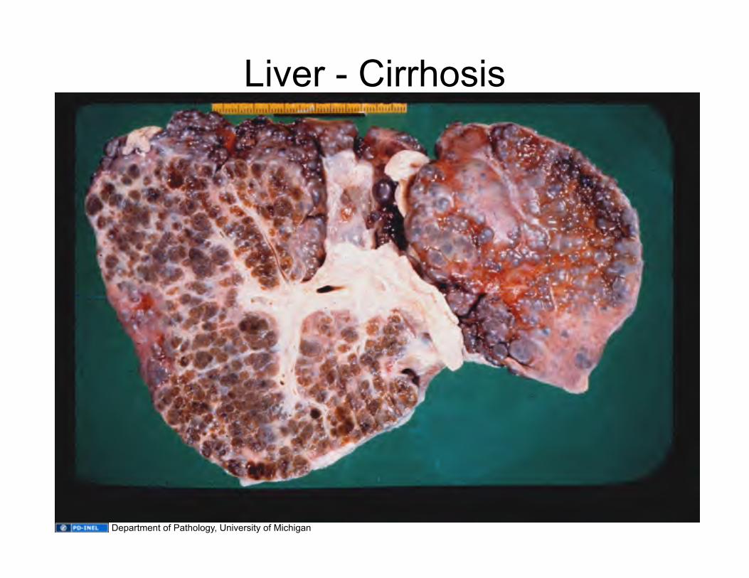
Liver - Cirrhosis
Department of Pathology, University of Michigan
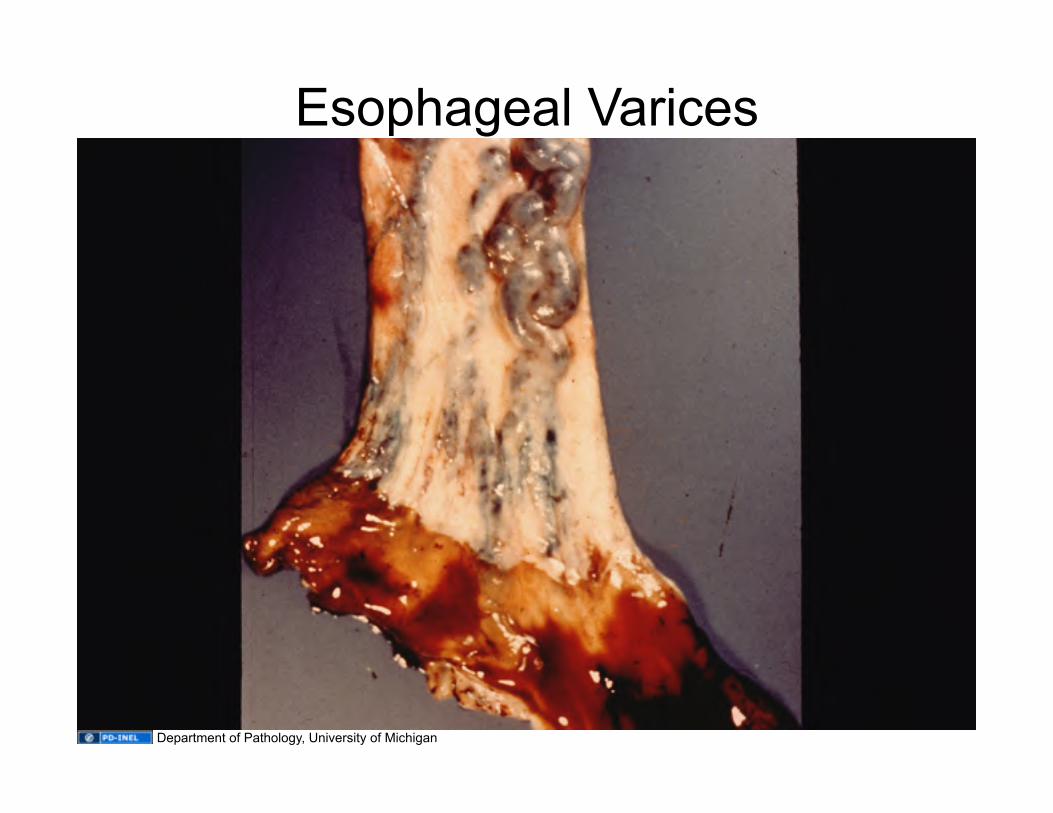
Esophageal Varices
Department of Pathology, University of Michigan
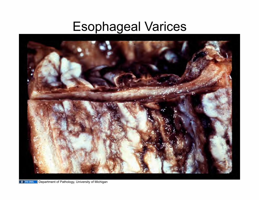
Esophageal Varices
Department of Pathology, University of Michigan
EDEMA
Accumulation of excess fluid in the interstitial spaces / body cavities
Effusion
Fluid accumulation in a body cavity Pleural Effusion Pericardial Effusion Peritoneal Effusion = Ascites Anasarca – Generalized edema, a water-logged body
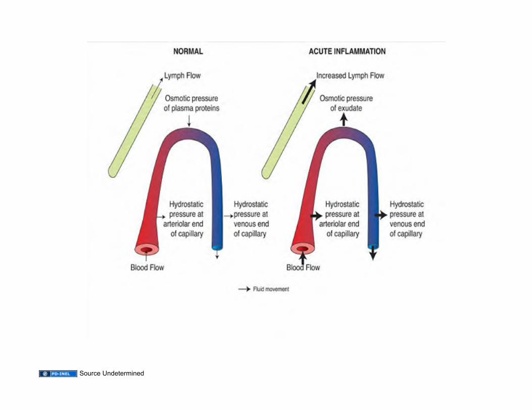
Source Undetermined
Fluid Collections
• Inflammatory collection = Exudate
• Non-inflammatory collection = Transudate
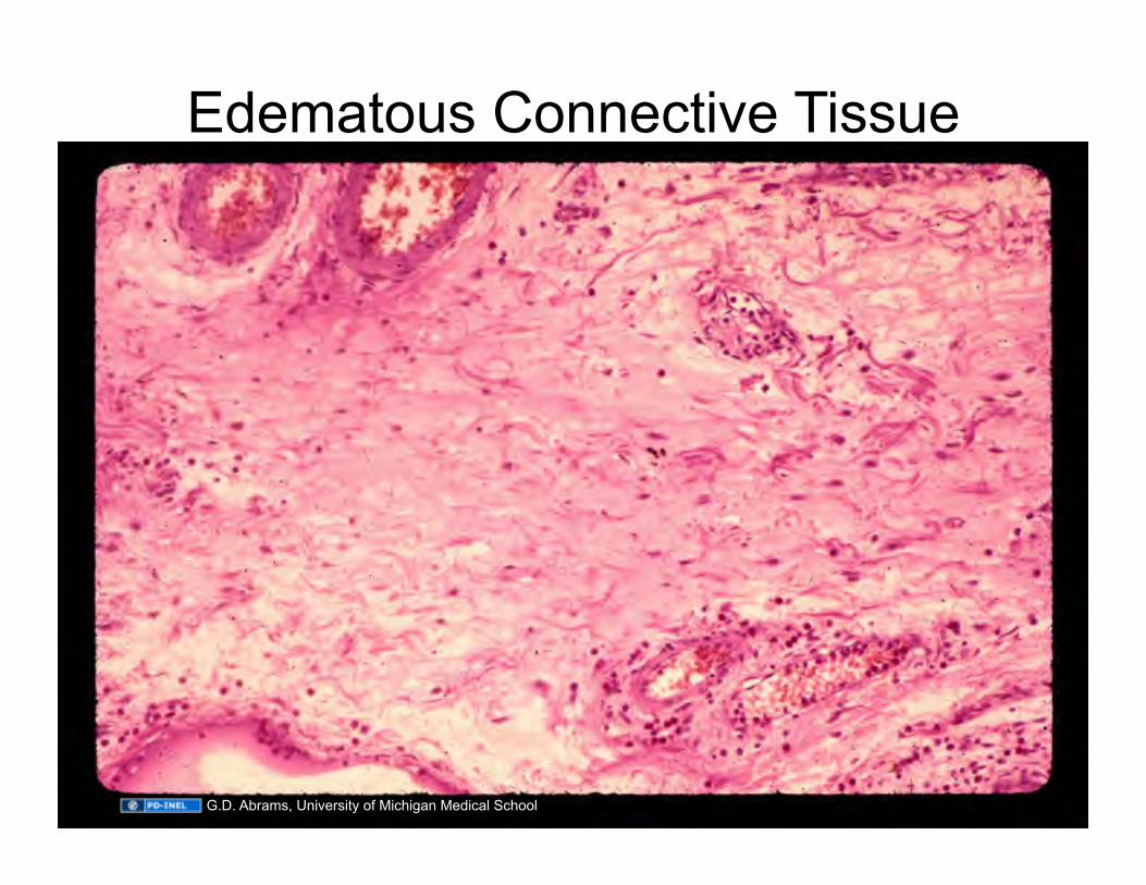
Edematous Connective Tissue
G.D. Abrams, University of Michigan Medical School
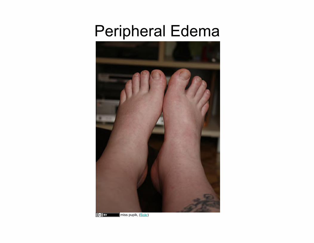
Peripheral Edema
miss pupik, (flickr)
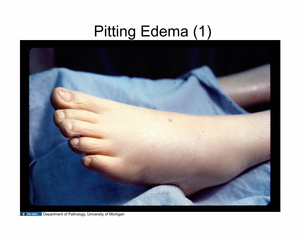
Pitting Edema (1)
Department of Pathology, University of Michigan
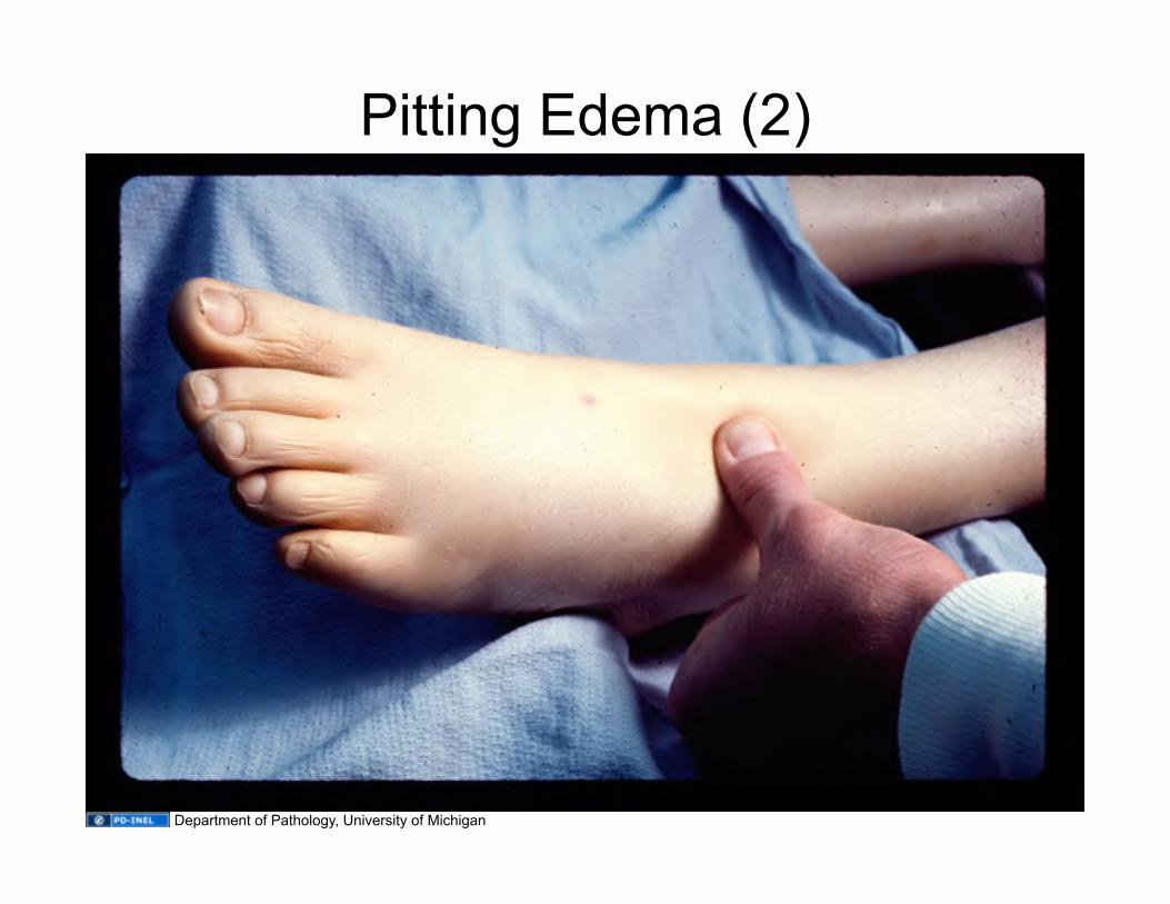
Pitting Edema (2)
Department of Pathology, University of Michigan
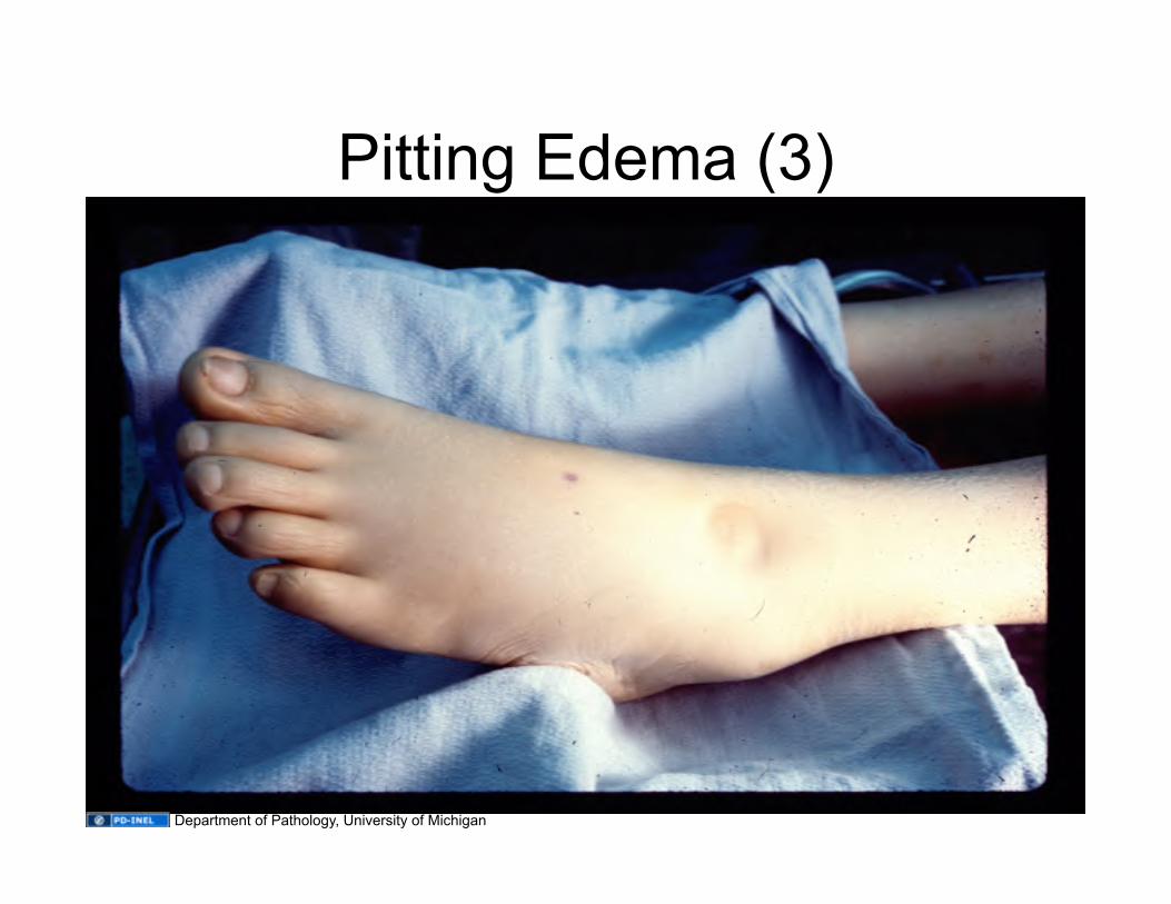
Pitting Edema (3)
Department of Pathology, University of Michigan
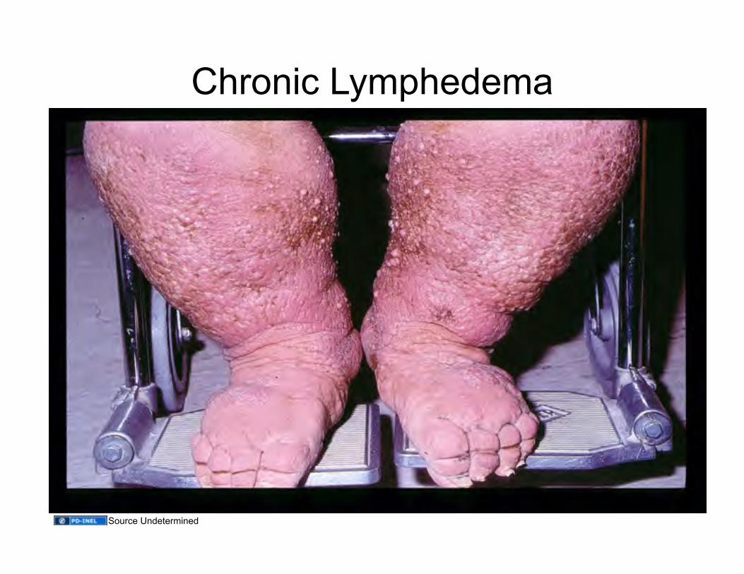
Chronic Lymphedema
Source Undetermined

Pulmonary Edema
G.D. Abrams, University of Michigan Medical School
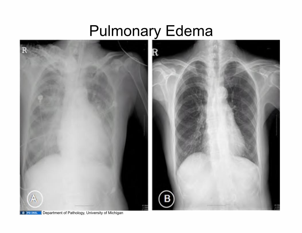
Pulmonary Edema
Department of Pathology, University of Michigan
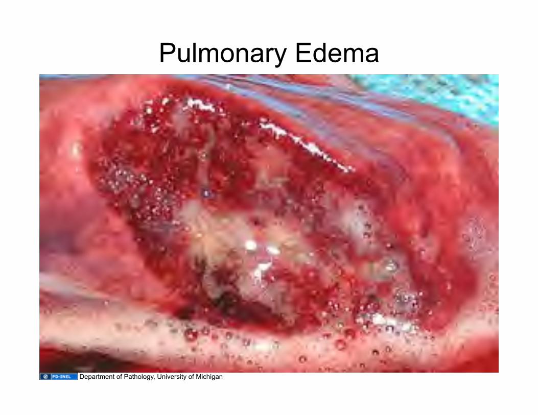
Pulmonary Edema
Department of Pathology, University of Michigan
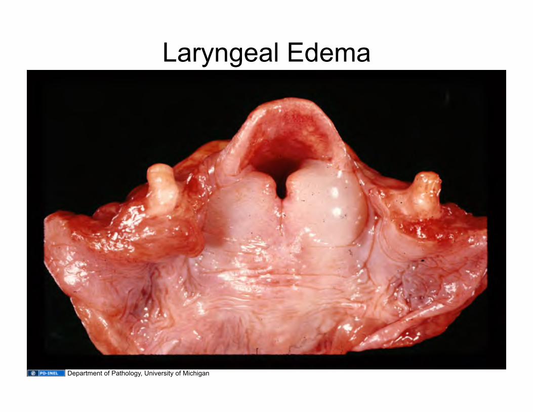
Laryngeal Edema
Department of Pathology, University of Michigan
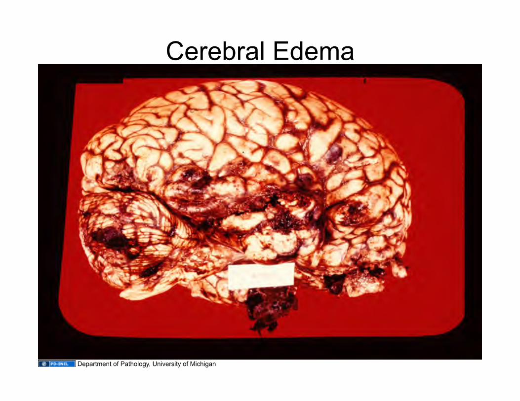
Cerebral Edema
Department of Pathology, University of Michigan

Slide 7: Source Undetermined Slide 8: Department of Pathology, University of Michigan Slide 9: Department of Pathology, University of Michigan Slide 10: G.D. Abrams, University of Michigan Medical School Slide 11: G.D. Abrams, University of Michigan Medical School Slide 12: G.D. Abrams, University of Michigan Medical School Slide 13: G.D. Abrams, University of Michigan Medical School Slide 14: G.D. Abrams, University of Michigan Medical School Slide 15: G.D. Abrams, University of Michigan Medical School Slide 16: G.D. Abrams, University of Michigan Medical School Slide 17: G.D. Abrams, University of Michigan Medical School Slide 18: G.D. Abrams, University of Michigan Medical School Slide 19: G.D. Abrams, University of Michigan Medical School Slide 20: G.D. Abrams, University of Michigan Medical School Slide 21: G.D. Abrams, University of Michigan Medical School Slide 22: Department of Pathology, University of Michigan Slide 23: Department of Pathology, University of Michigan Slide 24: Samir, Wikimedia Commons, http://en.wikipedia.org/wiki/File:Esophageal_varices_-_wale.jpg Slide 25: Gray’s Anatomy Slide 26: Department of Pathology, University of Michigan Slide 27: Department of Pathology, University of Michigan Slide 28: Department of Pathology, University of Michigan Slide 31: Source Undetermined Slide 33: G.D. Abrams, University of Michigan Medical School Slide 34: miss pupik, flickr, http://www.flickr.com/photos/miss_pupik/2418359498; CC:BY 2.0, http://creativecommons.org/licenses/by/2.0/deed.en Slide 35: Department of Pathology, University of Michigan Slide 36: Department of Pathology, University of Michigan Slide 37: Department of Pathology, University of Michigan Slide 38: G.D. Abrams, University of Michigan Medical School Slide 39: G.D. Abrams, University of Michigan Medical School Slide 40: Department of Pathology, University of Michigan Slide 41: Department of Pathology, University of Michigan Slide 42: Department of Pathology, University of Michigan Slide 43: Department of Pathology, University of Michigan
Additional Source Information for more information see: http://open.umich.edu/wiki/CitationPolicy