Embed Size (px)
Citation preview

Atrial fibrillation and flutter:Practical Management Tips
Internal Medicine Residency Program Noon Conference 2011

Learning Goals
A brief discussion of supraventricular tachycardia (SVT)
Review of AF and AFl physiology and EKG differentiation
Management of atrial fibrillation (AF) Management of atrial flutter (AFl)
Management of fib/flutter
Anticoagulation Cardioversion Rate control Rhythm control Ablation Cardiothoracic surgery
But, first, a brief diversion
Definition of supraventricular tachycardia (SVT)
Differentiating among types of SVT Differentiating AF from AFl
Supraventricular tachycardia
Abbreviated SVT “Supra” means “above” Supraventricular tachycardia comes from
above the ventricles DO NOT CONFUSE with NSVT (non-sustained
ventricular tachycardia) (Essentially) all narrow-complex tachycardia
has a supraventricular origin

SVT possible sites of origin
Sinus node Atria Atrioventricular node His bundle
Or some combination of the above

Supraventricular tachycardia
Sinus tachycardia Multifocal atrial tachcyardia Paroxysmal atrial tachycardia AV nodal reentrant tachycardia (AVNRT) Atrioventricular reentrant tachycardia (AVRT) Atrial fibrillation Atrial flutter
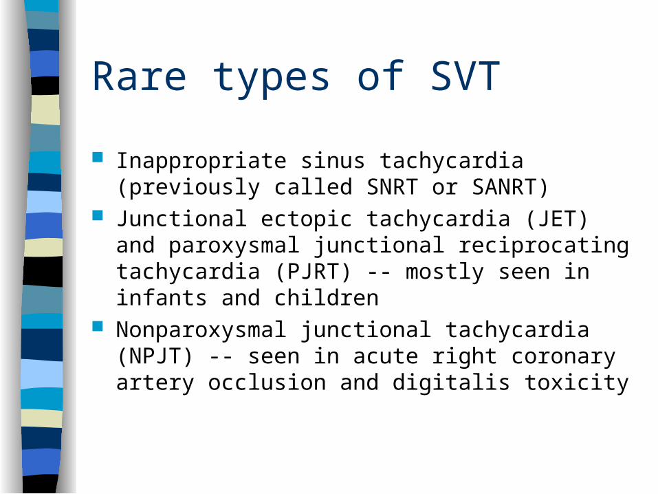
Rare types of SVT
Inappropriate sinus tachycardia (previously called SNRT or SANRT)
Junctional ectopic tachycardia (JET) and paroxysmal junctional reciprocating tachycardia (PJRT) -- mostly seen in infants and children
Nonparoxysmal junctional tachycardia (NPJT) -- seen in acute right coronary artery occlusion and digitalis toxicity
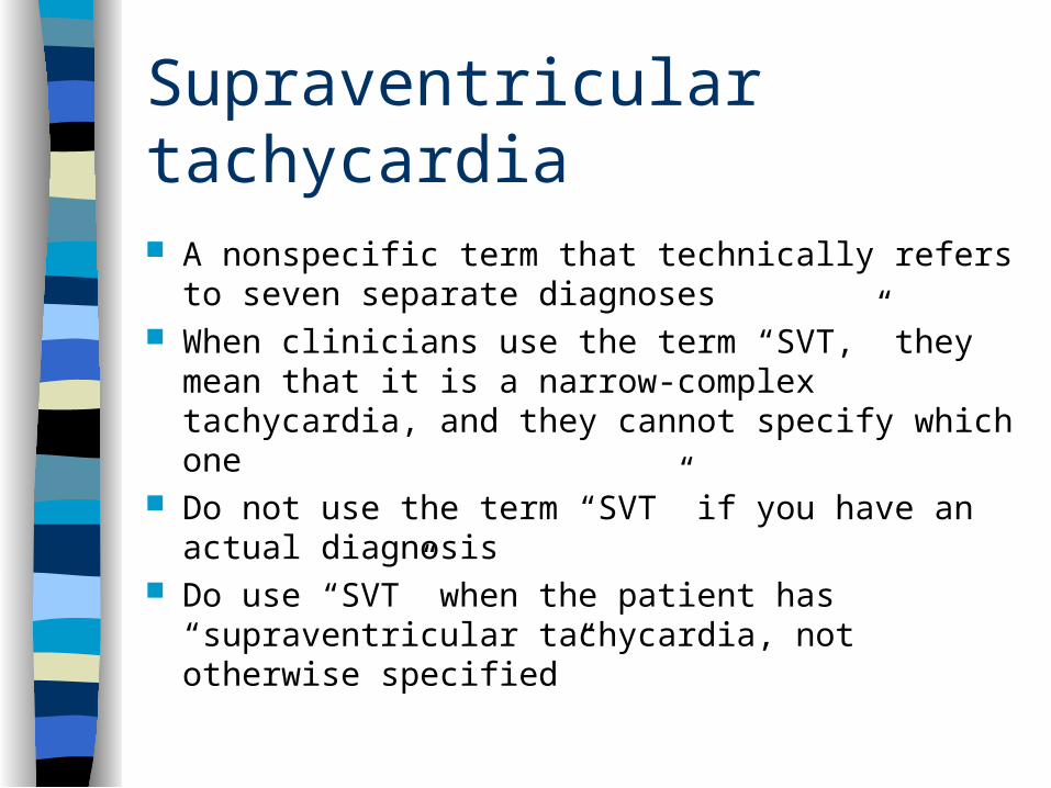
Supraventricular tachycardia
A nonspecific term that technically refers to seven separate diagnoses
When clinicians use the term “SVT,” they mean that it is a narrow-complex tachycardia, and they cannot specify which one
Do not use the term “SVT” if you have an actual diagnosis
Do use “SVT” when the patient has “supraventricular tachycardia, not otherwise specified”
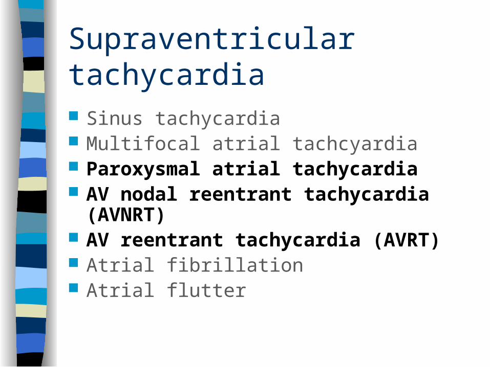
Supraventricular tachycardia
Sinus tachycardia Multifocal atrial tachcyardia Paroxysmal atrial tachycardia AV nodal reentrant tachycardia (AVNRT) AV reentrant tachycardia (AVRT) Atrial fibrillation Atrial flutter
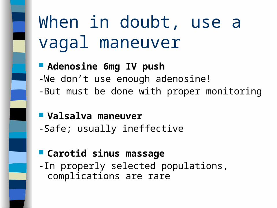
When in doubt, use a vagal maneuver Adenosine 6mg IV push-We don’t use enough adenosine!-But must be done with proper monitoring
Valsalva maneuver-Safe; usually ineffective
Carotid sinus massage-In properly selected populations, complications are
rare
Carotid Sinus Massage Contraindications Carotid bruit Prior stroke or transient ischemic attack,
unless imaging has shown no significant carotid disease
Myocardial infarction in the previous six months
History of serious cardiac arrhythmias (VT, VF)
Vagal maneuvers
Diagnostic-Usually you can learn which SVT it was by
doing a vagal maneuver
Therapeutic-Vagal maneuvers can terminate AVRT and
AVNRT

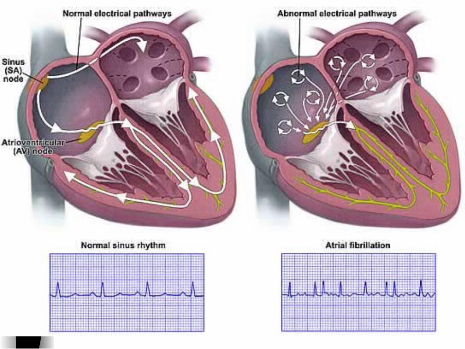
Differentiating AF from AFl
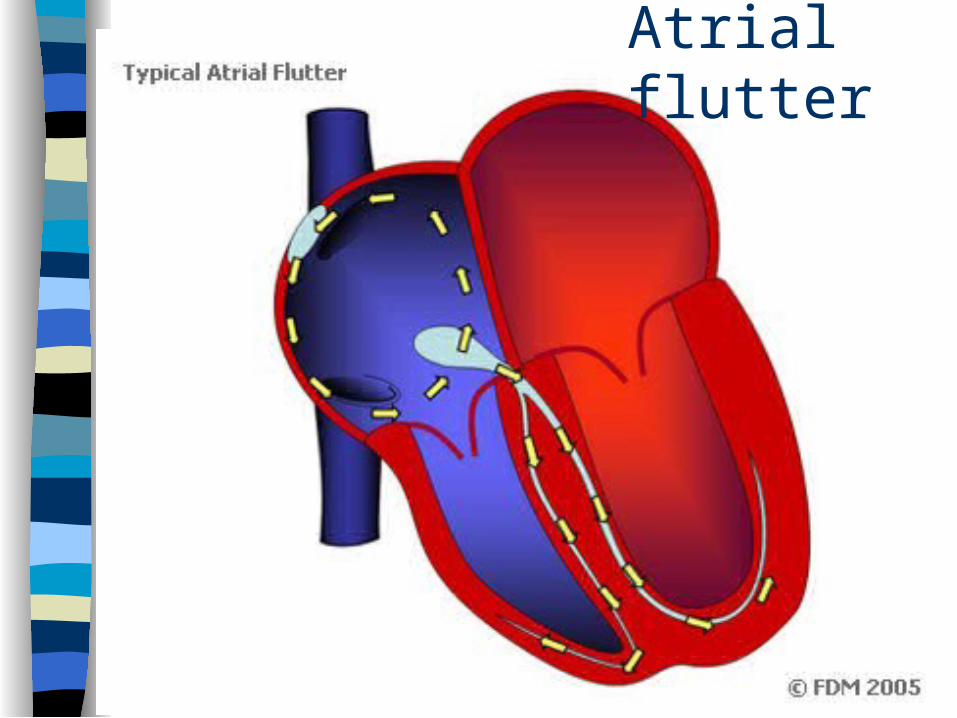
AFl is a macroreentrant atrial rhythm with a reentry circuit that involves a large area of atrial myocardium
AF is caused by multiple wandering wavelets, a hodgepodge of microreentrant circuits, often located in the pulmonary veins

Atrial flutter
Atrial flutter
P waves exhibit a “sawtooth” pattern referred to as flutter waves or “F” waves
Atrial rate is typically 250-350 beats per minute (bpm)
Atrial flutter, continued
Classically, atrial rate is 300 bpm with 2:1 AV conduction, leading to a heart rate of 150 bpm
But focus on the atrial pattern when diagnosing SVT--try to ignore the QRS complexes, just looking at the P (or F) waves at first
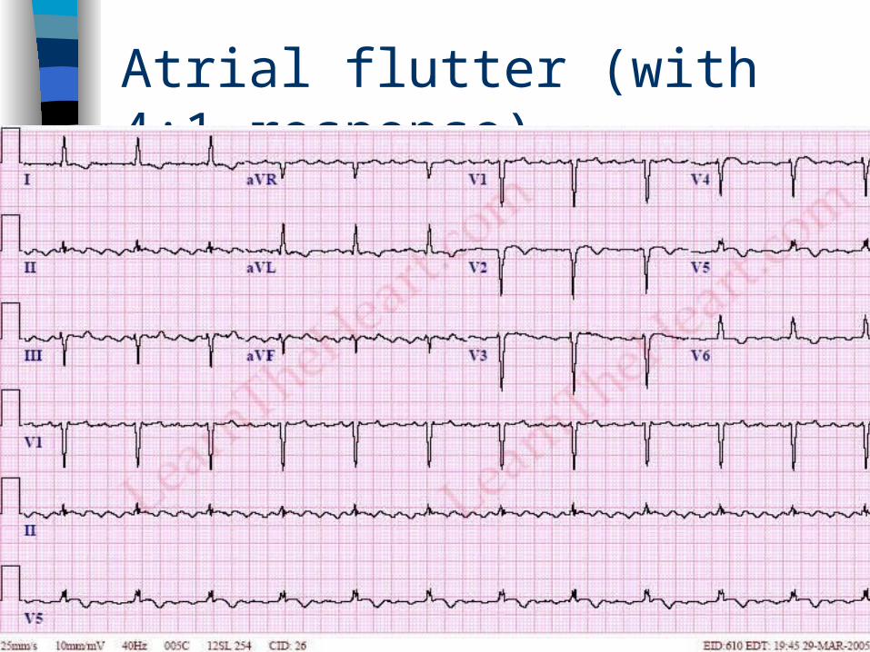
Atrial flutter (with 4:1 response)
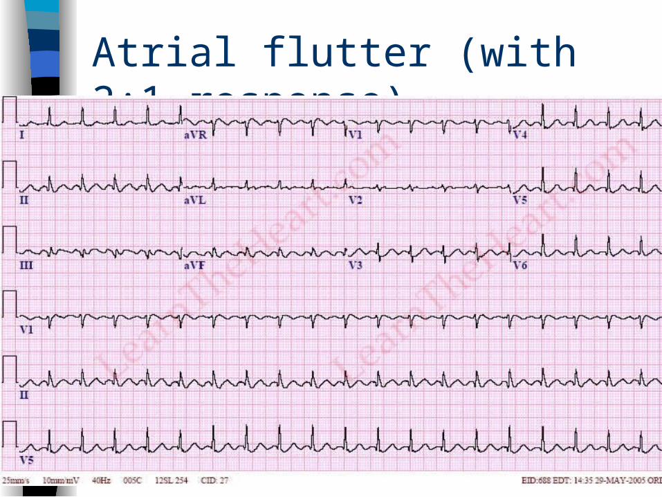
Atrial flutter (with 2:1 response)

Atrial flutter (variable response)
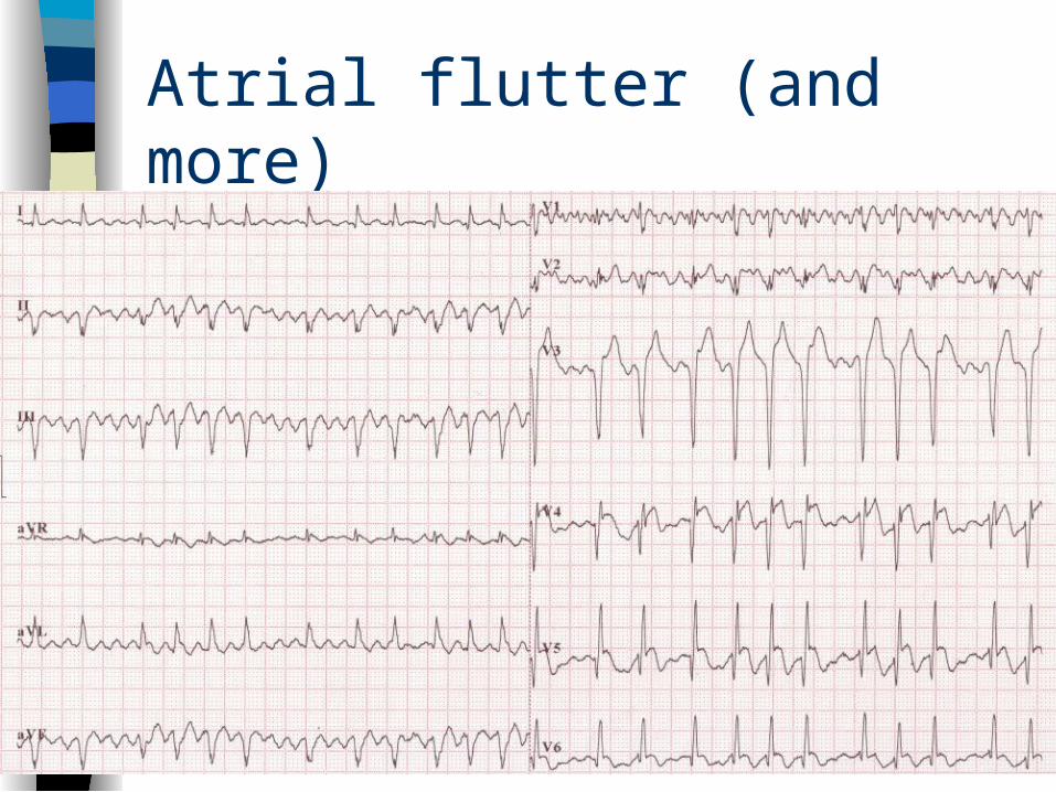
Atrial flutter (and more)

AF diagram
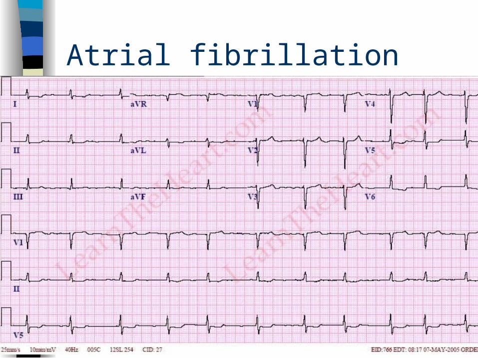
Atrial fibrillation
Rapid and irregular atrial activity at a rate of 350-600 impulses per minute
Usually irregularly irregular ventricular response
There are no P waves Sometimes the F waves are so fine, the
surface EKG cannot detect them

Atrial fibrillation
Atrial fibrillation
Atrial fibrillation terms
Paroxysmal Persistent Permanent “Lone”
Atrial fibrillation terms, cont.
Paroxysmal
-episodes terminate spontaneously in less than seven days
Persistent
-fails to terminate within seven days
Atrial fibrillation terms, cont.
Permanent-AF lasts for more than one year, and-Cardioversion has not been attempted or
has failed “Lone”-patients less than 60 years of age
without structural heart disease
Atrial fibrillation terms, cont.
This classification applies only when no clear reversible cause of AF.
If AF is clearly due to heart surgery, pericarditis, myocardial infarction, hyperthyroidism, pulmonary embolism, or other reversible causes, avoid this classification system
Management of fib/flutter
Anticoagulation Cardioversion Rate control Rhythm control Ablation Cardiothoracic surgery
Anticoagulation (AC)
Recommendations are essentially the same for AF and AFl
First, assess if the patient is high risk for cardioembolic stroke
Most patients with high risk should be on AC if they ever were seen in AF or AFl

High risk for cardioembolic stroke Rheumatic mitral stenosis-Mitral valve area less than 2.0 cm2
Prosthetic heart valves Hyperthyroid (?)-2006 ACC/AHA/ESC guidelines
recommend INR 2-3 in all patients until euthyroid; ACCP does not comment on this
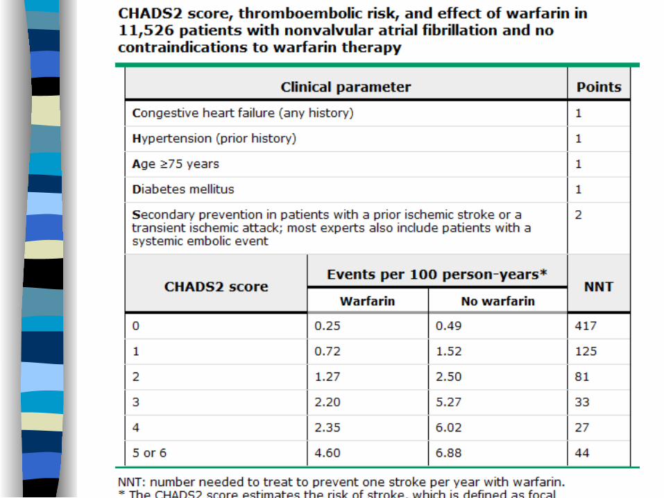
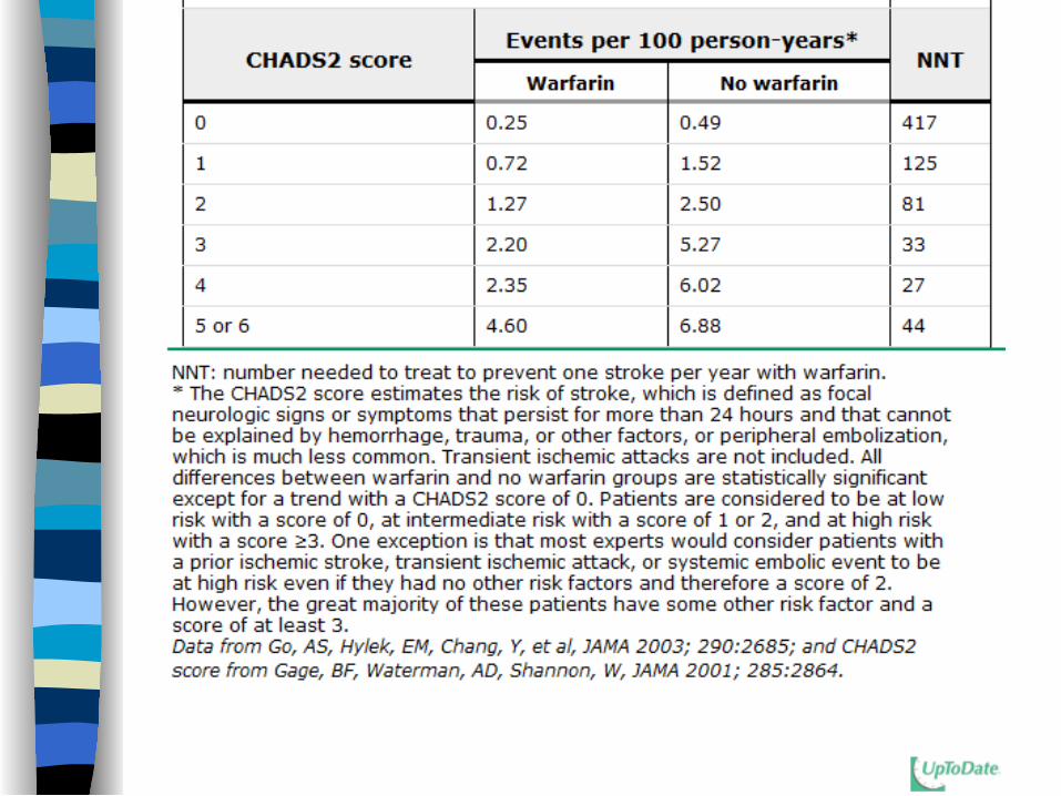
CHADS2 score
For use in patients without the high risk factors on the previous slide
There are other risk models, including the CHADS2-VASc score

CHADS2 score
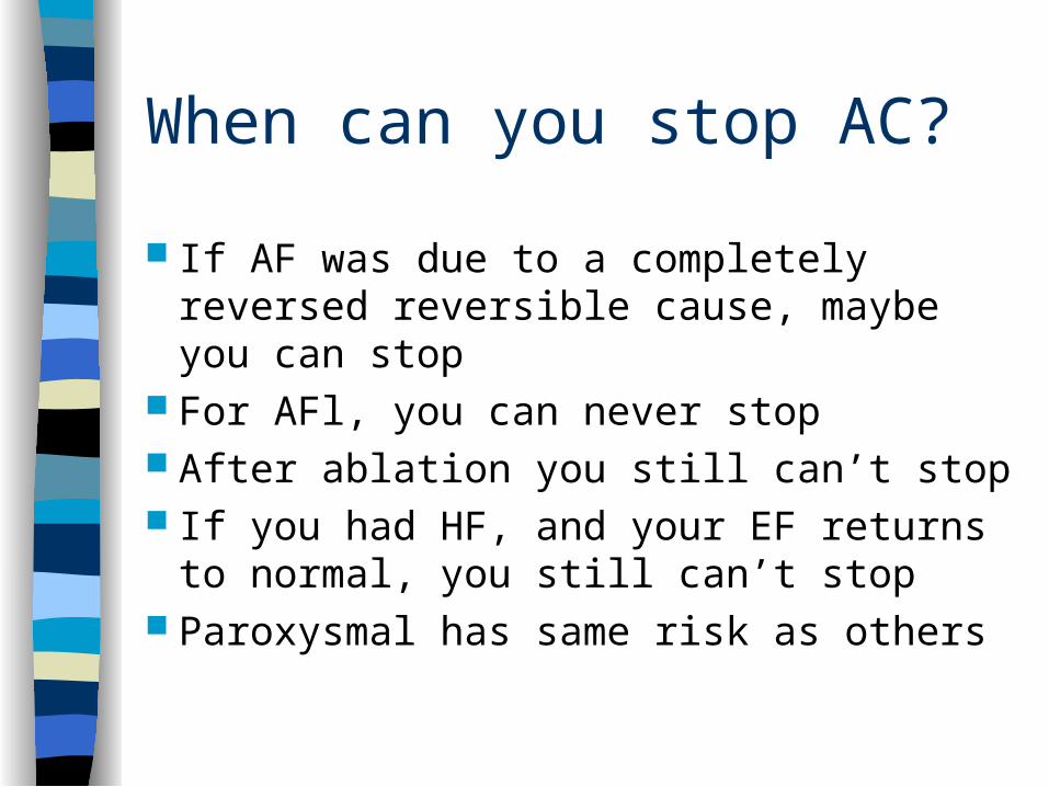
When can you stop AC?
If AF was due to a completely reversed reversible cause, maybe you can stop
For AFl, you can never stop After ablation you still can’t stop If you had HF, and your EF returns to
normal, you still can’t stop Paroxysmal has same risk as others
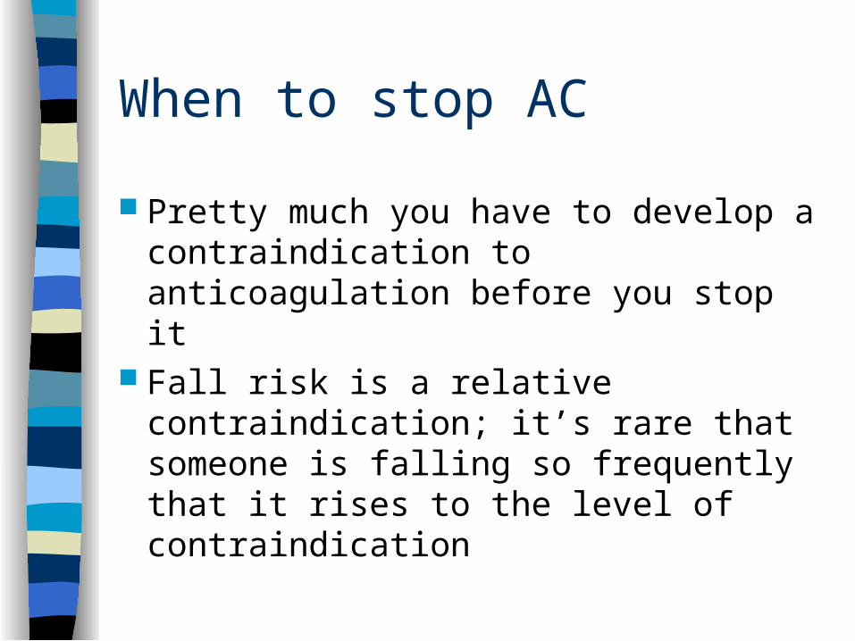
When to stop AC
Pretty much you have to develop a contraindication to anticoagulation before you stop it
Fall risk is a relative contraindication; it’s rare that someone is falling so frequently that it rises to the level of contraindication
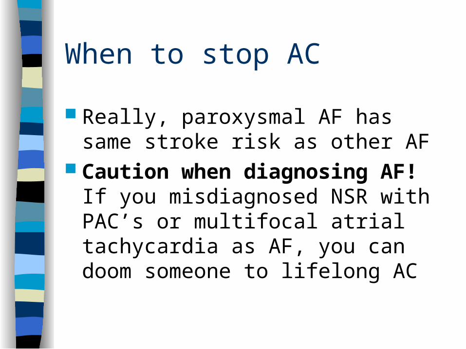
When to stop AC
Really, paroxysmal AF has same stroke risk as other AF
Caution when diagnosing AF! If you misdiagnosed NSR with PAC’s or multifocal atrial tachycardia as AF, you can doom someone to lifelong AC
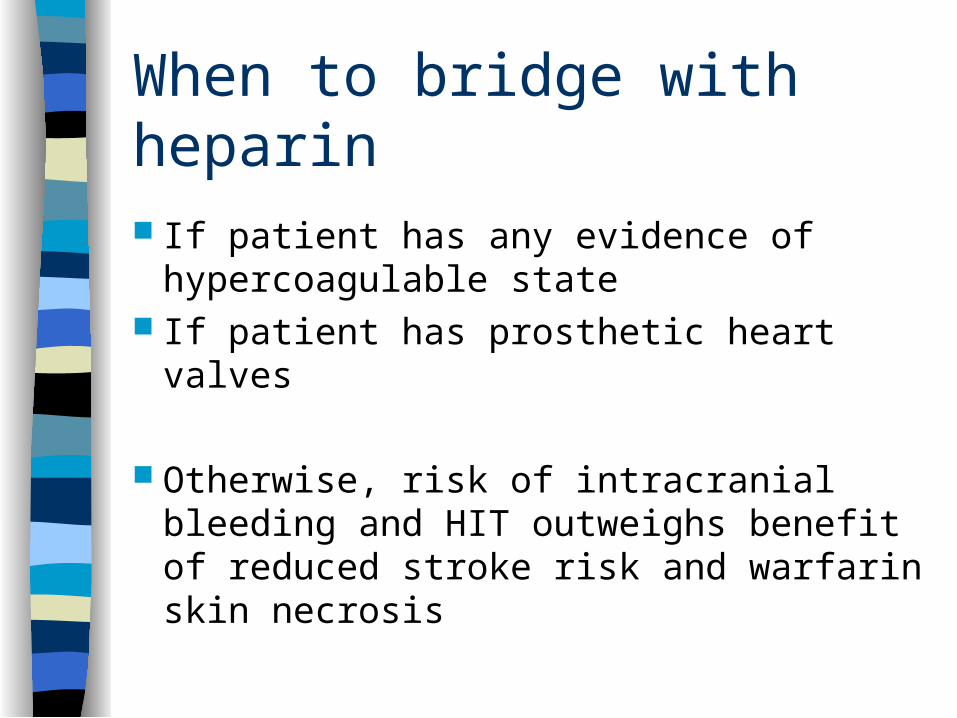
When to bridge with heparin
If patient has any evidence of hypercoagulable state
If patient has prosthetic heart valves
Otherwise, risk of intracranial bleeding and HIT outweighs benefit of reduced stroke risk and warfarin skin necrosis

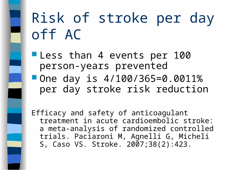
Risk of stroke per day off AC
Less than 4 events per 100 person-years prevented
One day is 4/100/365=0.0011% per day stroke risk reduction
Efficacy and safety of anticoagulant treatment in acute cardioembolic stroke: a meta-analysis of randomized controlled trials. Paciaroni M, Agnelli G, Micheli S, Caso VS. Stroke. 2007;38(2):423.
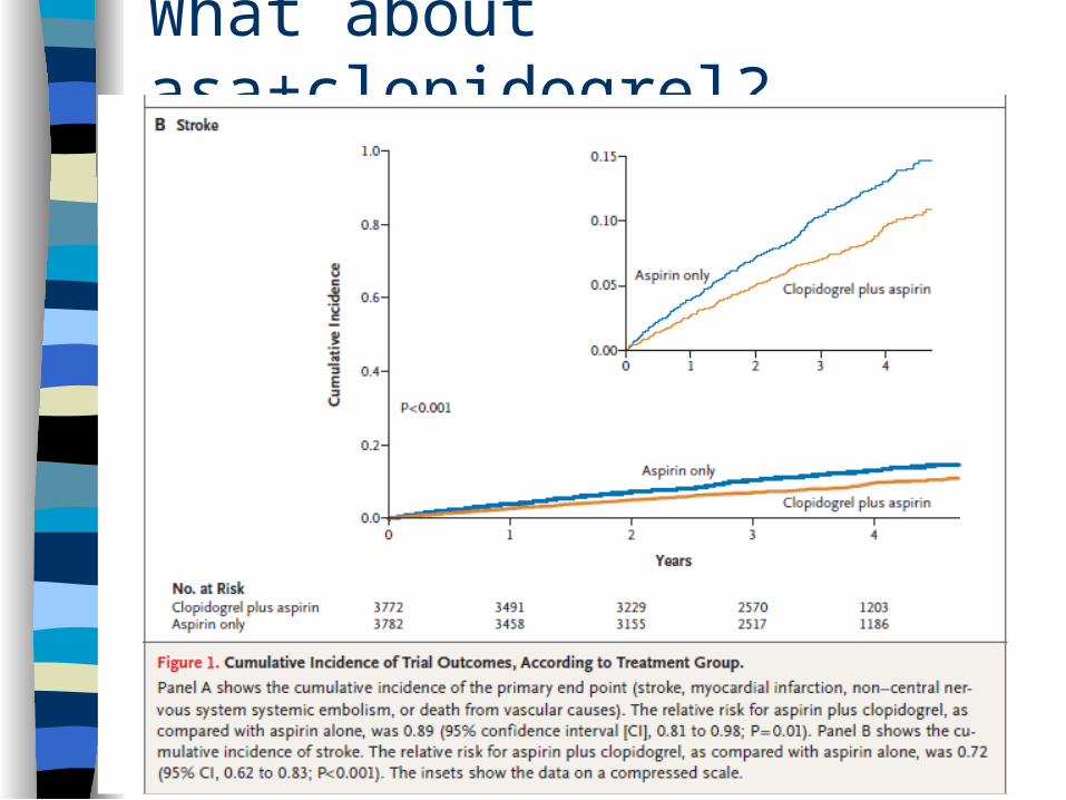
What about asa+clopidogrel?
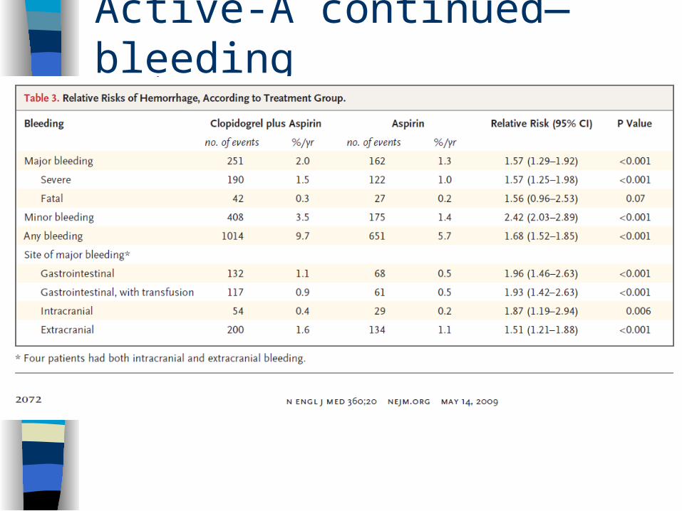
Active-A continued—bleeding
Take-home point
Warfarin>ASA+clopidogrel>ASA This applies to stroke prevention And to bleeding risk
Every AF patient who is off warfarin should be on aspirin unless there is a contraindication to ASA therapy
Clopidogrel + Warfarin = Bleed ASA+warfarin and ASA+clopidogrel
are relatively safe in most situations where both are indicated
Warfarin+clopidogrel has a relative risk of bleeding >3 times greater than warfarin alone
ASA+warfarin+clopidogrel has less rigorous data; evidence suggests >5 times greater than asa+clopidogrel alone
Dabigatran and Apixaban
Oral anticoagulant medications that do not require monitoring
Dabigatran approved for AC in AF/AFl in USA; NYS Medicaid and NBHN do not pay for it. Medicare part D does pay
Apixaban likely similar; not yet approved

You’re the night intern
78 W with HTN, DM, COPD, and history of paroxysmal AF
Nurse pages you to say that HR is now 172 beats per minute after albuterol
You ask, “what’s the blood pressure?” The nurse says she will check. You go
to the bedside.
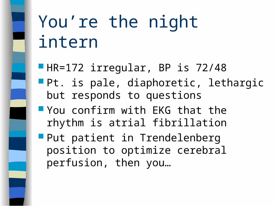
You’re the night intern
HR=172 irregular, BP is 72/48 Pt. is pale, diaphoretic, lethargic but
responds to questions You confirm with EKG that the rhythm is
atrial fibrillation Put patient in Trendelenberg position to
optimize cerebral perfusion, then you…
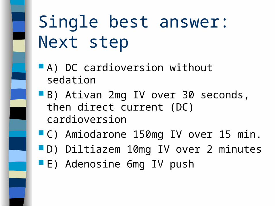
Single best answer: Next step
A) DC cardioversion without sedation B) Ativan 2mg IV over 30 seconds, then
direct current (DC) cardioversion C) Amiodarone 150mg IV over 15 min. D) Diltiazem 10mg IV over 2 minutes E) Adenosine 6mg IV push
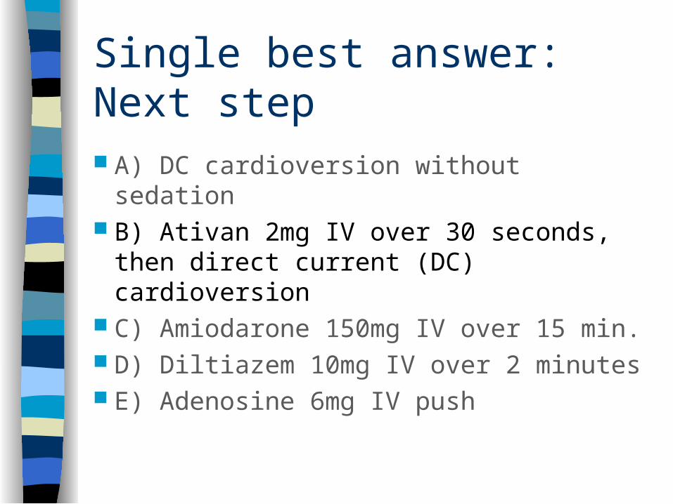
Single best answer: Next step
A) DC cardioversion without sedation B) Ativan 2mg IV over 30 seconds, then
direct current (DC) cardioversion C) Amiodarone 150mg IV over 15 min. D) Diltiazem 10mg IV over 2 minutes E) Adenosine 6mg IV push
Management of fib/flutter
Anticoagulation Cardioversion Rate control Rhythm control Ablation Cardiothoracic surgery
Indications for cardioversion
Hemodynamically unstable patient with any (non-sinus) tachycardia with RVR
Cardiovert first; page cardiology second When people are awake, sedate before
cardioversion (You learned this in ACLS)
Cardioversion when hemodynamically stable
Has never been shown to improve prognosis or reduce embolic risk, despite rigorous evaluation of this question in AFFIRM and RACE
Still, “every patient deserves a trial of sinus rhythm”
Cardioversion when hemodynamically stable
AFl is hard to rate control and easy to cardiovert
Heart failure (HF) may improve with restoration of atrial “kick” from cardioversion
Particularly important in severe diastolic HF, also beneficial in systolic HF
Cardioversion complication: Cardioembolism
Mostly in patients who are not anticogulated at time of cardioversion
Reduce risk by anticoagulating beforehand for 3-4 weeks
If not on A/C for ~4 weeks prior, do TEE
-If no thrombus on transesophageal echocardiogram, give heparin bolus, then cardiovert
Cardioversion complication: Cardioembolism
Continue anticoagulation for at least 4 weeks after cardioversion
If stroke risk is low enough, change from warfarin to ASA (+/- clopidogrel)
What if someone in paroxysmal AF self-cardioverts in front of you? Do they need 4 weeks of warfarin? If they self-cardiovert at home, will anyone know?
Cardioversion complications
ST-T changes, CK, troponin Myocardial stunning Transient hypotension Pulmonary edema Skin burns/Self-injury Ventricular fibrillation (not if SYNC on)
-Much more common in digitalis toxicity
DC Cardioversion, fine points
Usually should be done with one pad on front and one on patient’s back
Optimal current level to use in first shock is not known; lower in AFl
Biphasic is more successful than monophasic in terminating arrhythmias
Turn on the SYNC function

DC Cardioversion, fine points
If first attempt fails, try increasing the current to 200 joules
Then try changing the pad position Then try pretreatment with
antiarrhythmic drugs-might require long-term drug treatment Still, about 1/3 of DC cardioversion
efforts will fail
Chemical cardioversion
Usually with ibutilide Restricted to Cardiology use Lower cardioversion success rate than
electrical, but more comfortable (no sedation needed)
Caution in long QT Amiodarone IV is not cardioversion
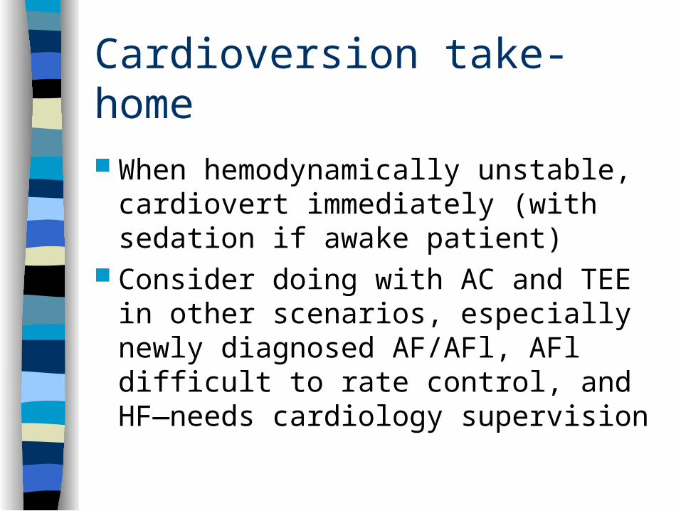
Cardioversion take-home
When hemodynamically unstable, cardiovert immediately (with sedation if awake patient)
Consider doing with AC and TEE in other scenarios, especially newly diagnosed AF/AFl, AFl difficult to rate control, and HF—needs cardiology supervision
Management of fib/flutter
Anticoagulation Cardioversion Rate control Rhythm control Ablation Cardiothoracic surgery
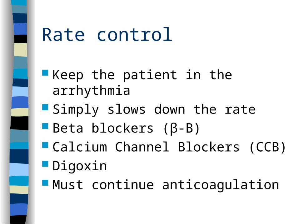
Rate control
Keep the patient in the arrhythmia Simply slows down the rate Beta blockers (β-B) Calcium Channel Blockers (CCB) Digoxin Must continue anticoagulation

Rhythm control
Goal is to keep patient in sinus rhythm First anticoagulate Then load rhythm control medication Then cardiovert (possibly with TEE) Monitor for antiarrhythmic side effects
Can you stop AC in rhythm control?
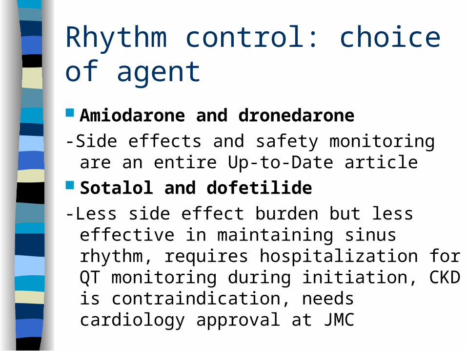
Rhythm control: choice of agent
Amiodarone and dronedarone
-Side effects and safety monitoring are an entire Up-to-Date article
Sotalol and dofetilide
-Less side effect burden but less effective in maintaining sinus rhythm, requires hospitalization for QT monitoring during initiation, CKD is contraindication, needs cardiology approval at JMC
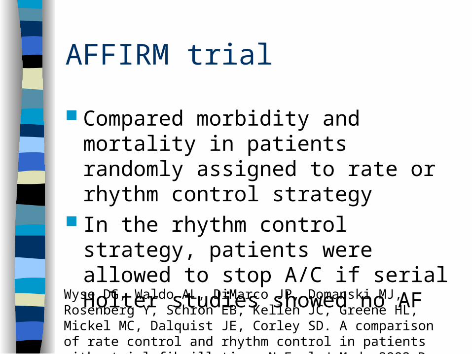
AFFIRM trial
Compared morbidity and mortality in patients randomly assigned to rate or rhythm control strategy
In the rhythm control strategy, patients were allowed to stop A/C if serial Holter studies showed no AF
Wyse DG, Waldo AL, DiMarco JP, Domanski MJ, Rosenberg Y, Schron EB, Kellen JC, Greene HL, Mickel MC, Dalquist JE, Corley SD. A comparison of rate control and rhythm control in patients with atrial fibrillation. N Engl J Med. 2002 Dec 5;347(23):1825-33.
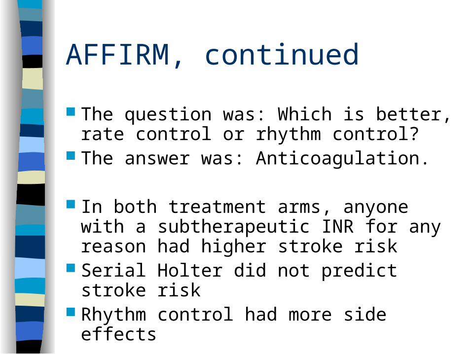
AFFIRM, continued
The question was: Which is better, rate control or rhythm control?
The answer was: Anticoagulation.
In both treatment arms, anyone with a subtherapeutic INR for any reason had higher stroke risk
Serial Holter did not predict stroke risk Rhythm control had more side effects
Management of fib/flutter
Anticoagulation Cardioversion Rate control Rhythm control Ablation Cardiothoracic surgery
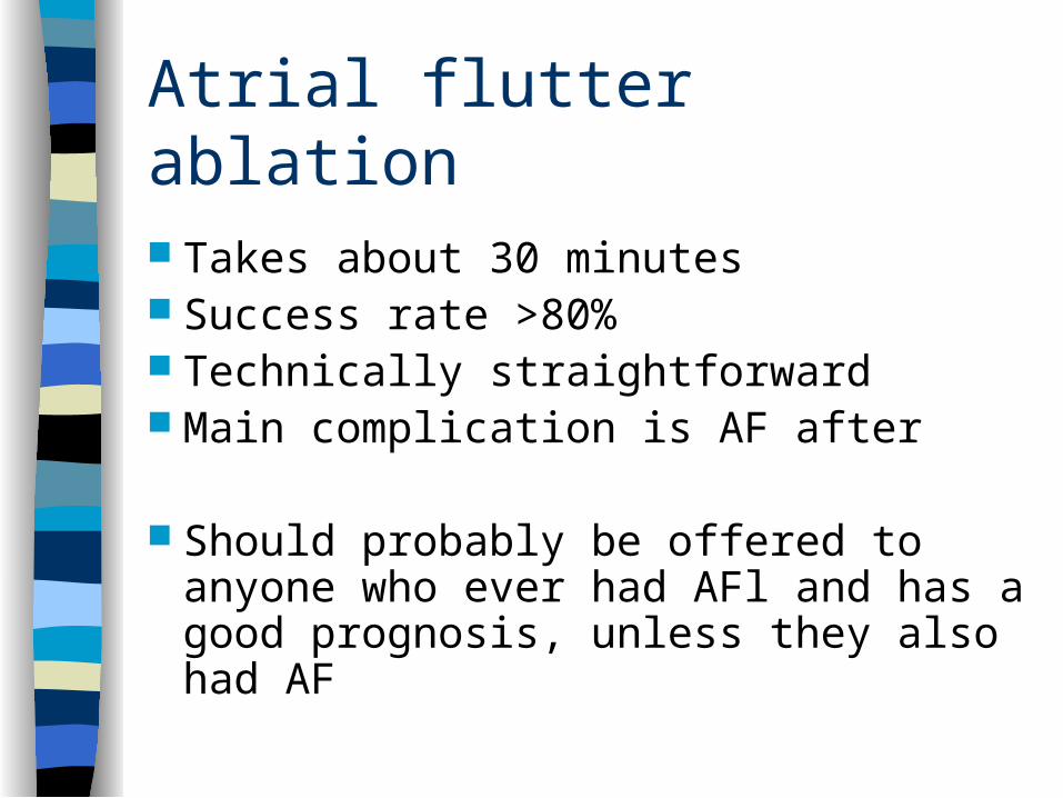
Atrial flutter ablation
Takes about 30 minutes Success rate >80% Technically straightforward Main complication is AF after
Should probably be offered to anyone who ever had AFl and has a good prognosis, unless they also had AF
Atrial flutter
Atrial fibrillation ablation
Also called pulmonary vein isolation Can last more than six hours Requires atrial septal puncture and
heparinization

AF diagram
Atrial fibrillation ablation
Main post-procedural complication is a special type of atrial tachycardia
No evidence that it’s safe to stop anticoagulation afterward
Often requires repeat ablation procedure to maintain sinus rhythm
Ablate and pace
Insert a biventricular pacemaker Ablate the AV node (on purpose) Very high procedural success rate But leaves the patient lifelong
pacemaker dependent Most useful in heart failure, particularly
tachycardia-induced cardiomyopathy Also in pacemaker-dependent patients
AF diagram
Management of fib/flutter
Anticoagulation Cardioversion Rate control Rhythm control Ablation Cardiothoracic surgery
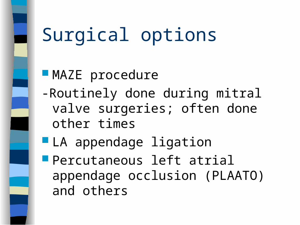
Surgical options
MAZE procedure
-Routinely done during mitral valve surgeries; often done other times
LA appendage ligation Percutaneous left atrial appendage
occlusion (PLAATO) and others
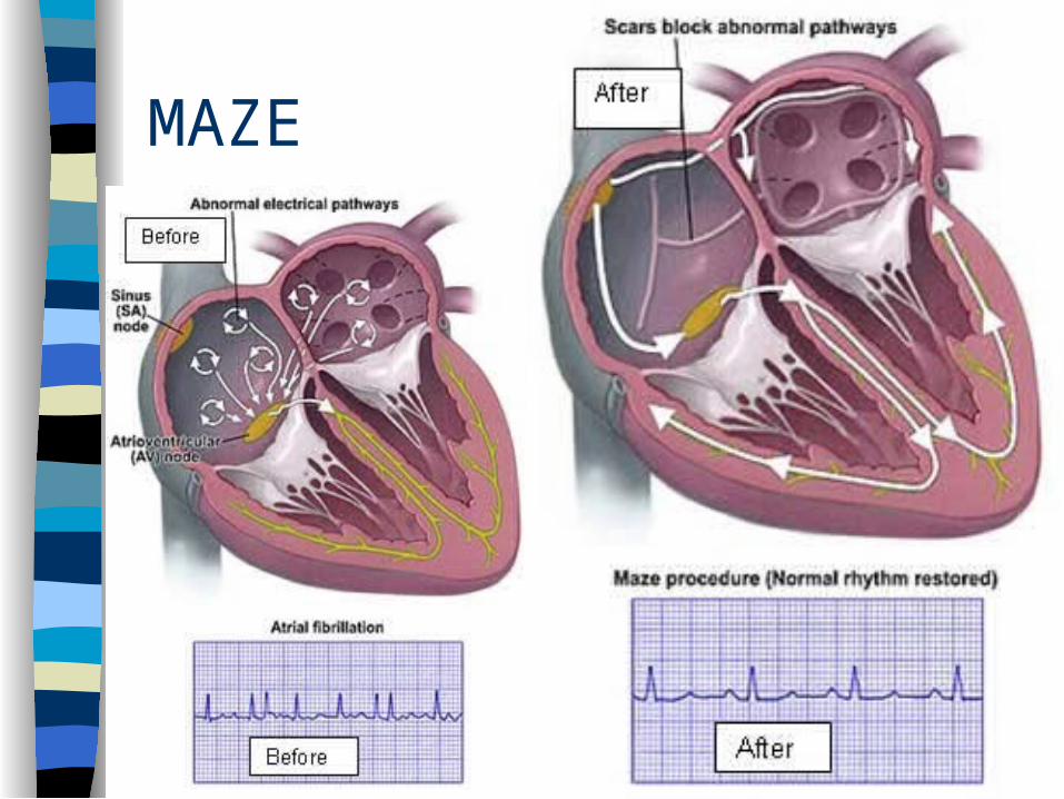
MAZE

PLAATO
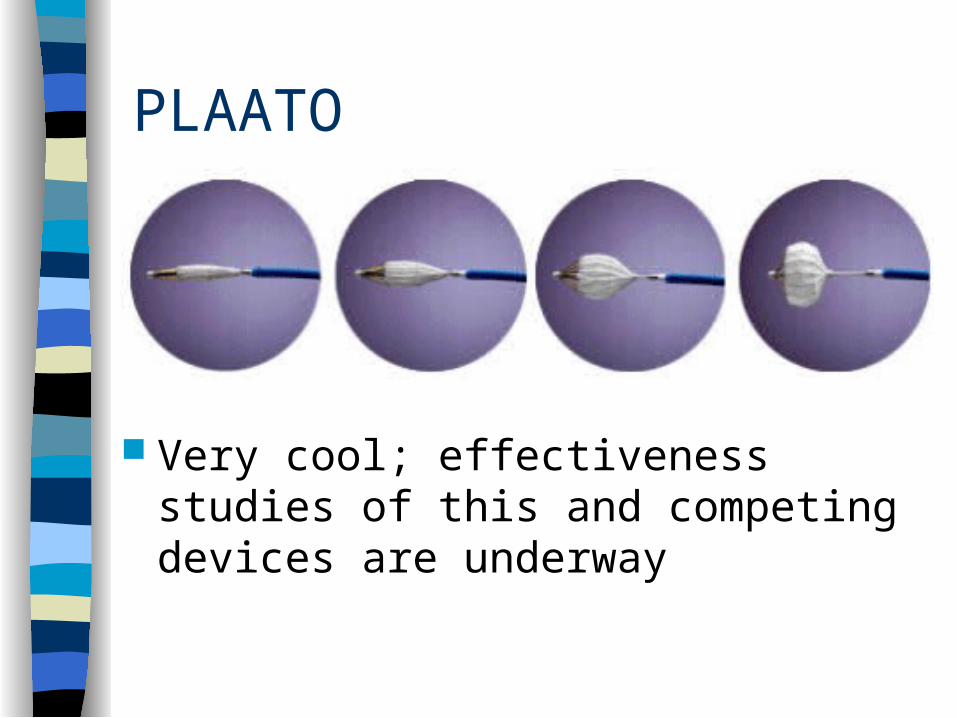
PLAATO
Very cool; effectiveness studies of this and competing devices are underway
Learning Goals
A brief discussion of supraventricular tachycardia (SVT)
Review of AF and AFl physiology and EKG differentiation
Management of atrial fibrillation (AF) Management of atrial flutter (AFl)
Conclusion
Anticoagulation, when indicated, is the most important treatment in most patients with AF/AFl; mostly warfarin/dabigatran
Stronger AC has less stroke but more bleed Cardiovert your hemodynamically unstable
patients right away Rate control for most patients Call cardiology for cardioversion, rhythm
control, ablation, or surgery if appropriate

Thank you
Insert humorous cartoon or scenic image on this slide


![Dysrhythmias (002) [Read-Only] - Aventri · Atrial AV node Ventricular Classification of Rhythm Abnormalities Supraventricular Atrial origin Atrial fibrillation Atrial flutter Atrial](https://img.dokumen.tips/doc/110x75/5f024baa7e708231d4038f22/dysrhythmias-002-read-only-aventri-atrial-av-node-ventricular-classification.jpg)