Embed Size (px)
Citation preview

ASSAY DEVELOPMENT IN DRUG
DISCOVERY
Indira Padmalayam, Ph.D.
Drug Discovery Division, Southern Research Institute, Birmingham, Alabama 35205
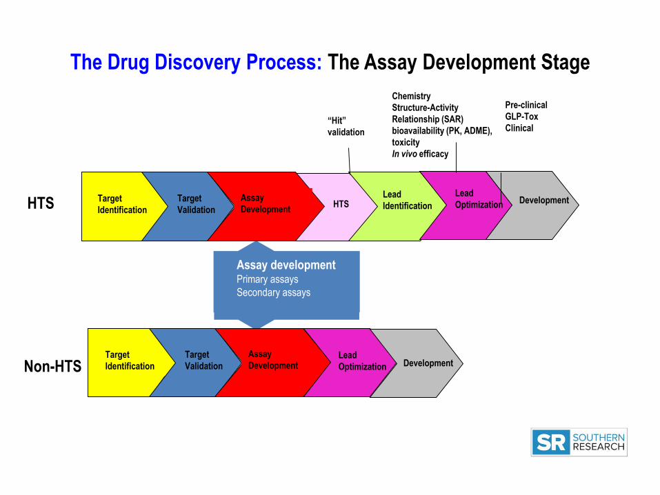
The Drug Discovery Process: The Assay Development Stage
Assay developmentPrimary assays
Secondary assays
Target
Identification
Target
Validation
Assay
DevelopmentHTS
Lead
Identification
Lead
Optimization Development
“Hit”
validation
Chemistry
Structure-Activity
Relationship (SAR)
bioavailability (PK, ADME),
toxicity
In vivo efficacy
Pre-clinical
GLP-Tox
Clinical
Target
Identification
Target
Validation
Assay
DevelopmentLead
Optimization Development
HTS
Non-HTS

Assay development: A critical part of the “hit” discovery process
“HITS”
A “hit” is a compound which
has the desired activity in a
compound screen and whose
activity is confirmed upon
retesting
Adapted frrom: Hughes, J.P. et.al. (2011), British Journal of
Pharmacology, 162 1239-1249
Secondary assaysPrimary assays

Assay
Development
Pre-
Roadblocks to faster assay development
Why is this a
bottleneck?Assay Development=months
HTS=weeks
Drug Discovery World, Summer 2010

Topics to be covered
• Paradigms in Drug Discovery
• Significance of Assay Development in Drug Discovery
• Assay types
• Assay formats
• Optimizing assays for HTS
Factors to be considered (will be covered in detail in HTS lecture)
• Assay development examples

Paradigms in Drug Discovery
Physiology-based Target-based
Target is unknown
Physiological/phenotypic read-outs
Cell-based assays
Known target
Read-outs are based on activity or expression of target
Biochemical or Cell-based assays
The two paradigms are not mutually exclusive: Drug Discovery programs can use two-pronged
approach.

Physiology-based drug discovery
Example : Viral CPE (Cytopathic Effect) assay
Green: uninfected cells
Red: host cells+virus
Blue: host cells+virus treated with
an antiviral compound, Ribavarin
Slide:courtesy, Jim Noah, SRI
1
8
15S1
S3
S5
S7
S9
S11
S13
S15
S17
S19
S21
S23
0
20000
40000
60000
80000
100000
120000
140000
Influenza CPE assay - Preliminary CV Plate
Ho
st c
ell
viab
ility

Target-based Drug Discovery
Science, 2000
Enzymes:
•Kinases
Receptor Tyrosine Kinase
Non-receptor tyrosine
kinase
Serine Threonine kinase
•Phosphatases
•Proteases:
Serine proteases
Zinc proteases
Receptors:
Ion channel receptors
GPCRs
Nuclear receptors

Assay Development for Drug Discovery
Target identified Assay Development HTS
and Validated
To ensure successful Assay Development:
-Ask the right “question” (the biological problem being addressed)
-Make sure that the “wording of the question” is correct (choose the correct assay, read-out and sequence of assays).
Key Questions:
1. Are we trying to inhibit or activate the target?
2. What function of the target are we trying to regulate?
3. What are the types of assays that are available to measure the function?
4. Primary assay vs Secondary assays
5. Which assay will work best in terms of translatability to HTS
6. Which are the post-HTS assays that will drive Structure Activity Relationship (SAR)

Key Considerations in Assay Development
The three “Rs”-Relevance
-Robustness-Reliability/Reproducibility
Cost
“The quality of an assay determines the quality of the data: compromising on assay development
can have substantial downstream consequences”
Practicality/Feasibility
Automation

Types of Assays
Assays in Drug Discovery
Biochemical assays Cell-based assays
Target-based Phenotype-based
Measure function of a purified target
Activity assays: Enzymes (e.g. kinases, proteases)
Binding assays: Receptors
(e.g. Nuclear receptors, Kinase receptors, ion
channels, GPCRs)
Identify compounds that modulate activity /binding of the target protein
Recombinant (engineered) proteins, proteins isolated from crude cell lysatesMonitor a surrogate read-out
Examples: Kinase/ATPase assays, protease assays, protein interaction assays
Measure function of the target in the context of the cell
-Transcriptional read-outs, second messenger levels,
cell viability (cell death/apoptosis), proliferation
Measure expression of the target
mRNA levels, protein expression and localization
Provide a functional read-out of compound activity (as a functional consequence of target engagement)
Examples: reporter assays, viability assays, GPCR and ion channel assays, qPCR

Biochemical versus Cell-based assays
Advantages:
-More physiological, amenable to systems approach
-Can simultaneously assay for compound properties
(membrane permeability, toxicity, off-target effects)
Disadvantages:
-Complex
-High rate of noise
-Exclusion of less soluble/permeable compounds
Advantages:
-Simple
-More consistency
-Direct measurement of target engagement
-Can measure compound characteristics such as Kd, Ki etc.
-Increased specificity of compounds
Disadvantages:
-May be non-physiological
-Not possible to determine compound properties such as membrane permeability, toxicity, off-target effects
Cell-based assaysBiochemical assays

Causes of Assay Variation
Biochemical Assays
• pH
• Temperature
• Ion concentration
• Reagent Solubility
• Reagent Stability
• Reagent Aggregation
• Order of reagent addition
• Instrumentation
Cell Based Assays
Same as for biochemical assays
Plus:
• Cell culture plastics
• Culture media
• Culture conditions
• Serum
• Cell cycle
• Passage number

Common Assay formats
• Fluorescence
• Luminescence

Fluorescence-based assays
Based on excitation of a fluorophore
Variety of assays using fluorescence
- Simple assays where protein of interest is conjugated to fluorophore, or where the protein of interest generates a fluorescent product
- Reporter assays
- Advantages: High sensitivity, ease of set-up and operation
- Disadvantages: Prone to false positives due to auto fluorescence of compound
Examples:
- FRET, TR-FRET
- Fluorescence polarization (FP)
1 to 10 nano secs

Fluorescence-based assays (contd.)
FRET: Florescence Resonance Energy Transfer
Half life: 1 to 10 nano secs
• Principle: Two fluorophores: Donor and Acceptor
-Based on transfer of energy between donor and acceptor
-Distance is critical
Far: No energy transfer no FRET
Close: Energy transfer from donor to acceptor FRET
• Use: Protein-protein, antigen-antibody, DNA-DNA, DNA-
protein
• Advantages:
-Homogenous assay format
-Reduced assay time and cost (HTS friendly)
• Disadvantage:
-Short half life of fluorophore results in high background

Fluorescence-based assays (contd.)
• “Improved”version of FRET
- Uses long lived fluorophores and time-
resolved detection to reduce backgound
-Rare earth elements (Lanthanides):
Samarium (Sm), Europium (Eu),Terbium
(Tb), and Dysprosium (Dy)
• Advantage:
-Low background; better signal to noise
• Disadvantage:
-Lanthanides have poor ability to absorb
light, so have to be complexed with
organic moieties that can harvest light
and transfer it to them.
TR-FRET: Time Resolved-Florescence Resonance Energy Transfer

Fluorescence-based assays (contd.)
Principle:
Small, unbound fluorophore: fast rotation, light is emitted in a plane different from excitation light.
Fluorophore bound to protein: Slow rotation, light is emitted in the same plane as excitation light.
Applications: Study molecular interactions e.g. protein-protein, receptor-ligand, DNA-protein, tyrosine Kinase assays.
Advantages: Highly sensitive (low picomolar range), homogenous assays, multiple measurements can be made on the same sample, because there is no change in samples during the assay.
Disadvantage: More optimization of assay may be needed to ensure saturation of all target binding sites with the fluorophore-labeled ligand (to ensure displacement by unlabeled ligand)
Fluorescence Polarization (FP) assay

Fluorescence-based assays (contd.)
Un-labeled compounds
ATP binding pocket
Cy3 labeled
compound
FP
read
FP assay
Screen

Luminescence-based assays
• Chemical reaction produces light
• Bioluminescence is the production and
emission of light by a living organism (e.g.
luciferase by firefly)
Luciferin + ATP Luciferyl adenylate+PPi
Luciferyl + O2
AdenylateOxyluciferin +AMP +Light
Luciferase
Example: Luciferase reporter assay:

Examples of biochemical assays

Assay for kinase activity
ADP Hunter assay
Kinase targetSubstrate Phosphorylated Substrate+ ADP
Fluorescent
product
Kinase targetSubstrate
Inhibitors
Phosphorylated Substrate+ ADPFluorescent
product
Reduced signal

Assay for protein-protein interaction
AlphaScreen technologyAmplified Luminescent Proximity Homogenous Assay
4 µsec
4 µsec
200 nm
BAAlpha Donor
Bead
Alpha
Acceptor
Bead
Emission at 520-
620 nmExcitation at
680 nm
1O2

Alphascreen Assay: Measuring Tau-Fyn interactions in Alzheimer’s disease
• Interactions between Tau and Fyn are implicated in Alzheimer’s disease
• Donor bead labeled with glutathione
– Binds to Fyn-GST
• Acceptor bead chelated to nickel
– Binds to a His-tag on Tau protein
Figure: Courtesy Erik Roberson, ADDA Tau-Fyn project

Examples of cell-based assays

Cell based assays
• Proliferation
Fluorescent dyes
• Viability
-Assays that measure ATP
content (Cell Titer Glo), viral CPE
(bioluminescence-based)
-Apoptosis assays
(bioluminescence/fluorescence)
• Migration
-Scratch assay
• Reporter Gene assays
-Transcriptional activity/expression
Cell Proliferation(non-specific dye)
Cell migration
“scratch assay”
1
8
15S1
S3
S5
S7
S9
S11
S13
S15
S17
S19
S21
S23
0
20000
40000
60000
80000
100000
120000
140000
Influenza CPE assay - Preliminary CV Plate
Cell viability(CPE assay)

Assays for G protein-coupled receptors (GPCRs)
• GPCRs are the targets for majority of best-selling drugs and approx 40% of prescription drugs (e.g. Zantac, Clarinex, Zyprexa)
• GPCRs are proteins with 7 transmembrane domains
• Assay will depend on the type of GPCR being targeted. 3 type of G proteins (Gs, Gi/G0 and Gq)
• Non-G protein mediated responses
(β-Arrestin recruitment)
• Assays are designed to measure second messengers such as cAMP, Ca2+
• Measurement of proximal or distal
responses
• HTS friendly assays

High Content Screening
High content screens:
• Facilitates validation of effects of
compounds at the cellular and subcellular
level (expression, localization,
morphological changes in cells)
• Macromolecules (e.g. proteins, RNA) are
labeled with fluorescent tags
• Technology uses automated digital
microscopy, flow cytometry and IT-
systems for analysis and storage of data
• Slower than HTS
• Popular as secondary screens in drug
discovery programs

10µM 1µM 0.1µM 0.01µM
MDA-MB-231 breast cancer cells were co-cultured with 3T3L1 fibroblasts and treated with Cytochalasin-D, an inhibitor of actin
polymerization. Migration of cells through the matrigel layer was measured at various heights of the Z stack
Cytochalasin-D concentration response on cell migration
Example 1: Cell migration assays: Oncology
0.001 0.01 0.1 1 10 1000
25
50
75
100
Concentration (uM)
% In
hib
itio
n
Cytochalasin-D 48hrsIC50 = 0.12uM
0
0.2
0.4
0.6
0.8
1
Cel
l In
vasi
on
Rat
io
High Content Screening in Drug Discovery

5 nMDMSO 20 nM 80 nM 0.32 µM 1.25 µM 5 µM 10 µM
0100200300400500600700800
Mean total neurite length per cell (µm)
0
50
100
150
200
Mean length of longest neurite per cell (µm)
20 µM
High Content Screening in Drug Discovery
Example 2: Neurite Outgrowth Assay: Alzheimer’s disease
Aβ concentration response on neurite outgrowth

Assay development: From the bench to HTS
From This To This
When you go
THE RULES CHANGE

Everything is done in Microtiter Plates96, 384, 1536, 3456
96-well100-200µl
384-well25-50µl
1536-well4-10 µl
3456-well1-2 µl

• What are you aiming for in an HTS assay:
• To have a reasonable chance, to believe the results of a single
determination, i.e. one well
• For that you need:
• Reproducibility from well to well
• Reproducibility from assay plate to assay plate
• Reproducibility from day to day
• How do you know if your assay is ready for HTS?
Assay development: From the bench to HTS (HTS 101)Assay development: From the bench to HTS

ASSAY QC PARAMETERS
• “Z” factor: Key measure of readiness of an assay for HTS
Z > 0.5
• Coefficient of variance (CV) : Measure of dispersion
• Signal to background (S/B) and Signal to noise (S/N): Measures of
signal strength
S/B = Mean of sample S/N = Mean of sample- Mean of control
Mean of control Standard Deviation of control
3SD of sample + 3SD of control
mean of sample – mean of controlZ = 1 –
% CV =SD X 100
Mean
Will be covered in HTS lecture next week

ASSAY DEVELOPMENT CASE STUDIES

Case 1: Identification of a small molecule inhibitor of mitotic spindle bipolarity
Identification of
Monastrol, inhibitor
of mitotic kinesin, Eg5
Mayer, TU eta al, Science 286, 971-974 (1999)
Primary assay: Phenotype-based Counter screen Secondary assay: To select mitosis inhibitors

CASE 2: Identifying compounds that inhibit TXNIP expression
Assay developmentPrimary and secondary assays
HTS primary screen
(single dose)
“Hits”
Hit validation and determination
of potency (IC50)(counter screens, concentration
response curves)
Secondary phenotypic assays
Hit validation and determination of potency1. Cytotox assay to eliminate compounds with toxicity
2. Concentration response in primary assay for potency (IC50)
3. Chemistry analysis to identify scaffolds of interest
Counter screen: To eliminate non-specific luciferase inhibitors
Primary HTS assay: Luciferase reporter assay with TXNIP
promoter
HTS primary screen: 10K pilot screen followed by 300K
full screen(single dose)
3000 “Hits”
Chemistry analysis
1258 “Hits”
651 “Hits”
157 “Hits”
Secondary assay: TXNIP qPCR
37 “Hits”
Test in other confirmatory assays at UAB
ADDA project: TXNIP as a target for diabetes
TXNIP = Thioredoxin-interacting protein
13 “Hits”

CASE 3: Identifying compounds that activate HO-1 expression
Hit validation and determination of potency1. Counter screen using a mutant form of the HO-1 promoter
2. Cytotox assay to eliminate compounds with toxicity
2. Concentration response in primary assay for potency (IC50)
3. Chemistry analysis to identify scaffolds of interest
Chemical clustering analysis
Primary HTS assay: Luciferase reporter assay with HO-1
promoter containing enhancer element
HTS primary screen: 10K pilot screen followed by ~155K
full screen(single dose)
2000 “Hits”
“Analoging”: selection and purchase of analogs of hits
747 “Hits”
Secondary assays: HO-1 qPCR and Western blot
80 Compounds
Test in other confirmatory assays (qPCR and In Cell Western)
28 “Hits” representing six chemical clusters
Assay developmentPrimary and secondary assays
HTS primary screen
(single dose)
“Hits”
Hit validation and determination
of potency (IC50)(counter screens, concentration
response curves)
Secondary phenotypic assays
ADDA project: HO-1 as a target for multiple conditions
(chronic kidney disease, transplant rejection)
HO-1 = Heme Oxygenase-1

NCGC Online Resources
The National Chemical Genomics Center provides a
comprehensive online manual for assay development and
validation with HTS in mind.
For more information go to:
http://assay.nih.gov/assay/index.php/Table_of_Contents
Manuscript helpful for HTS Assay Development:
Inglese, J. et.al. (2007), Nature Chemical Biology 3(8) 466-479

“The subject is not exhausted but we are.”
George Bernard Shaw
![ADME FOR THERAPEUTIC BIOLOGICS: WHAT CAN WE LEVERAGE … · 2020-01-13 · 2 ADME FOR THERAPEUTIC BIOLOGICS complex clinical trials [1, 6–8]. In 1990s, poor human PK and bioavailability](https://img.dokumen.tips/doc/110x75/5f0d390c7e708231d4394738/adme-for-therapeutic-biologics-what-can-we-leverage-2020-01-13-2-adme-for-therapeutic.jpg)