Embed Size (px)
Citation preview
Original Article
Arthroscopic Anterior Cruciate Ligament Reconstruction
Using Bone-patellar Tendon-bone Graft
Abstract:We have studied 30 patients with ACL ruptures. Among them 83%(25) were males & 17%(5) were females. Mean age
was 31 years. All of them underwent Arthroscopic ACL Reconstruction using BTB graft. All patients were regularly
followed up at 6 weeks, 3 months, 6 months & 1 year. The assessment was done on basis of LYSHOLM KNEE SCORE.
Average score at 6 weeks was 70.67(fair), at 3 months 85.33(fair to good), at 6 months 92.6(good to excellent) and at 1
year 96.13(good to excellent).
Keywords: ACL Reconstruction, Bone-patellar tendon-Bone (BTB) graft
IntroductionAnterior cruciate ligament (ACL) is an intra-articular, extrasynovial structure present in the central complex of the knee joint which along with other structures in and around knee joint controls, limits motion and maintains static and dynamic equilibrium of knee joint [12,3]. ACL is commonly injured in athletic activities and in road traffic accidents example: when a sudden loading or tension is placed on the ligament as when a running athlete plants a foot to suddenly decelerate or change direction [1,4]. ACL has a poor capacity of healing. The need for surgical correction of ACL injuries arises because, untreated complete injury to the ligament leads to progressive symptomatic instability leading to recurrent injury and damage to the menisci and articular cartilage thus resulting in early osteoarthritis [5,6,7]. Arthroscopic guided ACL Reconstruction (ACLR) has multiple advantages over open ACLR. Numerous authors have described successful reconstruction of ACL (ACLR) with use of autografts (e.g. Patellar tendon, hamstring tendons, distally based ilio tibial band (ITB), fascia late etc) and allografts (e.g. Achillis tendon, tibialis anterior, patellar tendon, hamstring tendons etc) [8,-12]. The Bone-Patellar tendon-Bone autograft the most commonly used autograft for reconstruction [13-17,24-25] and it is widely accepted as the gold standard technique with a high success rate [17-20,24-25].
Material and methodsIn this study, 30 cases of ACL tear, admitted under
Orthopaedic Department of D.Y.Patil Hospital, Kolhapur, treated with bone-patellar tendon-bone graft, will be studied, to know the prognosis. In all the cases, primarily routine investigation, urine routine & microscopic examination, chest X-ray, blood sugar level, bleeding time and clotting time will be carried out. Then examination of the affected knee on clinical basis carried out. And Final diagnosis is made by MRI. After the patient is clinically and radiologically (magnetic resonance imaging) diagnosed to have tear, and after meeting inclusion criteria, the patients were taken up for ACL reconstruction. All the patients were followed up at regular interval i.e. 6 weeks, 3 months, 6 months and 1 year (prospective study). In our study we have used autologous, ipsilateral bone-patellar tendon-bone graft in all the patients for ACL reconstruction. In all the patients the graft is fixed with titanium interference on femoral and tibial sides.SURGICAL PROCEDURE The anterior cruciate ligament was reconstructed with a single-incision, arthroscopic assisted techniques. Prophylactic antibiotic was given prior to the skin incision. The portals used for arthroscopy included the anteromedial portal and anterolateral portal. We used to do diagnostic arthroscopy prior to harvesting of the graft and any meniscal pathology will be addressed.The bone-patellar tendon-bone autograft was harvested via a longitudinal incision (usually 4-5 cm in length) over the patellar tendon. The graft was prepared into a bone-patellar tendon-bone construct with the leading suture on the patellar side [22-23].The notch was prepared using a curette and motorized shaver until the over the-top position and femoral ACL footprint were clearly demonstrated. The tibial stump was cleaned leaving a short amount of stump for reference and covering the graft. The tibial guide pin was inserted to the posterior half of the remnant using the Acufex-elbow-tipped tibial guide and tibial tunnel reamed according to the size of the graft. With the knee flexed at 90 degrees, a guide pin was passed through the
1Dept. of Orthopaedics, Dr D. Y. Patil Medical College and Hospital,
Kolhapur
Address of Correspondence
Dr PG Kulkarni.
Department of Orthopaedics, Dr D. Y. Patil Medical
College and Hospital, Kolhapur
Mail: [email protected]
Journal of Trauma & Orthopaedic Surgery 2016;11(1):21-25
Journal of Trauma & Orthopaedic Surgery | Jan-March 2016 | Volume 11 |Issue 1 | Page 21-25
Copyright © 2016 by The Maharashtra Orthopaedic Association |
21
1 1Utkal Dudhwala , P.G. Kulkarni

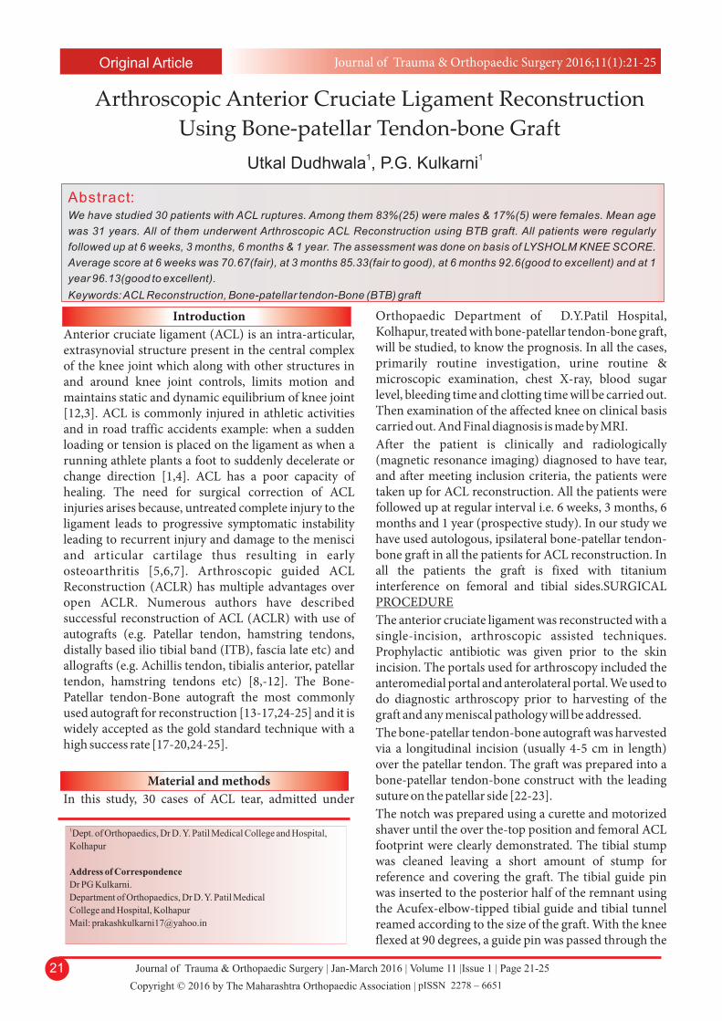
tibial tunnel to the femoral tunnel position. The femoral tunnel was reamed according to the size of the graft.Using a suture passing pin, the graft was passed through the tibial tunnel into the femoral tunnel and the suture passing pin passing out distal to the anterolateral skin of the thigh.The fixation method for patellar tendon graft was using cannulated interference screws usually 7 x 25mm, 8 x 25mm and rarely 9 × 25mm. The femoral site was fixed at 120 degrees knee flexion with the screw guide pin passed through the tibial tunnel. After femoral fixation, tension was applied to the tibial bone block suture and the knee passed through several cycles of flexion-extension to pretension the graft. The tibial site was fixed at 20 degrees knee flexion.The interference screw fixation method over the femoral tunnel is called as orifice fixation. All over the world this method is still suggested to be gold standard. The point is raised that the screw goes away from the graft but it is a positive point because the interference screw holds the graft at the orifice by its head and then goes away from the graft to avoid the damage to the graft and interface in between graft and bone is kept free.After the procedure, an intra-articular vacuum drain was placed into the joint. The drain was removed at 48 hours postoperatively. The knee was placed in a compressive dressing and long knee brace locked in full extension. Rehabilitation was a per protocol in Fig 4.Evaluation of results: All the patients were evaluated periodically at 6 weeks, 3 months, 6 months & 1 year.
The standard protocol of LYSHOLM KNEE SCORING SYSTEM is used for evaluation of the results of the surgery during follow up. At each follow up along with subjective evaluation, the following clinical examinations were also done.-Ligament laxity was assessed using Anterior Drawer Test, Lachman Test and pivot shift test. -Range of motion of the operated knee was noted and compared with the opposite knee. -Knee extension or straight leg raising (quadriceps power) was assessed.
Results We have used the Lysholm score for subjective evaluation of all our patients at each follow up. The following are the parameters and the maximum points given for each. Parameters (100 points). Limp (5 points), Support (5 points), Stair climbing (10 points), Squatting (5 points), Instability (25 points), Pain (25 points), Swelling (10 points), Locking sensation of knee (15 points). In our study Lysholm score was done at 6 weeks, 3 months, 6 months and 1 year. Average Lysholm score at 6 weeks was 70.67, at 3 months 85.33, at 6 months 92.6 and at 1 year 96.13.Other parameters were also used to evaluate the patients clinicallyi.Instability was assessed using anterior drawer test.ii.Range of motion of the knee was compared with the contra lateral side. iii.Quadriceps muscle strength was assessed by using MRC grading for muscle.
22Journal of Trauma & Orthopaedic Surgery | Jan-March 2016 | Volume 11 |Issue 1 | Page 21-25
www.moajournal.com Dudhwala and Kulkarni
Figure 1:Position and Making portals of Arthroscopy
Figure 2: Arthroscopic view of complete ACL tear and harvested Graft
Figure 3: After harvesting graft - Inserting the Tibial Screw- Arthroscopic view
23Journal of Trauma & Orthopaedic Surgery | Jan-March 2016 | Volume 11 |Issue 1 | Page 21-25
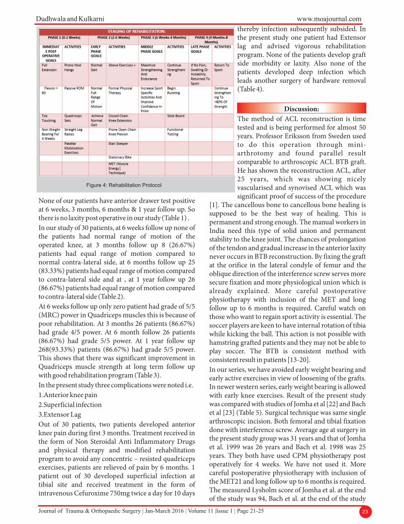
None of our patients have anterior drawer test positive at 6 weeks, 3 months, 6 months & 1 year follow up. So there is no laxity post operative in our study (Table 1) .In our study of 30 patients, at 6 weeks follow up none of the patients had normal range of motion of the operated knee, at 3 months follow up 8 (26.67%) patients had equal range of motion compared to normal contra-lateral side, at 6 months follow up 25 (83.33%) patients had equal range of motion compared to contra-lateral side and at , at 1 year follow up 26 (86.67%) patients had equal range of motion compared to contra-lateral side (Table 2).At 6 weeks follow up only zero patient had grade of 5/5 (MRC) power in Quadriceps muscles this is because of poor rehabilitation. At 3 months 26 patients (86.67%) had grade 4/5 power. At 6 month follow 26 patients (86.67%) had grade 5/5 power. At 1 year follow up 268(93.33%) patients (86.67%) had grade 5/5 power. This shows that there was significant improvement in Quadriceps muscle strength at long term follow up with good rehabilitation program (Table 3). In the present study three complications were noted i.e.1.Anterior knee pain 2.Superficial infection 3.Extensor Lag Out of 30 patients, two patients developed anterior knee pain during first 3 months. Treatment received in the form of Non Steroidal Anti Inflammatory Drugs and physical therapy and modified rehabilitation program to avoid any concentric – resisted quadriceps exercises, patients are relieved of pain by 6 months. 1 patient out of 30 developed superficial infection at tibial site and received treatment in the form of intravenous Cefuroxime 750mg twice a day for 10 days
thereby infection subsequently subsided. In the present study one patient had Extensor lag and advised vigorous rehabilitation program. None of the patients develop graft side morbidity or laxity. Also none of the patients developed deep infection which leads another surgery of hardware removal (Table 4).
Discussion:The method of ACL reconstruction is time tested and is being performed for almost 50 years. Professor Eriksson from Sweden used to do this operation through mini-arthrotomy and found parallel result comparable to arthroscopic ACL BTB graft. He has shown the reconstruction ACL, after 25 years, which was showing nicely vascularised and synovised ACL which was significant proof of success of the procedure
[1]. The cancellous bone to cancellous bone healing is supposed to be the best way of healing. This is permanent and strong enough. The manual workers in India need this type of solid union and permanent stability to the knee joint. The chances of prolongation of the tendon and gradual increase in the anterior laxity never occurs in BTB reconstruction. By fixing the graft at the orifice in the lateral condyle of femur and the oblique direction of the interference screw serves more secure fixation and more physiological union which is already explained. More careful postoperative physiotherapy with inclusion of the MET and long follow up to 6 months is required. Careful watch on those who want to regain sport activity is essential. The soccer players are keen to have internal rotation of tibia while kicking the ball. This action is not possible with hamstring grafted patients and they may not be able to play soccer. The BTB is consistent method with consistent result in patients [13-20].In our series, we have avoided early weight bearing and early active exercises in view of loosening of the grafts. In newer western series, early weight bearing is allowed with early knee exercises. Result of the present study was compared with studies of Jomha et al [22] and Bach et al [23] (Table 5). Surgical technique was same single arthroscopic incision. Both femoral and tibial fixation done with interference screw. Average age at surgery in the present study group was 31 years and that of Jomha et al. 1999 was 26 years and Bach et al. 1998 was 25 years. They both have used CPM physiotherapy post operatively for 4 weeks. We have not used it. More careful postoperative physiotherapy with inclusion of the MET21 and long follow up to 6 months is required. The measured Lysholm score of Jomha et al. at the end of the study was 94, Bach et al. at the end of the study
www.moajournal.com Dudhwala and Kulkarni
Figure 4: Rehabilitation Protocol
24Journal of Trauma & Orthopaedic Surgery | Jan-March 2016 | Volume 11 |Issue 1 | Page 21-25
www.moajournal.com Dudhwala and Kulkarni
was 90 and our study average Lyshom score at the end of the study is 96.In our series, early passive assisted knee flexion was recommended to get flexion up to 90 degrees before the patient is discharged and CPM is not used in any of the patients. The incidence of complications like knee pain and weakness in extensor mechanism was very rare in present series. The extension lag was due to insufficient postoperative physiotherapy and not because of middle third tendon loss. In present series only one patient had extension lag and that was also corrected after careful physiotherapy.There should be no objection in going for di f ferent procedures to perform ACL reconstruction, those newer methods can teach us many different things but ultimately the procedure which is more time tested remains stable. The enthusiasm of newer surgeons to blame or praise certain procedures becomes less signif icant when excel lent result with fundamental procedures are evident. It is not the surgeon who decides the stability of the procedure but it is the stability of the knee which decides the best and good. Ultimately it is the patient's satisfaction which overpowers the complexity of the research.
Conclusion
Autologous ipsilateral bone patellar tendon bone graft is a gold standard graft choice in arthroscopic ACL reconstruction because it is a permanent and a stable procedure for the stability of the knee.
1. Robert H Miller : Knee injuries : in Campbell vol 2, 9th edition.
2. Kennedy J.C. Weinberg H, W. WilsionA.S: The anatomy and function of ACL. JBJS, 56-A, 223-235, 1974.
3. Schultz R.A, Miller D.C., Kerr C.S. et al : Mechanoreceptors, in human cruciate ligaments, JBJS, 66-A, 1072-1076,
4. Abbott LC, Saunders, JB, Bost FC, Anderson, CE: Injuries to the ligaments of the
knee joint, J Bone Joint Surg 26: 503,194
5. Arnold JA, Coker TP, Heaton LM, et al.Natural history of anterior cruciate tears, Am J Sports Med 7: 305, 1979.
6. Noyes FR, Mooar PA, Matthews DS Butler DL: The symptomatic anterior cruciate –deficient knee. I. The long term functional disability in athletically active individual, J Bone Joint Surg 65-A : 154, 1983
References
25Journal of Trauma & Orthopaedic Surgery | Jan-March 2016 | Volume 11 |Issue 1 | Page 21-25
www.moajournal.com Dudhwala and Kulkarni 7. McGintyJB: Arthroscopic surgery in sports injuries, OrthopClin North Am 11: 787,
1980.
8. Campbell WC: Reconstruction of the ligaments of the Knee , Am J Surg 43:473, 1939.
9. Jones K.G: Reconstruction of the anterior cruciate ligament using the central one –third of the patellar ligament, J Bone, joint Surg 52-A: 1302, 1970.
10. Insall JN, Joseph DM, Aglietti P, Campbell RD Jr. Bone –block iliotibial band transfer for anterior cruciate insufficiency, JBone Joint Surg 63-A : 560, 1981.
11. Clancy WG, Nelson DA, Reider B. Narechania RG: Anterior cruciate ligament reconstruction using one –third of the patellar ligament, augmented by extra – articular tendon transfer, J Bone joint Surg 64-A, 352, 1982.
12. Puddu G: method for reconstruction of anterior cruciate ligament using the semitendinous tendon, Am J Sports Med 8: 402, 1980.
13. Shaieb MD, Kan DM, Chang SK, Marumoto JM and Richardson AB. A prospective randomized comparison of patellar versus semitendinosus and gracillis tendon autografts for anterior cruciate ligament reconstruction. Am J Sports Med 2002; 30: 214-20.
14. Barret GR, Noojin FK, Hartzog CW, Nash SR. Reconstruction of the anterior cruciate ligament in females. A comparison of hamstring versus patellar tendon autograft. Arthroscopy 2002; 18: 46-54.
15. Ejerhed L, Kartus J, Sernert N, Kohler K, Karlsson J. Patellar tendon or semitendinosus tendon autografts for anterior cruciate ligament reconstruction: A prospective randomized study with a two-year followup. Am J Sports Med 2003; 31: 19-25
16. Jansson KA, Linko E, Sandelin J, Harilainen A. A prospective randomized study of patellar versus hamstring tendon autografts for anterior cruciate ligament reconstruction. Am J Sports Med. 2003; 31: 12-8.
17. Pinczewski LA, Deehan DJ, Salmon LJ, Russell VJ, and Clingeleffer A. A five-year comparison of patellar tendon versus four-strand hamstring tendon autograft for arthroscopic reconstruction of the anterior cruciate ligament. Am J Sports Med 2002; 30: 523-36.
18. Beynnon BD, Johnson RJ, Fleming BC, Kannus P, Kaplan M, Samani J, and Renstrom P. Anterior cruciate ligament replacement: comparison of bone-patellar tendon-bone grafts with two strand hamstring grafts. J Bone Joint Surg (Am) 2002; 84: 1503-13.
19. Aglietti P, Buzzi R, Zaccherotti G, DeBiase P. Patellar tendon versus doubled semitendinosus and gracillis tendons for anterior cruciate ligament reconstruction. Am J Sports Med 1994; 22: 211-8.
20. Marder RA, Raskind JR, Carroll M. Prospective evaluation of arthroscopically assisted anterior cruciate ligament reconstruction. Patellar tendon versus semitendinosis and gracillis tendons. Am J Sports Med 1991; 19: 478-84.
21. Leon Chaitow. Muscle Energy Technique: Advance soft tissue techniques: Third Edion.
22. Jomha NM, Pinczewski LA, Clingeleffer A, Otto A. Arthroscopic reconstruction of anterior cruciate ligament with patellar-tendon autograft and interference screw fixation. The results at seven years. J Bone Joint Surg (Br) 1999; 81: 775-9.
23. Bach BR, Tradonsky S, Bojchuk J, Levy ME, Bush-Joseph CA, Khan NH. Arthroscopically assisted ACL reconstruction using patellar tendon autograft. Fiveto nine-year follow-up evaluation. Am J Sports Med 1998; 26: 20-9.
24. Eriksson E; Reconstruction of the anterior cruciate ligament, OrthopClin North Am 7:167, 1976.
25. PG Kulkarni. Arthroscopic evaluation and analysis of 200 patients with knee problems. MJDYPU; September 2008 ; vol 3;45-49.
How to Cite this ArticleDudhwala U, Kulkarni PG. Arthroscopic Anterior Cruciate Ligament Reconstruction Using Bone-Patellar Tendon-Bone Graft. Journal of Trauma and Orthopaedic Surgery Jan-March 2016;11(1):21-25.
Conflict of Interest: NILSource of Support: NIL


