Embed Size (px)
Citation preview
Arkansas
4-H Veterinary Science


Urinalysis
1
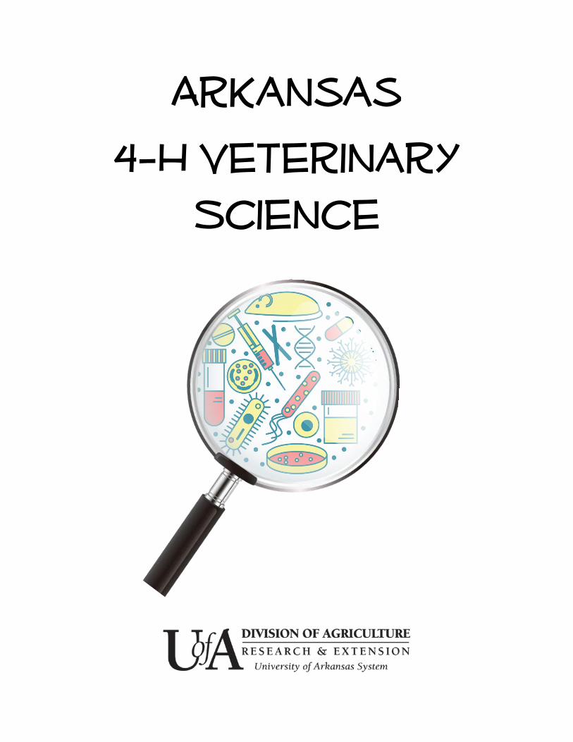
Why Urine?
Urine is the end product of a filtering process thatremoves waste from the body
The color of urine can give you information abouthydration level as well as possible underlying disease
A urinalysis should be performed at least yearly forhealthy pets, and more often for older animals andthose with existing or chronic health issues
Important elements of a urinalysis include a visualinspection of the urine sample, a dipstick test, andmicroscopic evaluation of urine sediment
2
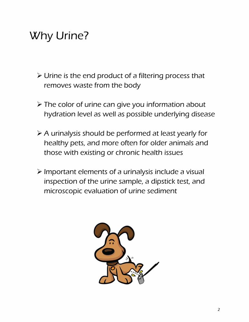
The Urinary System
The urinary tract consists of the kidneys, the ureters, the bladder, the urethra, and finally, the urethral opening at either the end of the penis or just within the vagina
Kidneys filter out waste products from the blood
Ureters connect the kidneys to the bladder
The urethra is a tube that is controlled by a sphincter muscle that empties the bladder to the outside world
3
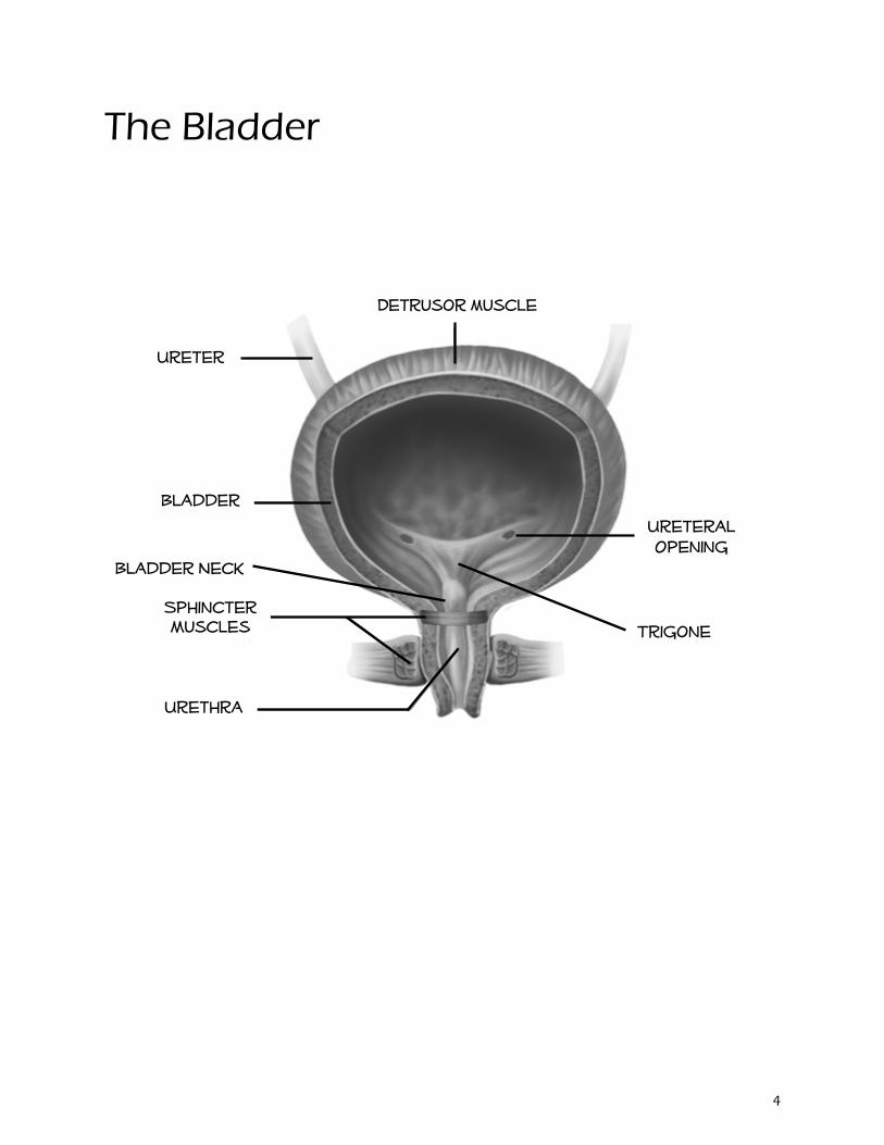
The Bladder
Trigone
Ureteral Opening
Detrusor muscle
Ureter
Bladder
Sphincter Muscles
Urethra
Bladder Neck
4
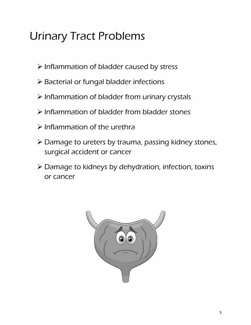
Urinary Tract Problems
Inflammation of bladder caused by stress
Bacterial or fungal bladder infections
Inflammation of bladder from urinary crystals
Inflammation of bladder from bladder stones
Inflammation of the urethra
Damage to ureters by trauma, passing kidney stones,surgical accident or cancer
Damage to kidneys by dehydration, infection, toxinsor cancer
5
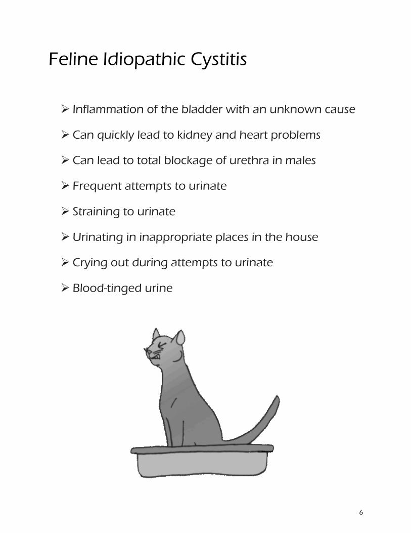
Feline Idiopathic Cystitis
Inflammation of the bladder with an unknown cause
Can quickly lead to kidney and heart problems
Can lead to total blockage of urethra in males
Frequent attempts to urinate
Straining to urinate
Urinating in inappropriate places in the house
Crying out during attempts to urinate
Blood-tinged urine
6
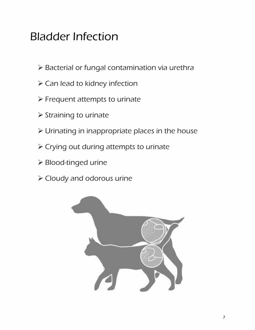
Bladder Infection
Bacterial or fungal contamination via urethra
Can lead to kidney infection
Frequent attempts to urinate
Straining to urinate
Urinating in inappropriate places in the house
Crying out during attempts to urinate
Blood-tinged urine
Cloudy and odorous urine
7
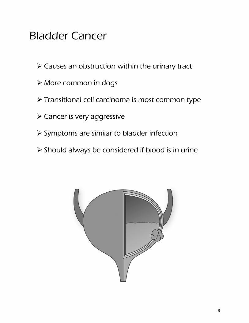
Bladder Cancer
Causes an obstruction within the urinary tract
More common in dogs
Transitional cell carcinoma is most common type
Cancer is very aggressive
Symptoms are similar to bladder infection
Should always be considered if blood is in urine
8
Visual Characteristics of Urine
Color
Clear to yellow is normal
Dark yellow to brownish yellow indicates dehydration
Brown to dark brown indicates muscle damage
Pink, Orange or Dark Red indicates blood
Clarity
Clear is normal
Cloudy may indicate infection or inflammation
Precipitates may indicate neoplasia (cancer)
Urine stream
Urination should occur in a steady stream
A slow stream indicates a problem
Leakage (incontinence) indicates a problem
Posturing to urinate without urine is called anuria
9
The Dipstick Test
Urine pH is affected by many variables, including time since the last meal, diet, a number of medications, lung and kidney function, and renal and systemic diseases.
Blood in urine can occur with disease anywhere in the urogenital tract.
Leukocytes (white blood cells) in urine indicates active inflammation in the urogenital tract.
Glucose in urine means that either the glucose in blood is elevated or there is a kidney disease that prevents full reabsorption of glucose.
Bilirubin in urine occurs with hemolysis (break down of red blood cells) or liver disease.
Protein in urine is due to pre-glomerular, glomerular, or postglomerular disease. The glomerulus is a cluster of capillaries around the end of a kidney tubule, where waste products are filtered from the blood.
Ketones in urine are formed when the body is unable to get sufficient energy from glucose and must metabolize large quantities of fatty acids instead.
Specific gravity is an indirect measure of kidney function.
10
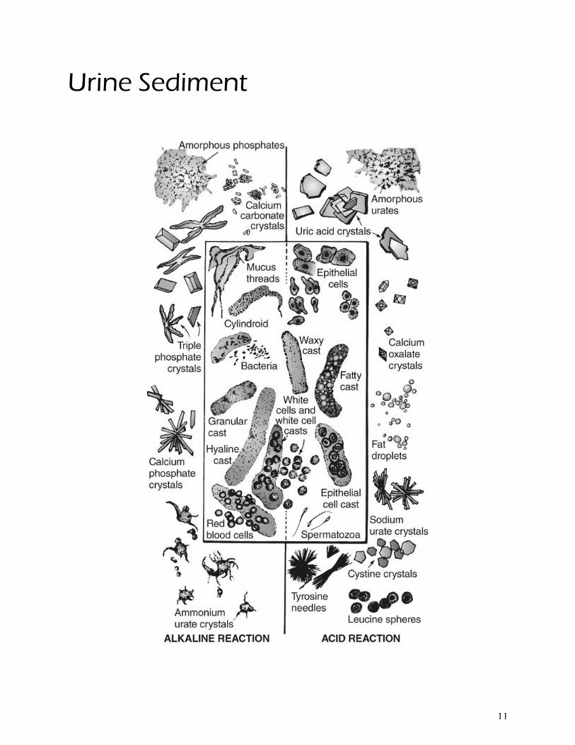
Urine Sediment
11

12

Color wheel of poop
13
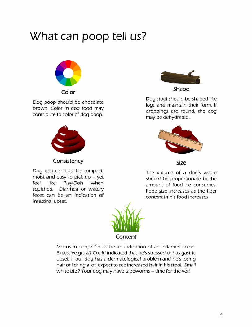
What can poop tell us?
Color
Dog poop should be chocolate brown. Color in dog food may contribute to color of dog poop.
Shape
Dog stool should be shaped like logs and maintain their form. If droppings are round, the dog may be dehydrated.
Consistency
Dog poop should be compact, moist and easy to pick up – yet feel like Play-Doh when squished. Diarrhea or watery feces can be an indication of intestinal upset.
Size
The volume of a dog’s waste should be proportionate to the amount of food he consumes. Poop size increases as the fiber content in his food increases.
Content
Mucus in poop? Could be an indication of an inflamed colon. Excessive grass? Could indicated that he’s stressed or has gastric upset. If our dog has a dermatological problem and he’s losing hair or licking a lot, expect to see increased hair in his stool. Small white bits? Your dog may have tapeworms – time for the vet!
14

What does the color of poop mean?
15
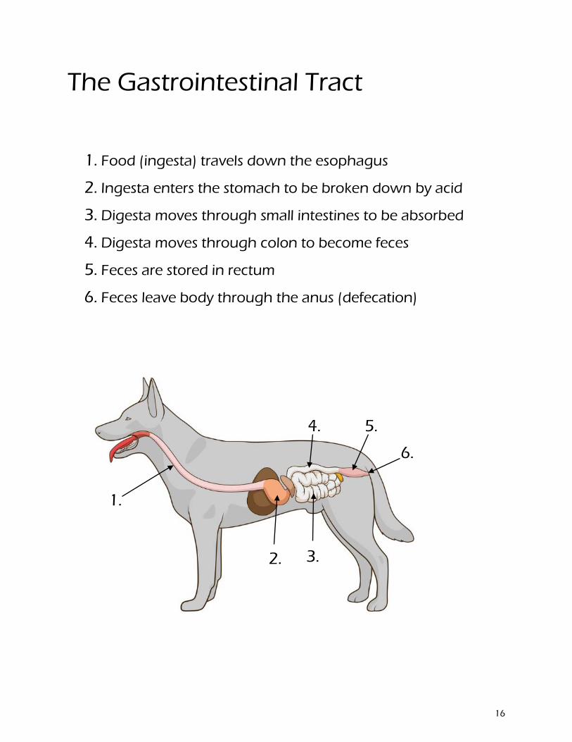
The Gastrointestinal Tract
1. Food (ingesta) travels down the esophagus
2. Ingesta enters the stomach to be broken down by acid
3. Digesta moves through small intestines to be absorbed
4. Digesta moves through colon to become feces
5. Feces are stored in rectum
6. Feces leave body through the anus (defecation)
1.
2. 3.
4. 5.
6.
16

The Anal Glands
Anal sacs expel pheromone during defecation
Also called scent glands
Secretion is used for animal identification (territory)
Inflammation can block ducts
Sacs can rupture if not expressed regularly!
17

Types of Bacteria in Feces
Gram-Positive bacteria in feces
Clostridia s Enterococcus
Gram-Negative bacteria in feces
Campylobacter Escheria coli Salmonella Proteus
18
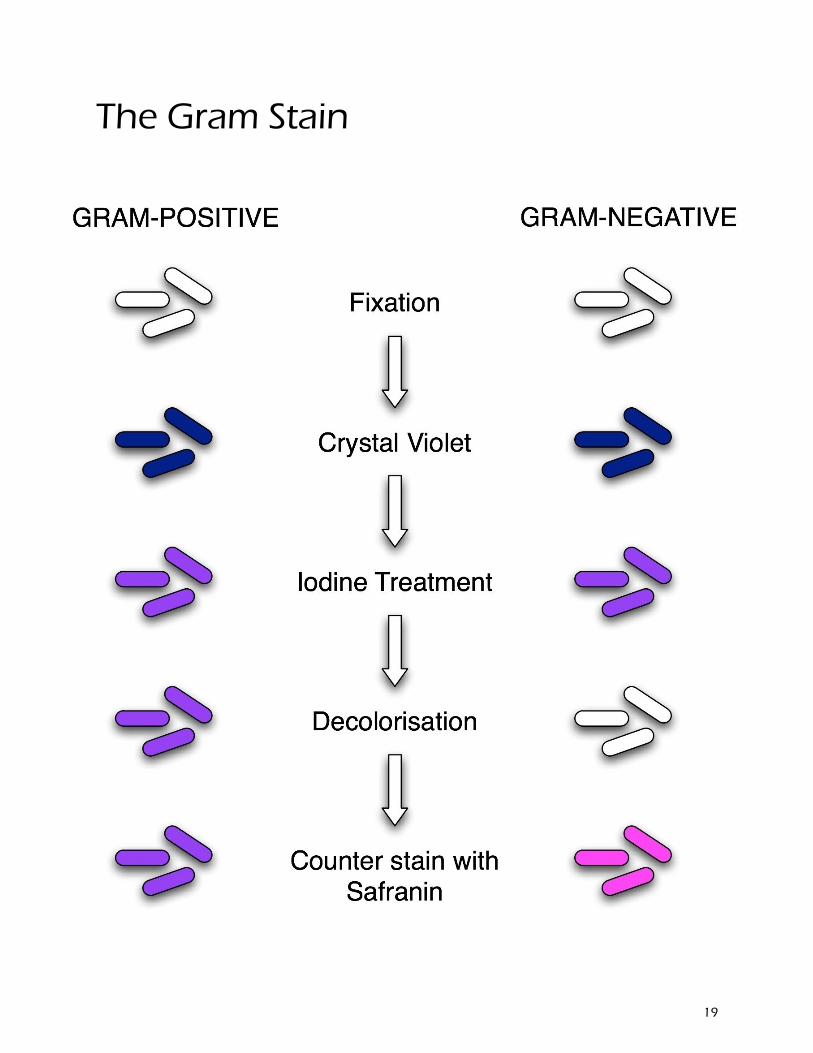
The Gram Stain
19

20
Stress and the Immune system
21
The Big Picture
Stress is a biological and psychological response to athreatening event
Also known as the flight-or-flight response
Stress hormones are released due to a communicationbetween the brain and adrenal glands
Adrenaline increases heart rate and cortisol releasessugar stores – both hormones act to prepare the bodyfor fight-or-flight
The immune system is a collection of billions of cellsthat travel through the bloodstream
The main types of immune cells are white blood cells
Stress hormones can suppress the immune system bylowering the number of white blood cells
22
Stress and the Digestive System
Stress responses have an effect on digestion
During stress digestion is inhibited
After stress digestive activity increases
Increased digestive activity can cause diarrhea orulcers
Release of stress hormones may also cause ulcers byincreasing stomach acid production
Cats are more susceptible to ulcers
Most mammals are susceptible to stress-relateddiarrhea
23
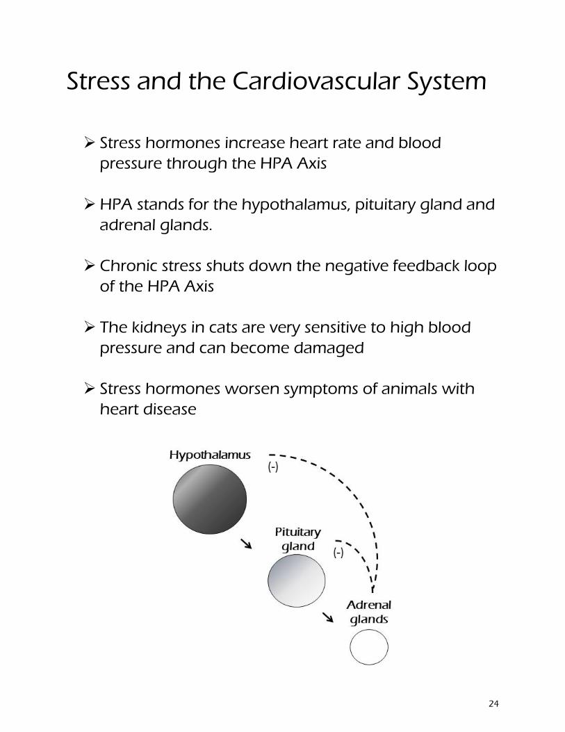
Stress and the Cardiovascular System
Stress hormones increase heart rate and bloodpressure through the HPA Axis
HPA stands for the hypothalamus, pituitary gland andadrenal glands.
Chronic stress shuts down the negative feedback loopof the HPA Axis
The kidneys in cats are very sensitive to high bloodpressure and can become damaged
Stress hormones worsen symptoms of animals withheart disease
24
25
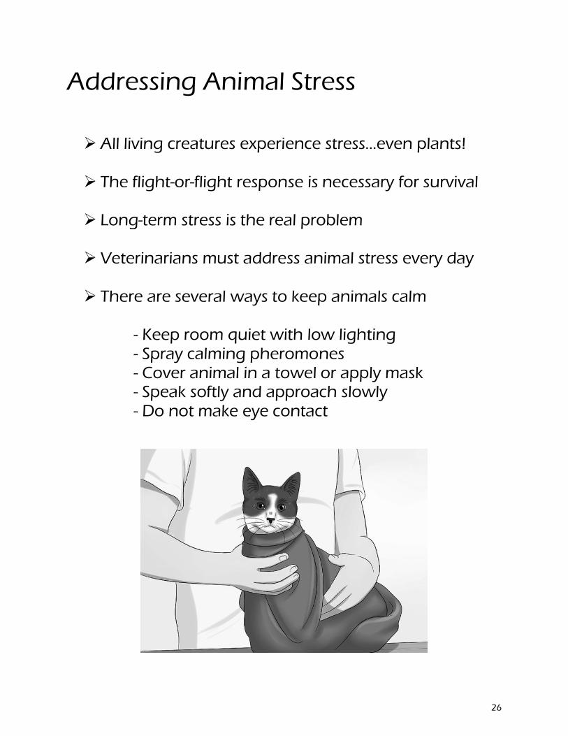
Addressing Animal Stress
All living creatures experience stress…even plants!
The flight-or-flight response is necessary for survival
Long-term stress is the real problem
Veterinarians must address animal stress every day
There are several ways to keep animals calm
- Keep room quiet with low lighting - Spray calming pheromones - Cover animal in a towel or apply mask - Speak softly and approach slowly - Do not make eye contact
26
The Immune System
A story by MEGAN LLEWELLYN
Augusta university
27
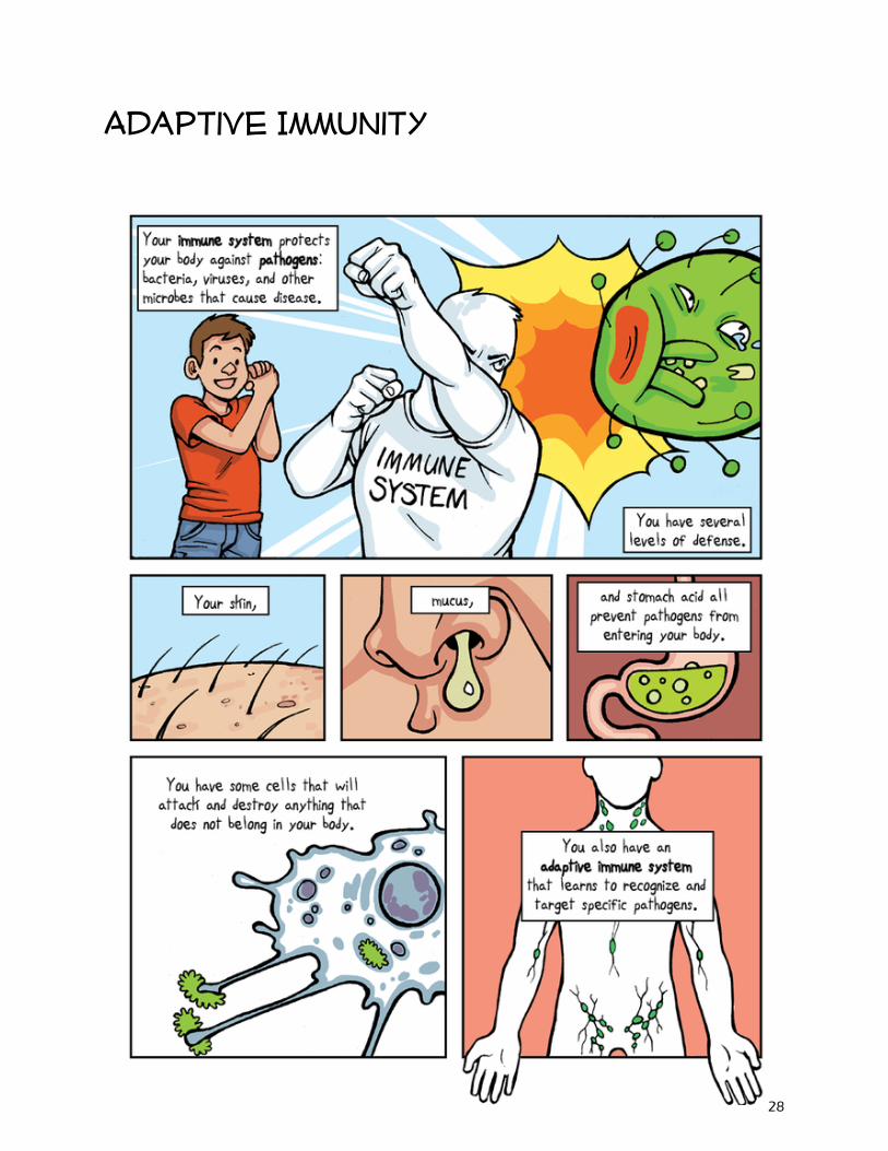
Adaptive Immunity
28
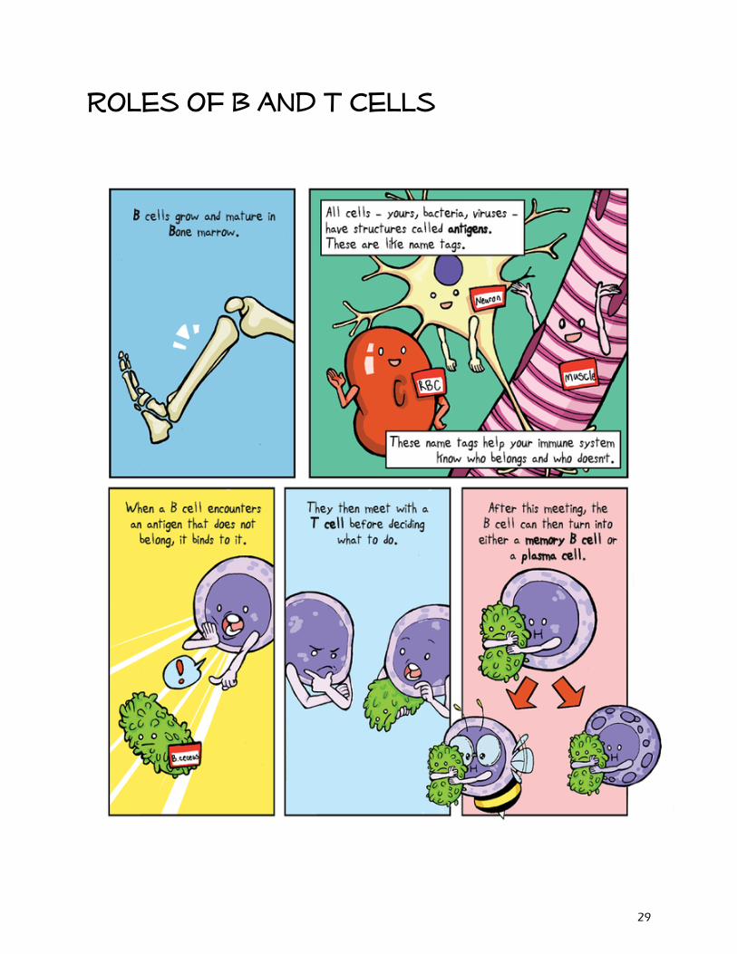
Roles of B and T Cells
29
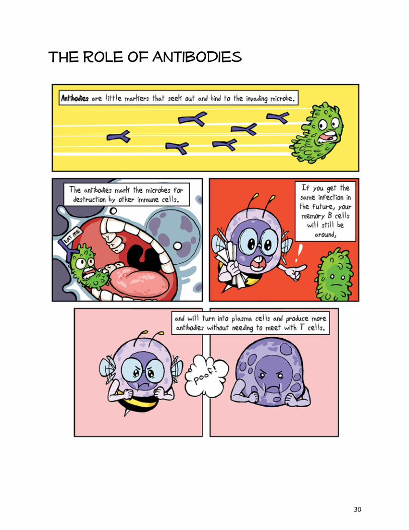
The role of antibodies
30
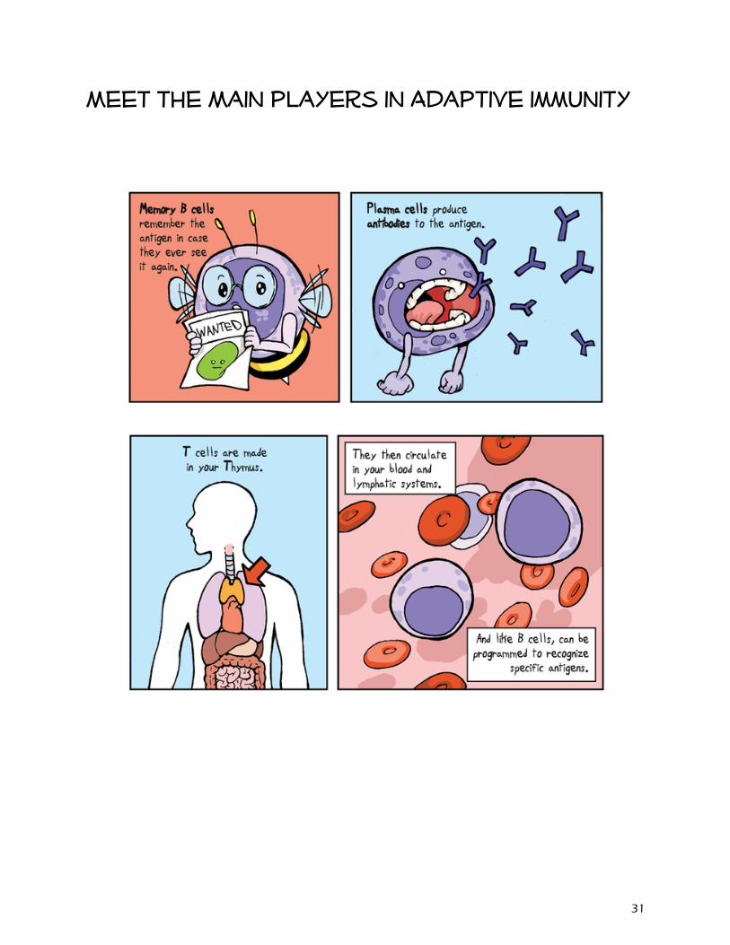
Meet the Main players in Adaptive immunity
31
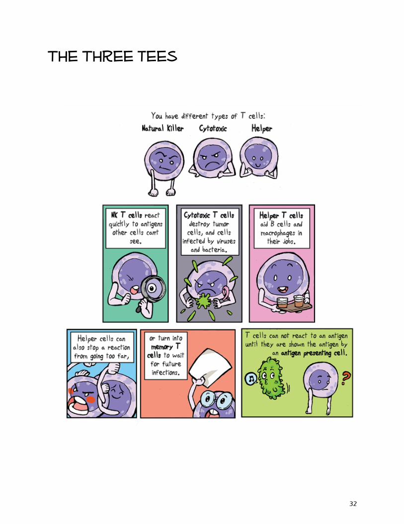
The Three tees
32
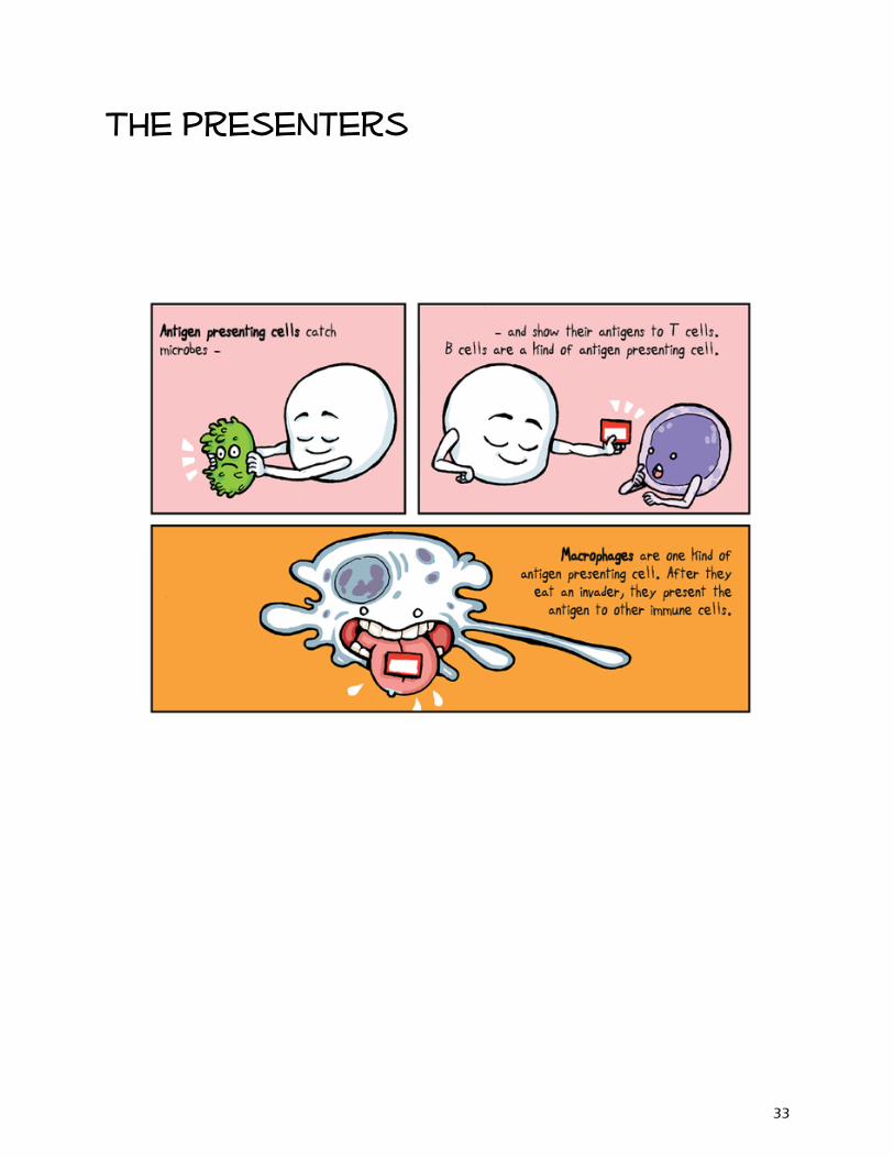
The presenters
33
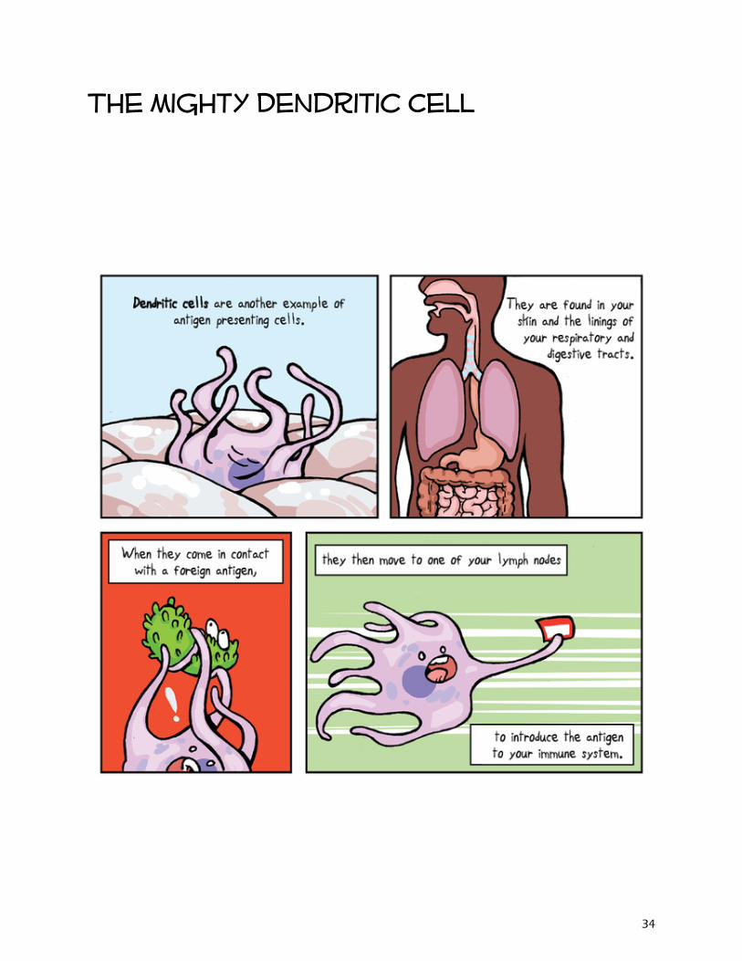
The mighty Dendritic Cell
34
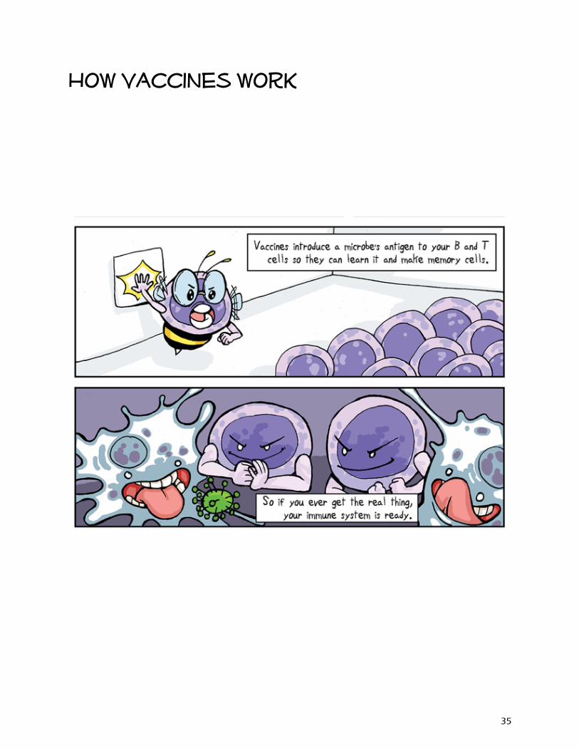
How Vaccines work
35
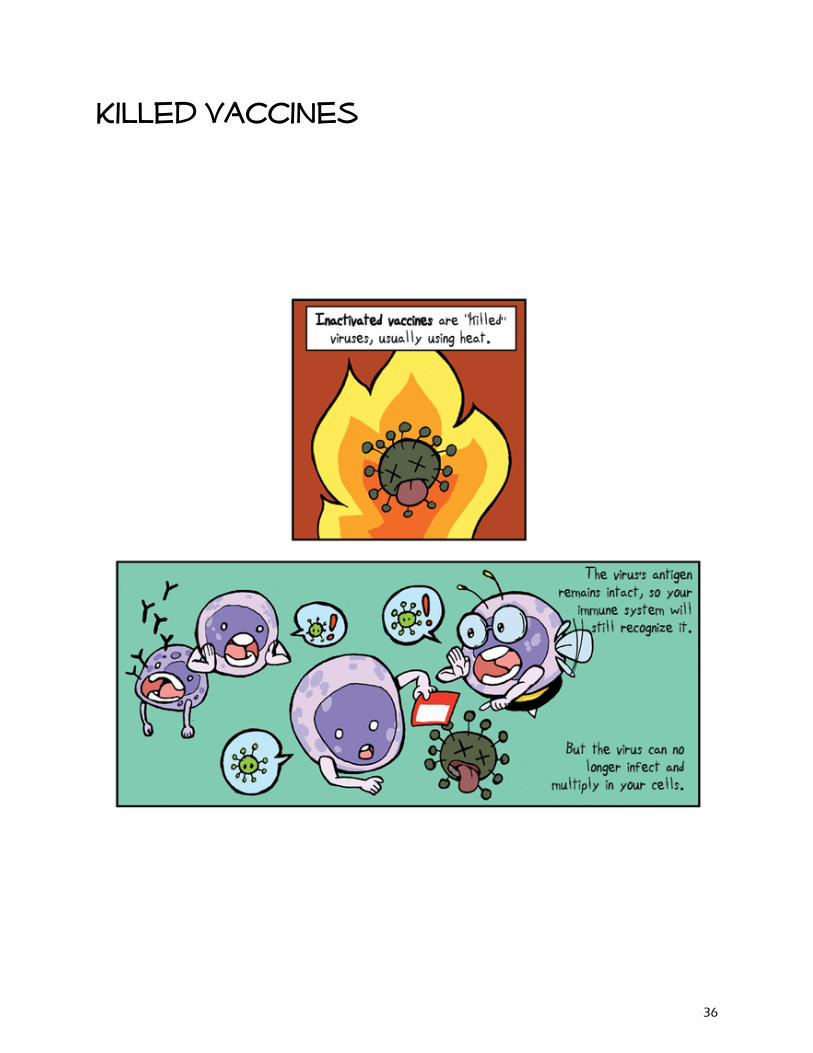
Killed vaccines
36
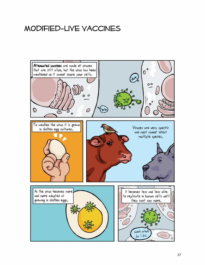
Modified-live Vaccines
37
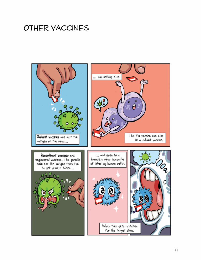
Other Vaccines
38
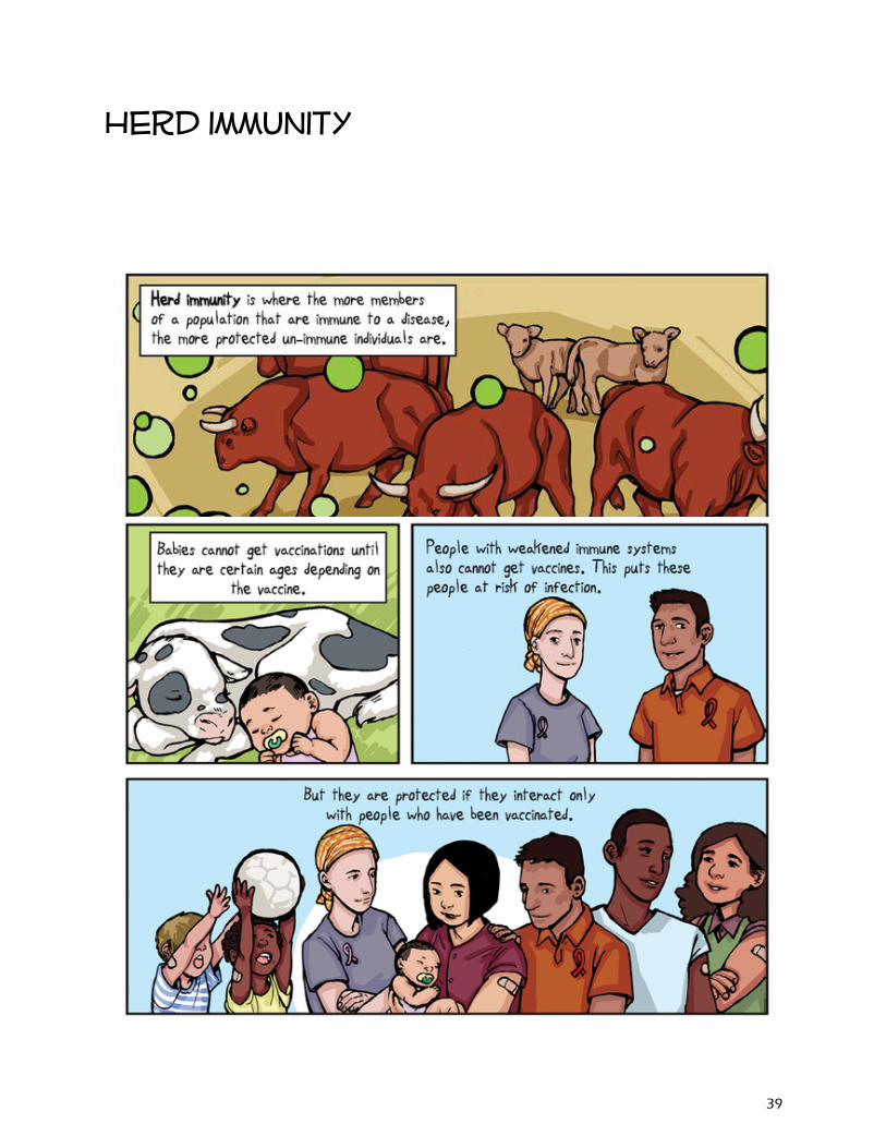
Herd immunity
39
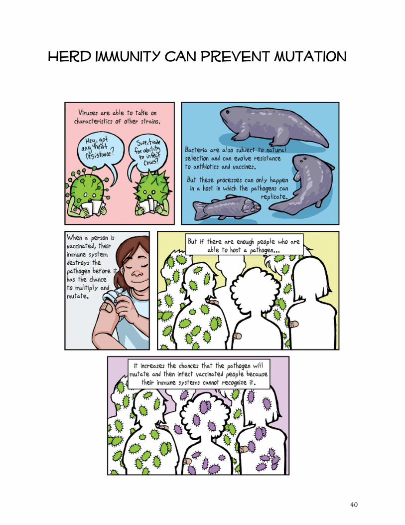
Herd immunity can prevent mutation
40
Principles of acupuncture
41

What is acupuncture?
Points on skin surface connect to nerves
Needles are inserted into skin to stimulate nerves
Procedure is performed to create a healing response
Considered alternative or holistic medicine
Contact points have specific actions when stimulated
Can be used with or without electric current
Alieves symptoms of several diseases and disorders
Needles are painless to the patient
42
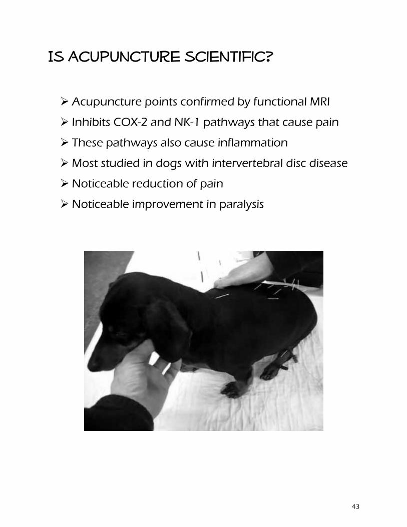
Is acupuncture scientific?
Acupuncture points confirmed by functional MRI
Inhibits COX-2 and NK-1 pathways that cause pain
These pathways also cause inflammation
Most studied in dogs with intervertebral disc disease
Noticeable reduction of pain
Noticeable improvement in paralysis
43
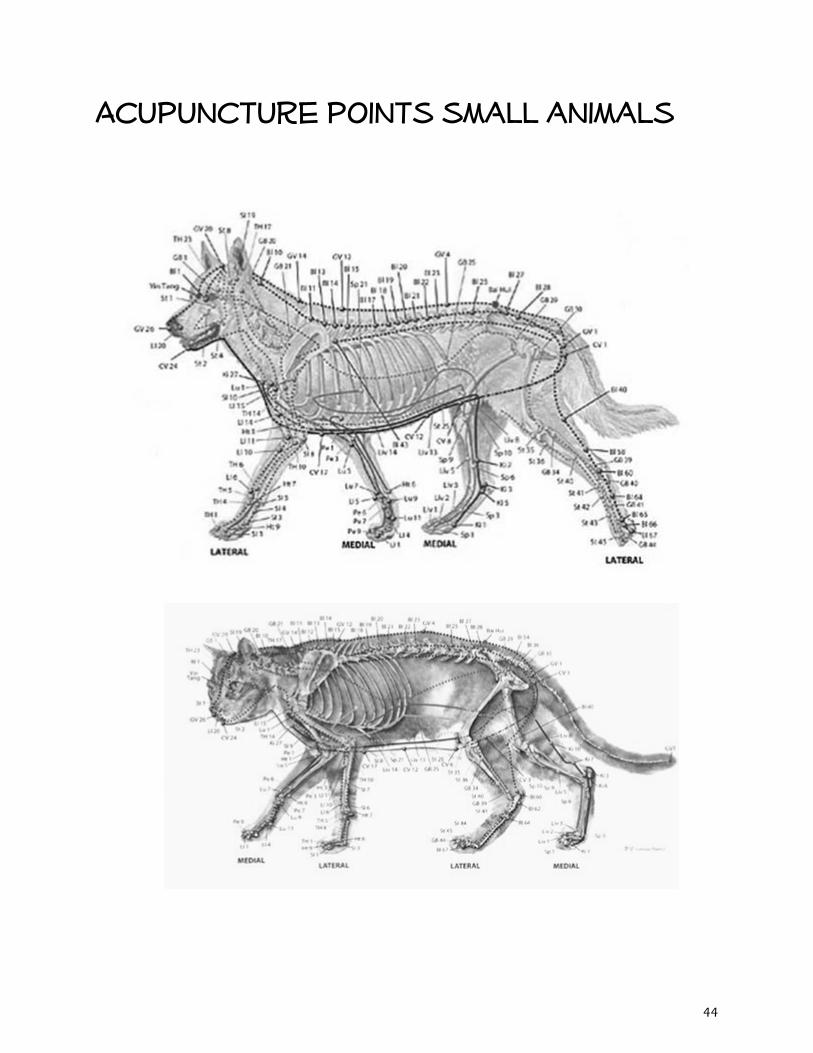
Acupuncture points small animals
44
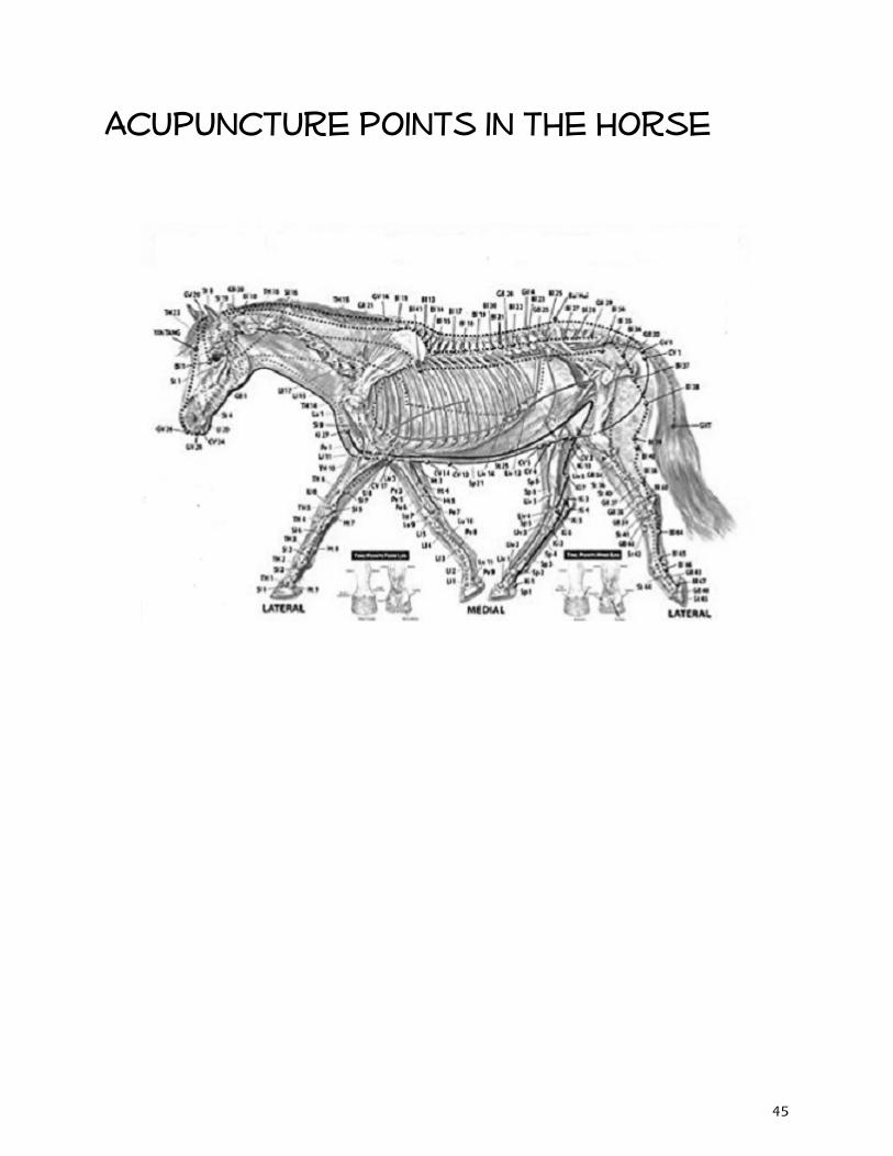
Acupuncture points in the horse
45

46
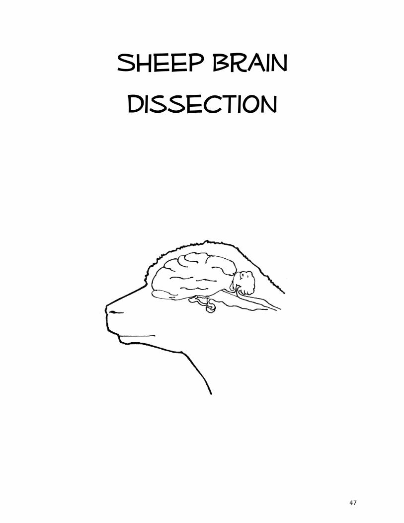
Sheep Brain
Dissection
47
Dissection guide
1. Set the brain down so the flatter side, with the white spinal cord at one end,rests on the dissection pan. Notice that the brain has two halves, or hemispheres. Can you tell the difference between the cerebrum and the cerebellum? Do the ridges (called gyri) and grooves (sulci) in the tissue look different? How does the surface feel?
2. Turn the brain over. You’ll probably be able to identify the medulla, pons,midbrain, optic chiasm, and olfactory bulbs. Find the olfactory bulb on each hemisphere. These will be slightly smoother and a different shade than the tissue around them. The olfactory bulbs control the sense of smell. The nerves to the nose are no longer connected, but you can see nubby ends where they were. The nerves to your mouth and lower body are attached to the medulla; the nerves to your eyes are connected to the optic chiasm.
3. Place the brain with the curved top side of the cerebrum facing up. Use ascalpel to slice through the brain along the center line, starting at the cerebrum and going down through the cerebellum, spinal cord, medulla, and pons. Separate the two halves of the brain and lay them with the inside facing up.
4. Use the labeled picture to identify the corpus callosum, medulla, pons,midbrain, and the place where pituitary gland attaches to the brain. (In many preserved specimens the pituitary gland is no longer present. It is not pictured.) Use your fingers or a teasing needle to gently probe the parts and see how they are connected to each other. What does that opening inside the corpus callosum lead to? How many different kinds of tissue can you see and feel?
48
5. Look closely at the inside of the cerebellum. You should see a branching ‘tree’of lighter tissue surrounded by darker tissue. The branches are white matter, which is made up of nerve axons. The darker tissue is gray matter, which is a collection of nerve cell bodies. You can see gray and white matter in the cerebrum, too, if you cut into a portion of it.
6. You can also use the letter labels on the anatomy picture to identify thefollowing:
a. The corpus callosum is a bundle of white fibers that connects the twohemispheres of the brain, providing coordination between the two.
b. The medulla is located right under the cerebellum. In this the nerves crossover so the left hemisphere controls the right side of the body and viceversa. This area of the brain controls the vital functions like heartbeat andrespiration (breathing).
c. The pons is next to the medulla. It serves as a bridge between the medullaand the upper brainstem, and it relays messages between the cerebrumand the cerebellum.
d. The pituitary gland, which produces important hormones, is a sac-like areathat attaches to the brain between the pons and the optic chiasm. This mayor may not be present on your specimen.
e. Ventricles contain cerebrospinal fluidf. The occipital lobe receives and interprets visual sensory messagesg. The temporal lobe is involved in hearing and smell. You can find this by
looking on the outside of one of the hemispheres. You will see a horizontalgroove called the lateral fissure. The temporal lobe is the section of thecerebrum below this line.
h. The frontal lobe also plays a part in smell, plus dealing with motor functioni. The parietal lobe handles all the sensory info except for vision, hearing, and
smell.j. The thalamus is a ‘relay station’ for sensory information. It receives
messages from the nerve axons and then transmits them to theappropriate parts of the brain.
k. The pineal gland produces important hormones.
49

50

Rashes and other itchy stuff
51
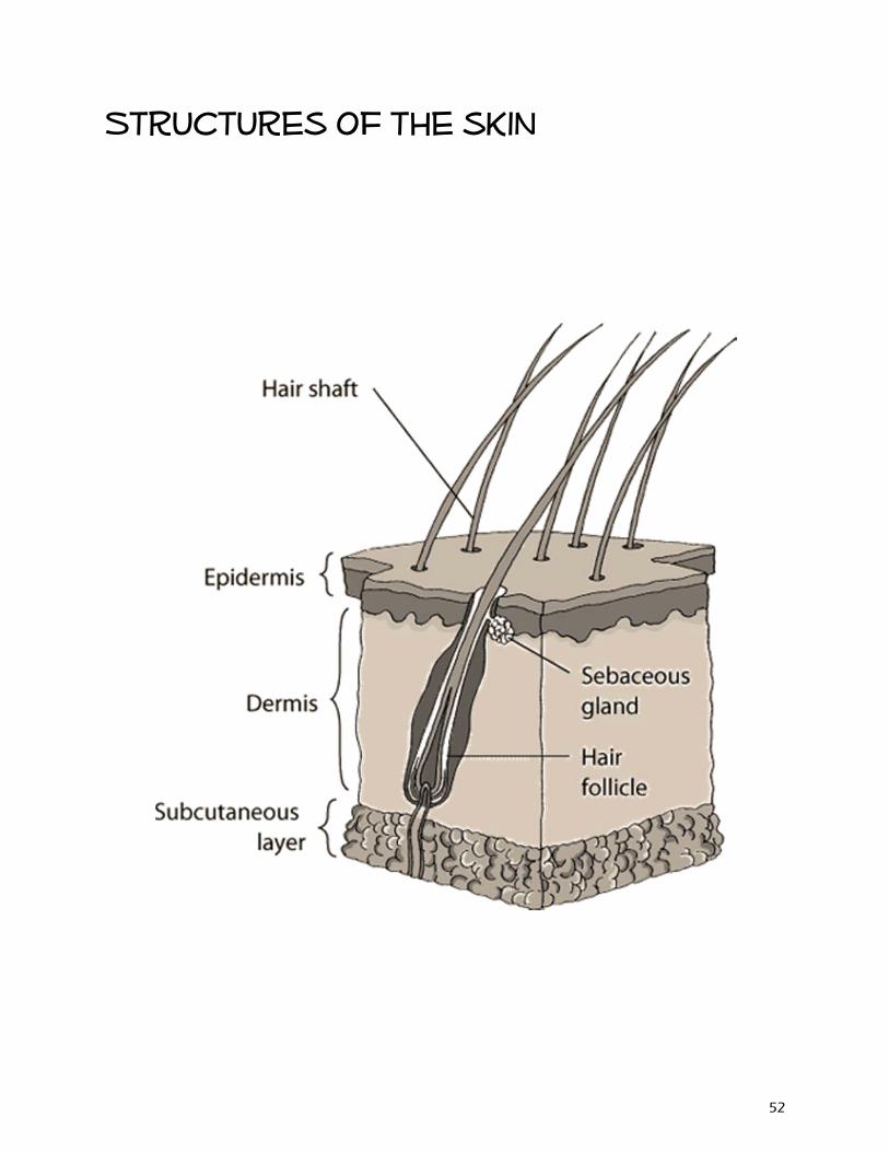
Structures of the skin
52
What is dermatitis?
Dermatitis is inflammation of the skin
Incited by allergies, infection, mites, hormones or
medication
Skin can become red, painful, itchy, crusty, flaky, oily or smelly
Caused by a combination of several factors:
- Skin barrier dysfunction
- Cell mediated immune responses
- IgE mediated hypersensitivity
- Environmental factors
53
Dermatology terms
Otitis externa – inflammation of the outer ear canal
Atopic dermatitis – inflammation caused by allergens
Pododermatitis – inflammation of paws
Conjunctivitis – inflammation of eyelids
Acute moist dermatitis – commonly known as hot spots
Pyoderma – bacterial skin infection
Dermatophytosis – fungal skin infection
Demodicosis – inflammation caused by Demodex mite
Flea allergy dermatitis – inflammation due to flea bites
54
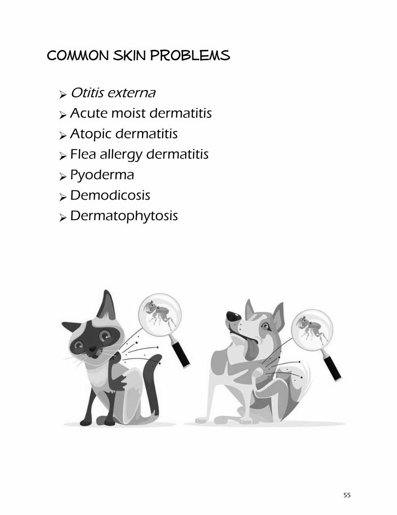
Common skin problems
Otitis externa
Acute moist dermatitis
Atopic dermatitis
Flea allergy dermatitis
Pyoderma
Demodicosis
Dermatophytosis
55
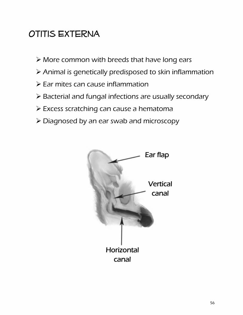
Otitis Externa
More common with breeds that have long ears
Animal is genetically predisposed to skin inflammation
Ear mites can cause inflammation
Bacterial and fungal infections are usually secondary
Excess scratching can cause a hematoma
Diagnosed by an ear swab and microscopy
Vertical canal
Ear flap
Horizontal canal
56
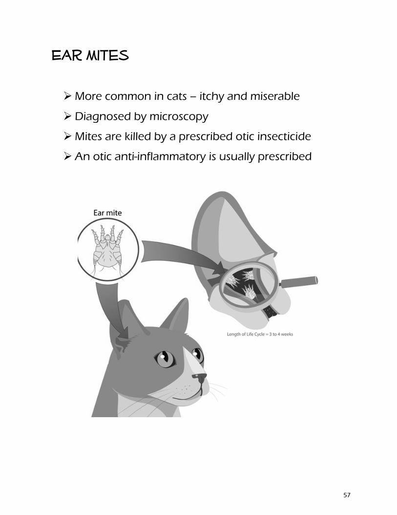
Ear mites
More common in cats – itchy and miserable
Diagnosed by microscopy
Mites are killed by a prescribed otic insecticide
An otic anti-inflammatory is usually prescribed
57
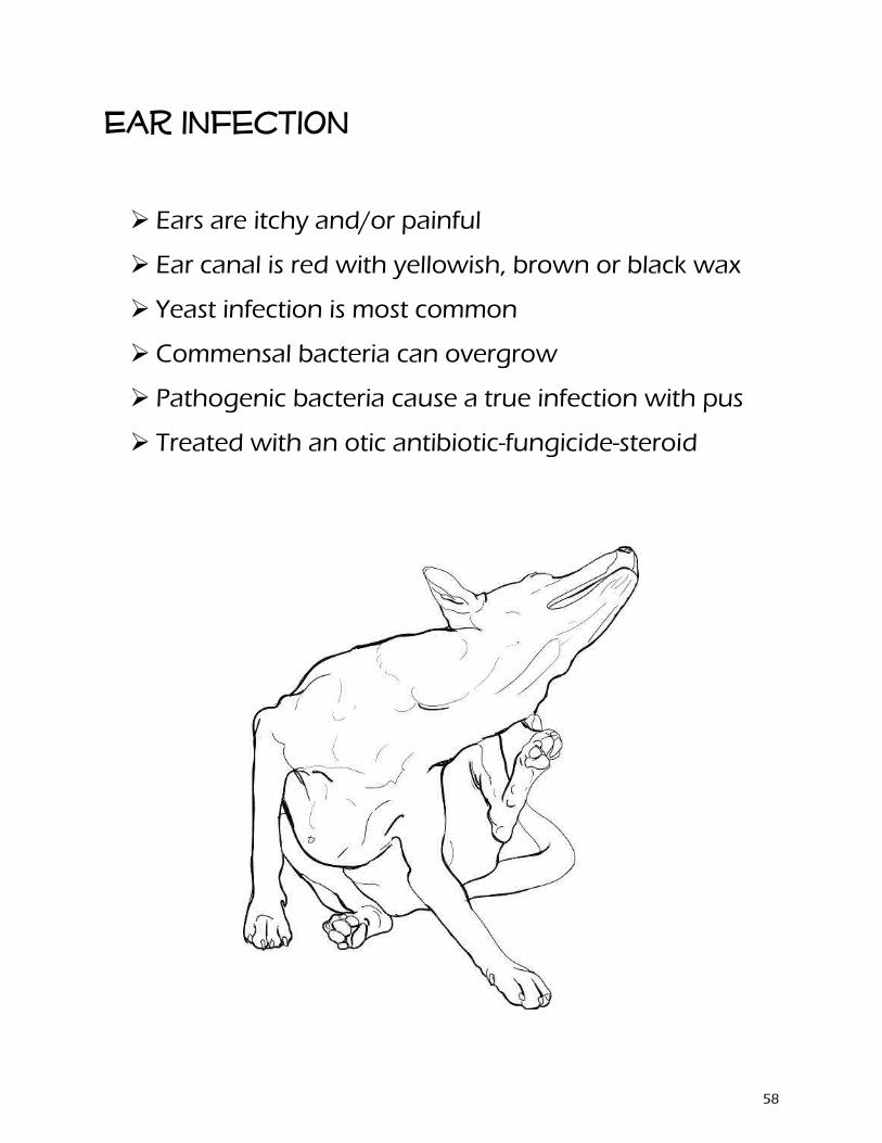
Ear infection
Ears are itchy and/or painful
Ear canal is red with yellowish, brown or black wax
Yeast infection is most common
Commensal bacteria can overgrow
Pathogenic bacteria cause a true infection with pus
Treated with an otic antibiotic-fungicide-steroid
58
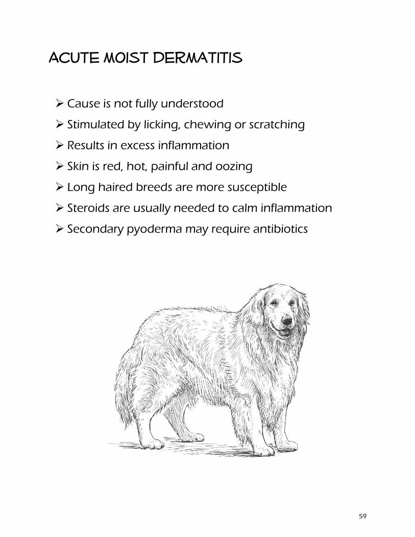
Acute moist dermatitis
Cause is not fully understood
Stimulated by licking, chewing or scratching
Results in excess inflammation
Skin is red, hot, painful and oozing
Long haired breeds are more susceptible
Steroids are usually needed to calm inflammation
Secondary pyoderma may require antibiotics
59
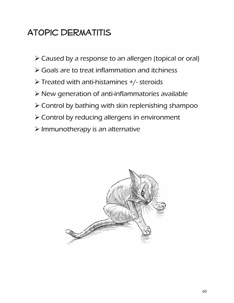
Atopic dermatitis
Caused by a response to an allergen (topical or oral)
Goals are to treat inflammation and itchiness
Treated with anti-histamines +/- steroids
New generation of anti-inflammatories available
Control by bathing with skin replenishing shampoo
Control by reducing allergens in environment
Immunotherapy is an alternative
60
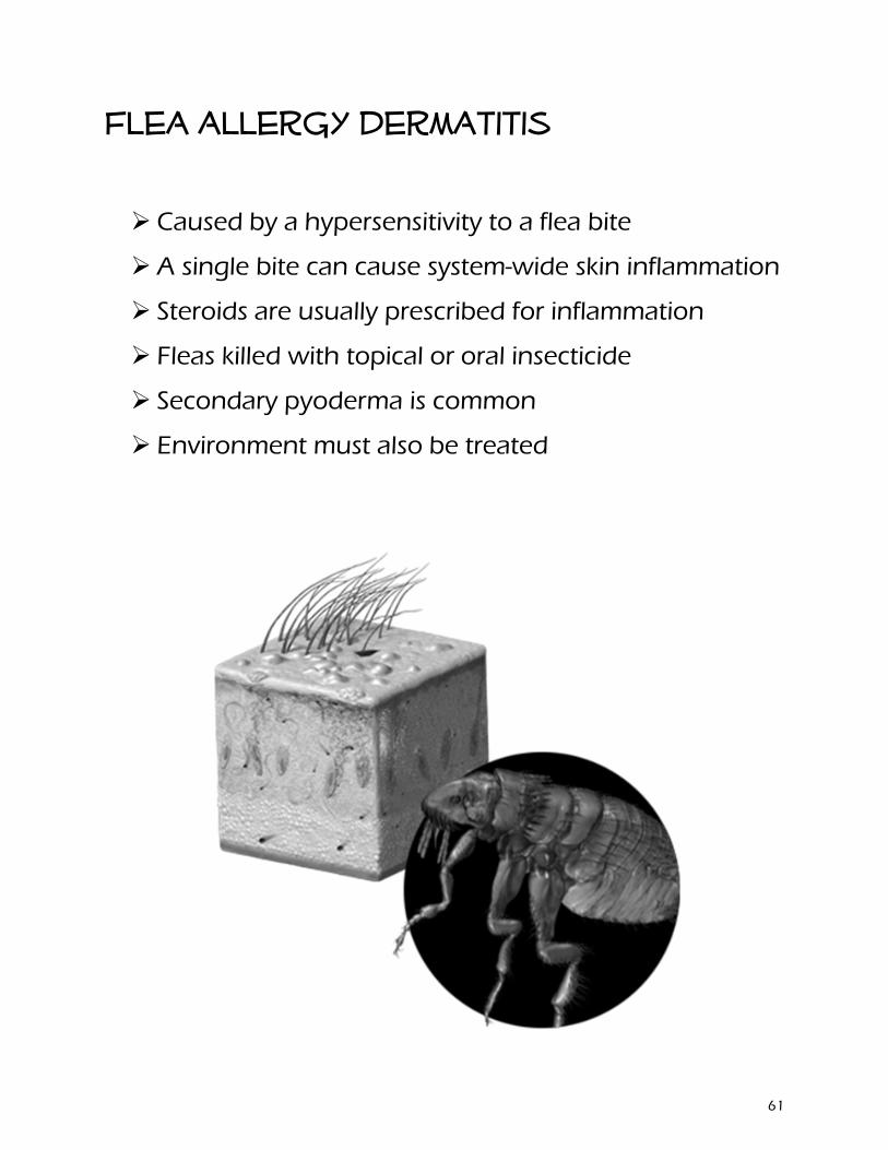
Flea allergy dermatitis
Caused by a hypersensitivity to a flea bite
A single bite can cause system-wide skin inflammation
Steroids are usually prescribed for inflammation
Fleas killed with topical or oral insecticide
Secondary pyoderma is common
Environment must also be treated
61
Pyoderma
Often secondary to dermatitis
Superficial and deep forms
Often seen on abdomen of puppies
Diagnosed by skin swab
Staph infections are most common
Treated with topical or oral antibiotics
Controlled with bathing in antibacterial shampoo
62
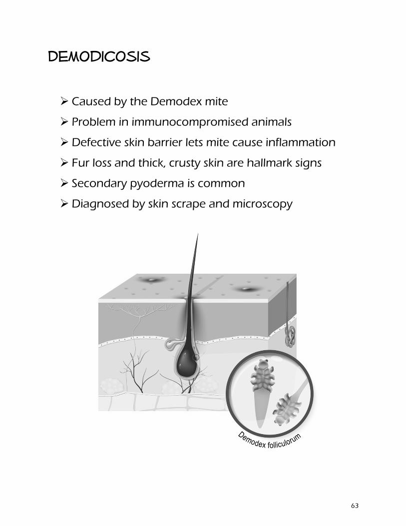
Demodicosis
Caused by the Demodex mite
Problem in immunocompromised animals
Defective skin barrier lets mite cause inflammation
Fur loss and thick, crusty skin are hallmark signs
Secondary pyoderma is common
Diagnosed by skin scrape and microscopy
63
Dermatophytosis
Also called ringworm, but is caused by a fungus
Common in cats, but any animal can be infected
Itchy, contagious and zoonotic!
Lesion is round and red +/- dark edge
Fur usually falls our around region
Diagnosed by ultraviolet light or fungal culture
Treat with oral or topical antifungal
Remove spores from environment
64
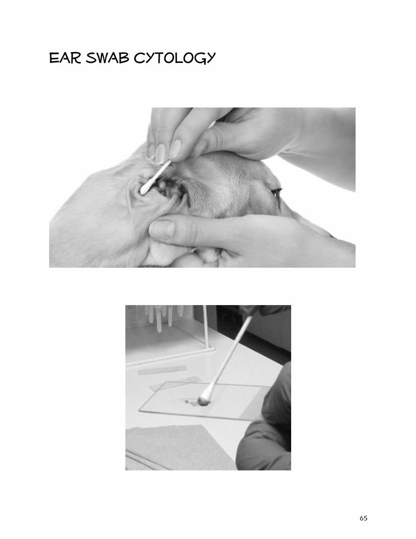
Ear swab cytology
65

66
Horse
67
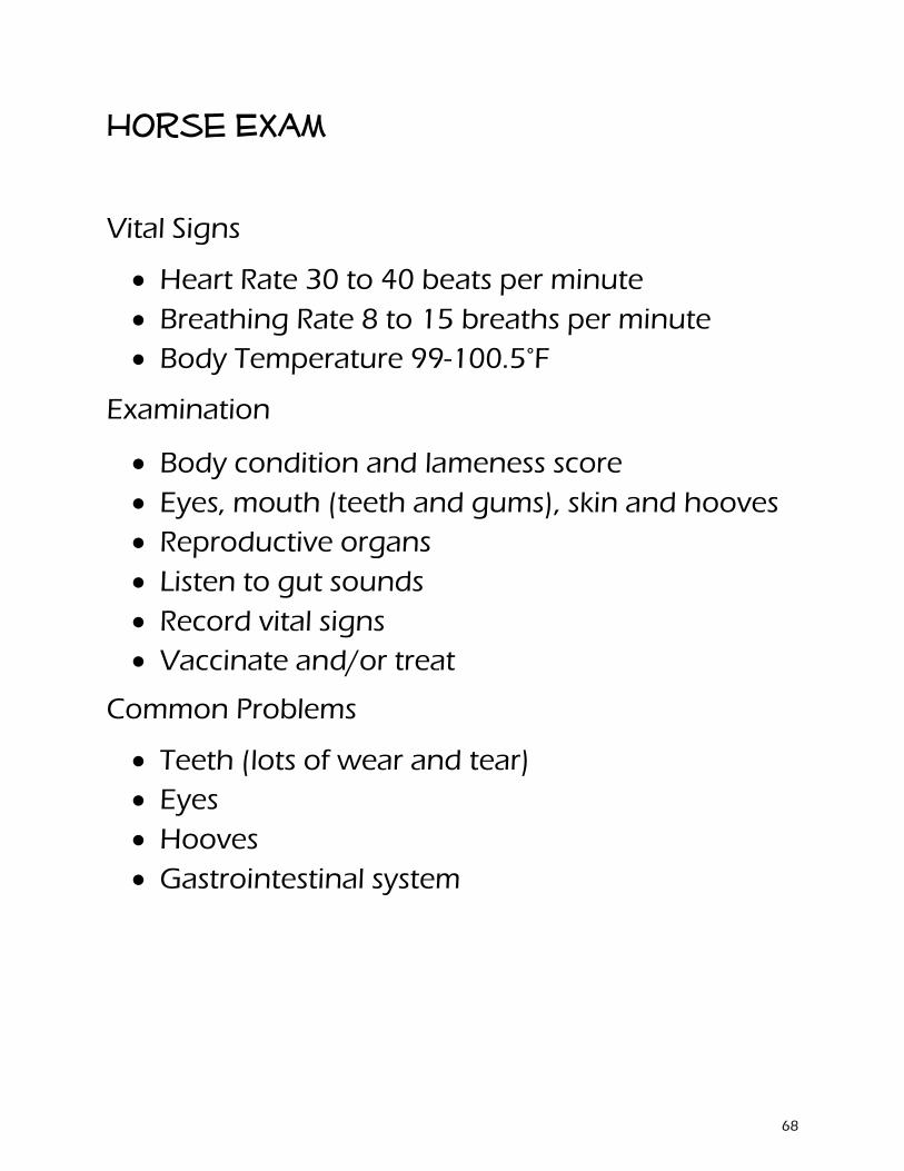
Horse Exam
Vital Signs
• Heart Rate 30 to 40 beats per minute• Breathing Rate 8 to 15 breaths per minute• Body Temperature 99-100.5°F
Examination
• Body condition and lameness score• Eyes, mouth (teeth and gums), skin and hooves• Reproductive organs• Listen to gut sounds• Record vital signs• Vaccinate and/or treat
Common Problems
• Teeth (lots of wear and tear)• Eyes• Hooves• Gastrointestinal system
68
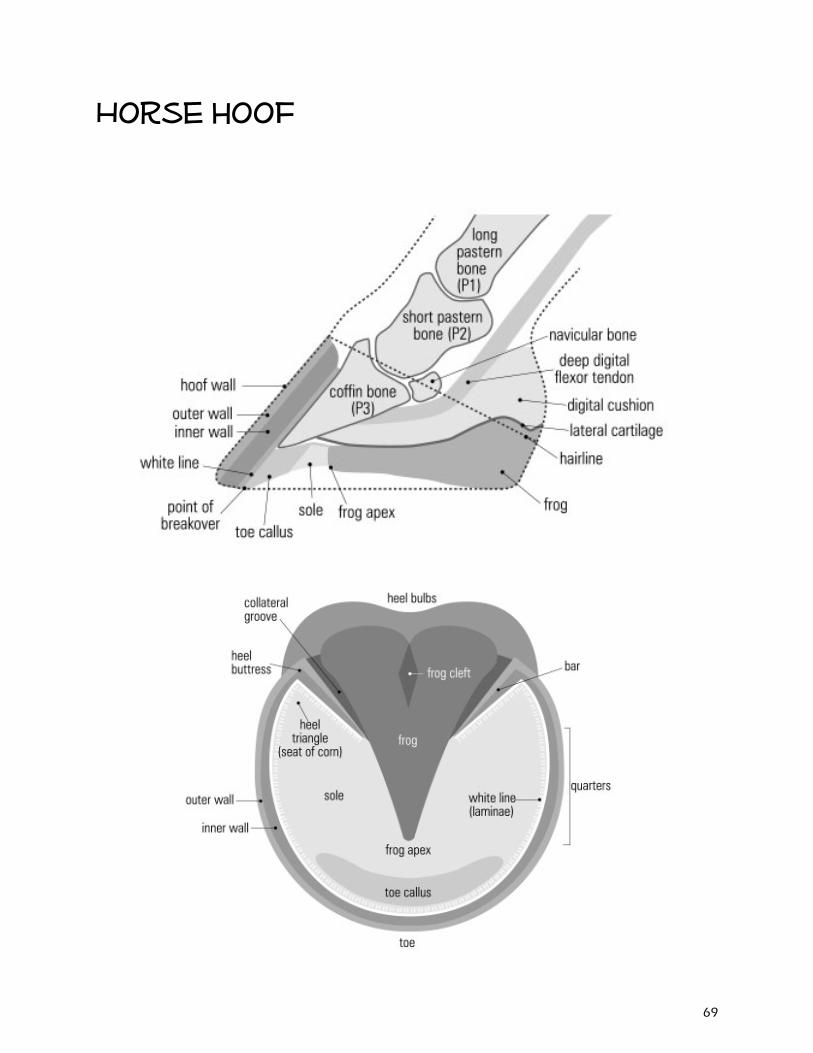
Horse hoof
69
Laminitis
Also called Founder
Inflammation of the laminae inside the hoof
Results from the disruption of blood flow to laminae
If severe, bone and hoof wall can separate
Can affect all or one foot
Front feet are affected more often
Inflammation often starts somewhere else in the body
- Digestive upsets from diet - Sudden access to excessive amounts of lush forage - Toxins released within the horse's system - High fever or illness - Severe colic - Retained placenta in the mare after foaling - Excessive concussion to the feet - Excessive weight-bearing on one leg due to injury - Various primary foot diseases - Bedding that contains black walnut shavings - Prolonged use of corticosteroids
70
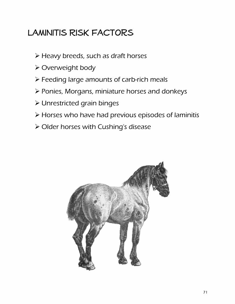
Laminitis Risk Factors
Heavy breeds, such as draft horses
Overweight body
Feeding large amounts of carb-rich meals
Ponies, Morgans, miniature horses and donkeys
Unrestricted grain binges
Horses who have had previous episodes of laminitis
Older horses with Cushing's disease
71
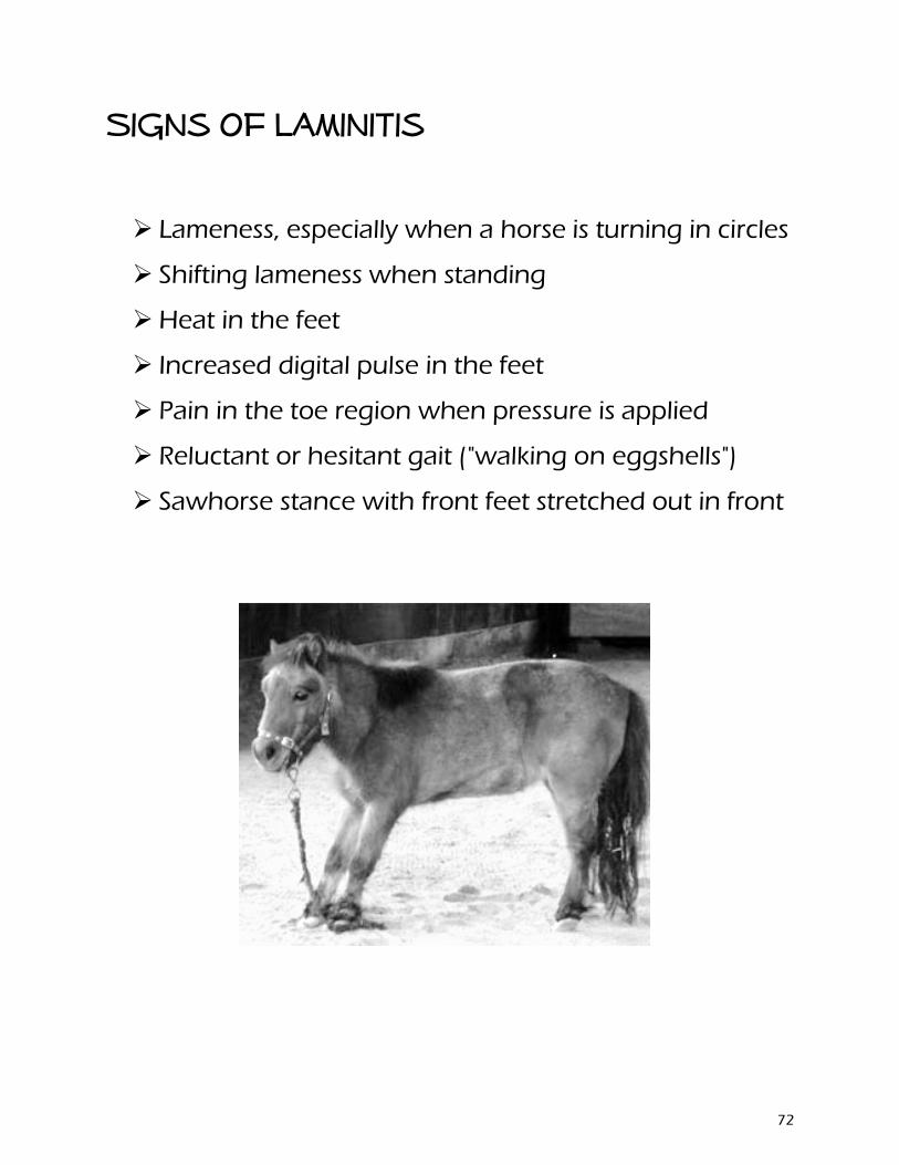
Signs of laminitis
Lameness, especially when a horse is turning in circles
Shifting lameness when standing
Heat in the feet
Increased digital pulse in the feet
Pain in the toe region when pressure is applied
Reluctant or hesitant gait ("walking on eggshells")
Sawhorse stance with front feet stretched out in front
72
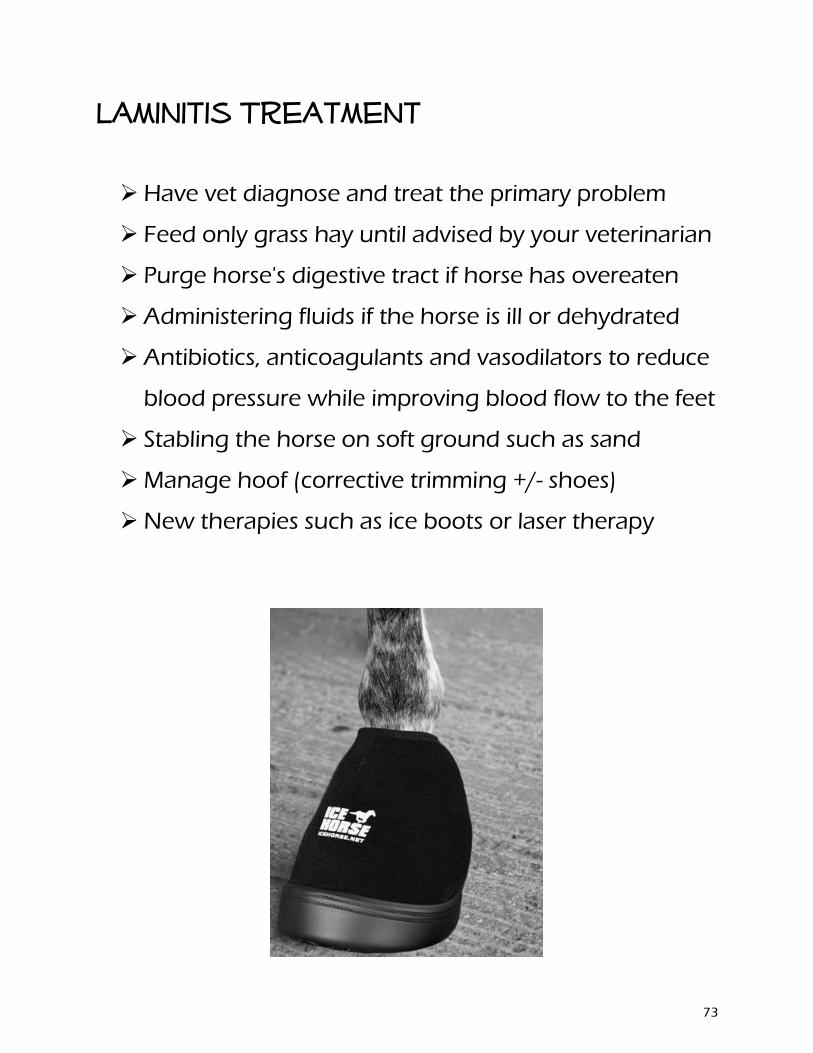
Laminitis Treatment
Have vet diagnose and treat the primary problem
Feed only grass hay until advised by your veterinarian
Purge horse's digestive tract if horse has overeaten
Administering fluids if the horse is ill or dehydrated
Antibiotics, anticoagulants and vasodilators to reduce
blood pressure while improving blood flow to the feet
Stabling the horse on soft ground such as sand
Manage hoof (corrective trimming +/- shoes)
New therapies such as ice boots or laser therapy
73
Laminitis management
A modified diet that provides adequate nutrition based on high-quality forage, digestible fiber (beet pulp) and oil. Avoid excess carbohydrates, especially from grain.
Routine hoof care, including regular trimming and, in some cases, therapeutic shoeing (additional radiographs may be needed to monitor progress).
A good health-maintenance schedule, including parasite control and vaccinations, to reduce the horse's susceptibility to illness or disease
A nutritional supplement formulated to promote hoof health.
Avoid grazing lush pastures, especially between late morning and late afternoon hours, since plant sugars are the highest during these times. Restrict pasture intake during spring or anytime the pasture suddenly greens up
74
Camelids
75
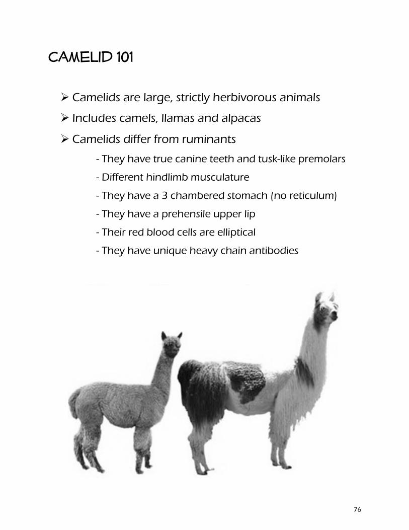
Camelid 101
Camelids are large, strictly herbivorous animals
Includes camels, llamas and alpacas
Camelids differ from ruminants
- They have true canine teeth and tusk-like premolars
- Different hindlimb musculature
- They have a 3 chambered stomach (no reticulum)
- They have a prehensile upper lip
- Their red blood cells are elliptical
- They have unique heavy chain antibodies
76
Llamas
Scientific name is Lama glama
Average height is 5.6 to 5.9 ft
Average weight is 290 and 440 lb
A baby llama is called a cria
Llamas typically live for 15 to 25 years
Females are induced ovulators
The gestation period of a llama is 11.5 months
Male llamas are excellent livestock guard animals
Llama vital signs
Normal rectal temperature 99.0° to 101.5°F
Average heart rate is 48 to 60 beats per minute
Average breathing rate is 12 to 30 breaths per minute
77
Alpaca
Scientific name is Vicugna pacos
Average height is 2.7 to 3.2 ft
Average weight is 110 to190 lb
A baby alpaca is also called a cria
Alpacas typically live for 15 to 20 years
Females are also induced ovulators
The gestation period of an alpaca is 11.5 months
Alpacas are social animals that live in groups
Alpaca Vital signs Normal rectal temperature 99.5° to 102.5°F
Average heart rate is 70 to120 beats per minute
Average breathing rate is 6 to 20 breaths per minute
78
Differences between camelids
Their ears: Alpaca ears have short spear-shaped ears while llamas have much longer, banana-shaped ears.
Their size: Alpacas generally weigh in at around 150 pounds while llamas can get as heavy as 400 pounds. At the shoulder, an average alpaca stands between 34 and 36 inches, while a llama generally ranges between 42 and 46 inches.
Their faces: Llamas have a longer face; an alpaca’s face is a bit blunter, giving them a “smooshed in” look.
Their purpose: For more than 5,000 years alpacas have been bred for fiber (and in Peru for meat as well), while llamas have been bred for the same amount of time as pack animals and for meat.
Their hair: The alpaca produces a much finer fiber than the llama. The alpaca also produces more fleece than its larger cousin and in a much greater variety of colors. Llamas also generally do not have as much hair on their head and face as alpacas do.
Their dispositions: Alpacas are very much herd animals, while llamas are more independent minded. Alpacas also tend to be a bit more skittish than llamas, which are often used as guard animals for alpacas, sheep, and other small livestock.
79

80

The University of Arkansas System Division of Agriculture offers all its Extension and Research programs and services without regard to race, color, sex, gender identity, sexual orientation, national origin, religion, age,
disability, marital or veteran status, genetic information, or any other legally protected status, and is an Affirmative Action/Equal Opportunity Employer.