Embed Size (px)
Citation preview
Archives of Biochemistry and Biophysics 502 (2010) 31–38
Contents lists available at ScienceDirect
Archives of Biochemistry and Biophysics
journal homepage: www.elsevier .com/ locate/yabbi
Characterization of recombinant UDP-galactopyranose mutasefrom Aspergillus fumigatus
Michelle Oppenheimer a, Myles B. Poulin b, Todd L. Lowary b, Richard F. Helm a, Pablo Sobrado a,*
a Department of Biochemistry, Virginia Tech, Blacksburg, VA 24061, USAb Alberta Ingenuity Centre for Carbohydrate Science and Department of Chemistry, University of Alberta, Edmonton, AB, Canada
a r t i c l e i n f o
Article history:Received 25 February 2010and in revised form 29 June 2010Available online 6 July 2010
Keywords:UDP-galactopyranose mutaseGalactofuranoseAspergillosisFlavoenzymeNon-redox reaction
0003-9861/$ - see front matter Published by Elsevierdoi:10.1016/j.abb.2010.06.035
* Corresponding author. Fax: +1 540 231 9070.E-mail address: [email protected] (P. Sobrado).
1 Abbreviations used: ABPA, allergic bronchopulmonapulmonary aspergillosis; ICUs, intensive care units; UDGalp, UDP-galactopyranose; UDP-Galf, UDP-galactofuraUGM, UDP-galactopyranose mutase; HPLC, high presSDS–PAGE, sodium dodecyl polyacrylamide gel electroylsulfonyl fluoride; PCR, polymerase chain reaction; FADSTD-NMR, saturation transfer difference nuclear magnhydroxyethyl)-1-piperazineethanesulfonic acid.
a b s t r a c t
UDP-galactopyranose mutase (UGM) is a flavin-containing enzyme that catalyzes the conversion of UDP-galactopyranose to UDP-galactofuranose, the precursor of galactofuranose, which is an important cellwall component in Aspergillus fumigatus and other pathogenic microbes. A. fumigatus UGM (AfUGM)was expressed in Escherichia coli and purified to homogeneity. The enzyme was shown to function as ahomotetramer by size-exclusion chromatography and to contain �50% of the flavin in the active reducedform. A kcat value of 72 ± 4 s�1 and a KM value of 110 ± 15 lM were determined with UDP-galactofuranoseas substrate. In the oxidized state, AfUGM does not bind UDP-galactopyranose, while UDP and UDP-glu-cose bind with Kd values of 33 ± 9 lM and 90 ± 30 lM, respectively. Functional and structural differencesbetween the bacterial and eukaryotic UGMs are discussed.
Published by Elsevier Inc.
Introduction
Human pathogenic fungi of the genus Aspergillus are responsi-ble for severe human diseases ranging from allergic reactions andlung infections to sepsis and death [1]. There are hundreds ofmembers of the Aspergillus genus, but only a few have been identi-fied as pathogenic to humans, with Aspergillus fumigatus and A. ni-ger being the most common [1,2]. Among the diseases related toAspergillus infection, allergic bronchopulmonary aspergillosis(ABPA)1 and invasive pulmonary aspergillosis (IPA) represent signif-icant health threats to immunocompetent and immunocomprom-ized persons [3,4]. IPA infections are commonly observed inpatients receiving chemotherapy, organ transplants, and in late-stage AIDS [4]. A 0.3–5.8% increase in IPA infections in patientsadmitted to intensive care units (ICUs) has been reported in recentyears and IPA infections are accompanied by a high mortality rateof 50–70% [5].
Chemotherapeutic treatments against Aspergillus spp. includecompounds that inhibit the biosynthesis of cell wall components
Inc.
ry aspergillosis; IPA, invasiveP, uridine diphosphate; UDP-nose; UDP-Glc, UDP-glucose;sure liquid chromatography;phoresis; PMSF, phenylmeth-, flavin adenine dinucleotide;etic resonance; HEPES, 4-(2-
such as b-1, 3-glucan. Inhibition of cell wall biosynthesis rendersthe fungi more susceptible to osmotic stress, leading to cell lysis[6]. Cell wall biosynthesis in Aspergillus spp. is an attractive tar-get for chemical intervention since enzymes in this biosyntheticpathway are absent in humans [7]. A major component of thecell wall of A. fumigatus is galactofuranose (Galf), the 5-mem-bered ring form of galactose [8]. The presence of Galf in A. fumig-atus has been known for many years and was first identified as acomponent of galactomannan by immunodetection in patientssuffering from IPA [9]. Galf has also been shown to be a majorcomponent of saccharide structures in secreted molecules, mem-brane lipids, and glycosyl phosphoinositol (GPI)-anchored lipo-phosphogalactomannan in Aspergillus spp. [9]. In bacteria, thebiosynthesis of Galf has been well characterized and involvesthe action of UDP-galactopyranose mutase (UGM), a flavin-dependent enzyme that catalyzes the conversion of UDP-galacto-pyranose (UDP-Galp) to UDP-galactofuranose (UDP-Galf)(Scheme 1) [10]. UGM homologues have been identified ineukaryotic organisms such as A. fumigatus, A. niger, Leishmaniamajor, and Trypanosoma cruzi [11–13]. A. fumigatus UGM(AfUGM) has been shown to be an important virulence factor.Screening for genes involved in cell wall biosynthesis in A. nigeridentified UGM activity as important in cell wall biogenesis [14].Deletion of the AfUGM gene leads to attenuated virulence, dimin-ished cell wall thickness, and increased sensitivity to antifungalagents [13]. These results clearly indicate that AfUGM is anattractive target for the identification of new antifungal drugs.Here, we report the functional expression and characterizationof the recombinant AfUGM enzyme.
Scheme 1.
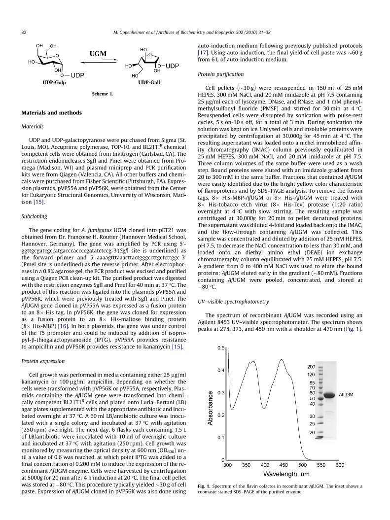
Fig. 1. Spectrum of the flavin cofactor in recombinant AfUGM. The inset shows acoomasie stained SDS–PAGE of the purified enzyme.
32 M. Oppenheimer et al. / Archives of Biochemistry and Biophysics 502 (2010) 31–38
Materials and methods
Materials
UDP and UDP-galactopyranose were purchased from Sigma (St.Louis, MO). Accuprime polymerase, TOP-10, and BL21TIR chemicalcompetent cells were obtained from Invitrogen (Carlsbad, CA). Therestriction endonucleases SgfI and PmeI were obtained from Pro-mega (Madison, WI) and plasmid miniprep and PCR purificationkits were from Qiagen (Valencia, CA). All other buffers and chemi-cals were purchased from Fisher Scientific (Pittsburgh, PA). Expres-sion plasmids, pVP55A and pVP56K, were obtained from the Centerfor Eukaryotic Structural Genomics, University of Wisconsin, Mad-ison [15].
Subcloning
The gene coding for A. fumigatus UGM cloned into pET21 wasobtained from Dr. Françoise H. Routier (Hannover Medical School,Hannover, Germany). The gene was amplified by PCR using 50-ggttgcgatcgccatgacccaccccgatatctccg-30(SgfI site is underlined) asthe forward primer and 50-aaaagtttaaacttactgggccttgctcttggc-30
(PmeI site is underlined) as the reverse primer. After electrophor-eses in a 0.8% agarose gel, the PCR product was excised and purifiedusing a Qiagen PCR clean-up kit. The purified product was digestedwith the restriction enzymes SgfI and PmeI for 40 min at 37 �C. Theproduct of this reaction was ligated into the plasmids pVP55A andpVP56K, which were previously treated with SgfI and PmeI. TheAfUGM gene cloned in pVP55A was expressed as a fusion proteinto an 8� His tag. In pVP56K, the gene was cloned for expressionas a fusion protein to an 8� His-maltose binding protein(8� His-MBP) [16]. In both plasmids, the gene was under controlof the T5 promoter and could be induced by addition of isopro-pyl-b-thiogalactopyranoside (IPTG). pVP55A provides resistanceto ampicillin and pVP56K provides resistance to kanamycin [15].
Protein expression
Cell growth was performed in media containing either 25 lg/mlkanamycin or 100 lg/ml ampicillin, depending on whether thecells were transformed with pVP56K or pVP55A, respectively. Plas-mids containing the AfUGM gene were transformed into chemi-cally competent BL21T1R cells and plated onto Luria–Bertani (LB)agar plates supplemented with the appropriate antibiotic and incu-bated overnight at 37 �C. A 60 ml LB/antibiotic culture was inocu-lated with a single colony and incubated at 37 �C with agitation(250 rpm) overnight. The next day, 6 flasks each containing 1.5 Lof LB/antibiotic were inoculated with 10 ml of overnight cultureand incubated at 37 �C with agitation (250 rpm). Cell growth wasmonitored by measuring the optical density at 600 nm (OD600) un-til a value of 0.6 was reached, at which point IPTG was added to afinal concentration of 0.200 mM to induce the expression of the re-combinant AfUGM enzyme. Cells were harvested by centrifugationat 5000g for 20 min after 4 h induction at 20 �C. The final cell pelletwas stored at �80 �C. This procedure typically yielded �30 g of cellpaste. Expression of AfUGM cloned in pVP56K was also done using
auto-induction medium following previously published protocols[17]. Using auto-induction, the final yield of cell paste was �60 gfrom 6 L of auto-induction medium.
Protein purification
Cell pellets (�30 g) were resuspended in 150 ml of 25 mMHEPES, 300 mM NaCl, and 20 mM imidazole at pH 7.5 containing25 lg/ml each of lysozyme, DNase, and RNase, and 1 mM phenyl-methylsulfonyl fluoride (PMSF) and stirred for 30 min at 4 �C.Resuspended cells were disrupted by sonication with pulse-restcycles, 5 s on-10 s off, for a total of 3 min. During sonication thesolution was kept on ice. Unlysed cells and insoluble proteins wereprecipitated by centrifugation at 30,000g for 45 min at 4 �C. Theresulting supernatant was loaded onto a nickel immobilized affin-ity chromatography (IMAC) column previously equilibrated in25 mM HEPES, 300 mM NaCl, and 20 mM imidazole at pH 7.5.Three column volumes of the same buffer were used as a washstep. Bound proteins were eluted with an imidazole gradient from20 to 300 mM in the same buffer. Fractions that contained AfUGMwere easily identified due to the bright yellow color characteristicof flavoproteins and by SDS–PAGE analysis. To remove the fusiontags, 8� His-MBP-AfUGM or 8� His-AfUGM were treated with8� His-tobacco etch virus (8� His-Tev) protease (1:20 ratio)overnight at 4 �C with slow stirring. The resulting sample wascentrifuged at 30,000g for 20 min to pellet denatured proteins.The supernatant was diluted 4-fold and loaded back onto the IMAC,and the flow-through containing AfUGM was collected. Thissample was concentrated and diluted by addition of 25 mM HEPES,pH 7.5, to decrease the NaCl concentration to less than 30 mM, andloaded onto an diethyl amino ethyl (DEAE) ion exchangechromatography column equilibrated with 25 mM HEPES, pH 7.5.A gradient from 0 to 400 mM NaCl was used to elute the boundproteins; AfUGM eluted early in the gradient (�80 mM). Fractionscontaining AfUGM were pooled, concentrated, and stored at�80 �C.
UV–visible spectrophotometry
The spectrum of recombinant AfUGM was recorded using anAgilent 8453 UV–visible spectrophotometer. The spectrum showspeaks at 278, 373, and 450 nm with a shoulder at 470 nm (Fig. 1).
M. Oppenheimer et al. / Archives of Biochemistry and Biophysics 502 (2010) 31–38 33
Flavin oxidation/reduction studies
AfUGM (10–15 lM) was reduced by addition of excess dithio-nite and spectra were monitored until all excess dithionite wasconsumed. The cuvette was exposed to air to allow the reduced en-zyme to react with molecular oxygen. The spectra were recordedfor at least 30 min. After no further flavin oxidation was observed,0.5 mM UDP or UDP-Galp was added to the reaction and monitoredfor 20 min until no further changes in the flavin spectrum were ob-served. To determine the extinction coefficient, protein was heatdenatured at 95 �C for 10 min and centrifuged in a table top centri-fuge for 10 min to pellet the precipitated protein. A spectrum of theresulting solution was recorded.
Activity assay
The activity of recombinant AfUGM was tested by monitoringthe formation of UDP-Galf from UDP-Galp by HPLC. The assaywas performed in 0.1 ml of 25 mM HEPES, 125 mM NaCl, 20 mMdithionite, 0.5 mM UDP-Galp at pH 7.5, and the reaction was initi-ated by addition of 50 nM AfUGM. Concentration of AfUGM wasdetermined using the flavin extinction coefficient at 450 nm,e450 = 10.6 mM�1 cm�1. The reaction was incubated at 37 �C for10 min and terminated by heat denaturation, 95 �C for 5 min, ina DNA engine thermo cycler (BioRad, Hercules, CA). The resultingmixture was injected on a PA-100 (Dionex) HPLC column. The sam-ple was eluted isocratically with 75 mM KH2PO4, pH 4.5. Absor-bance at 262 nm was monitored to identify fractions containingsubstrate and product. Under these conditions, UDP-Galp elutedat 25.8 min and UDP-Galf at 32.8 min. The extent of conversionwas determined by comparing the integration of the substrateand product peaks. Activity of AfUGM was also monitored in the re-verse direction, monitoring UDP-Galf isomerization to UDP-Galp.UDP-Galf was synthesized using published protocols [18,19].Increasing amounts of UDP-Galf (20–800 lM) were incubated with50 nM AfUGM for 1 min at 37 �C, then the reaction was stopped byheat denaturation at 95 �C for 5 min. The formation of UDP-Galpwas determined by HPLC as indicated above.
Determination of active redox state
To determine if AfUGM was active in the oxidized or reducedstate, the enzyme was assayed with 0.5 mM UDP-Galp in the pres-ence or absence of 20 mM dithionite as described above. To ensurethat AfUGM was completely oxidized, the enzyme was reactedwith 0.5 mM of cytochrome c, ferricyanide, or dichloroindophenol(DCIP) before the activity was measured.
Molecular weight determination
The molecular mass of the recombinant AfUGM in solution wasdetermined using size-exclusion chromatography. The purifiedAfUGM was loaded onto a Superdex 200 column (GE Healthcare)equilibrated with 25 mM HEPES at pH 7.5 containing 125 mMNaCl. Using a set of protein standards (aproprotein (6.5 kDa), ribo-nuclease (13 kDa), carbonic anhydrase (29 kDa), ovalbumin(43 kDa), canolbumin (75 kDa), aldolase (158 kDa), and ferritin(440 kDa)), a standard curve was obtained by plotting the log ofmolecular weight versus Kav for the standards [20]. The molecularweight of AfUGM was determined to be 275,000 ± 20,000 Da, sug-gesting that in solution AfUGM functions as a tetramer.
Fluorescence assay
Fluorescence of AfUGM (20 lM) was measured with a Spectra-Max M5e plate reader (Molecular Devices) using 450 nm as the
excitation wavelength and the fluorescence spectrum was ob-tained from 500 to 700 nm. Ligand binding was monitored by mea-suring the changes in fluorescence at 520 nm upon addition ofUDP, UDP-Galp, or UDP-Glucose (UDP-Glc). The data wereanalyzed by subtracting the fluorescence value at 520 nm in theabsence of ligand (S), and dividing this value by the maximumfluorescence value (Smax). The data were fit to S ¼ðS�max½Ligand�Þ=ðKd þ ½Ligand�Þwhere Kd is the dissociation constant.
Circular dichroism spectroscopy
Circular dichroism (CD) spectra were recorded on a Jasco J-815spectropolarimeter. For acquisition of far-UV CD spectra, AfUGM(1.1 lM) was placed in a buffer consisting of 10 mM potassiumphosphate, pH 7.5, and 50 mM KF. Experiments were performedin a 1-mm path length quartz cell at room temperature. The CDspectra was obtained from the average of five scans from 190 to250 nm using a bandwidth of 1-nm and a response time of 1 s ata scan speed of 20 nm/min. Buffer backgrounds were subtractedfrom the protein spectra. Spectra were deconvoluted to estimatesecondary structure content with the online server DichroWeb[21].
Mass spectrum analysis
Purified AfUGM was precipitated by addition of equal volumesof methanol and precipitated by centrifugation at 10,000g. Thedenatured protein was washed with methanol to remove excessbuffer and salt. The sample was analyzed using a liquid chromatog-raphy electrospray ionization-mass spectrometer (LC-MS). Todetermine the type of flavin bound to AfUGM, the cofactor was re-moved by heating the protein at 95 �C for 5 min and precipitatingthe denatured protein by centrifugation for 10 min at 10,000g. Thesupernatant was analyzed by matrix-assisted laser desorption/ion-ization (MALDI) mass spectrometer.
Results
Expression and purification of recombinant AfUGM
The gene coding for AfUGM was cloned into two plasmids,pVP55A and pVP56K, for the expression of the recombinant proteinwith two fusion tags, an 8� His and an 8� His-MBP, respectively.Expression was done either using LB or auto-induction media[17]. When AfUGM was expressed as a fusion to 8� His, the proteinwas soluble. However, the solubility was enhanced by expressionof the protein as a fusion to 8� His-MBP. Recombinant AfUGMwas isolated using a nickel IMAC, followed by removal of the fusiontag by treatment with Tev protease, which also contained an8� His tag. This allowed for the isolation of free AfUGM from theTev and 8� His tag by loading the sample back onto an IMACand collecting the yellow flow-through. A final step using a DEAEcolumn was needed to obtain highly pure AfUGM (Fig. 1). In gen-eral, the purification of 8� His-AfUGM yielded 2 mg of proteinper gram of cell paste and a 2-fold increase was obtained with8� His-MBP-AfUGM. We generally obtained twice the amount ofcell mass and purified protein using the auto-induction method.
Redox active form of AfUGM
Initial characterization of bacterial UGM reported that the re-combinant enzyme from Escherichia coli was active in the oxidizedstate [22]. Later, it was shown that bacterial UGMs are two ordersof magnitude more active in the reduced form, demonstratingthat this is the active state [23]. Our initial activity assays with
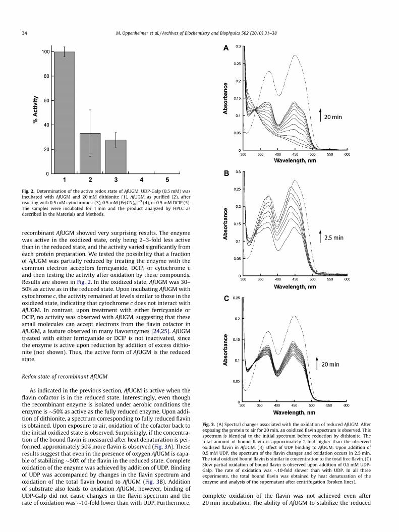
Fig. 2. Determination of the active redox state of AfUGM. UDP-Galp (0.5 mM) wasincubated with AfUGM and 20 mM dithionite (1), AfUGM as purified (2), afterreacting with 0.5 mM cytochrome c (3), 0.5 mM [Fe(CN)6]�3 (4), or 0.5 mM DCIP (5).The samples were incubated for 1 min and the product analyzed by HPLC asdescribed in the Materials and Methods.
34 M. Oppenheimer et al. / Archives of Biochemistry and Biophysics 502 (2010) 31–38
recombinant AfUGM showed very surprising results. The enzymewas active in the oxidized state, only being 2–3-fold less activethan in the reduced state, and the activity varied significantly fromeach protein preparation. We tested the possibility that a fractionof AfUGM was partially reduced by treating the enzyme with thecommon electron acceptors ferricyanide, DCIP, or cytochrome cand then testing the activity after oxidation by these compounds.Results are shown in Fig. 2. In the oxidized state, AfUGM was 30–50% as active as in the reduced state. Upon incubating AfUGM withcytochrome c, the activity remained at levels similar to those in theoxidized state, indicating that cytochrome c does not interact withAfUGM. In contrast, upon treatment with either ferricyanide orDCIP, no activity was observed with AfUGM, suggesting that thesesmall molecules can accept electrons from the flavin cofactor inAfUGM, a feature observed in many flavoenzymes [24,25]. AfUGMtreated with either ferricyanide or DCIP is not inactivated, sincethe enzyme is active upon reduction by addition of excess dithio-nite (not shown). Thus, the active form of AfUGM is the reducedstate.
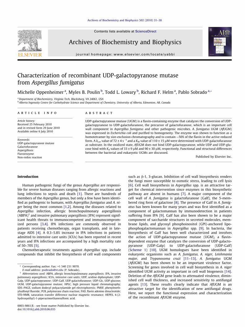
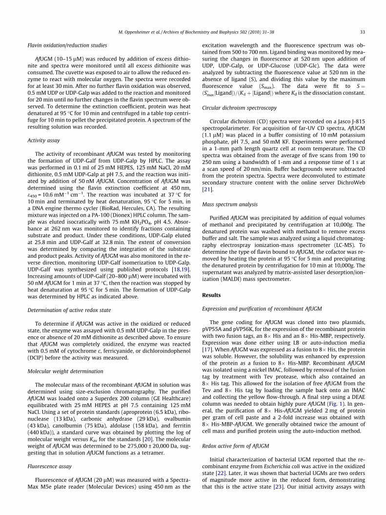
Fig. 3. (A) Spectral changes associated with the oxidation of reduced AfUGM. Afterexposing the protein to air for 20 min, an oxidized flavin spectrum is observed. Thisspectrum is identical to the initial spectrum before reduction by dithionite. Thetotal amount of bound flavin is approximately 2-fold higher than the observedoxidized flavin in AfUGM. (B) Effect of UDP binding to AfUGM. Upon addition of0.5 mM UDP, the spectrum of the flavin changes and oxidation occurs in 2.5 min.The total oxidized bound flavin is similar in concentration to the total free flavin. (C)Slow partial oxidation of bound flavin is observed upon addition of 0.5 mM UDP-Galp. The rate of oxidation was �10-fold slower than with UDP. In all threeexperiments, the total bound flavin was obtained by heat denaturation of theenzyme and analysis of the supernatant after centrifugation (broken lines).
Redox state of recombinant AfUGM
As indicated in the previous section, AfUGM is active when theflavin cofactor is in the reduced state. Interestingly, even thoughthe recombinant enzyme is isolated under aerobic conditions theenzyme is �50% as active as the fully reduced enzyme. Upon addi-tion of dithionite, a spectrum corresponding to fully reduced flavinis obtained. Upon exposure to air, oxidation of the cofactor back tothe initial oxidized state is observed. Surprisingly, if the concentra-tion of the bound flavin is measured after heat denaturation is per-formed, approximately 50% more flavin is observed (Fig. 3A). Theseresults suggest that even in the presence of oxygen AfUGM is capa-ble of stabilizing �50% of the flavin in the reduced state. Completeoxidation of the enzyme was achieved by addition of UDP. Bindingof UDP was accompanied by changes in the flavin spectrum andoxidation of the total flavin bound to AfUGM (Fig. 3B). Additionof substrate also leads to oxidation AfUGM, however, binding ofUDP-Galp did not cause changes in the flavin spectrum and therate of oxidation was �10-fold lower than with UDP. Furthermore,
complete oxidation of the flavin was not achieved even after20 min incubation. The ability of AfUGM to stabilize the reduced
Table 1Kinetic parameters of UDP-galactopyranose mutasesa.
M. Oppenheimer et al. / Archives of Biochemistry and Biophysics 502 (2010) 31–38 35
form of the flavin is a unique feature not previously reported inUGM enzymes.
Species kcat, s�1 KM, lM kcat/KM, lM�1 s�1 Ref.
A. fumigatus 72 ± 4 110 ± 15 0.65 ± 0.09 This workE. coli 27 22 1.22 [37]K. pneomoniae 5.5 ± 0.66 43 ± 6 0.12 ± 0.02 [43]D. radiodurans 66 ± 2.4 55 ± 7.0 1.18 [34]
a All the kinetic parameters are with UDP-galactofuranose as substrate in thepresence of 5–20 mM dithionite.
Enzyme activity
The activity of recombinant AfUGM was determined with UDP-Galf to accurately measure the initial rates at various substrateconcentrations. The activity of AfUGM follows a saturation kineticbehavior and the data was fit to the Michaelis–Menten equation(Fig. 4). Kinetic parameters are summarized in Table 1. Althoughthere are some differences in the kcat and KM values, the catalyticefficiencies of this eukaryotic UGM is within 5-fold of the valuesreported for other prokaryotic UGMs.
Oligomeric state of eukaryotic AfUGM
The molecular weight of AfUGM was determined by size-exclu-sion chromatography. Our analysis shows that this protein appearsto be active as a tetramer (Fig. 5 and Table 2). For comparison, wealso determined the molecular weight of Mycobacterium tuberculo-sis UGM (MtUGM). It was determined that in solution MtUGMfunctions as a dimer. This is consistent with the X-ray crystallogra-phy data showing homodimeric structures for MtUGM and other
Fig. 4. Steady-state kinetic characterization of AfUGM. Activity determined bymonitoring the formation UDP-Galp from UDP-Galf. (A) Chromatogram of the HPLCtrace at 262 nm showing the elution times of UDP-Galf (35.8 min) and UDP-Galp(25.8 min). (B) Initial rates of the formation of UDP-Galp as a function of UDP-Galf.The line is fit to the Michaelis–Menten equation.
Fig. 5. Size exclusion chromatography. The elution volumes for aprotinin (1),ribonuclease (2), ovalbumin (3), conalbumin (4), aldolase (5), and ferritin (6) wereused to calculate the Kav values (Kav = (Ve�Vo)/ (Vt Vo), where Vo is the void volumeof the column, Vt is the total volume of the column, and Ve is the elution volume ofthe protein). The Kav values for MtUGM and AfUGM are also plotted.
bacterial UGMs [23,26]. Thus, bacterial and eukaryotic UGMs ap-pear to differ in their quaternary structure.
Primary and secondary structures
Among the UGM from A. fumigatus, L. major, and T. cruzi, the se-quence identity is between 35% and 50%. In contrast, between thebacterial and eukaryotic enzymes the identity is less than 15%.Some of the conserved residues are those predicted to be in theFAD binding domain and those found to be in the active site of bac-terial UGMs (Fig. 6). We measured the CD spectrum to obtain infor-mation about the secondary structure of AfUGM (Fig. 7). Thespectrum shows characteristic features of folded proteins contain-ing high a-helix content, with a positive band at 193 nm and neg-ative bands at 208 and 222 nm [27]. Analysis of the spectrumpredicts that the protein contains 42% helix, 13% strand, and 45%coil. These values are similar to the secondary structure contentdetermined from known structures of bacterial UGMs (Table 3).
Ligand binding
To determine the affinity of AfUGM to various ligands, we mon-itored changes in flavin fluorescence upon substrate binding at520 nm (excitation at 450 nm). Upon ligand binding to oxidizedAfUGM, the flavin fluorescence increased (Fig. 8). This allowed usto calculate a Kd value for UDP and UDP-Glc (Table 4). Binding of
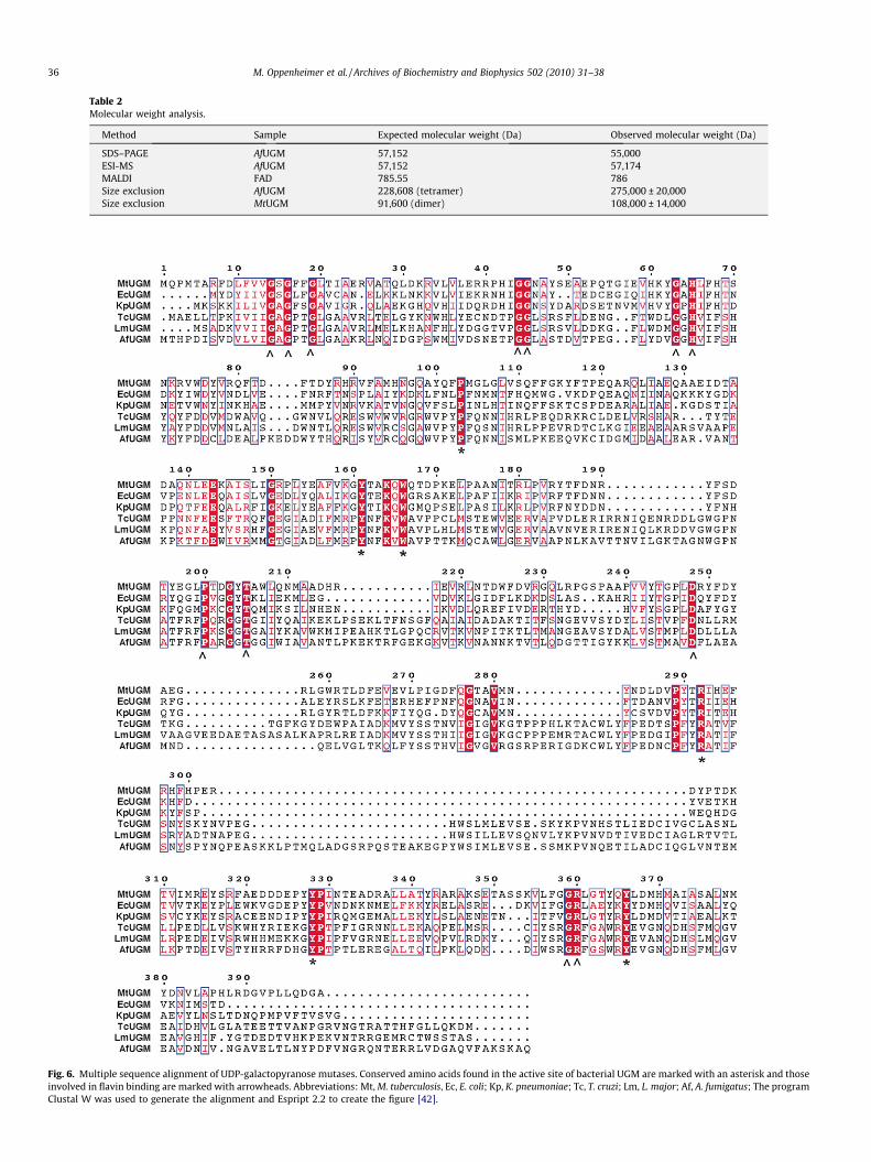
Table 2Molecular weight analysis.
Method Sample Expected molecular weight (Da) Observed molecular weight (Da)
SDS–PAGE AfUGM 57,152 55,000ESI-MS AfUGM 57,152 57,174MALDI FAD 785.55 786Size exclusion AfUGM 228,608 (tetramer) 275,000 ± 20,000Size exclusion MtUGM 91,600 (dimer) 108,000 ± 14,000
Fig. 6. Multiple sequence alignment of UDP-galactopyranose mutases. Conserved amino acids found in the active site of bacterial UGM are marked with an asterisk and thoseinvolved in flavin binding are marked with arrowheads. Abbreviations: Mt, M. tuberculosis, Ec, E. coli; Kp, K. pneumoniae; Tc, T. cruzi; Lm, L. major; Af, A. fumigatus; The programClustal W was used to generate the alignment and Espript 2.2 to create the figure [42].
36 M. Oppenheimer et al. / Archives of Biochemistry and Biophysics 502 (2010) 31–38
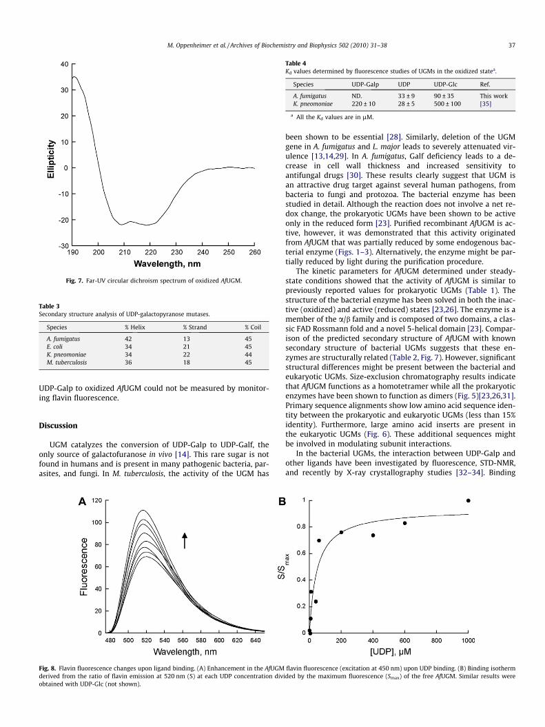
Fig. 7. Far-UV circular dichroism spectrum of oxidized AfUGM.
Table 3Secondary structure analysis of UDP-galactopyranose mutases.
Species % Helix % Strand % Coil
A. fumigatus 42 13 45E. coli 34 21 45K. pneomoniae 34 22 44M. tuberculosis 36 18 45
Table 4Kd values determined by fluorescence studies of UGMs in the oxidized statea.
Species UDP-Galp UDP UDP-Glc Ref.
A. fumigatus ND. 33 ± 9 90 ± 35 This workK. pneomoniae 220 ± 10 28 ± 5 500 ± 100 [35]
a All the Kd values are in lM.
M. Oppenheimer et al. / Archives of Biochemistry and Biophysics 502 (2010) 31–38 37
UDP-Galp to oxidized AfUGM could not be measured by monitor-ing flavin fluorescence.
Discussion
UGM catalyzes the conversion of UDP-Galp to UDP-Galf, theonly source of galactofuranose in vivo [14]. This rare sugar is notfound in humans and is present in many pathogenic bacteria, par-asites, and fungi. In M. tuberculosis, the activity of the UGM has
Fig. 8. Flavin fluorescence changes upon ligand binding. (A) Enhancement in the AfUGMderived from the ratio of flavin emission at 520 nm (S) at each UDP concentration divobtained with UDP-Glc (not shown).
been shown to be essential [28]. Similarly, deletion of the UGMgene in A. fumigatus and L. major leads to severely attenuated vir-ulence [13,14,29]. In A. fumigatus, Galf deficiency leads to a de-crease in cell wall thickness and increased sensitivity toantifungal drugs [30]. These results clearly suggest that UGM isan attractive drug target against several human pathogens, frombacteria to fungi and protozoa. The bacterial enzyme has beenstudied in detail. Although the reaction does not involve a net re-dox change, the prokaryotic UGMs have been shown to be activeonly in the reduced form [23]. Purified recombinant AfUGM is ac-tive, however, it was demonstrated that this activity originatedfrom AfUGM that was partially reduced by some endogenous bac-terial enzyme (Figs. 1–3). Alternatively, the enzyme might be par-tially reduced by light during the purification procedure.
The kinetic parameters for AfUGM determined under steady-state conditions showed that the activity of AfUGM is similar topreviously reported values for prokaryotic UGMs (Table 1). Thestructure of the bacterial enzyme has been solved in both the inac-tive (oxidized) and active (reduced) states [23,26]. The enzyme is amember of the a/b family and is composed of two domains, a clas-sic FAD Rossmann fold and a novel 5-helical domain [23]. Compar-ison of the predicted secondary structure of AfUGM with knownsecondary structure of bacterial UGMs suggests that these en-zymes are structurally related (Table 2, Fig. 7). However, significantstructural differences might be present between the bacterial andeukaryotic UGMs. Size-exclusion chromatography results indicatethat AfUGM functions as a homotetramer while all the prokaryoticenzymes have been shown to function as dimers (Fig. 5)[23,26,31].Primary sequence alignments show low amino acid sequence iden-tity between the prokaryotic and eukaryotic UGMs (less than 15%identity). Furthermore, large amino acid inserts are present inthe eukaryotic UGMs (Fig. 6). These additional sequences mightbe involved in modulating subunit interactions.
In the bacterial UGMs, the interaction between UDP-Galp andother ligands have been investigated by fluorescence, STD-NMR,and recently by X-ray crystallography studies [32–34]. Binding
flavin fluorescence (excitation at 450 nm) upon UDP binding. (B) Binding isothermided by the maximum fluorescence (Smax) of the free AfUGM. Similar results were
38 M. Oppenheimer et al. / Archives of Biochemistry and Biophysics 502 (2010) 31–38
assays following changes in the fluorescence of the oxidized flavinin KpUGM have shown that binding affinity for UDP is similar tothe value for AfUGM, while for UDP-Glc, the affinity for the bacte-rial enzyme is only 5-fold tighter, suggesting that the mode ofbinding for these substrates is somewhat similar between the bac-terial and eukaryotic UGMs. While binding of UDP-Galp to oxidizedKpUGM was measured by monitoring changes in the flavin fluores-cence, no changes with AfUGM upon binding of UDP-Galp were ob-served (Table 4). It was also observed that UDP binding causeschanges in the spectra of the oxidized flavin and triggers the oxida-tion of the reduced flavin. In contrast, changes in the flavin spectraare not observed upon binding of UDP-Galp (Fig. 3). Thus, this li-gand either has a very low affinity or binds to AfUGM in a confor-mation that does not affect the fluorescence of the flavin.
Thus, a different mechanism of substrate binding is employed inthe eukaryotic enzymes. Furthermore, increase in tryptophan fluo-rescence upon UDP and UDP-Galp binding were also reported forKpUGM [35]. These changes were not observed in AfUGM, furtherdemonstrating differences in the mechanism of binding/structurebetween the bacterial and eukaryotic enzymes (not shown).
Our results clearly show that the reduced enzyme is the activeform of the enzyme and that AfUGM can stabilize �50% of boundflavin in the reduced form. These results suggest a half-site reactiv-ity for this unique tetrameric enzyme, where the active reducedmonomers are protected from oxidation. UDP binding induces rel-atively rapid oxidation of the reduced flavin; however, in the pres-ence of UDP-Galp the oxidation is �10-fold slower and does notreach 100% of the total flavin. Thus, during catalysis, even underaerobic conditions, AfUGM stabilizes the reduced form of the flavin,which is essential for activity. The slow oxidation observed in thepresence of UDP-Galp might be compensated by reduction by thestill unknown electron transfer partner in vivo.
The mechanism by which UGM catalyses the conversion ofUDP-Galp to UDP-Galf is not well understood. Using oxygen posi-tional isotope exchange (PIX), it was demonstrated that the glyco-sidic bond is broken during catalysis [36,37]. Potentiometry studiessuggest that the semiquinone form of the flavin is formed and sta-bilized by substrate binding [38]. It was also shown that 5-deaza-flavin substituted prokaryotic UGM is inactive, indicating that anelectron transfer step was necessary for catalysis [39]. Recently,it was demonstrated that a covalent substrate-FAD adduct isformed between the anomeric carbon and the N5 atom of the flavin[40,41]. These data have led to two proposals describing the mech-anism by which UGM catalyzes the FAD-dependent ring contrac-tion to form UGM-Galf [10]. One mechanism invokes the flavinas a nucleophile that attacks the anomeric carbon to display UDP[40]. The other mechanism involves a single electron transfer fromthe reduced flavin to a postulated oxocarbenium sugar intermedi-ate followed by the formation of a flavin-sugar adduct [38,39].Availability of high levels of active AfUGM provides the opportu-nity to test the role of the flavin cofactor in this novel enzyme.These results will be important for the development of novel anti-fungal agents.
Acknowledgments
This work was supported in part by funds from the Fralin LifeScience Institute (PS), and the Alberta Ingenuity Centre for Carbo-hydrate Science and The National Sciences and Engineering Re-
search Council of Canada (TL). MBP is the recipient of aStudentship Award from Alberta Ingenuity.
References
[1] R.L. Kradin, E.J. Mark, Arch. Pathol. Lab. Med. 132 (2008) 606–614.[2] R.J. Trof, A. Beishuizen, Y.J. Debets-Ossenkopp, A.R. Girbes, A.B. Groeneveld,
Intensive Care Med. 33 (2007) 1694–1703.[3] C. Virnig, R.K. Bush, Curr. Opin. Pulm. Med. 13 (2007) 67–71.[4] S. Chong, K.S. Lee, C.A. Yi, M.J. Chung, T.S. Kim, J. Han, Eur. J. Radiol. 59 (2006)
371–383.[5] G. Chamilos, M. Luna, R.E. Lewis, G.P. Bodey, R. Chemaly, J.J. Tarrand, A. Safdar
Raad II, D.P. Kontoyiannis, Haematologica 91 (2006) 986–989.[6] D.W. Denning, J. Antimicrob, Chemotheraphy 49 (2002) 889–891.[7] T.F. Patterson, Curr. Infect. Dis. Rep. 8 (2006) 442–448.[8] J.P. Latge, Med. Mycol. (2009) S104–S109.[9] C. Lamarre, R. Beau, V. Balloy, T. Fontaine, J.W. Hoi, S. Guadagnini, N. Berkova,
M. Chignard, A. Beauvais, J.P. Latge, Cell Microbiol. 24 (2009).[10] M.R. Richards, T.L. Lowary, Chembiochem 10 (2009) 1920–1938.[11] S.M. Beverley, K.L. Owens, M. Showalter, C.L. Griffith, T.L. Doering, V.C. Jones,
M.R. McNeil, Eukaryotic Cell 4 (2005) 1147–1154.[12] H. Bakker, B. Kleczka, R. Gerardy-Schahn, F.H. Routier, Biol. Chem. 386 (2005)
657–661.[13] B. Kleczka, A.C. Lamerz, G. van Zandbergen, A. Wenzel, R. Gerardy-Schahn, M.
Wiese, F.H. Routier, J. Biol. Chem. 282 (2007) 10498–10505.[14] R.A. Damveld, A. Franken, M. Arentshorst, P.J. Punt, F.M. Klis, C.A. Van del
Hondel, A.F. RAM, Genetics 178 (2008) 873–881.[15] P.G. Blommel, P.A. Martin, R.L. Wrobel, E. Steffen, B.G. Fox, Protein Expr. Purif.
47 (2006) 562–570.[16] P. Sobrado, M.A. Goren, D. James, C.K. Amundson, B.G. Fox, Protein Expr. Purif.
58 (2008) 229–241.[17] P.G. Blommel, K.J. Becker, P. Duvnjak, B.G. Fox, Biotechnol. Prog. 23 (2007)
585–598.[18] N.L. Rose, R.B. Zheng, J. Pearcey, R. Zhou, G.C. Completo, T.L. Lowary,
Carbohydr. Res. 343 (2008) 2130–2139.[19] J.C. Errey, M.C. Mann, S.A. Fairhurst, L. Hill, M.R. McNeil, J.H. Naismith, J.M.
Percy, C. Whitfield, R.A. Field, Org. Biomol. Chem. 7 (2009) 1009–1016.[20] P. Andrews, Biochem. J. 91 (1964) 222–233.[21] L. Whitmore, B.A. Wallace, Nucleic Acids Res. 32 (2004) W668–673.[22] Q. Zhang, J. Am. Chem. Soc. 122 (2000) 9065–9070.[23] D.A. Sanders, A.G. Staines, S.A. McMahon, M.R. McNeil, C. Whitfield, J.H.
Naismith, Nat. Struct. Biol. 8 (2001) 858–863.[24] E. Pessione, S. Divari, E. Griva, M. Cavaletto, G.L. Rossi, G. Gilardi, C. Giunta, Eur.
J. Biochem. 265 (1999) 549–555.[25] P. Sobrado, S.C. Daubner, P.F. Fitzpatrick, Biochemistry 40 (2001) 994–1001.[26] K. Beis, V. Srikannathasan, H. Liu, S.W. Fullerton, V.A. Bamford, D.A. Sanders, C.
Whitfield, M.R. McNeil, J.H. Naismith, J. Mol. Biol. 348 (2005) 971–982.[27] N.J. Greenfield, Nat. Protoc. 1 (2006) 2876–2890.[28] F. Pan, M. Jackson, Y. Ma, M. McNeil, J. Bacteriol. 183 (2001) 3991–3998.[29] A.M. El-Ganiny, D.A. Sanders, S.G. Kaminskyj, Fungal Genet. Biol. 45 (2008)
1533–1542.[30] P.S. Schmalhorst, S. Krappmann, W. Vervecken, M. Rohde, M. Muller, G.H.
Braus, R. Contreras, A. Braun, H. Bakker, F.H. Routier, Eukaryotic Cell 7 (2008)1268–1277.
[31] S.A. McMahon, G.A. Leonard, L.V. Buchanan, M.F. Giraud, J.H. Naismith, ActaCrystallogr. 55 (1999) 399–402.
[32] Y. Yuan, X. Wen, D.A. Sanders, B.M. Pinto, Biochemistry 44 (2005) 14080–14089.
[33] Y. Yuan, D.W. Bleile, X. Wen, D.A. Sanders, K. Itoh, H.W. Liu, B.M. Pinto, J. Am.Chem. Soc. 130 (2008) 3157–3168.
[34] S.K. Partha, K.E. van Straaten, D.A. Sanders, J. Mol. Biol. 394 (2009) 864–877.[35] X. Yao, D.W. Bleile, Y. Yuan, J. Chao, K.P. Sarathy, D.A. Sanders, B.M. Pinto, M.A.
O’Neill, Proteins 74 (2008) 972–979.[36] J.N. Barlow, M.E. Girvin, J.S. Blanchard, J. Am. Chem. Soc. 121 (1999).[37] Q. Zhang, H. Liu, J. Am. Chem. Soc. 123 (2001) 6756–6766.[38] S.W. Fullerton, S. Daff, D.A. Sanders, W.J. Ingledew, C. Whitfield, S.K. Chapman,
J.H. Naismith, Biochemistry 42 (2003) 2104–2109.[39] Z. Huang, Q. Zhang, H.W. Liu, Bioorg. Chem. 31 (2003) 494–502.[40] M. Soltero-Higgin, E.E. Carlson, T.D. Gruber, L.L. Kiessling, Nat. Struct. Mol. Biol.
11 (2004) 539–543.[41] T.D. Gruber, W.M. Westler, L.L. Kiessling, K.T. Forest, Biochemistry (2009).[42] P. Gouet, E. Courcelle, D.I. Stuart, F. Metoz, Bioinformatics 15 (1999) 305–308
(Oxford, England).[43] J.M. Chad, K.P. Sarathy, T.D. Gruber, E. Addala, L.L. Kiessling, D.A. Sanders,
Biochemistry 46 (2007) 6723–6732.