Embed Size (px)
Citation preview

AQA Biology A-level
Topic 2: Cells Notes
www.pmt.education

Methods of studying cells
There are many methods for studying cells and one way in which this is done is through the use of microscopes of which there are two main types.
Light Microscopes
Light microscopes use a pair of convex glass lenses that can resolve images that are 0.2um apart. The reason for this is that this is wavelength of light and therefore restricts the resolution that a light microscope resolve to. This is compared to electron microscopes which can distinguish between items 0.1nm apart.
The magnification of an image as seen through a microscope can be calculated using the following equation:
Magnification = size of image/size of real object
Resolution is defined as the minimum distance apart that two objects can be distinguished as separate objects in an image. The greater the resolution the more clear the image will be.
Electron Microscopes
The limitation of light microscopes only resolving to a resolution of 0.2um means that electron microscopes can be used to look at objects that are closer than 0.2um apart. There are two main types of electron microscope, these are transmission electron microscopes (TEM) and scanning electron microscopes (SEM). Electron microscopes work in a similar way to light microscopes, but instead use a beam of electrons that are focused by electromagnets inside a vacuum environment. The vacuum environment is needed so that particles in the air do not deflect the electrons out of the beam alignment.
The following details how each type of electron microscope works:
1.Transmission Electron Microscope - a beam of electrons passes through a thin section of a specimen. Areas that absorb the electrons appear darker on the electron micrograph that is produced.
2.Scanning Electron Microscope - in a scanning electron microscope a beam of electrons passes across the surface and scatter. The pattern of scattering builds up a 3D image depending on the contours of the specimen.
There are some limitations though when using electron microscopes, the limitations for SEM and TEM are:
- The whole system must be in a vacuum so living specimens cannot be observed.
- A complex staining process is required which may introduce artefacts into the image.
- Specimens have to be very thin, particularly for TEM so that the electrons can pass through.
- SEM has a lower resolving power than TEM, but both have greater resolving power than a light microscope.
www.pmt.education

Cell Fractionation and Ultracentrifugation
Cell fractionation is the process in which different parts and organelles of a cell a separated so that they can be studied in detail. The most common method of cell fractionation is differential centrifugation.
The process of homogenation is detailed below:
1.The cells are first blended in an homogeniser forming the resultant fluid called the homogenate. This tube of homogenate is then placed in a centrifuge and spun at a slow speed.
2.The heaviest organelles, the nuclei, are forced to the bottom of the tube where a thin sediment or pellet forms.
3.The fluid at the top, called the supernatant, is removed which leaves just the sediment of the nuclei. The supernatant is then transferred to another tube and spun at a slightly faster speed. This time the pellet that forms contains the next heaviest organelle, the mitochondria.
4.This process continues so that each time the speed is increased the next heaviest organelle is sedimented and separated out.
The homogenate at the beginning is placed in a cold, buffered solution of the same water potential as the cells. This is to prevent the organelles from bursting under osmotic pressure, to inactivate any enzymes from breaking down organelles and so that the pH does not fluctuate.
www.pmt.education
Cell structure All living organisms are made of cells, of which there are several different types of cells, some of them sharing some common features. Humans are made up of eukaryotic cells. All eukaryotic cells contain a nucleus and membrane bound organelles. A more detailed structure of cells called the ultrastructure can be obtained by using a microscope.
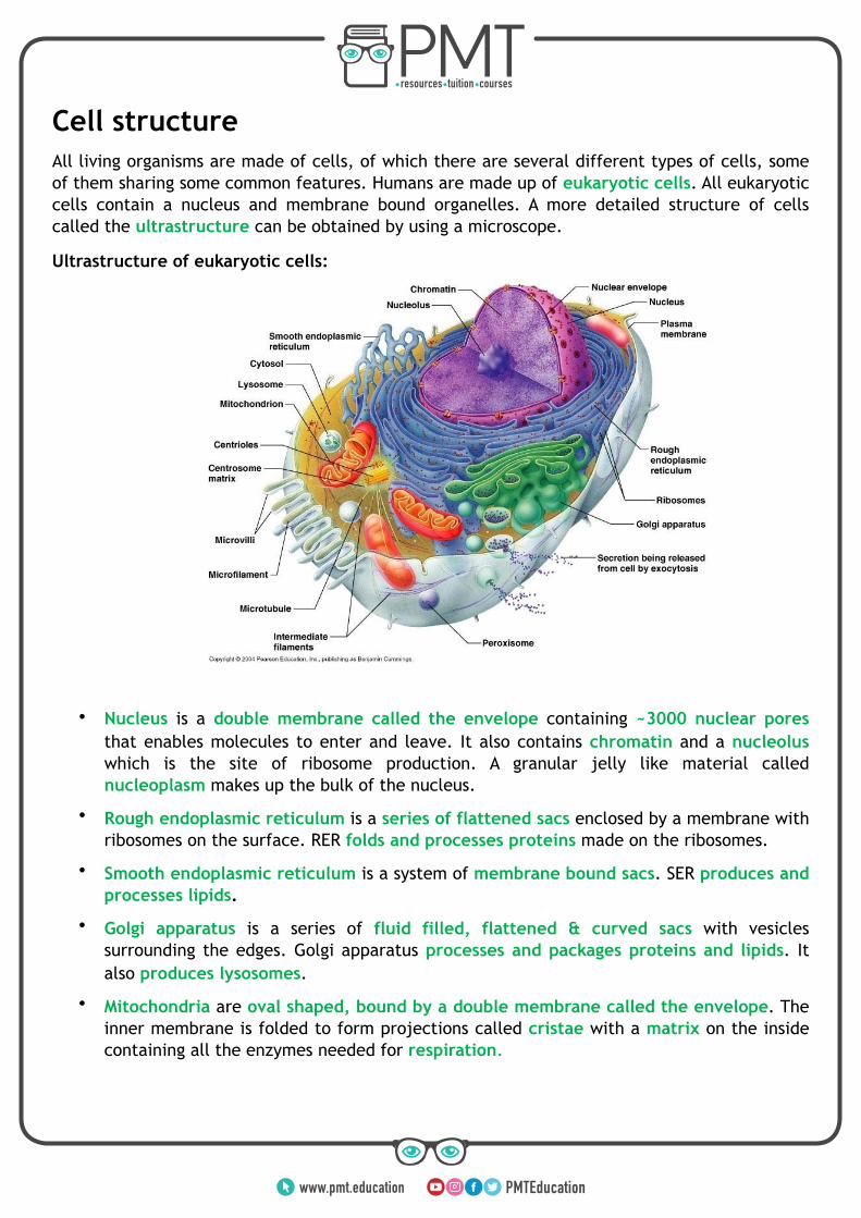
Ultrastructure of eukaryotic cells:
• Nucleus is a double membrane called the envelope containing ~3000 nuclear pores that enables molecules to enter and leave. It also contains chromatin and a nucleolus which is the site of ribosome production. A granular jelly like material called nucleoplasm makes up the bulk of the nucleus.
• Rough endoplasmic reticulum is a series of flattened sacs enclosed by a membrane with ribosomes on the surface. RER folds and processes proteins made on the ribosomes.
• Smooth endoplasmic reticulum is a system of membrane bound sacs. SER produces and processes lipids.
• Golgi apparatus is a series of fluid filled, flattened & curved sacs with vesicles surrounding the edges. Golgi apparatus processes and packages proteins and lipids. It also produces lysosomes.
• Mitochondria are oval shaped, bound by a double membrane called the envelope. The inner membrane is folded to form projections called cristae with a matrix on the inside containing all the enzymes needed for respiration.
www.pmt.education

• Centrioles are hollow cylinders containing a ring of microtubules arranged at right angles to each other. Centrioles are involved in producing spindle fibres for cell division.
• Ribosomes are composed of two sub units and are the site of protein production.
• Lysosomes are vesicles containing digestive enzymes bound by a single membrane.
Prokaryotic cells such as bacteria contain:
• Cell wall – Rigid outer covering made of peptidoglycan.
• Capsule – Protective slimy layer which helps the cell to retain moisture and adhere to surfaces.
• Plasmid – Circular piece of DNA.
• Flagellum - a tail like structure which rotates to move the cell.
• Pili - Hair-like structures which attach to other bacterial cells.
• Ribosomes - Site of protein production.
• Mesosomes - Infoldings of the inner membrane which contain enzymes required for respiration.
Viruses are non-living structures which consist of nucleic acid (either DNA or RNA) enclosed in a protective protein coat called the capsid, sometimes covered with a lipid layer called the envelope.
Cells of multicellular organisms are organised into tissues, tissues into organs and organs into systems.
Cell division – mitosis
The role of mitosis and the cell cycle is to produce identical daughter cells for growth and asexual reproduction. All the cells produced by mitosis are genetically identical therefore mitosis does not give rise to genetic variation.
There are three stages of the cell cycle:
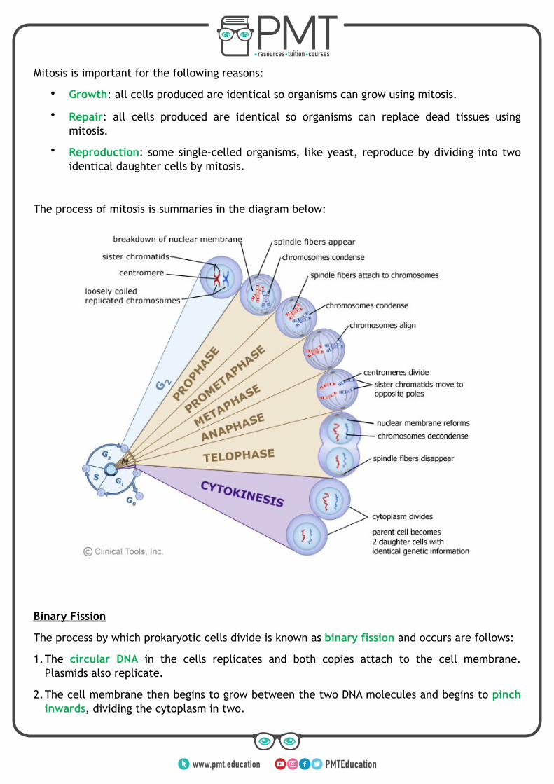
• Mitosis – mitosis is a form of cell division that produces identical cells, there are four stages of mitosis: prophase, metaphase, anaphase and telophase.
• Interphase – during this stage the cell grows and then prepares to divide – chromosomes and some organelles are replicated, chromosomes also begin to condense.
• Cytokinesis – during cytokinesis the parent and replicated organelles move to opposite sides of the cell and the cytoplasm divides thus producing two daughter cells.
www.pmt.education
Mitosis is important for the following reasons:
• Growth: all cells produced are identical so organisms can grow using mitosis.
• Repair: all cells produced are identical so organisms can replace dead tissues using mitosis.
• Reproduction: some single-celled organisms, like yeast, reproduce by dividing into two identical daughter cells by mitosis.
The process of mitosis is summaries in the diagram below:
Binary Fission
The process by which prokaryotic cells divide is known as binary fission and occurs are follows:
1.The circular DNA in the cells replicates and both copies attach to the cell membrane. Plasmids also replicate.
2.The cell membrane then begins to grow between the two DNA molecules and begins to pinch inwards, dividing the cytoplasm in two.
www.pmt.education

3.A new cell wall forms between the two DNA molecules dividing the original cell. The identical daughter cells each have a single copy of the circular DNA and a variable number of copies of the plasmids.
As viruses are non-living, they do not undergo cell division – following the injection of their nucleic acids into another cell, the infected host cell replicates the virus particles.
Biological membranes All cells and organelles are surrounded by a partially permeable membrane composed of a sea phospholipids with protein molecules between the phospholipid molecules. The main function of the membrane is controlling the movement of substances in and out of the cell/organelle. However, it also contains receptors for other molecules such as hormones and enables adjacent cells to stick together. The fluidity of the membrane and the mosaic arrangement of the protein give the structure of the membrane its name – fluid mosaic model.
The cell membrane is comprised of phospholipids in which the hydrophilic heads point outwards and the hydrophobic tails point inwards. This structure allows lipid soluble molecules to pass through the membrane, but not water soluble molecules. It also means that the membrane is flexible and self-sealing.
The membrane contains many other components, these are detailed below:
1. Proteins - these may be integrated throughout the membrane (intrinsic proteins) or may be on the surface
(extrinsic proteins). Intrinsic proteins include carrier proteins which allow
substances to cross the membrane. Proteins are present to aid movement across the membrane, provide mechanical support and act in conjunction with glycolipds as
receptors.
2. Cholesterol - Cholesterol is present to make the membrane more rigid and reduce the lateral movement of the
phospholipids. It also prevents the leakage of water and dissolved ions from the cell
as it is very hydrophobic.
3. Glycolipds - these are made up of a carbohydrate that is bound to lipids.
These extend from the surface of the cell and acts as a cell surface receptors for
certain molecules. They also allow cells to adhere to one another to form tissues.
4. Glycoproteins - These are carbohydrates that attach to extrinsic proteins and acts
a cell surface receptors and neurotransmitters. These allow cells to
recognise one another as well as attach to form tissues.
www.pmt.education

The movement of molecules through cell membrane depends on the properties of the molecule as well as the requirements of the cell. There are several types of movement:
• Diffusion is the passive movement of small, non-polar, lipid soluble molecules such as carbon dioxide and oxygen from an area of high concentration to an area of low concentration. The molecules move directly through the phospholipid bilayer.
• Facilitated diffusion requires a channel protein in the cell membrane to transport polar molecules, charged and water soluble molecules across the membrane.
• Osmosis is the diffusion of water molecules from an area of high water potential to an area to low water potential through a partially permeable membrane.
• Active transport can transport all types of molecules through carrier proteins from an area of low concentration to an area of high concentration. However, this process requires energy in the form of ATP.
• Exocytosis and endocytosis transport large particles. The particles are enclosed in vesicles made from the cell surface membrane and transported into the cell in endocytosis. In exocytosis, vesicles containing large particles are fused with the cell surface membrane and released from the cell.
• Co-Transport uses ions to move substances into and out of cells. This occurs particularly in epithelial cells of the ileum. Here sodium and potassium ions are pumped out of the epithelial cell by active transport into the blood leaving a lower concentration in the cell. This causes these ions to move in from the lumen by facilitated diffusion, which at the same time brings glucose and amino acids into the cell. These then diffuse from a high concentration in the epithelial cell to a low concentration in the blood.
The rate of gas exchange by diffusion becomes more rapid as: • The surface area of the surface
increases
• The diffusion distance decreases
• The diffusion gradient becomes more steep
• The temperature increases
Bacteria and viruses Bacteria and viruses are the main disease causing pathogens in humans. Even though they both cause disease, they vary in many ways. Their differences are as following:
• Bacteria are prokaryotic cells – their genetic information is stored in the form of a circular strand of DNA whereas viruses consist of just nucleic acid enclosed in a protein coat and their genetic material can take the form of DNA or RNA.
• Bacteria do not require a host to survive whereas viruses are entirely dependent on their hosts and cannot survive without them.
• Viruses are significantly smaller than bacteria.
• Bacteria have a cell membrane, cell wall and cytoplasm as well as other organelles such as ribosomes, plasmids, flagellum and pili whereas viruses possess no such structures.
www.pmt.education
An example of a bacterial disease is tuberculosis also known as TB. TB is caused by a bacteria called Mycobacterium tuberculosis which infects phagocytes in the lungs. The first infection is symptomless as the infected phagocytes are sealed in tubercles as a result of inflammatory response in the lungs. However, the bacteria lie dormant inside the tubercles as they are not destroyed by the immune system due to the tubercles being covered with a thick waxy coat. When the immune system becomes weakened, the bacteria become active again and slowly destroy the lung tissue thus leading to breathing problems, coughing, weight loss as well as fever. TB can potentially lead to death.
An example of a viral infection is HIV i.e. Human Immunodeficiency Virus which causes AIDS. The first symptoms of HIV include fevers, tiredness and headaches. After several weeks HIV antibodies appear in blood thus making a person HIV positive. After this period, the symptoms disappear until the immune system becomes weakened again thus leading to AIDS. Symptoms of AIDS include weight loss, diarrhoea, dementia, cancers and opportunistic infections such as TB.
Immune response Physical barriers to infection include:
• Skin which is a tough physical barrier consisting of keratin.
• Stomach acid (hydrochloric acid) which kills bacteria.
• Gut and skin flora – natural bacterial flora competes with pathogens for food and space.
The immune system responds to antigens which are found on the surface of cells. These are proteins that have a complex structure that is unique for each cell and therefore identifies the cell as self or non-self.
Non Specific Immune Response
Non-specific responses of the body to infection include:
• Inflammation – histamines released by damaged white tissues cause vasodilation which increases the flow of blood to the infected area and increases permeability of blood vessels. As a result of that antibodies, white blood cells and plasma leak out into the infected tissue and destroy the pathogen.
• Lysozyme action – lysozyme are enzymes found in secretions such as tears and mucus which kill bacterial cells by damaging their cell wall.
• Interferon – interferons prevent viruses spreading to uninfected cells by stopping protein synthesis in viruses.
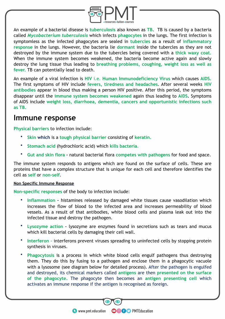
• Phagocytosis is a process in which white blood cells engulf pathogens thus destroying them. They do this by fusing to a pathogen and enclose them in a phagocytic vacuole with a lysosome (see diagram below for detailed process). After the pathogen is engulfed and destroyed, its chemical markers called antigens are then presented on the surface of the phagocyte. The phagocyte then becomes an antigen presenting cell which activates an immune response if the antigen is recognised as foreign.
www.pmt.education
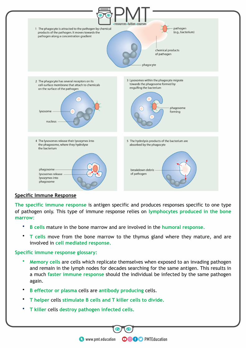
Specific Immune Response
The specific immune response is antigen specific and produces responses specific to one type of pathogen only. This type of immune response relies on lymphocytes produced in the bone marrow:
• B cells mature in the bone marrow and are involved in the humoral response.
• T cells move from the bone marrow to the thymus gland where they mature, and areinvolved in cell mediated response.
Specific immune response glossary:
• Memory cells are cells which replicate themselves when exposed to an invading pathogenand remain in the lymph nodes for decades searching for the same antigen. This results ina much faster immune response should the individual be infected by the same pathogenagain.
• B effector or plasma cells are antibody producing cells.
• T helper cells stimulate B cells and T killer cells to divide.
• T killer cells destroy pathogen infected cells.
www.pmt.education

Cell mediated response
Humoral response
www.pmt.education

Antibodies
In the humoral response plasma cells produce antibodies. These are made of four polypeptides chains forming a Y shaped structure. These are complementary to only a single antigen. They work by forming an antigen-antibody complex which serve as markers for phagocytes to destroy attached cells. Due to antibodies having two binding sites they can also clump cells together making them easier for phagocytes to find. This process is called agglutination.
Immunity Immunity can either be active or passive; active immunity results from the production of antibodies by the immune system in response to the presence of an antigen whereas passive immunity results from the introduction of antibodies from another person or animal. There are also two subtypes of immunity; natural or artificial:
• Natural active immunity arises from being exposed to an antigen/getting the diseasewhereas natural passive immunity is the result of crossing of mother’s antibodiesthrough the placenta and their presence in breast milk.
• Active artificial immunity is acquired through vaccinations which stimulate the immunesystem and lead to production of antibodies whereas passive artificial immunity is whereantibodies are injected into the body.
Vaccines
Vaccines are a way of introducing a pathogen into the body in order to produce an immune response. The pathogen may be dead of inactivated, but the antigens on its surface will still produce an immune response. This is an example of active immunity and results in the creation of memory B cells which will be able trigger a rapid secondary immune response should the same pathogen ever be detected again.
The success of a vaccination program is dependent on a number of factors such as:
- Cost of the vaccine
- Severity of the side effects
- Ease of production, transportation and administration
- Number of people who need to be vaccinated for herd immunity
The concept of herd immunity is that if you vaccinate enough people in the population then eventually the pathogen won’t be able to be transmitted from different hosts. This therefore means that those who aren’t vaccinated are protected by those around them who are.
www.pmt.education

However vaccines are not always useful in preventing a disease outbreak. This is because the antigen on the surface of the pathogen can change therefore removing immunity. An example of a virus that does this is Influenza. This virus rapidly changes it antigens and therefore immunity against this virus is only short lived.
Ethical Considerations
There are a number of ethical consideration to take into account when looking at vaccines, these include:
- Production and testing of vaccines may be done on animals
- The risks of the vaccine need to be balanced with the benefits
- The vaccine must be tested on humans first to determine toxicity
- Vaccinations are very expensive
- Should vaccinations be compulsory or should people be able to opt out of having a vaccination?
Monoclonal Antibodies
Due to there being many different types of antigens, each B cell will produce a different complementary antibody. It is therefore medically useful to be able to produce many clones of a single type of antibody. These are known as monoclonal antibodies.
They have many different uses are seen below:
Direct Therapy - Monoclonal antibodies that are specific to antigens found on the surface of cancerous cells can be used to target and then destroy the cells as part of an immune response. For example, herceptin targets breast cancer cells.
Indirect therapy - Drugs can be attached to monoclonal antibodies such as a cytotoxic drug. The antibody then is used to direct the drug towards the cells displaying a particular antigen rather than towards other cells.
Diagnosis - Particular antigens are targeted by antibodies to measure levels of that antigen in the body.
Pregnancy testing - Monoclonal antibodies in home pregnancy kits are specific to the hormone human chorionic gonadotrophin.
HIV
Human immunodeficiency virus (HIV) is a pathogen that can lead to the disease acquired immune deficiency syndrome (AIDS). HIV has the following structure:
- A lipid envelope with embedded attachment proteins.
- Inside a protein capsid where the genetic material (RNA) and reverse transcriptase enzymes are present. This catalyses the production of DNA from the RNA.
www.pmt.education

In order to replicate the HIV virus binds to the protein CD4 which is most frequently found on T-Helper cells. The capsid then fuses with the cell surface membrane and the RNA and reverse transcriptase enter the cell. The reverse transcriptase converts the RNA to DNA, which then moves into the nucleus of the cell. The cell now has the instructions to begin producing viral HIV components.
To i d e n t i f y H I V a n E n z y m e L i n k e d Immunosorbent Assay (ELISA) can be used. This detects the presence and quantity of the antigen found on HIV. An ELISA is shown in the digram below:
Antibiotics are ineffective against HIV and viruses. This is because many antibiotics work by preventing bacteria from making cells walls. Without this the bacterial cell cannot control the entry and exit of water and will therefore burst. However since viruses don’t have a cell wall and are reproduced within a host cell they are unaffected by antibiotics. Instead HIV is treated with antiretroviral drugs which keep the levels of HIV in the blood stream very low, reducing the impact on the hosts immune system.
www.pmt.education