Embed Size (px)
Citation preview

Employing Multiple Spectroscopic Techniques Simultaneously to Observe
Protein Unfolding
Brennan CullAdvisor: Dr. Justin J Link
Biophysics ProgramXavier UniversityCincinnati, OH

• A protein is a chain of amino acids
– Twenty different amino acids
• Proteins are essential to life
– Variety of biological functions• Catalysis, Structure, Protection (Immune System),
and Regulating Cell Division
• Diseases caused by protein misfolding
– Alzheimer’s Disease
– Huntington’s Disease
– Atherosclerosis
– Type II Diabetes
– Many types of cancers
Petsko, G. A., and Ringe, D. Protein Structure and Function. New
Science Press, 2004. Print
Background InformationEmail: [email protected]

PurposeDevelop a cost-efficient way to study the structure and stability of a protein as it unfolds
Overview of Procedure
• Titration that increases the concentration guanidine hydrochloride at each step in order to unfold the protein, cytochrome c, and a couple of its mutants
• Measure the following in one, automated scan:
– Circular Dichroism (CD)
– Absorbance
– Fluorescence
To help support or disclaim the results of published literature
Email: [email protected]

Equine Cytochrome c
• Model System
• Well characterized
• Relatively small in size
• Single Tryptophan
molecule
• Cofactor: Heme
group
Zang C., et al. (2009) J Am Chem Soc 131(8):2846–2852
Email: [email protected]

Mutants
W89
W66
W77W72
W82
W36
W23
W102W45
W15
W51
Location of the Tryptophan within Wild-Type Cytochrome c
W59
Email: [email protected]

Spectroscopic Techniques
Fluorescence monitors proximity of hemerelative to tryptophan
Zang C., et al. (2009) J Am ChemSoc 131(8):2846–2852
Absorbance monitors the local environment of the heme
Circular dichroism determines the components of secondary structure
W59
Email: [email protected]

Data Analysis (Wild-Type)
320 340 360 380 400 420
0.0
0.1
0.2
0.3
0.4
GdnH
Cl (
M)
Wavelength (nm)
Flu
or.
(A
U)
220 225 230 235 240
-100
-80
-60
-40
-20
0
GdnH
Cl (
M)
Wavelength (nm) C
D (
md
eg
)
380 400 420 440
0.0
0.2
0.4
0.6
0.8
1.0
1.2
1.4
GdnH
Cl (
M)
Wavelength (nm)
Ab
s.
(OD
)
222 nm403.1nm
350nm
0 1 2 3 4 5-0.1
0.0
0.1
0.2
0.3
0.4
Flu
or.
at
35
0n
m
GdnHCl (M)
Email: [email protected]

0 1 2 3 4 5-0.1
0.0
0.1
0.2
0.3
0.4
Flu
or.
at
35
0n
m
GdnHCl (M)
Data Analysis (Wild-Type Cross Section)
0 1 2 3 4 5-0.1
0.0
0.1
0.2
0.3
0.4
Flu
or.
at
35
0n
m
GdnHCl (M)
0 1 2 3 4 5
0.85
0.90
0.95
1.00
1.05
1.10
1.15
1.20
Ab
s.
at
40
3.1
nm
GdnHCl (M)
0 1 2 3 4 5
-100
-80
-60
-40
-20
0
CD
at
22
2n
m
GdnHCl (M)
0 1 2 3 4 5
0.85
0.90
0.95
1.00
1.05
1.10
1.15
1.20
Ab
s.
at
40
3.1
nm
GdnHCl (M)0 1 2 3 4 5
-100
-80
-60
-40
-20
0
CD
at
22
2n
m
GdnHCl (M)
𝑆𝑜𝑏𝑠
=𝐶𝑓 +𝑚𝑓 𝐺𝑑𝑛𝐻𝐶𝑙 + 𝐶𝑢 +𝑚𝑢 𝐺𝑑𝑛𝐻𝐶𝑙 𝑒
−Δ𝐺+𝑚𝑔[𝐺𝑑𝑛𝐻𝐶𝑙]
𝑅𝑇
1 + 𝑒−Δ𝐺+𝑚𝑔[𝐺𝑑𝑛𝐻𝐶𝑙]
𝑅𝑇
Email: [email protected]

0 1 2 3 4 5
0.0
0.2
0.4
0.6
0.8
1.0
Abs. at 403.1nm (OD)
Fra
cti
on
Un
fold
ed
GdnHCl (M)0 1 2 3 4 5
0.0
0.2
0.4
0.6
0.8
1.0
CD at 222nm (mdeg)
Fra
cti
on
Un
fold
ed
GndHCl (M)
0 1 2 3 4 5
0.0
0.2
0.4
0.6
0.8
1.0
Fluor. at 350nm (AU)
Fra
cti
on
Un
fold
ed
GdnHCl (M)
Technique ΔG (kcal/mol) Cm (M)
CD 7.83 2.30
Absorbance 7.12 2.25
Fluorescence 7.71 2.34
Published (CD) 7.27 2.42
Published (HX) 7.40 N/A
Knapp, JA and Pace CN. Biochemistry 13, 1289-94. (1974).
Maity, H et al. J Mol Biol 343, 223-33. (2004).
Data Analysis (Wild-Type Cross Section)
Email: [email protected]

Fraction Unfolded (Wild-Type)
Technique ΔG (kcal/mol) Cm (M)
CD 7.83 2.30
Absorbance 7.12 2.25
Fluorescence 7.71 2.34
Published (CD) 7.27 2.42
Published (HX) 7.40 N/A
Global Fit 7.83 2.30
0 1 2 3 4 5-0.2
0.0
0.2
0.4
0.6
0.8
1.0
Individual Fit
CD Fit
CD Data
Abs Fit
Abs Data
Fluor Fit
Fluor Data
Fra
cti
on
Un
fold
ed
GdnHCl (M)0 1 2 3 4 5
-0.2
0.0
0.2
0.4
0.6
0.8
1.0
Global FIt
Global Fit
CD
Absorbance
Fluorescence
Fra
cti
on
Un
fold
ed
GdnHCl (M)
Knapp, JA and Pace CN. Biochemistry 13, 1289-94. (1974).
Maity, H et al. J Mol Biol 343, 223-33. (2004).
Email: [email protected]

0 1 2 3 4 5
0.0
0.2
0.4
0.6
0.8
1.0
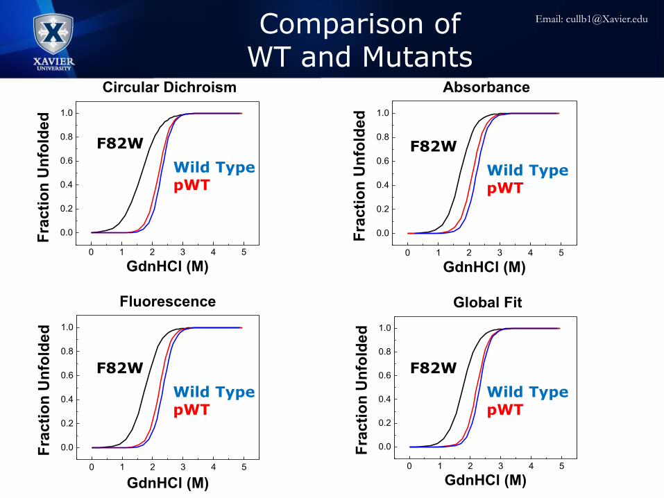
Circular Dichroism
Fra
cti
on
Un
fold
ed
GdnHCl (M)
F82W
pWT
Wild-Type
0 1 2 3 4 5
0.0
0.2
0.4
0.6
0.8
1.0
Global Fit
Fra
cti
on
Un
fold
ed
GdnHCl (M)
F82W
pWT
Wild-Type
0 1 2 3 4 5
0.0
0.2
0.4
0.6
0.8
1.0
Fluorescence
Fra
cti
on
Un
fold
ed
GdnHCl (M)
F82W
pWT
Wild-Type
0 1 2 3 4 5
0.0
0.2
0.4
0.6
0.8
1.0
Absorbance
Fra
cti
on
Un
fold
ed
GdnHCl (M)
F82W
pWT
Wild-Type
Wild TypepWT
F82W
Wild TypepWT
F82W
Wild TypepWT
F82W
Wild TypepWT
F82W
Comparison of WT and Mutants
Email: [email protected]

Acknowledgements
• Dr. Justin J Link
• Ben Kelty
• Previous Research Students
• Xavier University Physics Department
• John Hauck Foundation
Email: [email protected]
0 1 2 3 4 5
0.0
0.2
0.4
0.6
0.8
1.0
Circular Dichroism
Fra
cti
on
Un
fold
ed
GdnHCl (M)
F82W
pWT
Wild-Type
0 1 2 3 4 5
0.0
0.2
0.4
0.6
0.8
1.0
Global Fit
Fra
cti
on
Un
fold
ed
GdnHCl (M)
F82W
pWT
Wild-Type
0 1 2 3 4 5
0.0
0.2
0.4
0.6
0.8
1.0
Fluorescence
Fra
cti
on
Un
fold
ed
GdnHCl (M)
F82W
pWT
Wild-Type
0 1 2 3 4 5
0.0
0.2
0.4
0.6
0.8
1.0
Absorbance
Fra
cti
on
Un
fold
ed
GdnHCl (M)
F82W
pWT
Wild-Type
Comparison of WT and Mutants
Wild TypepWT
F82W
Wild TypepWT
F82W
Wild TypepWT
F82W
Wild TypepWT
F82W
Email: [email protected]