Embed Size (px)
Citation preview
Antitumor Activity of Gemcitabine and Oxaliplatin Is Augmented by
Thymoquinone in Pancreatic Cancer
Sanjeev Banerjee,1Ahmed O. Kaseb,
3Zhiwei Wang,
1Deujan Kong,
1Mussop Mohammad,
2
Subhash Padhye,1Fazlul H. Sarkar,
1and Ramzi M. Mohammad
2
1Department of Pathology and 2Division of Hematology and Oncology, Barbara Ann Karmanos Cancer Institute, Wayne State UniversitySchool of Medicine, Detroit, Michigan; and 3Department of Gastrointestinal Medical Oncology, University of TexasM. D. Anderson Cancer Center, Houston, Texas
Abstract
Previous studies have shown biological activity of thymoqui-none, an active compound extracted from Nigella sativa , inpancreatic cancer cells; however, preclinical animal studiesare lacking. Here, we report, for the first time, the chemosen-sitizing effect of thymoquinone to conventional chemothera-peutic agents both in vitro and in vivo using an orthotopicmodel of pancreatic cancer. In vitro studies revealed thatpreexposure of cells with thymoquinone (25 Mmol/L) for48 h followed by gemcitabine or oxaliplatin resulted in 60%to 80% growth inhibition compared with 15% to 25% whengemcitabine or oxaliplatin was used alone. Moreover, wefound that thymoquinone could potentiate the killing of pan-creatic cancer cells induced by chemotherapeutic agents bydown-regulation of nuclear factor-KB (NF-KB), Bcl-2 family,and NF-KB-dependent antiapoptotic genes (X-linked inhib-itors of apoptosis, survivin, and cyclooxygenase-2). As shownpreviously by our laboratory, NF-KB gets activated on expo-sure of pancreatic cancer cells to conventional chemother-apeutic agents; interestingly, thymoquinone was able todown-regulate NF-KB in vitro , resulting in chemosensitization.In addition to in vitro results, here we show for the first time,that thymoquinone in combination with gemcitabine and/oroxaliplatin is much more effective as an antitumor agentcompared with either agent alone. Most importantly, ourdata also showed that a specific target, such as NF-KB, wasinactivated in animal tumors pretreated with thymoquinonefollowed by gemcitabine and/or oxaliplatin. These resultsprovide strong in vivo molecular evidence in support of ourhypothesis that thymoquinone could abrogate gemcitabine-or oxaliplatin-induced activation of NF-KB, resulting in thechemosensitization of pancreatic tumors to conventionaltherapeutics. [Cancer Res 2009;69(13):5575–83]
Introduction
Pancreatic cancer is the fourth major cause of cancer-relateddeaths worldwide, with a 5-year survival rate of <3%. According toNIH-National Cancer Institute statistics, of the 38,000 new cases ofpancreatic cancer diagnosed in 2008, 35,000 are expected tosuccumb to this disease. Despite the availability of myriad oftreatment modalities, including preferred cytotoxic chemotherapy,dose-limiting toxicity to normal tissues and acquisition of acquired
resistance fails to transcend into optimal clinical benefit in termsof cure rate in an overwhelming majority of patients (1). Emergingevidence suggest DNA-binding ability of the transcription factornuclear factor-nB (NF-nB) as a major mechanism contributing tochemoresistant phenotype of pancreatic cancer (2–4) and thatseveral conventional cancer chemotherapeutic agents activate NF-nB leading to unfavorable clinical outcome (4–8). In recent years,naturally occurring compounds are being investigated becauseof their alleged therapeutic effects attributable to inactivation ofNF-nB and other survival signaling cascade within tumors andfewer associated toxicities (5, 9, 10). Here, we report, for the firsttime, the therapeutic benefit and mechanism of action of a nat-urally occurring compound thymoquinone in sensitizing pancreaticcancer cells to conventional cytotoxic drugs.Thymoquinone is the bioactive compound derived from black seed
(Nigella sativa) oil. In folklore medicine, the seed is reportedlyassociated with diverse therapeutic benefits as related to bronchialasthma, dysentery, headache, gastrointestinal problems, eczema,hypertension, and obesity. We recently reported in a comprehensivereview the therapeutic and chemopreventive potential of blackcumin seeds (11). In the context of cancer, thymoquinone has beenreported to exhibit antiproliferative effects on cell lines derived frombreast, colon, ovarian, larynx, lung, myeloblastic leukemia, andosteosarcoma (12–18). Additionally, thymoquinone inhibited hor-mone-refractory prostate cancer by targeting androgen receptor andtranscription factor E2F (19). Mechanistically, thymoquinone report-edly induced apoptosis in tumor cells by suppressing NF-nB, Aktactivation, and extracellular signal-regulated kinase signaling path-ways and also inhibits tumor angiogenesis (20, 21). These limitedstudies suggest that thymoquinone could be useful as an adjunct toconventional chemotherapeutics; however, in vivo studies in supportof this statement are lacking and our current findings filled that gap.Gemcitabine is the first-line conventional chemotherapy for
pancreatic cancer, but oxaliplatin is used as an alternative option(22, 23). Conventional dosing with either cytotoxic drug given atmaximum tolerated dose caused side effects and fails to respondadequately due to acquisition of chemoresistance phenomenon(23, 24). In the present study, first we sought to understand themolecular mechanism of action of thymoquinone in pancreaticcancer cells in inducing apoptosis and then examined the chemo-sensitization potential of thymoquinone in combination withoxaliplatin and gemcitabine to reduce viable cells and potentiateapoptosis using low concentrations of chemotherapeutic drugs.Importantly, we also tested our hypothesis in vivo using an ortho-tropic model of pancreatic cancer in severe combined immuno-deficient (SCID) animals. Using in vivo imaging data, we show, forthe first time, that thymoquinone pretreatment in combinationwith either gemcitabine or oxaliplatin caused greater antitumoractivity than each drug given alone. These results correlated with
Requests for reprints: Ramzi M. Mohammad, Room 732, HWCRC Building, 4100John R. Street, Detroit, MI 48201. Phone: 313-576-8329; Fax: 313-576-8389; E-mail:[email protected].
I2009 American Association for Cancer Research.doi:10.1158/0008-5472.CAN-08-4235
www.aacrjournals.org 5575 Cancer Res 2009; 69: (13). July 1, 2009
Research Article
Retrac
ted S
eptem
ber 1
4, 20
18
Research. on May 15, 2020. © 2009 American Association for Cancercancerres.aacrjournals.org Downloaded from
Published OnlineFirst June 23, 2009; DOI: 10.1158/0008-5472.CAN-08-4235
Research. on May 15, 2020. © 2009 American Association for Cancercancerres.aacrjournals.org Downloaded from
Published OnlineFirst June 23, 2009; DOI: 10.1158/0008-5472.CAN-08-4235
Research. on May 15, 2020. © 2009 American Association for Cancercancerres.aacrjournals.org Downloaded from
Published OnlineFirst June 23, 2009; DOI: 10.1158/0008-5472.CAN-08-4235

down-regulation of NF-nB activity and its downstream proteinssuch as survivin, Bcl-xL, and X-linked inhibitor of apoptosis (XIAP)in tumor extracts.
Materials and Methods
Cell culture. The human pancreatic cancer cell lines BxPC-3 and HPACwere obtained from the American Type Culture Collection. Human
pancreatic ductal epithelial cells and COLO-357 were obtained from M. D.
Anderson Cancer Center. The cell lines were maintained in continuous
exponential growth in DMEM (Life Technologies) supplemented with 10%fetal bovine serum, 100 units/mL penicillin, and 10 mg/mL streptomycin in
a humidified incubator containing 5% CO2 in air at 37jC.Antibodies were obtained from the following commercial sources:
caspase-3, caspase-9, cytochrome c , cytochrome c oxidase, and p65 (Cell
Signaling); anti-mouse Bcl-2, Bcl-xL, Bax, Mcl-1, and anti-retinoblastoma
antibody (Santa Cruz Biotechnology); anti-poly(ADP-ribose) polymerase
(PARP) antibody (Biomol Research); and anti-h-actin (Sigma). Thymoqui-none (Sigma) was dissolved in DMSO to make 20 mmol/L stock solution.
Oxaliplatin and gemcitabine were obtained from the Barbara Ann
Karmanos Cancer Institute pharmacy.
Cell viability inhibition by thymoquinone. Cells (3 � 103 per well) in96-well culture plates were seeded and replaced next day with fresh medium
containing thymoquinone (0-50 Amol/L) diluted from stock solution. After
72 h, MTT solution was added and incubated further for 2 h. MTT formazanformed by metabolically viable cells was dissolved in isopropanol, and
absorbance was measured at 595 nm on a plate reader (TECAN).
Cell viability inhibition by cytotoxic agents. Cells were plated and
incubated with medium containing thymoquinone (25 Amol/L) for 48 h andexposed to 500 nmol/L gemcitabine or 62 Amol/L oxaliplatin for an
additional 24 h. The effect of thymoquinone pretreatment on cell viability
was examined by MTT as described above. To investigate the synergistic
effect between thymoquinone, gemcitabine, and oxaliplatin, cells wereexposed to drugs in fixed ratio as above and combination index (CI) was
calculated using CalcuSyn software (Biosoft).
Quantification of apoptosis. The Cell Apoptosis ELISA Detection Kit(Roche) was used to detect apoptosis in pancreatic cancer according to the
manufacturer’s protocol.
Protein extraction and Western blot analysis. HPAC cells were plated
and allowed to attach for 36 h. Thymoquinone was directly added to cellcultures at the indicated concentrations and incubated for 72 h. Total
protein (40 Ag) was separated on SDS-PAGE, electrotransferred, and probed
with specific antibodies.
Analysis of cytochrome c release. Mitochondria and cytosolic fractionswere obtained as described previously (6) to detect cytochrome c release by
Western immunoblotting.
Caspase activity assays. Caspase-9 and caspase-3 activities were
measured in HPAC cells and tissue lysates by a colorimetric assay accordingto the manufacturer’s protocol (R&D Systems).
Determination of prostaglandin E2. HPAC cells were seeded in 50 mm
dishes and treated with thymoquinone (0-50 Amol/L) for 72 h in serum-freemedium. Conditioned medium was collected and analyzed for prostaglan-
din E2 (PGE2) using PGE2 high-sensitivity immunoassay kit (R&D Systems).
Cell cycle analysis. HPAC cells were treated with thymoquinone for
48 h followed by either gemcitabine (500 nmol/L) or oxaliplatin (62 Amol/L)for 24 h. The fixed cells were stained with propidium iodide for 30 min and
analyzed by flow cytometry.
Electrophoretic mobility shift assay. Nuclear extracts were prepared
from treated samples and electrophoretic mobility shift assay was done byincubating 10 Ag nuclear extract with IRDye-700-labeled NF-nB oligonucle-
otide as described earlier (6).
Experimental animals and orthotopic implantation of tumor cells.Female ICR/SCID mice were purchased from Taconic Farms. HPAC cells
were harvested from subconfluent cultures, washed in serum-free medium,
and resuspended in PBS. Cells (1 � 106) in 15 AL PBS were injected into the
parenchyma of pancreas with a 27-gauge hypodermic needle.
Experimental protocol. Mice were randomized into the following treat-ment groups (n = 7): (a) untreated control; (b) thymoquinone 3 mg/mouse
given daily by intragastric intubation for 25 days; (c) oxaliplatin 5 mg/kg
body weight, intraperitoneally, given twice per week; (d) gemcitabine
50 mg/kg body weight, intraperitoneally, given thrice per week; and (e) and( f ) thymoquinone and gemcitabine or oxaliplatin following schedule as for
individual treatments. All mice were sacrificed on day 35 following last dose
of thymoquinone, and their body weight was recorded. On autopsy, the
pancreas was excised neatly and weighed. One part of the tissue was fixed informalin and another part was frozen in liquid nitrogen.
Mice imaging. EGF-IRDye-800CW (epidermal growth factor receptor
antibody) was delivered via tail vein (1 nmol/L per mice) 96 h before eu-
thanizing the animals. Near IR fluorescence imaging of live animals wasdone using Odyssey Infrared Imaging System.
Tumor tissue nuclear protein extraction and electrophoreticmobility shift assay. Nuclear protein extracts were prepared from tumortissues and electrophoretic mobility shift assay was done by incubating
12 Ag nuclear extract with IRDye-700-labeled NF-nB oligonucleotide as
described earlier (5).
Histopathology and immunohistochemistry. H&E and immunohisto-chemistry for Ki-67 and terminal deoxynucleotidyl transferase-mediated
dUTP nick end labeling (TUNEL) for apoptosis were done on paraffin-
embedded tissue sections.
Statistical analysis. Data are represented as meanF SD for the absolutevalues or percent of controls as indicated in the vertical axis legend of
Figs. 1 to 6. Statistical significance was determined by Student’s t test.
P < 0.05 were considered statistically significant.
Results
Effect of thymoquinone on cell viability and apoptosisinduction. In Fig. 1A , thymoquinone inhibited cell viability in adose-dependent manner. In almost all pancreatic cancer cell lines,cell viability was reduced up to 70% with 50 Amol/L thymoquinonetreatment for 72 h. In contrast, treatment of human pancreaticductal epithelial cells resulted in minimal loss of viable cell whenexposed to identical concentrations of thymoquinone for a similarperiod. BxPC-3 cells harboring wild-type K-ras oncogene andmutated p53 was sensitive to the effect of thymoquinone in asmuch as pancreatic cancer cells with mutant k-ras and wild-typep53 (HPAC and COLO-357). Further, to assess whether the loss ofcell viability could in part be due to apoptosis, we evaluatedapoptosis using histone-DNA ELISA. Figure 1B shows a significantincrease in apoptotic cells, which closely parallels the loss of viablecells following thymoquinone treatment. In contrast, humanpancreatic ductal epithelial cells were minimally affected toapoptosis by equivalent concentrations of thymoquinone.
Thymoquinone inhibits antiapoptotic molecules in HPACcells. To understand the molecular basis of thymoquinone-inducedapoptosis, we performed Western immunoblotting using HPACcells as representative. As shown in Fig. 2A , the proapoptoticprotein Bax was markedly induced, whereas Bcl-2 and Bcl-xL weresignificantly inhibited in a concentration-dependent manner,indicating that the apoptotic effects of thymoquinone are partlycaused by up-regulating the Bax/Bcl-2 or Bax/Bcl-xL protein ratio,which is a critical determinant of apoptosis. Additionally, weassessed other antiapoptotic molecules Mcl-1, survivin, and XIAP.Relative to control, Mcl-1, survivin, and XIAP expression was down-regulated in cells exposed to thymoquinone. These results provideadditional molecular evidence of apoptosis induction by thymo-quinone treatment.Western immunoblotting also revealed thymoquinone treatment
resulted in appearance of cleaved active component of caspase-3,caspase-9, and PARP in HPAC cells (Fig. 2A). In parallel, caspase-3
Cancer Research
Cancer Res 2009; 69: (13). July 1, 2009 5576 www.aacrjournals.org
Retrac
ted S
eptem
ber 1
4, 20
18
Research. on May 15, 2020. © 2009 American Association for Cancercancerres.aacrjournals.org Downloaded from
Published OnlineFirst June 23, 2009; DOI: 10.1158/0008-5472.CAN-08-4235

and caspase-9 activities were also significantly elevated (Fig. 2B).An upstream event in activation of the caspase cascade is therelease of cytochrome c from mitochondria. Thymoquinone in-duced release of cytochrome c in dose-responsive manner (Fig. 2A),suggesting that thymoquinone-induced apoptosis in HPAC cells ismediated at least in part by the mitochondrial pathway.
Inhibition of cyclooxygenase-2 expression and PGE2 accu-mulation by thymoquinone. The cyclooxygenase-2 (COX-2)enzyme is overexpressed in pancreatic cancer and is involved inthe inhibition of apoptosis, potentiation of cell growth, and angio-genesis and thus considered as a putative target for drug dev-elopment (25). To determine whether thymoquinone reduces theexpression of COX-2 and consequently the production of PGE2,COX-2-expressing HPAC cells were treated with thymoquinone.Western immunoblotting showed a significant decrease in COX-2protein expression in a dose-dependent manner (Fig. 2A). Further,to confirm whether the catalytic activity of COX-2 and thus theproduction of PGE2 were inhibited by thymoquinone or not, culturesupernatant was collected from exponentially growing HPAC cellsin the presence of thymoquinone, and the PGE2 levels were mea-sured. As shown in Fig. 2C , the synthesis of PGE2 was significantlydecreased in a dose-dependent manner after thymoquinonetreatment, which well correlated with the down-regulation ofCOX-2 expression. These results are consistent with the ability ofthymoquinone in inhibiting cell growth and induction of apoptosis.
Thymoquinone inhibits activation of NF-KB. To investigatewhether thymoquinone could abrogate constitutively active NF-nBin pancreatic cancer cells, cells were treated with increasing dosesof thymoquinone (0-40 Amol/L) for 48 h and subjected to gel shiftassay (electrophoretic mobility shift assay). Thymoquinone resultedin a concentration-dependent decrease in NF-nB DNA-bindingactivity in HPAC and BxPC-3 cells (Fig. 2D), which was furtherconfirmed by supershift experiment. These results are consistentwith down-regulation of COX-2, survivin, and Bcl family proteinsbecause these proteins are transcriptionally regulated by NF-nBand contribute to the survival of cells by inhibiting apoptosis.
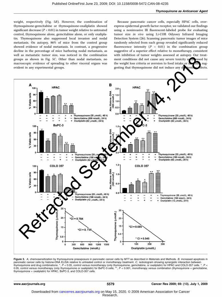
Thymoquinone sensitizes pancreatic cancer cells to chemo-therapeutic agents. The effect of gemcitabine or oxaliplatin onthe viability of pancreatic cancer cells was determined by MTTassay (data not shown). Subsequent studies were undertaken toexamine whether cells pretreated with thymoquinone could bemore sensitive to gemcitabine and oxaliplatin. Cells were eithertreated with thymoquinone (25 Amol/L alone for 48 h) followed by24 h incubation with suboptimal dose of either drugs, gemcitabineand oxaliplatin, and cell viability was determined (Fig. 3A). Wenoticed treatment of cells with either thymoquinone for 48 h orgemcitabine and oxaliplatin alone for 24 h caused 15% to 50% lossof viable pancreatic cancer cells depending on cell type. However,pretreatment with thymoquinone followed by cytotoxic chemo-therapeutic drugs resulted in 65% to 80% (P < 0.001) loss of viablecells in all pancreatic cancer cell evaluated, which we believe maybe associated with less toxic side effects on normal cells.Interestingly, pretreatment with thymoquinone proved moreefficacious in sensitizing the cells to cytotoxic agents comparedwith cotreatment or washing away thymoquinone before addingdrugs (data not shown). Further, to confirm synergism, we deter-mined CI values for two combination treatment groups. CI is aquantitative measure of the degree of drug interaction; CI < 1indicates synergism, CI > 1 indicates antagonism, and CI = 1indicates additive effect. Our results show that cells pretreated withthymoquinone showed synergistic loss of the cell viability whencombined with gemcitabine or oxaliplatin (CI = 0.735 and 0.784 forthymoquinone and gemcitabine and 0.545 and 0.685 for thymo-quinone and oxaliplatin).
Thymoquinone sensitizes HPAC cells to apoptosis bygemcitabine and oxaliplatin. We next confirmed whetherenhanced cytotoxicity by thymoquinone pretreatment was medi-ated by apoptosis. Accordingly, relative to single agent, thymoqui-none pretreatment (25 Amol/L) followed by either gemcitabine oroxaliplatin elicited significantly (V30-50%; P < 0.01) higher apop-tosis in all investigated pancreatic cancer cell lines, suggesting thatthe loss of viable cells by thymoquinone and gemcitabine/or oxa-liplatin results due to the induction of cell death pathway (Fig. 3D).
Thymoquinone pretreatment enhances apoptosis by oxali-platin and gemcitabine in HPAC cells. To understand sensitiza-tion mechanism and apoptosis by thymoquinone pretreatment,we assessed by Western immunoblotting the status on caspase-3,caspase-9, and PARP cleavage and modulation of other prosur-vival molecules (Fig. 4A). Cells were exposed to thymoquinone(25 Amol/L) for 48 h before addition of gemcitabine or oxaliplatinfor 24 h. Our data showed that whereas treatment of HPAC cellswith single agents showed appearance of cleaved caspase-3, 9, andPARP, thymoquinone pretreatment significantly augmented andshowed comparatively strong band for cleaved caspase-3, caspase-9,and PARP (Fig. 4A). Additionally, caspase-3 activity was alsoobserved significantly elevated in the combination treatment group
Figure 1. Evaluation of cell viability by MTT (A ) and histone DNA ELISAfor apoptosis (B ) by thymoquinone treatment for 72 h. *, P < 0.05; **, P <0.01. HPDE, human pancreatic ductal epithelial cells.
Thymoquinone as Anticancer Agent
www.aacrjournals.org 5577 Cancer Res 2009; 69: (13). July 1, 2009
Retrac
ted S
eptem
ber 1
4, 20
18
Research. on May 15, 2020. © 2009 American Association for Cancercancerres.aacrjournals.org Downloaded from
Published OnlineFirst June 23, 2009; DOI: 10.1158/0008-5472.CAN-08-4235

(Fig. 4C). Based on our results showing apoptosis, we evaluatedother prosurvival molecules such as Bcl-xL, survivin, and XIAP.Our results showed significant down-regulation of these proteinsconsistent with results obtained for PARP and caspases, indicatingthat thymoquinone indeed synergize the cytotoxic effect of gem-citabine and oxaliplatin (Fig. 4A).
Thymoquinone down-regulates NF-KB activation stimulatedby gemcitabine and oxaliplatin. HPAC cells were exposed to25 Amol/L thymoquinone for 48 h followed by 3 h of eithergemcitabine (500 nmol/L) or oxaliplatin (62 Amol/L), and theirnuclear extracts were subjected to NF-nB DNA-binding activity assayby electrophoretic mobility shift assay. Consistent with previouslypublished data (5, 6), we found that gemcitabine and oxaliplatintreatment alone for 3 h induced NF-nB DNA-binding activity(Fig. 4B). Interestingly, pretreatment with 25 Amol/L thymoquinoneabrogated gemcitabine- or oxaliplatin-induced activation of NF-nB(Fig. 4C). These results show that thymoquinone not only down-regulates preexisting basal levels of NF-nB DNA-binding activityin unstimulated pancreatic cancer cells but also could inhibit gem-citabine- or oxaliplatin-induced NF-nB activation, and we stronglybelieve the chemosensitizing effect of thymoquinone is in part dueto inactivation of NF-nB and its downstream genes.
Effect of thymoquinone exposure on cell cycle distribu-tion. Untreated HPAC cells showed a relatively normal pattern,with most cells in the S phase (49.37%), a lower G0-G1 phase(32.96%), and G2-M (17.66%) peak of the cell cycle. The changein the cell cycle distribution of HPAC cells treated for 48 h with25 Amol/L thymoquinone followed by 24 h treatment with either
gemcitabine (500 nmol/L) or oxaliplatin (62 Amol/L) is shown inFig. 4D . Gemcitabine (500 nmol/L) alone caused G0-G1 cell cyclearrest (46.40% versus 32.96% in control), whereas oxaliplatin (62Amol/L) alone resulted in S-phase arrest (62.96% versus 32.96%in control) after 24 h treatment. Interestingly, thymoquinonepretreatment led to increased cell population at the G0-G1 phasefrom 46.40% to 56.61% in thymoquinone-gemcitabine group.Thymoquinone-oxaliplatin augmented S-phase arrest from62.96% to 73.28% in pancreatic cancer cells, whereas theproportion of G2-M-phase cells decreased. This indicates thatthymoquinone pretreatment potentates arrest of cells inprogression of the cell cycle.
Thymoquinone enhances in vivo therapeutic effect ofgemcitabine and oxaliplatin on HPAC orthotopic tumor.Building on aforementioned results, which strongly support betterkilling of pancreatic cancer cells when preexposed to thymoqui-none, we evaluated therapeutic advantage of thymoquinone andgemcitabine or oxaliplatin in SCID mice bearing orthotopicallyimplanted HPAC cells. Such studies have never been documentedin vivo to the best of our knowledge. A dose of 3.0 mg/dthymoquinone/mouse was selected for intragastric administration,whereas gemcitabine (50 mg/kg body weight) and oxaliplatin(5 mg/kg body weight) dosages were based on previously publishedreports and administered as depicted in Fig. 5A . The efficacy oftreatment was determined by considering mean pancreatic tumorweight immediately following euthanization. Administration ofthymoquinone caused 38% reduction in tumor weight. Gemcita-bine or oxaliplatin alone caused 66% and 58% reduction in tumor
Figure 2. A, Western blot depicting alterations in apoptosis-related proteins and COX-2 in lysates prepared from HPAC cells after treatment for 72 h. B, percentchanges in caspase-3 and caspase-9 activities relative to untreated control. C, PGE2 quantitation by ELISA in conditioned medium from HPAC cells. D, gel shift assayshowing down-regulation of NF-nB in pancreatic cancer cells. TQ, thymoquinone.
Cancer Research
Cancer Res 2009; 69: (13). July 1, 2009 5578 www.aacrjournals.org
Retrac
ted S
eptem
ber 1
4, 20
18
Research. on May 15, 2020. © 2009 American Association for Cancercancerres.aacrjournals.org Downloaded from
Published OnlineFirst June 23, 2009; DOI: 10.1158/0008-5472.CAN-08-4235
weight, respectively (Fig. 5B). However, the combination ofthymoquinone-gemcitabine or thymoquinone-oxaliplatin showedsignificant decrease (P < 0.01) in tumor weight relative to untreatedcontrol, thymoquinone alone, gemcitabine alone, or only oxalipla-tin. Thymoquinone also suppressed local invasion and nodalmetastasis. On autopsy, 86% of mice from the control groupshowed evidence of nodal metastasis. In contrast, a progressivedecline in the percentage of mice harboring nodal metastasis, aswell as metastatic tumor size, was noticed in the combinationgroups as shown in Fig. 5C . Other than nodal metastasis, nomacroscopic evidence of spreading to other visceral organs wasevident in any experimental groups.
Because pancreatic cancer cells, especially HPAC cells, over-express epidermal growth factor receptor, we validated our findingsusing a noninvasive IR fluorescent-labeled probe for evaluatingtumor size in vivo using Li-COR Odyssey Infrared ImagingDetection System (26). Scanning pancreatic tumor images of micerandomly selected from each group revealed significantly reducedfluorescence intensity (P < 0.01) in the combination groupsuggestive of a superior effect relative to monotherapy, consistentwith inhibition of tumor weights assessed at autopsy. Our treat-ment conditions did not cause any severe toxicity as assessed bythe weight loss criteria or aversion to food intake or diarrhea, sug-gesting that thymoquinone did not induce any deleterious effects.
Figure 3. A, chemosensitization by thymoquinone preexposure in pancreatic cancer cells by MTT as described in Materials and Methods. B, increased apoptosis inpancreatic cancer cells by histone-DNA ELISA relative to untreated control or monotherapy treatment. C, isobologram showing synergistic interaction betweenthymoquinone and drug combinations. *, P < 0.05, control versus monotherapy (only thymoquinone, gemcitabine, or oxaliplatin) for HPAC and COLO-357 cells. *, P <0.05, control versus monotherapy (only thymoquinone or oxaliplatin) for BxPC-3 cells; **, P < 0.001, monotherapy versus combination (thymoquinone + gemcitabine,thymoquinone + oxaliplatin) for HPAC, BxPC-3, and COLO-357 cells.
Thymoquinone as Anticancer Agent
www.aacrjournals.org 5579 Cancer Res 2009; 69: (13). July 1, 2009
Retrac
ted S
eptem
ber 1
4, 20
18
Research. on May 15, 2020. © 2009 American Association for Cancercancerres.aacrjournals.org Downloaded from
Published OnlineFirst June 23, 2009; DOI: 10.1158/0008-5472.CAN-08-4235

These results, for the first time, confirm the chemosensitizationefficacy of thymoquinone in an in vivo orthotopic model.
NF-KB DNA-binding activity and antiapoptotic proteinexpression in vivo. Our results clearly show that NF-nB in tumorsamples was moderately down-regulated by thymoquinone alone,but unlike in vitro situation, neither gemcitabine- nor oxaliplatin-treated animals revealed any overtly induction of NF-nB DNA-binding activity relative to control specimens. However, similar to ourin vitro studies, constitutively active NF-nB was abrogated in tumorssamples from mice treated with thymoquinone in combination witheither gemcitabine or oxaliplatin (Fig. 6B). These results are in partsimilar to our in vitro findings and complement our hypothesis thatthe inactivation of NF-nB is, at least, one of the molecularmechanisms by which thymoquinone potentiates gemcitabine- oroxaliplatin-induced antitumor activity in vivo . Tumors also revealeddown-regulation of a few important NF-nB-regulatedmolecules suchas Bcl-xL, survivin, and XIAP proteins and also caspase-3 activity,providing evidence of apoptosis within tumors (Fig. 6B and C).
Tumor histology and immunohistochemistry. H&E evalua-tion of the tumors from all groups showed high-grade carcinoma
associated with tumor apoptosis and necrosis (Fig. 6D, left column).In the control and thymoquinone group, the tumor was largelyviable with high mitosis and minimal intratumoral stroma. Incontrast, in the group receiving combined treatment, large areas ofcell debris due to marked tumor destruction in the tissue sectionswere seen. The peripheral tumor is composed of smaller nests orclusters of neoplastic cells. Similar but milder changes were also seenin the tumors from the group treated with gemcitabine or oxaliplatinalone. Immunohistochemistry revealed significant reduction inKi-67-positive cells in tumors derived from mice treated withthymoquinone and gemcitabine (P < 0.05) or thymoquinone andoxaliplatin (P < 0.05) compared with single-agent-treated mice.Significant differences in percentage of TUNEL-positive cells werealso noted in tumors derived from the combination group relative tosingle-agent-treated animals (P < 0.05).
Discussion
Loss of viability and induction of apoptotic cell death are twomajor mechanisms by which chemotherapeutic agents kill cancer
Figure 4. A, Western blot showing down-regulation of antiapoptotic and prosurvival molecules in HPAC cells under conditions of preexposure to thymoquinonefollowed by either cytotoxic chemotherapeutic drug. B, NF-nB DNA binding in HPAC cells in presence and/or absence of thymoquinone and gemcitabine or oxaliplatinas detailed in Materials and Methods. Thymoquinone pretreatment down-regulated gemcitabine- and oxaliplatin-induced NF-nB in HPAC cells. Western immunoblottingfor retinoblastoma protein in the nuclear extract was used as loading control. C, caspase-3 activity relative to untreated control in lysates from HPAC cells treated asdescribed above, *, P < 0.01, treatment versus control. D, effect of thymoquinone on % cells in different phases of cell cycle by flow cytometry in HPAC cells.
Cancer Research
Cancer Res 2009; 69: (13). July 1, 2009 5580 www.aacrjournals.org
Retrac
ted S
eptem
ber 1
4, 20
18
Research. on May 15, 2020. © 2009 American Association for Cancercancerres.aacrjournals.org Downloaded from
Published OnlineFirst June 23, 2009; DOI: 10.1158/0008-5472.CAN-08-4235

cells. Unfortunately, in pancreatic tumors, acquisition of drugresistance during chemotherapy constitutes a major impedimentand challenge in curing patients. In clinical setting, suboptimaltherapeutic benefit was achieved when prognostic combinations ofdifferent classes of chemotherapeutic agents were attempted owingto dose-limiting toxicities (27). Extensive studies from our laboratoryand elsewhere acknowledge the underlying resistance to therapeutic
response and aggressiveness of pancreatic cancer is partly due toconstitutive activation of the transcription factor, NF-nB (5, 28, 29).We therefore conceptualized that both de novo and acquiredresistance to therapy could be attenuated using combination therapybased on sound rationale and hypothesized that thymoquinonemight be an novel chemosensitizing agent, which mechanistically atmolecular level could be due to inactivation of the DNA-bindingactivity of NF-nB resulting in inactivation of multiple downstreamsurvival factors. Additionally, Bcl-2 and Bcl-xL proteins predomi-nantly overexpressed in pancreatic cancer cells also reportedlycontribute to pancreatic cancer chemoresistance, which can be sup-pressed by NF-nB inhibition (30). Emerging evidence also indicatesthat overexpression of XIAP and survivin, members of IAP proteinsin human pancreatic cancer, is associated with poor prognosis andincreased tumor recurrence (31, 32). Survivin has been validated as atherapeutic target because of its dual function in inhibitingapoptosis as well as its role in regulation of mitosis in concert withdifferent cell cycle regulators (33, 34). Small interfering RNA directedagainst survivin leading to enhanced pancreatic cancer chemosen-sitivity to gemcitabine has been reported (35). Additionally, expres-sion of these proteins has also been shown to be associated withresistance to chemotherapy and poor prognosis in carcinomas of thelung, breast, ovary, and esophagus (36–39).To understand how thymoquinone inhibits cell viability and
induce apoptosis, we noticed that thymoquinone per se was effec-tive as a general inducer of apoptosis in pancreatic cancer bydown-regulating several antiapoptotic proteins using a dose thathad no effect on normal human pancreatic ductal epithelial cells.Corollary to our hypothesis, we present evidence documentingsignificant reduction of tumors in vivo by combination of thy-moquinone and gemcitabine or oxaliplatin that were foundassociated with the inhibition of antiapoptotic Bcl-xL. Thymoqui-none per se was effective in down-regulating IAP proteins, XIAPand survivin, not only in pancreatic cancer cells in vitro but also inpreclinical in vivo conditions along with gemcitabine andoxaliplatin. Furthermore, our observations with cell cycle progres-sion revealed that preexposure to thymoquinone augmentedgemcitabine-induced G0-G1-phase cell cycle arrest, whereas thy-moquinone and oxaliplatin resulted in increased S-phase cell cyclearrest. It is conceivable that thymoquinone prevented the prog-ression of cell cycle through the M phase resulting from inhibitionof survivin, leading them to undergo apoptosis.COX-2 and its synthesized product, PGE2, is also overexpressed
in pancreatic cancer and therefore considered as promisingchemotherapeutic target for treatment and reversal of chemo-resistance phenotype (25, 40–42). The effect of thymoquinone onCOX-2 expression and PGE2 production in a mouse model ofallergic airway inflammation has been reported (43). In line withthis, significant down-regulation of COX-2 protein expression andthe level of PGE2 were observed in HPAC cells treated withthymoquinone. Because the promoter sequence of COX-2 containbinding sites for NF-nB, it is conceivable that thymoquinonecomplemented down-regulation of COX-2 by inhibition of NF-nB.These observations highlight therapeutic benefit of thymoquinoneas natural inhibitor of COX-2 and PGE2, which open avenues forinclusion of thymoquinone as natural adjuvant in pancreaticcancer treatment.Interestingly, our in vitro results were recapitulated in vivo in an
orthotopic pancreatic cancer model, wherein thymoquinonesignificantly augmented antitumor efficacy of chemotherapeutics.Although none of the mice from combination treatment group were
Figure 5. A, flow chart for in vivo experimental design and treatment schedule.B, isolated pancreatic tumor weight between different groups of mice. C,metastasis frequency and tumor size distribution between different treatmentgroups of mice.
Thymoquinone as Anticancer Agent
www.aacrjournals.org 5581 Cancer Res 2009; 69: (13). July 1, 2009
Retrac
ted S
eptem
ber 1
4, 20
18
Research. on May 15, 2020. © 2009 American Association for Cancercancerres.aacrjournals.org Downloaded from
Published OnlineFirst June 23, 2009; DOI: 10.1158/0008-5472.CAN-08-4235

found tumor-free, the therapeutic effect was significant comparedwith single-drug treatment. These findings draw a parallel withincreased TUNEL staining and reduced Ki-67 immunoreactivityindicative of apoptosis and reduced cellular proliferation withintumors. These features are of significant value in predictingimproved therapeutic outcome and warrant further studies.In conclusion, we have presented evidence showing that cells
with de novo and acquired resistance to chemotherapeuticdrugs (such as gemcitabine and oxaliplatin) could be reversedby thymoquinone pretreatment. Thymoquinone down-regulatedantiapoptotic and prosurvival proteins that are transcriptionallyregulated by NF-nB, resulting in loss of pancreatic cancer cellsto survive and proliferate. Our in vitro findings along within vivo results provide confidence in support of further
development of thymoquinone as an adjunct to conventionalchemotherapeutics by targeted inactivation of NF-nB fortreatment of human pancreatic tumors in the future andinitiation of clinical trial.
Disclosure of Potential Conflicts of Interest
No potential conflicts of interest were disclosed.
Acknowledgments
Received 11/5/08; revised 4/17/09; accepted 5/5/09; published OnlineFirst 6/23/09.The costs of publication of this article were defrayed in part by the payment of page
charges. This article must therefore be hereby marked advertisement in accordancewith 18 U.S.C. Section 1734 solely to indicate this fact.
Figure 6. A, IR fluorescence imaging of pancreatic tumors in SCID mice showing therapeutic benefit of thymoquinone pretreatment and chemotherapy. Reducedfluorescence of IRDye-800CW EGF-targeting agent in the combination group parallel with reduced tumor size. Bottom, quantification of imaging data using Odysseysoftware. B, gel shift assay for NF-nB on two randomly selected primary pancreatic tumors from each group and Western immunoblotting in tumor lysates (right ).C, percent change in caspase-3 activity relative to control in primary tumor extracts. *, P < 0.01, monotherapy versus combination. D, H&E and immunohistochemistryfor apoptosis by TUNEL and Ki-67 in tumor tissue sections.
Cancer Research
Cancer Res 2009; 69: (13). July 1, 2009 5582 www.aacrjournals.org
Retrac
ted S
eptem
ber 1
4, 20
18
Research. on May 15, 2020. © 2009 American Association for Cancercancerres.aacrjournals.org Downloaded from
Published OnlineFirst June 23, 2009; DOI: 10.1158/0008-5472.CAN-08-4235

Thymoquinone as Anticancer Agent
www.aacrjournals.org 5583 Cancer Res 2009; 69: (13). July 1, 2009
References
1. Bardeesy N, DePinho RA. Pancreatic cancer biologyand genetics. Nat Rev Cancer 2002;2:897–909.
2. Lage H, Dietel M. Multiple mechanisms conferdifferent drug-resistant phenotypes in pancreatic carci-noma cells. J Cancer Res Clin Oncol 2002;128:349–57.
3. Arlt A, Gehrz A, Muerkoster S, et al. Role of NF-nB andAkt/PI3K in the resistance of pancreatic carcinoma celllines against gemcitabine-induced cell death. Oncogene2003;22:3243–51.
4. Zalatnai A, Molnar J. Review. Molecular background ofchemoresistance in pancreatic cancer. In Vivo 2007;21:339–47.
5. Banerjee S, Zhang Y, Ali S, et al. Molecular evidence forincreased antitumor activity of gemcitabine by genisteinin vitro and in vivo using an orthotopic model ofpancreatic cancer. Cancer Res 2005;65:9064–72.
6. Banerjee S, Zhang Y, Wang Z, et al. In vitro and in vivomolecular evidence of genistein action in augmentingthe efficacy of cisplatin in pancreatic cancer. Int JCancer 2007;120:906–17.
7. Das KC, White CW. Activation of NF-nB by antineo-plastic agents. Role of protein kinase C. J Biol Chem1997;272:14914–20.
8. Chuang SE, Yeh PY, Lu YS, et al. Basal levels andpatterns of anticancer drug-induced activation ofnuclear factor-nB (NF-nB), and its attenuation bytamoxifen, dexamethasone, and curcumin in carcinomacells. Biochem Pharmacol 2002;63:1709–16.
9. Sarkar FH, Li Y. NF-nB: a potential target for cancerchemoprevention and therapy. Front Biosci 2008;13:2950–9.
10. Sarkar FH, Li YW. Targeting multiple signal pathwaysby chemopreventive agents for cancer prevention andtherapy. Acta Pharmacol Sin 2007;28:1305–15.
11. Padhye S, Banerjee S, Ahmad A, Mohammad R,Sarkar FH. From here to eternity—the secret ofpharaohs: therapeutic potential of black cumin seedsand beyond. Cancer Ther 2008;6:495–510.
12. Norwood AA, Tan M, May M, Tucci M, Benghuzzi H.Comparison of potential chemotherapeutic agents, 5-fluoruracil, green tea, and thymoquinone on coloncancer cells. Biomed Sci Instrum 2006;42:350–6.
13. Gali-Muhtasib H, Kuester D, Mawrin C, et al. Thymo-quinone triggers inactivation of the stress responsepathway sensor CHEK1 and contributes to apoptosis incolorectal cancer cells. Cancer Res 2008;68:5609–18.
14. Shoieb AM, Elgayyar M, Dudrick PS, Bell JL, TithofPK. In vitro inhibition of growth and induction ofapoptosis in cancer cell lines by thymoquinone. Int JOncol 2003;22:107–13.
15. Wilson-Simpson F, Vance S, Benghuzzi H. Physiolog-
ical responses of ES-2 ovarian cell line followingadministration of epigallocatechin-3-gallate (EGCG),thymoquinone (TQ), and selenium (SE). Biomed SciInstrum 2007;43:378–83.
16. El-Mahdy MA, Zhu Q, Wang QE, Wani G, Wani AA.Thymoquinone induces apoptosis through activation ofcaspase-8 and mitochondrial events in p53-null myelo-blastic leukemia HL-60 cells. Int J Cancer 2005;117:409–17.
17. Roepke M, Diestel A, Bajbouj K, et al. Lack of p53augments thymoquinone-induced apoptosis and cas-pase activation in human osteosarcoma cells. CancerBiol Ther 2007;6:160–9.
18. Rooney S, Ryan MF. Effects of a-hederin andthymoquinone, constituents of Nigella sativa , on humancancer cell lines. Anticancer Res 2005;25:2199–204.
19. Kaseb AO, Chinnakannu K, Chen D, et al. Androgenreceptor and E2F-1 targeted thymoquinone therapy forhormone-refractory prostate cancer. Cancer Res 2007;67:7782–8.
20. Yi T, Cho SG, Yi Z, et al. Thymoquinone inhibitstumor angiogenesis and tumor growth through sup-pressing AKT and extracellular signal-regulated kinasesignaling pathways. Mol Cancer Ther 2008;7:1789–96.
21. Sethi G, Ahn KS, Aggarwal BB. Targeting nuclearfactor-nB activation pathway by thymoquinone: role insuppression of antiapoptotic gene products and enhance-ment of apoptosis. Mol Cancer Res 2008;6:1059–70.
22. O’Reilly EM, Abou-Alfa GK. Cytotoxic therapy foradvanced pancreatic adenocarcinoma. Semin Oncol2007;34:347–53.
23. Xiong HQ, Carr K, Abbruzzese JL. Cytotoxic chemo-therapy for pancreatic cancer: advances to date andfuture directions. Drugs 2006;66:1059–72.
24. Ducreux M, Boige V, Malka D. Treatment of advancedpancreatic cancer. Semin Oncol 2007;34:S25–30.
25. Sarkar FH, Adsule S, Li Y, Padhye S. Back to thefuture: COX-2 inhibitors for chemoprevention andcancer therapy. Mini Rev Med Chem 2007;7:599–608.
26. Ali S, Banerjee S, Ahmad A, El-Rayes BF, Philip PA,Sarkar FH. Apoptosis-inducing effect of erlotinib ispotentiated by 3,3¶-diindolylmethane in vitro and in vivousing an orthotopic model of pancreatic cancer. MolCancer Ther 2008;7:1708–19.
27. el-Rayes BF, Shields AF, Vaitkevicius V, Philip PA.Developments in the systemic therapy of pancreaticcancer. Cancer Invest 2003;21:73–86.
28. Sarkar FH, Li Y, Wang Z, Kong D. NF-nB signalingpathway and its therapeutic implications in humandiseases. Int Rev Immunol 2008;27:293–319.
29. Sarkar FH, Banerjee S, Li Y. Pancreatic cancer:pathogenesis, prevention and treatment. Toxicol ApplPharmacol 2007;224:326–36.
30. Bai J, Sui J, Demirjian A, Vollmer CM, Jr., Marasco
3W, Callery MP. Predominant Bcl-XL knockdown dis-ables antiapoptotic mechanisms: tumor necrosisfactor-related apoptosis-inducing ligand-based triplechemotherapy overcomes chemoresistance in pan-creatic cancer cells in vitro . Cancer Res 2005;65:2344–52.
31. Tamm I, Kornblau SM, Segall H, et al. Expression andprognostic significance of IAP-family genes in humancancers and myeloid leukemias. Clin Cancer Res 2000;6:1796–803.
32. Lee MA, Park GS, Lee HJ, et al. Survivin expressionand its clinical significance in pancreatic cancer. BMCCancer 2005;5:127.
33. Altieri DC. Validating survivin as a cancer therapeu-tic target. Nat Rev Cancer 2003;3:46–54.
34. Li F, Ambrosini G, Chu EY, et al. Control of apoptosisand mitotic spindle checkpoint by survivin. Nature 1998;396:580–4.
35. Liu WS, Yan HJ, Qin RY, et al. siRNA directed againstsurvivin enhances pancreatic cancer cell gemcitabinechemosensitivity. Dig Dis Sci 2009;54:89–96.
36. Monzo M, Rosell R, Felip E, et al. A novel anti-apoptosis gene: re-expression of survivin messengerRNA as a prognosis marker in non-small-cell lungcancers. J Clin Oncol 1999;17:2100–4.
37. Tanaka K, Iwamoto S, Gon G, Nohara T, Iwamoto M,Tanigawa N. Expression of survivin and its relationshipto loss of apoptosis in breast carcinomas. Clin CancerRes 2000;6:127–34.
38. Ferrandina G, Legge F, Martinelli E, et al. Survivinexpression in ovarian cancer and its correlation withclinico-pathological, surgical and apoptosis-relatedparameters. Br J Cancer 2005;92:271–7.
39. Kato J, Kuwabara Y, Mitani M, et al. Expression ofsurvivin in esophageal cancer: correlation with theprognosis and response to chemotherapy. Int J Cancer2001;95:92–5.
40. Albazaz R, Verbeke CS, Rahman SH, McMahon MJ.Cyclooxygenase-2 expression associated with severity ofPanIN lesions: a possible link between chronic pancrea-titis and pancreatic cancer. Pancreatology 2005;5:361–9.
41. Yip-Schneider MT, Barnard DS, Billings SD, et al.Cyclooxygenase-2 expression in human pancreaticadenocarcinomas. Carcinogenesis 2000;21:139–46.
42. Chu J, Lloyd FL, Trifan OC, Knapp B, Rizzo MT.Potential involvement of the cyclooxygenase-2 pathwayin the regulation of tumor-associated angiogenesis andgrowth in pancreatic cancer. Mol Cancer Ther 2003;2:1–7.
43. El Mezayen R, El Gazzar M, Nicolls MR, Marecki JC,Dreskin SC, Nomiyama H. Effect of thymoquinone oncyclooxygenase expression and prostaglandin produc-tion in a mouse model of allergic airway inflammation.Immunol Lett 2006;106:72–81.
Retrac
ted S
eptem
ber 1
4, 20
18
Research. on May 15, 2020. © 2009 American Association for Cancercancerres.aacrjournals.org Downloaded from
Published OnlineFirst June 23, 2009; DOI: 10.1158/0008-5472.CAN-08-4235

Retraction
Retraction: Antitumor Activity ofGemcitabine and Oxaliplatin Is Augmentedby Thymoquinone in Pancreatic Cancer
This article (1) has been retracted at the request of the editors. Following aninstitutional review by Wayne State University (Detroit, MI), the primary affiliationfor several of the authors, it was determined that the Bcl-xL andMcl-1 bands in Fig. 2Awere altered and was unable to verify that the original scanned images represent theexperiments described in the article. As a result of these findings, the institutionrecommended retraction and, upon internal review, the editors agree with thisrecommendation.
A copy of this Retraction Notice was sent to the last known email addresses for six ofthe eight authors. Two authors (A.O. Kaseb and R.M. Mohammad) agreed to theretraction; four authors (S. Banerjee, Z. Wang, M. Mohammad, and F.H. Sarkar) didnot respond; the two remaining authors (D. Kong and S. Padhye) could not belocated.
Reference1. Banerjee S, Kaseb AO, Wang Z, Kong D, Mohammad M, Padhye S, et al. Antitumor activity of
gemcitabine and oxaliplatin is augmented by thymoquinone in pancreatic cancer. Cancer Res2009;69:5575–83.
Published online September 14, 2018.doi: 10.1158/0008-5472.CAN-18-1174�2018 American Association for Cancer Research.
CancerResearch
Cancer Res; 78(18) September 15, 20185468
2009;69:5575-5583. Published OnlineFirst June 23, 2009.Cancer Res Sanjeev Banerjee, Ahmed O. Kaseb, Zhiwei Wang, et al. Augmented by Thymoquinone in Pancreatic CancerAntitumor Activity of Gemcitabine and Oxaliplatin Is
Updated version
10.1158/0008-5472.CAN-08-4235doi:
Access the most recent version of this article at:
Cited articles
http://cancerres.aacrjournals.org/content/69/13/5575.full#ref-list-1
This article cites 43 articles, 14 of which you can access for free at:
Citing articles
http://cancerres.aacrjournals.org/content/69/13/5575.full#related-urls
This article has been cited by 4 HighWire-hosted articles. Access the articles at:
E-mail alerts related to this article or journal.Sign up to receive free email-alerts
Subscriptions
Reprints and
To order reprints of this article or to subscribe to the journal, contact the AACR Publications
Permissions
Rightslink site. (CCC)Click on "Request Permissions" which will take you to the Copyright Clearance Center's
.http://cancerres.aacrjournals.org/content/69/13/5575To request permission to re-use all or part of this article, use this link
Research. on May 15, 2020. © 2009 American Association for Cancercancerres.aacrjournals.org Downloaded from
Published OnlineFirst June 23, 2009; DOI: 10.1158/0008-5472.CAN-08-4235