Embed Size (px)
Citation preview

ISSN 1990�7508, Biochemistry (Moscow) Supplement Series B: Biomedical Chemistry, 2011, Vol. 5, No. 2, pp. 95–102. © Pleiades Publishing, Ltd., 2011.Original Russian Text © S.A. Okorochenkov, G.A. Zheltukhina, V.E. Nebol’sin, 2011, published in Biomeditsinskaya Khimiya.
95
* INTRODUCTION
Endogenous antimicrobial peptides (AMPs) arecomponents of the most ancient defense system oforganisms against pathogens [1]. They have beenfound in almost all living organisms from bacteria toman [2]. During evolution multicellular organismsdeveloped the whole AMP arsenal protecting themagainst various pathogens including Gram�positiveand Gram�negative bacteria [2], DNA and RNAviruses [3, 4], fungi [5, 6], and protozoa [7, 8]. Antimi�crobial peptides are characterized by a high rate of thebactericide effect [9, 10], which is usually attributed toAMP�induced pore formation in the bacterial mem�brane [11]. The increasing occurrence of bacterialresistance to existing antibiotics is driving a renewedinterest on antimicrobial peptides. AMPs are nowconsidered as novel perspective antimicrobial agents[2] due to the lack of microbial resistance developmentto AMPs [12]. However, despite of biological activityof numerous AMPs only some of them are employedclinically [13, 14]. Limited applicability is associatedwith the hemolytic effect to most AMPs, rapid degra�dation in vivo [1] and also rather high cost. At the sametime some AMPs are free of these shortcomings [2]and therefore they may be used as the novel antimicro�bial agents.
1. ANTIMICROBIAL PEPTIDES POSESSING ANTIBACTERIAL ACTIVITY
It is rather difficult to refer particular antibiotics tocertain class due to existence of several classifications
* To whom correspondence should be addressed.
[15]. This problem also exists in the field of medico�biological science specialized on AMPs and some�times AMPs are referred to lantibiotics [1] (bacterialpolypeptides that contain polycyclic thioether aminoacids such as lanthionine and methyl�lanthionineand/or unsaturated fatty acid residues) [16]. In thisreview we consider AMPs that consist of amino acidsand lack nonpeptide fragments (e.g. carbohydrate,fatty acid residues, etc.).
Usually, AMPs exhibit low selectivity; they aretoxic to both bacterial and eukaryotic cells (e.g.magainin [17] and indolicidin [18]). However, someAMPs (e.g. pseudins [19]) in their antimicrobial con�centrations do not possess the hemolytic effect.Understanding of mechanisms by which AMPs recog�nize pathogen cells is an important precondition fordesign of AMP�based drugs. Differences in composi�tion of membranes of bacterial and eukaryotic cells arethe main factor determining AMP selectivity. Theexternal surface of eukaryotic cell membranes consistsof zwitterionic phospholipids, while bacterial cellmembranes contain high proportion of negativelycharged phospholipids on both external and internalsurfaces of the lipid bilayer [20]. Acidic features of theexternal surface of prokaryotic cells membrane deter�mine its preferential interaction with positivelycharged AMPs. Lack of cholesterol in bacterial cellmembranes is the second factor determining AMPselectivity [20]. It determines higher fluidity of thelipid bilayer of bacterial membranes and so pore for�mation by AMPs occurs more easily than in eukary�otic cell membranes. In addition, a high negativetransmembrane potential across the inner membrane
Antimicrobial Peptides: the Mode of Action and Perspectives of Practical Application
S. A. Okorochenkova, G. A. Zheltukhinaa*, and V. E. Nebol’sinb
aM. V. Lomonosov Moscow State Academy of Fine Chemical Technology, pr. Vernadskogo 86, Moscow, 119571 Russia; tel.: +7�495�936�89�03; e�mail: [email protected]
bLTD “Pharmenterprises”, Moscow, RussiaReceived December 22, 2009
Abstract—This review deals with antimicrobial peptides (AMPs) that demonstrate antibacterial, antiviral,and antifungal activity. It considers structure and mechanism of AMP interaction with lipid membrane andintracellular targets of pathogens. Special attention is paid to modern state and perspectives of AMP practicalapplication and also to approaches that increase efficacy and reduce toxicity of AMP by chemical modifica�tion of their structure.
Keywords: antimicrobial peptides, mode of action, practical application.
DOI: 10.1134/S1990750811020120

96
BIOCHEMISTRY (MOSCOW) SUPPLEMENT SERIES B: BIOMEDICAL CHEMISTRY Vol. 5 No. 2 2011
OKOROCHENKOV et al.
of bacterial cells also promotes pore formation byAMPs [21].
Usually, AMPs are amphiphilic compounds withwell defined hydrophilic and hydrophobic parts [22].Apart a few exceptions AMP molecules contain sev�eral lysine and/or arginine residues and at physiologi�cal pH values they are positively charged [22]. Struc�turally, AMPs may be subdivided into two large groups:cyclic peptides stabilized by �S�S� bonds and linearpeptides (Fig. 1).
The liner peptides may be further subdivided into“typical” peptides forming α�helical secondary struc�ture and linear peptides with atypical secondary struc�tures. The latter group of peptides are usually enrichedwith Pro, Arg, or Trp residues. Most peptides havedomains with various secondary structures and sowhen some peptide is referred to α�helical AMPs thisimplies predominance of α�helical domains in itsstructure [23].
In the lipid bilayer “typical” linear peptides mayexist as α�helical and also as β�sheet structures. Boththese structures demonstrate amphiphilic properties,and hydrophobic and hydrophilic parts are located onopposite sides of the polypeptide backbone.Amphiphilic features of AMPs promote such interac�tion with membranes, when polar peptide regionsinteract with polar head group of phospholipids whilehydrophobic regions interact with membrane fattyacid residues via hydrophobic interactions [22].
Results of early studies suggested existence ofdirect interrelationship between formation of helicalstructures and lytic activity of AMPs towards microor�
ganisms [24]. Hower, later it was demonstrated thatthe helical structure is more essential for cytotoxicactivity towards eukaryotic cells. For example, cyto�toxicity studies of pardaxin and melittin diastereomershave shown that loss of α�helical structure resulted indisappearance of the hemolytic activity, whereas theantibacterial activity against Gram�positive andGram�negative bacteria remained the same [25]. Flu�orescent studies on model membrane systems mim�icking outer membranes of prokaryotic and eukaryoticcells confirmed results of the cytotoxic studies [25].
Positively charged regions are important character�istic features of antibacterial peptides. Using theseregions AMPs find corresponding targets, microbialmembranes. Peptides bind to negatively charged mol�ecules of lipopolysaccharides (LPS), one of essentialcomponents of the outer membrane of Gram�negativebacteria [27]. Perturbations appeared in the LPS layerform co�called “vertical self�regulating channel”,which facilitates peptide penetration to the plasmamembrane [21, 28]. It is suggested that AMPs form“cracks” in the LPS layer (Fig. 2a) or bind to LPS sites(Fig. 2 b) responsible for interaction with divalent cat�ions, Ca2+ and Mg2+, required for stabilization of thecell surface and cross�binding the negative charges ofLPS [29].
Passing through the outer membrane of the bacte�rial cell AMPs bind to the negatively charged surfaceof the cytoplasmic membrane. Transfer of peptidesfrom the aqueous phase into a lipid bilayer environ�ment is accompanied by changes in the peptide con�formation [2, 30]. It remains unknown, at whichmoment the peptide adopts conformation typical for a
a b c
de
f
Fig. 1. Conformation of various antimicrobial peptides:(a)–β�defensin�2 (mixed conformation); (b)—tanatin(loop conformation), (c)–β�polyphemusin (β� hairpin),(d)—defensin�1 (mixed conformation), (e)—magainin(α�helix, (f)— indolicidin (extended conformation)(adapted from [24]).
a b
LPS
Outer
Cytoplasmic
membrane
membrane
c d
e
f
Fig. 2. Proposed mechanism of interaction of antimicro�bial peptides with the cell wall of Gram�negative bacteria(adapted from [26]).
BIOCHEMISTRY (MOSCOW) SUPPLEMENT SERIES B: BIOMEDICAL CHEMISTRY Vol. 5 No. 2 2011
ANTIMICROBIAL PEPTIDES: ACTION AND PRACTICAL APPLICATION 97
particular lipid environment (during transfer throughthe outer membrane or during insertion into the cyto�plasmic membrane). After insertion AMPs form vari�ous aggregates (Fig. 2d; hypothesis on the structure ofthese aggregates are considered below) or flip�flopexchange (Fig. 2e). After the flip�flop exchange AMPmolecules may then dissociate into the internalmedium of the bacterial cell and interact with suchintracellular polyanions as DNA or RNA (Fig. 2f)[31].
Results of numerous studies indicate that after pen�etration into membranes of pathogenic microorgan�isms AMPs form aggregates [32, 33]. Interaction ofpeptide molecular with each other and with mem�brane lipids results in formation of complex structures;this represents a part of mechanism of actionemployed by many AMPs. The ability of peptides toform aggregate in the lipid membrane is determined bytheir amino acid sequences. For example, peptidescontaining hydrophobic and hydrophilic domains may
“direct” orientation of their hydrophobic and hydro�philic surfaces towards corresponding membranecomponents and neighboring peptide molecules [23].Peptide associates may form selective [34] or nonse�lective [35] pores or channels. For example, pore wallsmay be lined by hydrophilic regions of the lining pep�tide molecules, whereas hydrophobic regions will beoriented towards acyl chains of phospholipids andhydrophilic compounds will preferentially passthrough such pore [34].
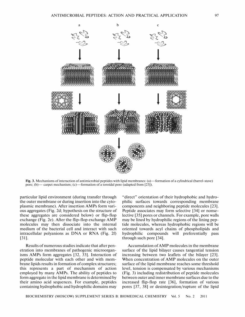
Accumulation of AMP molecules in the membranesurface of the lipid bilayer causes tangential tensionincreasing between two leaflets of the bilayer [23].When concentration of AMP molecules on the outersurface of the lipid membrane reaches some thresholdlevel, tension is compensated by various mechanisms(Fig. 3) including redistribution of peptide moleculesbetween outer and inner membrane surfaces due to theincreased flip�flop rate [36], formation of variouspores [37, 38] or desintegration/rupture of the lipid
a b c
Fig. 3. Mechanisms of interaction of antimicrobial peptides with lipid membranes: (a)—formation of a cylindrical (barrel�stave)pore; (b)— carpet mechanism; (c)—formation of a toroidal pore (adapted from [23]).
98
BIOCHEMISTRY (MOSCOW) SUPPLEMENT SERIES B: BIOMEDICAL CHEMISTRY Vol. 5 No. 2 2011
OKOROCHENKOV et al.
membrane [39]. Below we consider these mechanismsin more details.
1.1. The Barrel�Stave Model (Fig. 3a)
In the case of domination of hydrophobic interac�tions peptide chains are inserted into the lipid bilayerand adopt orientation perpendicular to the bilayer sur�face. Subsequent recruitment of other polypeptidechains results in the increase of the transmembranebundle of peptide molecules and formation of the bar�rel�stave type pore. The hydrophobic face of the pep�tide chain interacts with lipid acyl chains and thehydrophilic face forms the interior of the pore [27]. Aminimal length of the peptide sequence required forrealization of this model is about 22 residues for α�helical peptides and about 8 residues for peptides withthe beta�pleated sheet structure [40]. The number ofAMP molecules involved into pore formation mayvary and depend on AMP concentration. For exam�ple, alamethicin forms pores that consist of 3–11 pep�tides with an average diameter of 4 nm [41, 42]. Suchtype of interaction is typical for small number of pep�tides (alamethicin, pardaxin) characterized by verylow selectivity and also by toxicity to normal cells [40].
1.2. The Carpet Model (Fig. 3b)
This mechanism is realized in the case of verystrong electrostatic interactions between positivelycharged regions of peptide molecules and negativelycharged phospholipid polar head groups. Polypeptidechains are oriented parallel to the bilayer surface [44].Accumulation of peptide above some critical thresholdconcentration on the lipid membrane causes its rup�ture followed by micelle formation [45, 46]. This isaccompanied by formation of large (about 25 nm) tor�oidal pores (see more details about toroidal poresbelow) [35]. In this model pore formation is an inter�mediate step before the collapse of the membrane andsuch pore is not a stable structure [11].
1.3. The Toroidal Model (Fig. 3c)
According to this model, compensation of themembrane tension induced by insertion of peptidechain into the membrane occurs due to continuousbend of one membrane leaflet to the other one andassociation of membrane surfaces; this is accompa�nied by formation of toroidal pore [47]. Peptide mole�cules are inserted ]from the membrane surface into thehydrophobic part of the lipid bilayer. This results information of a pore structure, in which hydrophilicregions of peptide molecules and lipid head groupstogether form the pore wall (in contrast to the toroidalpore, the barrel�stave type pore is formed only by pep�tide molecules). Usually, toroidal pores have largersizes than barrel�stave type pores [11]. For example,the toroidal pore formed by magainin (and consisting
of 4—7 peptide molecules and about 90 phospholipidmolecules) has inner and outer diameters of 3—5 and7.0—8.4 nm, respectively [48].
Studies of mechanisms underlying AMP toxicitytowards pathogens and host cells represent an impor�tant problem. It was long thought that AMPs killedmicroorganisms by forming numerous unrecoverabledamages in their membranes. Indeed, AMPs mayform pores in microbial membranes as describedabove. A microbe “attacked” by AMP molecules candie due to leakage of ions and metabolites and also dueto membrane depolarization (followed by its dysfunc�tion), inhibition of cell respiration and also biopoly�mer synthesis. However, recent data [49, 50] suggestthat AMP�induced cell death may involve othermechanisms, for example, interaction of AMPs withintracellular targets. Thus, AMP�induced mecha�nisms of cell death require further studies and specifi�cation.
Below we consider processes occurring in the cellinteracting with AMPs. The cytoplasmic membranemaintains normal functioning of microorganisms. Itprovides selective permeability, maintains the electro�chemical gradient, electron transport, and oxidativephosphorylation (in the eukaryotic pathogens such asfungi, this process occurs on the inner mitochondrialmembrane), synthesis and conjugation of peptidogly�can, chitin, and other biopolymers. This suggests thatAMP�induced dysfunction of the outer and/or plasmamembrane may cause impairments in one or a fewthese functions, which result (directly or indirectly) incell death.
However, in many cases AMP�induced death ofbacteria cannot be attributed only to membrane dys�function [51]. For example, in the case of cytotoxicityof various peptides towards S. aureus there was no cor�relation between cytotoxicity and damage of bacterialmembrane integrity. The other study [52] demon�strated that gramicidin S caused rapid depolarizationof the cytoplasmic membrane of Pseudomonas aerugi�nosa. Nevertheless, this bacterium exhibited resistanceto this peptide. On the other hand, toxic effects ofpolymixins B and E1 were associated with membranedepolarization of Pseudomonas aeruginosa cells. Thissuggests that AMP�induced damages of membraneintegrity and bacterial cell death may represent inde�pendent events.
Inhibition of synthesis of peptidoglycans, chitin,and other macromolecules also represents an impor�tant mechanism of AMP action. For example, normalfunctioning of the bacterial cell membrane is associ�ated with peptidoglycan biosynthesis. Activated pepti�doglycan precursors are transported across the cyto�plasmic membrane and conjugate with each other inclose proximity of this membrane. Cationic peptidescause membrane perturbations and thus cause impair�ments in the peptidoglycan biosynthesis cycle by director indirect inhibition of its synthesis and translocation
BIOCHEMISTRY (MOSCOW) SUPPLEMENT SERIES B: BIOMEDICAL CHEMISTRY Vol. 5 No. 2 2011
ANTIMICROBIAL PEPTIDES: ACTION AND PRACTICAL APPLICATION 99
of peptidoglycan precursors and/or their conjugation.Considering high peptidoglycan content in the mem�brane of Gram�negative microorganisms it appearsthat they should be especially susceptible to the AMPeffect employing this mechanism. For example, it wasdemonstrated that the AMP plasmin inhibits pepti�doglycan synthesis in E. coli cells [53].
Although cell membrane damages are the keymoment in the AMP�induced cell death, results ofsome studies indicate that peptide interaction withsome intracellular targets also plays an important rolein the mechanism of AMP cytotoxicity [54�56]. Forexample, in some cases there was rather prolongedperiod before death of microorganisms induced byAMPs; this suggests the cell death did not occur via themembrane lytic mechanism. The study of cytotoxicityof the antimicrobial peptide tPMP demonstrated [57]that S. aureus cells treated with this peptide remainedviable for rather long period after damage of theirmembrane integrity. Subsequent death of these cellsoccurred due to direct inhibition of nucleic acid bio�synthesis by tPMP.
Thus, AMPs demonstrating antibacterial activityare abundant and the most studied class of antimicro�bial peptides. However, it should be noted that despiteconstant interest of the scientific community in thisclass of compounds only some of them are employedclinically. Increased attention to antibacterial AMPs isdetermined by the development of bacterial resistanceto them and therefore design of new AMP�based anti�biotics attracts researches.
2. ANTIMICROBIAL PEPTIDES POSSESSING ANTIVIRAL ACTIVITY
Besides antibacterial activity some AMPs alsoexhibit noticeable antiviral properties [24]. For exam�ple, it was demonstrated that the antiviral activity ofnumerous AMPs (e.g. lactoferrins) is determined bytheir binding to heparan sulfate, one of the most abun�dant components of the cell surface [58]. Heparan sul�fate is a proteoglycan, with its protein componentbeing covalently attached to one or a few sulfated glu�cosaminoglycan chains. Heparan sulfate is the mostnegatively charged component of the cell surface andthis determines preferential binding of positivelycharged extracellular ligands [59] and also manypathogens including viruses [60]. Binding to heparansulfate is the first step required for penetration of manyviruses into the cell [61, 62]. There is evidence [63, 64]that blockade of cell surface heparan sulfate attenu�ated viral infection of these cells. It was also demon�strated that recombinant cells with decreased expres�sion of heparan sulfate or chondroitin sulfate were lesssusceptible to herpes simplex virus (HSV) infection(by 80 and 60%, respectively) [65]. The increased con�tent of cell�surface heparan sulfate is an importantprecondition for penetration of hepatitis C virus intothe cell [66].
All these facts suggest that temporal blockade ofcell�surface heparan sulfate may prevent developmentof viral infections in the organism. AMPs demonstrat�ing antiviral properties have strong positive charge andtherefore they easily bind to cell�surface heparan sul�fate and prevent viral binding. For example, it wasdemonstrated that the antimicrobial peptide, melittin,prevented healthy cells against HSV infection [67].According to isothermal titration calorimetry experi�ments, melittin exhibits increased affinity to heparansulfate [68]. Lactoferrin and its analogues also exhibitincreased affinity to heparan sulfate [69]. Cells treatedwith lactoferrin analogues were less susceptible toHSV than untreated cells, but treatment of the HSVsuspension with these peptides did not inhibit viralactivity [69]. The antiviral activity of the cathelicidinfamily of AMP is also attributed to peptide binding toheparan sulfate [70].
On the contrary, antiviral activity of some AMPs isdetermined by direct interaction with the viral parti�cle. Usually, AMPs bind to viral envelope glycopro�teins. For example, AMP defensins can bind to aden�ovirus particles. Defensins inhibit virus disassembly atthe vertex region thereby restricting the release of aninternal capsid protein, pVI, which is required forendosomal membrane penetration during cell entry.Thus, defensins prevent the release of adenovirus par�ticles from endosomes, resulting in accumulation ofvirions in the lysosomal region where they are inacti�vated [71]. α�Defensin peptides can also inhibitadsorption of polyoma virus by binding to viral surfaceglycoproteins; they can also inhibit assembly of virusparticles [72].
In addition, some AMPs may interact with the virallipid envelope and cause its lysis, destabilization orpore formation [24]. For example, the AMP, indolici�din, inactivates HIV�1 by damaging the virion mem�brane [73]. Another peptide, dermaseptin, demon�strated similar activity [74]. This peptide did not influ�ence directly the HSV membrane but inhibited virusadsorption on normal cells [75].
It should be noted that despite rather intensivestudies of AMP antiviral activity the mechanism ofselected AMP toxicity towards viruses still requiresfurther investigation.
3. ANTIMICROBIAL PEPTIDES POSSESSING ANTIFUNGAL ACTIVITY
Many AMPs demonstrate significant antiviralactivity against microscopic fungi [24] by inducingtheir lysis [5]. However, structure function relation�ship activity for antifungal AMPs is less studied com�pared with structure function relationship activity ofantibacterial AMPs. For example, various peptidescharacterized by completely different structural motifsdemonstrate antibacterial activity. These include theantifungal peptide, containing 5 disulfide bridges,
100
BIOCHEMISTRY (MOSCOW) SUPPLEMENT SERIES B: BIOMEDICAL CHEMISTRY Vol. 5 No. 2 2011
OKOROCHENKOV et al.
which was isolated from the Eucommia ulmoidesplant [76], the peptide P18 possessing α�helical struc�ture [77], the “extended” peptide indolicidin [78], andβ�sheet pleated coleopteran isolated from Acrocinuslongimanus [79].
The antifungal activity of AMPs is intensively stud�ied in many laboratories [80�83]. Interestingly, inter�action of AMPs with pathogenic fungi cannot bedescribed by a universal mechanism (typical for AMPinteraction with viruses and bacteria). For example, itwas demonstrated [83] that peptide fragments fromthe lactoferrin sequence exhibit significant antifungalactivity towards Candida albicans; they inhibit biosyn�thesis of outer membrane components and cause ATLleak from the fungal cells. However, pleurocidin, iso�lated from Pseudopleuronectes americanus acts bymembrane lysis [81]. Plant defensins bind to someouter membrane components of microscopic fungiand thus violate transmembrane transport and inhibitsynthesis of their outer membrane components [80].Piscidin�2 isolated from mast cells hybrid striped bassacts on the fungal plasma membrane by forming poresand causing cell death [82].
Fungicide activity of some AMPs represents apoint of discussions. For example, it was suggested[84] that toxicity of the AMP, histatin 5, towardsC. albicans, C. neoformans and some other fungi isdetermined by formation of reactive oxygen species(ROS) in the fungal cells induced by this peptide.Authors [84] proposed a hypothesis, by which afterpenetration inside cells of microscopic fungi histatin5 enters mitochondria and inhibits coenzyme Q cycle;this results in accumulation of ROS in the fungal celland cell death due to oxidation of intracellular sub�strates by the accumulated ROS. According to anotherviewpoint [85] histatin 5 does not cause the increase ofROS in the fungal cells and the fungicide effect of thispeptide is determined by ATP leakage from fungalcells. These authors [85] assert that wrong conclusionsmade in [84] could be attributed to an unreliablemethod used for ROS detection in that study.
4. PERSPECTIVES OF PRACTICAL APPLICATION OF AMPS
High cost, sensitivity to proteolytic enzymes, andalso the hemolytic effect typical for many AMPs arethe main obstacles for AMP application in clinicalpractice [1]. Nevertheless, AMP�based drug prepara�tions are developed by many pharmaceutical compa�nies. Below we consider some examples of such prep�arations.
4.1. Omiganan
The active component is an analogue of the 13�res�idue antimicrobial peptide indolicidin. It is producedby Microbiologix Biotech. Phase III clinical trials ofOmiganan as a drug decreasing colonization of vein
catheters by microorganisms causing catheter�relatedbloodstream infections gave different results in variousgroups of patients; therefore its applicability in clinicalpractice is still questionable [86]. Repeated trialsrevealed that Omiganan 1% gel prevented cathetercolonization by all known bacteria and microscopicfungi [87].
4.2. MX594AN
The active substance is an analogue of the 13�resi�due antimicrobial peptide indolicidin. It is producedby Microbiologix Biotech. MX594AN successfullypassed through Phase IIb clinical trials as the drug fortreatment of acne formation and now it is under PhaseIII clinical trials [86].
4.3 hIF1�11
This is a 11�residue peptide from the N�terminalpart of human lactoferrin developed by AM�Pharma.Currently, it is under Phase II clinical trials as antifun�gal and antimicrobial drug [1, 88].
4.4. P113/P113D
The acting substance is a 12�residue peptide, mod�ified histatin [89]. This drug produced by Demer�gen/Pacgen is active against oral candidoses.P113/P113D passed through Phase II clinical trials.Its inhalation drug dosage form has been prepared forPhase III clinical trial [1].
In addition to drugs considered in this secationthere are some other AMP�based preparations , whichare now passing earlier phases of clinical trials [1, 86].
CONCLUSIONS
Although AMPs exhibit high activity against vari�ous pathogens in vitro they are not widely used in clin�ical trials due to high cost, proteolytic susceptibilityand hymolytic activity. This explains why design ofmodified AMPs, which would be free of these short�comings, attracts much attention [90]. The main strat�egies used for optimization of AMP structures toobtain novel biocide agents include synthesis of cyclicAMP analogues [91], insertion of a fluorine atom orthe trifluoromethyl group [92], synthesis of branchedAMPs (dendrimers) [93, 94] and also synthesis ofAMPs immobilized on various polymer matrices [95].Thus, AMPs remain in the spotlight of many researchgroups, interested in scientific and practical aspects oftheir effects and application as novel antibiotics.
ACKNOWLEDGMENTS
This work was supported by the Analytical Depart�mental Target Program “The Development of Scien�tific Potential of Higher School” no. 2.1.1./2889.
BIOCHEMISTRY (MOSCOW) SUPPLEMENT SERIES B: BIOMEDICAL CHEMISTRY Vol. 5 No. 2 2011
ANTIMICROBIAL PEPTIDES: ACTION AND PRACTICAL APPLICATION 101
REFERENCES
1. Giuliani, A., Pirri, G., and Nicoletto, S., Cent. Eur. J.Biol., 2007, vol. 2, no. 1, pp. 1⎯33.
2. Yeaman, M. and Yount, N.Y., Pharmacol. Rev., 2003,vol. 55, no. 1, pp. 27⎯55.
3. Bastian, A. and Schafer, H., Regul. Pept., 2001,vol. 101, pp. 157⎯161.
4. Horne, W., Wiethoff, C., Cui, C., Wilcoxen, K.,Amorin, M., Ghadiri, M., and Nemerow, G., Bioorg.Med. Chem., 2005, vol. 13, pp. 5145⎯5153.
5. De Lucca, A. and Walsh, T., Antimicrob. AgentsChemother., 1999, vol. 43, pp. 1⎯11.
6. Lustig, F., Hoebeke, J., Ostergren�Lunden, G., Velge�Roussel, F., Bondjers, G., Olsson, U., Ruetschi, U.,and Fager, G., Biochemistry, 1996, vol. 35, pp. 12077⎯
12085.7. Alberola, J., Rodriguez, A., Francino, O., Roura, X.,
Rivas, L., and Andreu, D., Antimicrob. AgentsChemother., 2004, vol. 48, pp. 641⎯643.
8. Zasloff, M., Proc. Natl. Acad. Sci. USA, 1987, vol. 84,pp. 5449–5453.
9. Pranting, M., Negrea, A., Rhen, M., and Andersson, D.,Antimicrob. Agents Chemother., 2008, vol. 52, no. 8,pp. 2734–2741.
10. Zasloff, M., J. Am. Soc. Nephrol., 2007, vol. 18,pp. 2810⎯2816.
11. Brogden, K.,Nat. Rev. Microbiol., 2005, vol. 3, pp. 238⎯
250.12. Bals, R., Respir. Res., 2000, vol. 1, pp. 141–150. 13. Faber, C., Stallmann, H., Lyaruu, D., Joosten, U., Von
Eiff, C., Van Nieuw Amerongen, A., and Wuisman, P.I.,Antimicrob. Agents Chemother., 2005, vol. 49, pp. 2438⎯
2444.14. Nibbering, P., Ravensbergen, E., Welling, M., Van Ber�
kel, L., Van Berkel, P., Pauwels, E., and Nuijens, J.,Infect. Immun., 2001, vol. 69, pp. 1469⎯1476.
15. Egorov, N.S., Osnovy ucheniya ob antibiotikah, (Princi�ples of Teaching about Antibiotics), Moscow: Nauka,2004, pp. 22⎯48.
16. Egorov, N.S. and Baranova, I.P., Antib. Khimoter., 1999,no. 6, pp. 33⎯40.
17. Imura, Y., Choda, N., and Matsuzaki, K., (2008) Bio�phys. J., 2008, vol. 95, pp. 5757–5765.
18. Smirnova, M., Afonin, V., Shpen’, V., Tyagotin, Yu.,and Kolodkin, N., Russ. J. Bioorg. Chem., 2004, vol. 30,pp. 458⎯465.
19. Olson, L., Soto, A., Knoop, F., and Conlon, J., Bio�chem. Biophys. Res. Commun., 2001, vol. 288, pp. 1001⎯
1005. 20. Gunstone, F., Harwood, J., and Dijkstra, A., in The
Lipid Handbook, NY: CRC Press, 2007, pp. 134⎯141. 21. Hancock, R., Lancet, 1997, vol. 349, pp. 419⎯422.22. Powers, J.�P. and Hancock, R., Peptides, 2003, vol. 24,
pp. 1681⎯1691.23. Toke, O., Biopolymers, 2005, vol. 80, pp. 717⎯735.24. Jenssen, H., Hamill, P., and Hancock, R., Clin. Micro�
biol. Rev., 2006, vol. 19, pp. 491⎯511.25. Shai, Y. and Oren, Z., Biochemistry, 1997, vol. 36,
pp. 1826⎯1835.
26. Hancock, R. and Chapple, D., Antimicrob. AgentsChemother., 1999, vol. 43, pp. 1317–1323.
27. Morris, M., Depollier, J., Mery, J., Heitz, F., and Div�ita, G., Nat. Biotechnol., 2001, vol. 19, pp. 1173⎯1176.
28. Piers, K., Brown, M., and Hancock, R., Antimicrob.Agents Chemother., 1994, vol. 38, pp. 2311⎯2316.
29. Silva, A. Jr. and Teschke, O., Biochim. Biophys. Acta,2003, vol. 1643, pp. 95⎯103.
30. Ovchinnikova, T., Shenkarev, Z., Balandin, S.,Nadezhdin, K., Paramonov, A., Kokryakov, V., andArseniev, A., Biopolymers, 2008, vol. 89, pp. 455⎯464.
31. Zanetti, M., Litteri, L., Gennaro, R., Horstmann, H.,and Romeo, D., J. Cell. Biol., 1990, vol. 111, pp. 1363⎯
1371. 32. Takeuchi, K., Takahashi, H., Sugai, M., Iwai, H.,
Kohno, T., Sekimizu, K., Natori, S., and Shimada, I.,J. Biol. Chem., 2004, vol. 279, pp. 4981–4987.
33. Mani, R., Cady, S., Tang, M., Waring, A., Lehrer, R.,and Hong, M., Proc. Natl. Acad. Sci. USA, 2006,vol. 103, pp. 16242–16247.
34. Christensen, B., Fink, J., Merrifield, R., and Mauzer�all, D., Proc. Natl. Acad. Sci. USA, 1988, vol. 85pp. 5072⎯5076.
35. Ladokhin, A., Selsted, M., and White, S., Biophys. J.,1997, vol. 72, pp. 1762⎯1766.
36. Zhao, H., Mattila, J.�P., Holopainen, J., and Kin�nunen, P., Biophys. J., 2001, vol. 81, pp. 2979⎯2991.
37. Sengupta, D., Hari, L., Mark, A., and Marrink, S.�J.,Biochim. Biophys. Acta�Biomembranes, 2008, vol. 1778,pp. 2308⎯2317.
38. Bessin, Y., Saint, N., Marri, L., Marchini, D., andMolle, G., Biochim. Biophys. Acta � Biomembranes,2004, vol. 1667, pp. 148⎯156.
39. Papo, N. and Shai, Y.,Biochemistry, 2003, vol. 42,pp. 458⎯466.
40. Mateo, C., Villalain, J., and Gonzales�Ros, J., in Pro�tein�Lipid Interactions: New Approaches and EmergingConcepts, Heidelberg: Springer�Verlag, 2006, p. 190.
41. Spaar, A., Munster, C., and Salditt, T., Biophys. J.,2004, vol. 87, pp. 396⎯407.
42. He, K., Ludtke, S., Huang, H., and Worcester, D., Bio�chemistry, 1995, vol. 34, pp. 15614⎯15618.
43. Bechinger, B., Biochim. Biophys. Acta, 1999, vol. 1462,pp. 157–183.
44. Pouny, Y., Rapaport, D., Mor, A., Nicolas, P., andShai, Y., Biochemistry, 1992, vol. 31, pp. 12416–12423.
45. Shai, Y., Biochim. Biophys. Acta, 1999, vol. 1462,pp. 55–70.
46. Ladokhin, A. and White, S., Biochim. Biophys. Acta,2001, vol. 1514, pp. 253–260.
47. Matsuzaki, K., Murase, O., Fujii, N., and Miyajima, K., Bio�chemistry, 1996, vol. 35, pp. 11361–11368.
48. Matsuzaki, K., Sugishita, K., Harada, M., Fujii, N.,and Miyajima, K., Biochim. Biophys. Acta, 1997,vol. 1327, pp. 119–130.
49. Gennaro, R. and Zanetti, M., Biopolymers, 2000,vol. 55, pp. 31–49.
50. Park, C., Yi, K., Matsuzaki, K., Kim, M., and Kim, S.,Proc. Natl. Acad. Sci. USA, 2000, vol. 97, pp. 8245–8250.
102
BIOCHEMISTRY (MOSCOW) SUPPLEMENT SERIES B: BIOMEDICAL CHEMISTRY Vol. 5 No. 2 2011
OKOROCHENKOV et al.
51. Koo, S.P., Bayer, A., and Yeaman, M., Infect. Immun.,2001, vol. 69, pp. 4916⎯4922.
52. Zhang, L., Rozek, A., and Hancock, R., J. Biol. Chem.,2001, vol. 276, pp. 35714⎯35722.
53. Chitnis, S. and Prasad, K., FEMS Microbiol. Lett.,1990, vol. 60, pp. 281⎯284.
54. Lehrer, I., Barton, A., Daher, K.A, Harwig, S., Ganz, T., andSelsted, M., J. Clin. Invest., 1989, vol. 84, pp. 553⎯561.
55. Park, C., Kim, H., and Kim, S., Biochem. Biophys. Res.Commun., 1998, vol. 244, pp. 253⎯257.
56. Sharma, S., Verma, I., and Khuller, G., Arch. Micro�biol., 1999, vol. 171, pp. 338⎯342.
57. Xiong, Y., Bayer, A., and Yeaman, M., J. Infect. Dis.,2002, vol. 186, pp. 668⎯677.
58. Bernfield, M., Kokenyesi, R., Kato, M., Hinkes, M.,Spring, J., Gallo, R., and Lose, E., Annu. Rev. CellBiol., 1992, vol. 8, pp. 365⎯393.
59. Parish, C., Nat. Rev. Immunol., 2006, vol. 6, pp. 633⎯
643.60. Rostand, K.S. and Esko, J., Infect. Immun., 1997,
vol. 65, pp. 1⎯8. 61. James, S., Gibbs, B., Toney, K., and Bennett, H., Anal.
Biochem., 1994, vol. 217, pp. 84⎯90. 62. Tamamura, H., Xu, Y., Hattori, T., Zhang, X., Arakaki, R.,
Kanbara, K., Omagari, A., Otaka, A., Ibuka, T.,Yamamoto, N., Nakashima, H., and Fujii, N., Bio�chem. Biophys. Res. Commun., 1998, vol. 253, pp. 877⎯
882.63. Shieh, M., Wudunn, D., Montgomery, R., Esko, J.,
and Spear, P., J. Cell Biol., 1992, vol. 116, pp. 1273⎯
1281. 64. Wudunn, D. and Spear, P., J. Virol, 1989, vol. 63,
pp. 52⎯58.65. Mardberg, K., Trybala, E., Tufaro, K., and Bergstrom, T., J.
Gen. Virol., 2002, vol. 83, pp. 291⎯300.66. Bals, R., Wang, X., Wu, Z., Freeman, T., Bafna, V.,
Zasloff, M., and Wilson, J., J. Clin. Invest, 1998, vol. 102,pp. 874⎯880.
67. Baghian, A., Jaynes, J., Enright, F., and Kousoulas, K., Pep�tides, 1997, vol. 18, pp. 173⎯183.
68. Klocek, G. and Seelig, J., Biochemistry, 2008, vol. 47,pp. 2841⎯2849.
69. Andersen, J., Jenssen, H., Sandvik, K., and Gutteberg, T., J.Med. Virol., 2004, vol. 74, pp. 262⎯271.
70. Kaneider, N., Djanani, A, and Wiedermann, C., Sci.World J., 2007, vol. 7, pp. 1832⎯1838.
71. Smith, J. and Nemerow, G., Cell Host Microbe, 2008,vol. 3, pp. 11⎯19.
72. Dugan, A., Maginnis, M., Jordan, J., Gasparovic, M.,Manley, K., Page, R., Williams, G., Porter, E., O’Hara, B.,and Atwood, W., J. Biol. Chem., 2008, vol. 283, pp. 31125⎯
31132.73. Robinson, W., Mcdougall, B., Tran, D., and Selsted, M., J.
Leukoc. Biol., 1998, vol. 63, pp. 94⎯100.
74. Lorin, C., Saidi, H., Belaid, A., Zairi, A., Baleux, F.,Hocini, H., Belec, L., Hani, K., and Tangy, F., Virol�ogy, 2005, vol. 334, pp. 264–275.
75. Benincasa, M., Skerlavaj, B., Gennaro, R., Pellegrini, A.,and Zanetti, M., Peptides, 2003, vol. 24, pp. 1723–1731.
76. Huang, R., Xiang, Y., Tu, G., Zhang, Y., and Wang, D.,Biochemistry, 2004, vol. 43, pp. 6005⎯6012.
77. Lee, D., Hahm, K., and Shin, S., Biotechnol. Lett.,2004, vol. 26, pp. 337⎯341.
78. Lee, D., Kim, H., Kim, S., Park, Y., Park, S., Jang, S.,and Hahm, K., Biochem. Biophys. Res. Commun., 2003,vol. 305, pp. 305⎯310.
79. Barbault, F., Landon, C., Guenneugues, M., Meyer, J.,Schott, V., Dimarcq, J., and Vovelle, F., Biochemistry,2003, vol. 42, pp. 14434⎯14442.
80. Thevissen, K., Ferket, K., Franöis, I., and Cammue, B.,Peptides, 2003, vol. 24, pp. 1705⎯1712.
81. Sung, W. and Lee, D., Biochem. Biophys. Res. Com�mun., 2008, vol. 369, pp. 858⎯861.
82. Sung, W.J.L., Lee, J., and Lee, D., Biochem. Biophys.Res. Commun., 2008, vol. 371, pp. 551⎯555.
83. Tanida, T., Okamoto, T., Ueta, E., Yamamoto, T., andOsaki, T., (2006) J. Antimicrob. Chemother., 2006,vol. 57, pp. 94⎯103.
84. Helmerhorst, E., Troxler, R., and Oppenheim, F., Proc.Natl. Acad. Sci. USA, 2001, vol. 98, pp. 14637⎯14642.
85. Veerman, E., Nazmi, K., van’t Hof, W., Bolscher, J.,Hertog, A., and Amerongen, A., Biochem. J., 2004,vol. 381, pp. 447⎯452.
86. Gordon, Y., Romanowski, E., and Mcdermott, A.,Curr. Eye Res., 2005, vol. 30, pp. 505⎯515.
87. Fritsche, T., Rhomberg, P., Sader, H., and Jones, R., J.Antimicrob. Chemother., 2008, vol. 61, pp. 1092⎯1098.
88. Dijkshoorn, L., Bogaards, S., Nemec, A., Van DenBroek, P., and Nibbering, P., J. Antimicrob. Chemother.,2004, vol. 48, pp. 4919⎯4921.
89. Umadevi, S., Linh, T., Nuria, S., Christopher, R.,Alan, A., Phillip, F., Janet, F., and David, R., Antimi�crob. Agents Chemother., 2001, vol. 45, pp. 3437⎯3444.
90. Knappe, D., Stegemann, C., Nimptisch, A., Kolobov, A.,Korableva, E., Shamova, O., Kokryakov, V., and Hoff�mann, R., Adv. Exp. Med. Biol., 2009, vol. 611, pp. 395⎯
396.91. Wessolowski, A., Bienert, M., and Dathe, M., J. Pept.
Res., 2004, vol. 64, pp. 159⎯169.92. Gimenez, D., Andreu, C., Del Olmo, M., Varea, T.,
Diaz, D., and Asensio, G., Bioorg. Med. Chem., 2006,vol. 14, pp. 6971⎯6978.
93. Lee, C., Mackay, J., Frechet, J., and Szoka, F., NatureBiotechnol., 2005, vol. 23, pp. 1517–1526.
94. Khrushcheva, A.., Kashparova, I., Klimenkova, L., andMitina, Yu., Russ. J. Bioorg. Chem., 2007, vol. 33,pp. 544⎯548
95. Bagheri, M., Beyermann, M., and Dathe, M., Antimi�crob. Agents Chemother., 2009, vol. 53, pp. 1132⎯1141.