Embed Size (px)
Citation preview
Antibacterial properties of nonwoven wound dressings coated with Manuka honey or methylglyoxal Sophie E L Bulman,* Giuseppe Tronci, Parikshit Goswami, Chris Carr and Stephen J Russell
Clothworkers’ Centre for Textile Materials Innovation for Healthcare (CCTMIH), Clothworkers Central Building, School of Design, University of Leeds, Leeds, West Yorkshire, LS2 9JT; [email protected]
* [email protected] ; Tel.: +44 7837 913 176
Abstract: Manuka honey (MH) is used as an antibacterial agent in bioactive wound dressings via direct
impregnation onto a suitable substrate. MH provides unique antibacterial activity when compared with
conventional honeys, owing partly to one of its constituents, methylglyoxal (MGO). Aiming to
investigate an antibiotic-free antimicrobial strategy, we studied the antibacterial activity of both MH
and MGO (at equivalent MGO concentrations) when applied as a physical coating to a nonwoven fabric
wound dressing. When physically coated on to a cellulosic hydroentangled nonwoven fabric, it was
found that concentrations of 0.0054 mg cm-2 of MGO in the form of MH and MGO was sufficient to
achieve 100 CFU% bacteria reduction against gram-positive Staphylococcus aureus and gram-negative
Klebsiella pneumoniae, based on BS EN ISO 20743:2007. A 3- to 20- fold increase in MGO concentration
(0.0170 ‒ 0.1 mg cm-2) was required to facilitate a good antibacterial effect (based on BS EN ISO
20645:2004) in terms of zone of inhibition and lack of growth under the sample. The minimum
inhibitory concentration (MIC) and minimum bactericidal concentration (MBC) was also assessed for
MGO in liquid form against three prevalent wound and healthcare-associated pathogens, i.e.
Staphylococcus aureus, gram-negative Pseudomonas aeruginosa and gram-positive Enterococcus faecalis.
Other than the case of MGO-containing fabrics, solutions with much higher MGO concentrations (128
mg L-1 ‒ 1024 mg L-1) were required to provide either a bacteriostatic or bactericidal effect. The results
presented in this study therefore demonstrate the relevance of MGO-based coating as an environment-
friendly strategy for the design of functional dressings with antibiotic-free antimicrobial chemistries.
Keywords: Manuka honey; Methylglyoxal; Nonwoven; Antibacterial; Wound dressing
1. Introduction
With increasing bacterial resistance to antibiotics [1-3] and concern to find alternative treatments,
[3-4] the antibacterial activity of MH is of growing interest and well documented. MH inhibits the
growth of clinically-relevant pathogens and biofilms found in wounds, including gram-positive strains
such as Methicillin-resistant Staphylococcus aureus (MRSA) [5, 6], and Streptococcus pyogenes [7], and
gram-negative strains including Esherichia.coli [8], Proteus mirabilis and Enterobacter cloacae [9], and
P.aeruginosa [10, 11]. Gastrointestinal pathogens [12] and oral infections [13] have also shown
susceptibility to MH. The effect of MH on cells required for healing, including fibroblasts and
keratinocytes also suggests that MH is considered a safe compound for topical treatment [14-15].
Methylglyoxal (MGO) is a keto-aldehye, found as a yellow liquid and present in a variety of beverages
and foods including wine, beer [16], bread [17], soya, coffee, teas [18] and notably, MH [19]. Mavric et

al [20] reported that MGO is responsible for the heightened and unique non-peroxide antibacterial
activity associated with MH, and the minimum inhibitory concentrations (MIC) of MGO in the form of
both MH and isolated synthetic compound required to have an antibacterial effect have been
established. For MGO, a MIC of 1.1 mM is required to induce an antibacterial effect, whilst a range of
MIC values has been observed in the case of MH, in light of inherent variations in MGO content. For
example, five MHs with MGO concentrations ranging between 347 to 761 ± 25 mg kg-1 were shown to
exhibit an antibacterial effect when the MH was diluted to 15 to 30% (w/v). These resulting MGO
concentrations correspond to MIC values between 1.1 mM and 1.8 mM, and therefore compare with
the 1.1 mM MIC value associated with synthetic MGO [20]. The antibacterial activity of MGO in the
form of solution [20, 21], hydrogel [21], polymer-based formulation [22], and poly (vinyl alcohol) fibres
[23] has also been studied. With respect to MH, MGO has attracted attention because of its ability to act
as a lone compound at defined concentration for the inhibition of bacterial growth, as well as its
carcinostatic properties [24-28] and anti-proliferative effects on leukaemia cells [29, 30].
The antibacterial effects of MH and MGO in the form of nonwoven fabric coating have not
previously been compared in terms of concentration per unit area. This is particularly important when
designing dressings, where the required concentration of the active compound per unit area should be
known. Nonwovens in this context relate to textile materials produced by drylaid methods, which are
most commonly employed to manufacture wound dressings [31, 20].
Therefore it is of interest to understand the degree to which MGO exhibits an equivalent
antibacterial effect to MH, aiming to identify a synthetically-defined alternative to MH towards the
design of antibacterial dressings. Consequently, the aim of this work was firstly to compare and
evaluate the antibacterial efficiency of both MH and MGO when applied as a coating to a nonwoven
fabric at equivalent MGO concentrations. Secondly, we wanted to determine the minimum inhibitory
concentration (MIC) and minimum bactericidal concentration (MBC) of MGO (isolated synthetic
compound) in liquid form against three of the most common wound pathogens including
Staphylococcus aureus (S.aureus), Peudomonas aeruginosa (P. aeruginosa) and Enterococcus faecalis (E.
faecalis) [33].
2. Results and Discussion
Sample nomenclature used in this study is as follows: samples are coded as ‘MH1, MH2, MGO 1
and MGO2’, whereby ‘MH or MGO’ identifies the additive that the nonwoven samples were prepares
with, ie. MH or MGO; and ‘1 or 2’ describes the concentration formulation, as described in Table 1.
‘NW’ and ‘WP’ indicate the coating-free nonwoven and woven polyester control samples, respectively.
Table 1 provides an overview of the different sample additive formulations.

Table 1: MGO concentration in either the coating solutions (Cs) or resulting coated nonwoven fabrics (Cf).
Sample ID Cs (mg g-1) Cf (mg cm-2)
MH1 0.11 0.0057 MH2 0.33 0.0169
MGO1 0.11 0.0054 MGO2 0.33 0.0170
2.1. Antibacterial performance of the nonwoven coated samples
2.1.1 Antibacterial performance of the nonwoven coated samples Using BS EN ISO 20743:2007
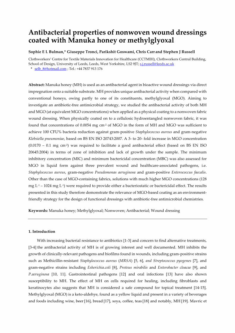
With reference to BS EN ISO 20743:2007 [34], the results in Tables 2 and 3 report the average
reduction of bacteria in colony forming units (CFU), for either S. aureus or K. Pneumoniae, respectively.
For the MH coated and synthetic MGO coated nonwovens, 100 CFU% reduction in bacteria was
achieved for all samples where the calculated concentration of MGO ranged from 0.0054 mg cm-2 to
0.0170 mg cm-2, regardless of the strain tested.. Interestingly, an average reduction of 97 CFU% in
bacteria was still reported for the nonwoven control against S.aureus (Table 2), whilst considerably high
growth (-252 CFU%) was reported when the same sample was challenged with K. pneumonia (Table 3).
This latter effect was still observed in the case of woven polyester control following contact with either
S. aureus or K. Pneumoniae (-22438 CFU% and -5635 CFU%).
Previously, it has been reported that TENCEL® or lyocell fibres are able to reduce the growth of S.aureus
considerably when compared with synthetic fibres such as polypropylene, polyester and polyacrylate
[35]. The previous study showed that the synthetic samples exhibited 100 to 1000 times higher bacteria
growth when compared with lyocell. It is conceivable that the reduced growth of bacteria observed
with lyocell fibres is associated with the behaviour of the fibres in water. In the case of the synthetic
fibres, there is limited penetration of water into the fibres and interactions are mainly at the surface
which is fully accessible to bacterial organisms. However, because of the nano-fibrillar structure of
lyocell fibres, water can be absorbed into the micro capillaries inside the fibre, such that there is a
reduced life sustaining environment for the bacteria to thrive [35]. It was reported that approximately
1,333,000 nanofibrils with a diameter of 10 nm are apparent in a single TENCEL fibre, thus contributing
to the highly absorbent characteristic nature of the fibre [36]. This behaviour is therefore a likely
explanation as to why a reduced bacterial count (97 CFU%) was observed for the NW control in the
case of S.aureus in the present study. Following these considerations, the thinner peptidoglycan and
additional lipopolysaccharide layer present in gram-negative K. pneumoniae compared to gram-positive
S. aureus [37] are likely to provide the former bacteria with increased adaptability on hydrated fibres in
the experimental conditions investigated, explaining why K. pneumoniae growth, rather than reduction,
was observed in contact with the nonwoven, similarly to the polyester, control (Table 3).
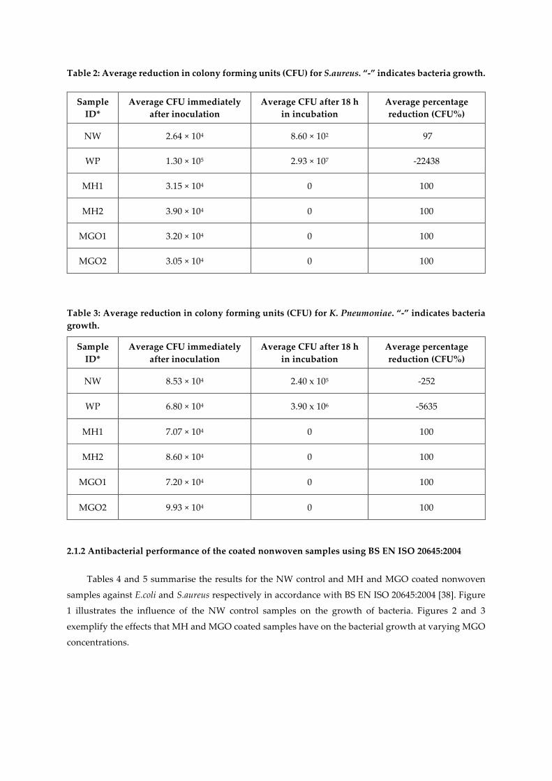
Table 2: Average reduction in colony forming units (CFU) for S.aureus. “-” indicates bacteria growth.
Sample ID*
Average CFU immediately after inoculation
Average CFU after 18 h in incubation
Average percentage reduction (CFU%)
NW 2.64 × 10⁴ 8.60 × 10² 97
WP 1.30 × 10⁵ 2.93 × 10⁷ -22438
MH1 3.15 × 10⁴ 0 100
MH2 3.90 × 10⁴ 0 100
MGO1 3.20 × 10⁴ 0 100
MGO2 3.05 × 10⁴ 0 100
Table 3: Average reduction in colony forming units (CFU) for K. Pneumoniae. “-” indicates bacteria growth.
Sample ID*
Average CFU immediately after inoculation
Average CFU after 18 h in incubation
Average percentage reduction (CFU%)
NW 8.53 × 10⁴ 2.40 x 10⁵ -252
WP 6.80 × 10⁴ 3.90 x 10⁶ -5635
MH1 7.07 × 10⁴ 0 100
MH2 8.60 × 10⁴ 0 100
MGO1 7.20 × 10⁴ 0 100
MGO2 9.93 × 10⁴ 0 100
2.1.2 Antibacterial performance of the coated nonwoven samples using BS EN ISO 20645:2004
Tables 4 and 5 summarise the results for the NW control and MH and MGO coated nonwoven
samples against E.coli and S.aureus respectively in accordance with BS EN ISO 20645:2004 [38]. Figure
1 illustrates the influence of the NW control samples on the growth of bacteria. Figures 2 and 3
exemplify the effects that MH and MGO coated samples have on the bacterial growth at varying MGO
concentrations.
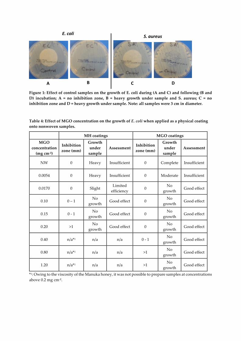
Figure 1: Effect of control samples on the growth of E. coli during (A and C) and following (B and D) incubation; A = no inhibition zone, B = heavy growth under sample and S. aureus; C = no inhibition zone and D = heavy growth under sample. Note: all samples were 3 cm in diameter.
Table 4: Effect of MGO concentration on the growth of E. coli when applied as a physical coating onto nonwoven samples.
MH coatings MGO coatings MGO
concentration (mg cm-²)
Inhibition zone (mm)
Growth under
sample Assessment
Inhibition zone (mm)
Growth under
sample Assessment
NW 0 Heavy Insufficient 0 Complete Insufficient
0.0054 0 Heavy Insufficient 0 Moderate Insufficient
0.0170 0 Slight Limited
efficiency 0
No growth
Good effect
0.10 0 – 1 No
growth Good effect 0
No growth
Good effect
0.15 0 - 1 No
growth Good effect 0
No growth
Good effect
0.20 >1 No
growth Good effect 0
No growth
Good effect
0.40 n/a*1 n/a n/a 0 - 1 No
growth Good effect
0.80 n/a*1 n/a n/a >1 No
growth Good effect
1.20 n/a*1 n/a n/a >1 No
growth Good effect
*1: Owing to the viscosity of the Manuka honey, it was not possible to prepare samples at concentrations above 0.2 mg cm-².
E.coli
A DCB
S.aureus
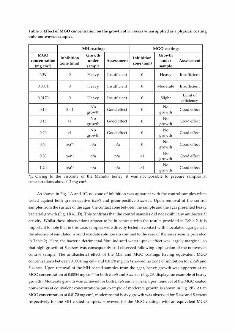
Table 5: Effect of MGO concentration on the growth of S. aureus when applied as a physical coating onto nonwoven samples.
MH coatings MGO coatings
MGO concentration
(mg cm-²)
Inhibition zone (mm)
Growth under
sample Assessment
Inhibition zone (mm)
Growth under
sample Assessment
NW 0 Heavy Insufficient 0 Heavy Insufficient
0.0054 0 Heavy Insufficient 0 Moderate Insufficient
0.0170 0 Heavy Insufficient 0 Slight Limit of
efficiency
0.10 0 – 1 No
growth Good effect 0
No growth
Good effect
0.15 >1 No
growth Good effect 0
No growth
Good effect
0.20 >1 No
growth Good effect 0
No growth
Good effect
0.40 n/a*1 n/a n/a 0 No
growth Good effect
0.80 n/a*1 n/a n/a >1 No
growth Good effect
1.20 n/a*1 n/a n/a >1 No
growth Good effect
*1: Owing to the viscosity of the Manuka honey, it was not possible to prepare samples at concentrations above 0.2 mg cm-².
As shown in Fig. 1A and 1C, no zone of inhibition was apparent with the control samples when
tested against both gram-negative E.coli and gram-positive S.aureus. Upon removal of the control
samples from the surface of the agar, the contact zone between the sample and the agar presented heavy
bacterial growth (Fig. 1B & 1D). This confirms that the control samples did not exhibit any antibacterial
activity. Whilst these observations appear to be in contrast with the results provided in Table 2, it is
important to note that in this case, samples were directly tested in contact with inoculated agar gels, in
the absence of simulated wound exudate solution (in contrast to the case of the assay results provided
in Table 2). Here, the bacteria detrimental fibre-induced water uptake effect was largely marginal, so
that high growth of S.aureus was consequently still observed following application of the nonwoven
control sample. The antibacterial effect of the MH and MGO coatings having equivalent MGO
concentrations between 0.0054 mg cm-2 and 0.0170 mg cm-2 showed no zone of inhibition for E.coli and
S.aureus. Upon removal of the MH coated samples from the agar, heavy growth was apparent at an
MGO concentration of 0.0054 mg cm-2 for both E.coli and S.aureus (Fig. 2A displays an example of heavy
growth). Moderate growth was achieved for both E.coli and S.aureus, upon removal of the MGO coated
nonwovens at equivalent concentrations (an example of moderate growth is shown in Fig. 2B). At an
MGO concentration of 0.0170 mg cm-2, moderate and heavy growth was observed for E.coli and S.aureus
respectively for the MH coated samples. However, for the MGO coatings with an equivalent MGO
concentration of 0.0170 mg cm-2, no growth and slight growth was evident against E.coli and S.aureus,
respectively (examples of slight growth and no growth are shown in Fig. 2C and Fig. 2D). These initial
evaluations at a concentration of 0.0054 mg cm-2 suggest an insufficient antibacterial effect was achieved
for both MH and MGO coatings. At a concentration of 0.0170 mg cm-2, limited efficacy was observed
for the MH coatings. However, for the MGO coatings with an MGO concentration of 0.0170 mg cm-2,
the antibacterial effect was shown to improve slightly and a good antibacterial effect and a limit of
efficiency was achieved for both E.coli and S.aureus respectively.
Figure 2: Examples of bacteria growth under either MH or MGO coated nonwoven samples. A = heavy growth of E. coli, B = medium growth of S. Aureus, C = slight growth of S. aureus and D = no growth of E. coli. Note: all coated samples were 3 cm in diameter.
It is important to note that where no growth or inhibition zone was apparent, a good antibacterial
effect may still be observed. This may be linked to the diffusion rate of the active compound from the
fabric [38] to the agar plate and the affinity of the fibres for moisture. Thus, it is likely that, within the
time frames investigated in this study, herein the hygroscopic, crystalline nano-fibrils of the TENCEL®
fibres [35] retain the MGO and honey coating, thereby limiting the diffusion of MGO into the agar at
these MGO concentrations. This situation may well be expected in this case, given that no additional
simulated wound exudate solution was applied. The minimal swelling of the fibres expected following
contact with the agar plate may well be directly related to a decreased MGO diffusion. This hypothesis
is supported when comparing data obtained in exudate-free conditions with the ones presented in
Tables 2 and 3, where complete bacteria killing was observed with the same MGO concentrations
following addition of simulated wound exudate solution. As the MGO concentration increases between
0.1 mg cm-2 and 1.2 mg cm-2, a good antibacterial effect is observed with both MH and MGO in all cases
(Table 4 and 5). For the MH coated samples, mean zones of inhibition of 0-1mm were apparent against
both E.coli and S.aureus at concentrations between 0.1 mg cm-2 and 0.2 mg cm-2. Fig. 3B displays an
example of an inhibition zone from 0-1mm. As the concentration of MGO doubled to 0.2 mg cm-2, the
mean zone of inhibition for E.coli and S.aureus increased to achieve a mean zone of >1mm. An example
of a mean zone of >1mm can be seen in Fig. 3C.
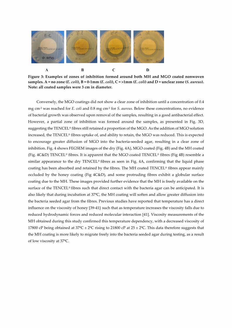
Figure 3: Examples of zones of inhibition formed around both MH and MGO coated nonwoven samples. A = no zone (E. coli), B = 0-1mm (E. coli), C = >1mm (E. coli) and D = unclear zone (S. aureus). Note: all coated samples were 3 cm in diameter.
Conversely, the MGO coatings did not show a clear zone of inhibition until a concentration of 0.4
mg cm-2 was reached for E. coli and 0.8 mg cm-2 for S. aureus. Below these concentrations, no evidence
of bacterial growth was observed upon removal of the samples, resulting in a good antibacterial effect.
However, a partial zone of inhibition was formed around the samples, as presented in Fig. 3D,
suggesting the TENCEL® fibres still retained a proportion of the MGO. As the addition of MGO solution
increased, the TENCEL® fibres uptake of, and ability to retain, the MGO was reduced. This is expected
to encourage greater diffusion of MGO into the bacteria-seeded agar, resulting in a clear zone of
inhibition. Fig. 4 shows FEGSEM images of the dry (Fig. 4A), MGO coated (Fig. 4B) and the MH coated
(Fig. 4C&D) TENCEL® fibres. It is apparent that the MGO coated TENCEL® fibres (Fig 4B) resemble a
similar appearance to the dry TENCEL® fibres as seen in Fig. 4A, confirming that the liquid phase
coating has been absorbed and retained by the fibres. The MH coated TENCEL® fibres appear mainly
occluded by the honey coating (Fig 4C&D), and some protruding fibres exhibit a globular surface
coating due to the MH. These images provided further evidence that the MH is freely available on the
surface of the TENCEL® fibres such that direct contact with the bacteria agar can be anticipated. It is
also likely that during incubation at 37ºC, the MH coating will soften and allow greater diffusion into
the bacteria seeded agar from the fibres. Previous studies have reported that temperature has a direct
influence on the viscosity of honey [39-41], such that as temperature increases the viscosity falls due to
reduced hydrodynamic forces and reduced molecular interaction [41]. Viscosity measurements of the
MH obtained during this study confirmed this temperature dependency, with a decreased viscosity of
17800 cP being obtained at 37ºC ± 2ºC rising to 21800 cP at 25 ± 2ºC. This data therefore suggests that
the MH coating is more likely to migrate freely into the bacteria seeded agar during testing, as a result
of low viscosity at 37ºC.
Figure 4: FEGSEM of dry TENCEL® fibres (A), synthetic MGO coated fibres (B) and Manuka honey coated fibres (C&D). Taken at a magnification of 500x (A, B & C) at 1000x (D). The concentration of MGO on both the MGO and MH coated samples was 0.1 mg cm². The diameter of the uncoated TENCEL fibres ranged between 10 and 15µm. MGO coated fibres ranged from 10µm to 25µm (this is due to the swelling of the TENCEL fibres after coating).
Previous investigation of the antibacterial activity of MH and MGO in a liquid form reported that
higher levels of MGO alone were required to inhibit the growth of P.aeroginosa when compared with
MH where equivalent MGO concentrations were apparent [42]. Secondly, the presence of hydrogen
peroxide in the MH may contribute to the heightened antibacterial effect [42-45].
Comparing the results obtained using both antibacterial methods, the concentration of MGO required
to produce an antibacterial effect was found to be slightly lower (0.0054 mg cm²) when assessed
according to BS EN ISO 20743:2007, compared to concentrations between 0.0170 mg cm² and 0.1 mg
cm² for E.coli and S.aureus respectively when using BS EN ISO 20645:2004. The lower MGO
concentration achieved using BS EN ISO 20743:2007 may be attributed to the addition of the liquid used
to simulate wound exudate (SCDLP) solution during testing [34]. This would result in the TENCEL®
fibres being exposed to higher moisture content, which could encourage hydration of the fibres and
facilitate extraction of the MGO. In the case of BS EN ISO 20645:2004 an insufficient moisture content
is available to initiate the diffusion of the MGO from the fibres [38]. It is only when the nonwoven
samples become increasingly hydrated that diffusion of MGO into the agar is promoted.
2.3. MIC and MBC of MGO against common wound pathogens
In addition to coatings on nonwoven fabric, Table 6 gives the results of the MIC and MBC of MGO
in liquid form against gram-positive S.aureus, gram-negative P.aeruginosa and gram-positive E.faecalis.
For P.aeruginosa the MIC against the ATCC strain and patient 1 strain was found to be 512 mg L-1. Twice
the concentration was required (1024 mg L-1) to inhibit the growth of the patient 2 strain. Upon
subculture of all three isolates, the MBC required to kill P.aeruginosa ATCC strain was doubled to 1024
mg L-1, while the two patient strains remained at 512 mg L-1. These concentrations were the highest
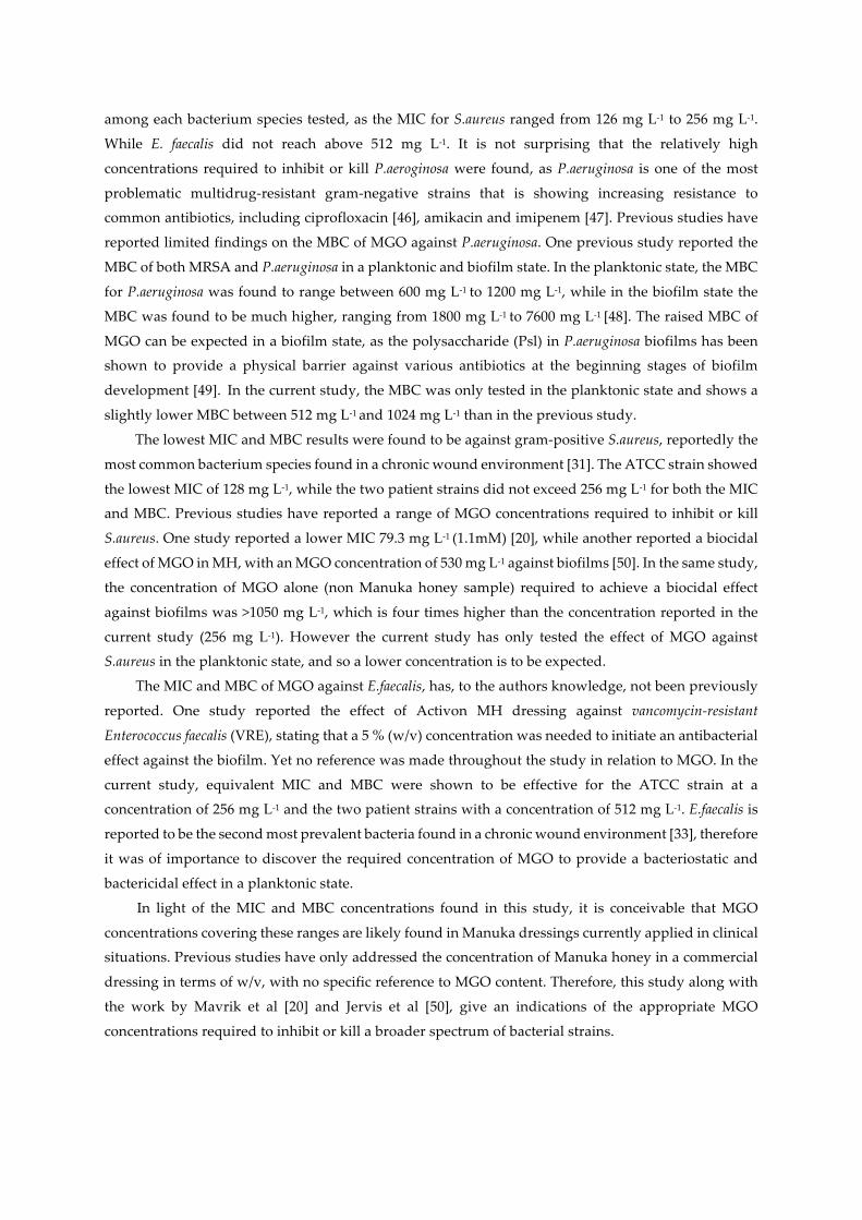
among each bacterium species tested, as the MIC for S.aureus ranged from 126 mg L-1 to 256 mg L-1.
While E. faecalis did not reach above 512 mg L-1. It is not surprising that the relatively high
concentrations required to inhibit or kill P.aeroginosa were found, as P.aeruginosa is one of the most
problematic multidrug-resistant gram-negative strains that is showing increasing resistance to
common antibiotics, including ciprofloxacin [46], amikacin and imipenem [47]. Previous studies have
reported limited findings on the MBC of MGO against P.aeruginosa. One previous study reported the
MBC of both MRSA and P.aeruginosa in a planktonic and biofilm state. In the planktonic state, the MBC
for P.aeruginosa was found to range between 600 mg L-1 to 1200 mg L-1, while in the biofilm state the
MBC was found to be much higher, ranging from 1800 mg L-1 to 7600 mg L-1 [48]. The raised MBC of
MGO can be expected in a biofilm state, as the polysaccharide (Psl) in P.aeruginosa biofilms has been
shown to provide a physical barrier against various antibiotics at the beginning stages of biofilm
development [49]. In the current study, the MBC was only tested in the planktonic state and shows a
slightly lower MBC between 512 mg L-1 and 1024 mg L-1 than in the previous study.
The lowest MIC and MBC results were found to be against gram-positive S.aureus, reportedly the
most common bacterium species found in a chronic wound environment [31]. The ATCC strain showed
the lowest MIC of 128 mg L-1, while the two patient strains did not exceed 256 mg L-1 for both the MIC
and MBC. Previous studies have reported a range of MGO concentrations required to inhibit or kill
S.aureus. One study reported a lower MIC 79.3 mg L-1 (1.1mM) [20], while another reported a biocidal
effect of MGO in MH, with an MGO concentration of 530 mg L-1 against biofilms [50]. In the same study,
the concentration of MGO alone (non Manuka honey sample) required to achieve a biocidal effect
against biofilms was >1050 mg L-1, which is four times higher than the concentration reported in the
current study (256 mg L-1). However the current study has only tested the effect of MGO against
S.aureus in the planktonic state, and so a lower concentration is to be expected.
The MIC and MBC of MGO against E.faecalis, has, to the authors knowledge, not been previously
reported. One study reported the effect of Activon MH dressing against vancomycin-resistant
Enterococcus faecalis (VRE), stating that a 5 % (w/v) concentration was needed to initiate an antibacterial
effect against the biofilm. Yet no reference was made throughout the study in relation to MGO. In the
current study, equivalent MIC and MBC were shown to be effective for the ATCC strain at a
concentration of 256 mg L-1 and the two patient strains with a concentration of 512 mg L-1. E.faecalis is
reported to be the second most prevalent bacteria found in a chronic wound environment [33], therefore
it was of importance to discover the required concentration of MGO to provide a bacteriostatic and
bactericidal effect in a planktonic state.
In light of the MIC and MBC concentrations found in this study, it is conceivable that MGO
concentrations covering these ranges are likely found in Manuka dressings currently applied in clinical
situations. Previous studies have only addressed the concentration of Manuka honey in a commercial
dressing in terms of w/v, with no specific reference to MGO content. Therefore, this study along with
the work by Mavrik et al [20] and Jervis et al [50], give an indications of the appropriate MGO
concentrations required to inhibit or kill a broader spectrum of bacterial strains.
Table 6: MIC and MBC (mg L-1) of MGO in liquid form against three common wound pathogens.
Test organism MIC (mg L-1) MIC (mM) MBC (mg L-1) MBC (mM)
Pseudomonas aeruginosa ATCC27853 512 7.1 1024 14.2
Pseudomonas aeruginosa patient 1 512 7.1 512 7.1
Pseudomonas aeruginosa patient 2 1024 14.2 1024 14.2
Staphylococcus aureus ATCC29213 128 1.8 256 3.6
Staphylococcus aureus patient 1 256 3.6 256 3.6
Staphylococcus aureus patient 2 256 3.6 256 3.6
Enterococcus faecalis ATCC21292 256 3.6 256 3.6
Enterococcus faecium (VRE) patient 1 512 7.1 512 7.1
Enterococcus faecium (VRE) patient 2 512 7.1 512 7.1
3. Materials and Methods
3.1 Materials
A 40 wt % MGO aqueous solution was purchased from Sigma Aldrich UK. Manuka honey 550+ was
purchased from Wellbeing UK and TENCEL® cellulose fibres with a linear density of 1.7 dtex and
length of 10 mm were obtained from Lenzing, Austria.
3.2 Preparations of MH and MGO coating solutions
A 20% (w/w) and a 60% (w/w) aqueous solution of MH was prepared by dissolving 100 g and 300
g of Manuka honey (MGO 550+) respectively, in distilled water and made up to 500 g. The concentration
of MGO of the two solutions was calculated as 0.11 mg g-1 and 0.33 mg g-1 respectively. A 40 wt% MGO
solution was diluted with distilled water to obtain equivalent concentrations of MGO. 3.3 Preparation of coated nonwoven dressings
Prior to the manufacture of the nonwoven samples, TENCEL® fibres were opened using a Shirley
fibre blender. Airlaid webs with a basis weight of 120 g m-2 were produced from 100% TENCEL® fibre
using a short fibre airlaying machine in which the fibres are sifted through a static screen aided by
rotating blades. The webs were mechanically bonded by hydroentanglement (water jet entanglement)
using an STL Hydrolace system with a 110 - 120 µm diameter jet strip and a jet pressure of 50 bar on
one side and 50 bar on the reverse. The hydroentangled webs were washed with warm water and fabric
detergent to remove residual chemical finish on the fibres after hydroentangling. Using a sample liquor
ratio of 1:50, the coatings were applied by immersing samples into the prepared MH and MGO coating
solutions for 10 min. The samples were then passed through a pad mangle at a pressure of 10 kg cm-2,
weighed and left to air dry at room temperature. A coating-free sample was also prepared and used as
a control (NW). The amount of MGO per unit area (mg cm-2) absorbed onto the coated nonwoven
samples was calculated to range between 0.0054 mg cm-2 and 0.0170 mg cm-2 as indicated in Table 1.
Following initial antibacterial testing at these relatively low concentrations, additional coated
nonwoven samples were prepared to provide six new MGO concentrations between 0.1 mg cm-2 and
1.2 mg cm-2. This was achieved by the addition of either MH or MGO to the pre-made coated
nonwovens. A 7 cm2 sample was placed in a weighing boat and weighed on a microbalance. The
addition of MH or MGO to the nonwoven sample equated to the required weight needed to give the
new range of MGO concentrations between 0.1 mg cm-2 and 1.2 mg cm-2. Prior to addition of the
Manuka honey 550+, the honey was heated in an incubator at 40ºC to allow it to soften and enable a
homogeneous distribution over the nonwoven sample. Note it was not possible to prepare MH samples
at concentrations above 0.2 mg cm-2 as the nonwoven samples could not retain additional material due
to the high density and viscous nature of the honey.
3.4 Characterisation of the nonwoven coated samples
3.4.1 Morphology of the coated nonwoven samples
The coated nonwoven samples were inspected using an FEI Quanta 200F Field Emission Scanning
Electron Microscope (FEGSEM). Prior to imaging, all samples were cut and mounted onto 25 mm
aluminium stubs and sputter coated with gold in a vacuum of 0.05 torr for 4 min at 20 mA. A voltage
of 15 kV and a vacuum pressure in the order of 10-6 mbar was achieved in the chamber. Magnifications
between x500 and x1000 were used in order to record morphological features of individual nonwoven
coated samples. 3.4.2 Viscosity measurements of the MH coatings
A Brookfield LV viscometer (DV-E) was used to measure the viscosity of MH (MGO 550+) solutions.
The solution viscosity of the MH was measured at a temperature of 25ºC ± 2ºC and 37 ºC ± 2ºC to assess
temperature dependency. In order for the chamber and solution to reach the specific temperature
required, 16.1 ml of the MH was decanted into the chamber and conditioned in an S1 500 Orbital
Incubator at the required temperature for 24 h prior to testing. The spindle was also conditioned to the
correct temperature. A speed of 6 r min-1 and a spindle size of 18 were used.
3.5 Antibacterial evaluation of MH and MGO-coated nonwoven samples
The antibacterial activity of the MH and MGO-coated nonwoven samples was determined using two
standard methods, BS EN ISO 20743:2007 (Textiles - Determination of antibacterial activity of
antibacterial finished products) [34] and BS EN ISO 20645:2004 (Textile fabrics - Determination of
antibacterial activity, agar diffusion plate test) [38]. The first method simulates the effect of an

antibacterial dressing in contact with contaminated wound exudate [51] and was used to indicate the
antibacterial effect at equivalent MGO concentrations. The second method made an assessment of the
MH and MGO coated nonwovens, as well as the NW using a bacteria seeded agar plate. A brief
description of each method is given below.
3.5.1 BS EN ISO 20743:2007 (Textiles - Determination of antibacterial activity of antibacterial finished products) Test pieces with a mass of 0.40 g ± 0.05 g were cut into suitable sizes for testing. Six control specimens
and six antibacterial specimens were prepared, based on standard protocols [34]. In this study bacteria
cultures of both S.aureus and K.Pneumonia were prepared to concentrations between 1-3·105 per 10 ml
in 1 in 20 nutrient broth. The test specimens were placed in sterile jars and inoculated with 0.2 ml of
bacterial suspension on several areas of the sample, taking care to prevent contact of the suspension
with the jar surface. Immediately after inoculation, 20 ml of SCDLP medium (simulated wound
exudate) was added to three of the control jars and three of the antibacterial sample jars. The jars were
sealed with caps and shaken in an arc of approximately 30 cm by hand for 30 sec. The number of
bacteria recovered from the samples was then determined using a standard serial dilution and pour
plate technique using peptone salt solution as the dilutant and enumeration agar. The remaining jars
were incubated at 37ºC for 24 h. After the incubation period, the number of bacteria that could be
recovered was determined using the equation in the standard. 3.5.2 BS EN ISO 20645:2004 (Textile fabrics - Determination of antibacterial activity, agar diffusion plate test)
A circular specimen of fabric with a diameter of 25 ± 5 mm was cut from the test sample. Two specimens
of the antibacterial fabric and two control specimens without addition of antibacterial treatment were
prepared, based on standard protocols [38]. The specimens were stored between 12 h to 24 h in
sterilized petri dishes at room temperature. Separate agar plates were inoculated with S.aureus and
E.coli bacterial species via streaking the plates with an inoculation loop from a solution containing 1-5
x 108 colony forming units per ml. The test specimen was placed onto the bacterial inoculated agar
surface using a sterilized pair of tweezers until the texture of the specimen was uniformly imprinted
onto the agar. The petri dishes were placed in the incubator for 24 h at 37°C ± 1°C. Immediately after
this period the petri dishes were examined for bacterial growth. If any zone of inhibition was formed
around the test specimens, the diameter of the zone was measured using a pair of calibrated callipers.
The microbial zone of inhibition was calculated using the equation in the standard. Table 7 shows the
criteria stipulated in the standard for defining the effect of an antibacterial treatment [38].
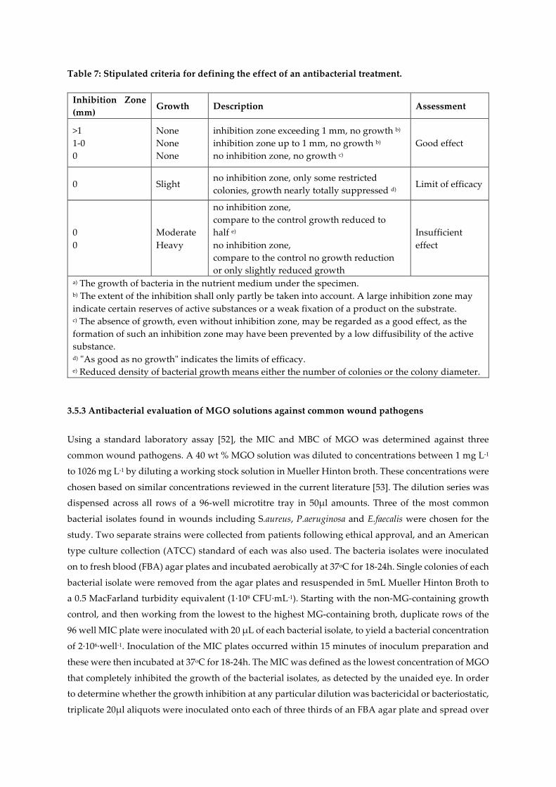
Table 7: Stipulated criteria for defining the effect of an antibacterial treatment.
Inhibition Zone (mm)
Growth Description Assessment
>1 1-0 0
None None None
inhibition zone exceeding 1 mm, no growth b) inhibition zone up to 1 mm, no growth b) no inhibition zone, no growth c)
Good effect
0 Slight no inhibition zone, only some restricted colonies, growth nearly totally suppressed d)
Limit of efficacy
0 0
Moderate Heavy
no inhibition zone, compare to the control growth reduced to half e) no inhibition zone, compare to the control no growth reduction or only slightly reduced growth
Insufficient effect
a) The growth of bacteria in the nutrient medium under the specimen. b) The extent of the inhibition shall only partly be taken into account. A large inhibition zone may indicate certain reserves of active substances or a weak fixation of a product on the substrate. c) The absence of growth, even without inhibition zone, may be regarded as a good effect, as the formation of such an inhibition zone may have been prevented by a low diffusibility of the active substance. d) "As good as no growth" indicates the limits of efficacy. e) Reduced density of bacterial growth means either the number of colonies or the colony diameter.
3.5.3 Antibacterial evaluation of MGO solutions against common wound pathogens
Using a standard laboratory assay [52], the MIC and MBC of MGO was determined against three
common wound pathogens. A 40 wt % MGO solution was diluted to concentrations between 1 mg L-1
to 1026 mg L-1 by diluting a working stock solution in Mueller Hinton broth. These concentrations were
chosen based on similar concentrations reviewed in the current literature [53]. The dilution series was
dispensed across all rows of a 96-well microtitre tray in 50µl amounts. Three of the most common
bacterial isolates found in wounds including S.aureus, P.aeruginosa and E.faecalis were chosen for the
study. Two separate strains were collected from patients following ethical approval, and an American
type culture collection (ATCC) standard of each was also used. The bacteria isolates were inoculated
on to fresh blood (FBA) agar plates and incubated aerobically at 37oC for 18-24h. Single colonies of each
bacterial isolate were removed from the agar plates and resuspended in 5mL Mueller Hinton Broth to
a 0.5 MacFarland turbidity equivalent (1·108 CFU·mL-1). Starting with the non-MG-containing growth
control, and then working from the lowest to the highest MG-containing broth, duplicate rows of the
96 well MIC plate were inoculated with 20 µL of each bacterial isolate, to yield a bacterial concentration
of 2·106·well-1. Inoculation of the MIC plates occurred within 15 minutes of inoculum preparation and
these were then incubated at 37oC for 18-24h. The MIC was defined as the lowest concentration of MGO
that completely inhibited the growth of the bacterial isolates, as detected by the unaided eye. In order
to determine whether the growth inhibition at any particular dilution was bactericidal or bacteriostatic,
triplicate 20µl aliquots were inoculated onto each of three thirds of an FBA agar plate and spread over
the surface of the FBA agar third with a sterile inoculating loop. Inoculated FBA plates were then
incubated at 37oC for 18-24h. The MBC was defined as the lowest concentration at which there was no
visible bacterial growth upon FBA.
4. Conclusions
This study provides the first comparison of equivalent MGO concentrations per unit area in MH
and MGO that are required to provide an antibacterial effect when applied as a physical coating to a
nonwoven wound dressing fabric. The antibacterial efficiency was investigated using both BS EN ISO
20743:2007 and BS EN ISO 20645:2004 to determine if synthetic MGO provided a comparable
antibacterial effect to MH. In the first instance, the bacteria inoculated samples were immersed in 20 ml
of simulated wound exudate fluid and it was found that an MGO concentration of 0.0054 mg cm-2 for
both MH and MGO was sufficient to achieve 100% reduction in bacteria when tested against Gram
positive S.aureus and Gram negative K.pneumonia.
Experiments using bacteria seeded agar plates, found that higher concentrations of MGO between
0.0170 mg cm-2 and 0.1 mg cm-2 were required to produce a good antibacterial effect against E.coli and
S.aureus. In the case of BS EN ISO 20743:2007, hygroscopic TENCEL® fibres are hydrated due to the
addition of 20 ml of SCDLP, which is likely to encourage MGO diffusion, as compared to BS EN ISO
20645:2004, where samples are only incubated with agar and limited moisture is available to facilitate
the diffusion mechanism.
The MH coated nonwovens produced zones of inhibition at relatively low MGO concentrations of
between 0.1 mg cm-2 and 0.2 mg cm-2, as compared with MGO-only coated nonwovens. Clear zones of
inhibition were not apparent until a MGO concentration threshold was reached of 0.4 mg cm-2 for E.coli
and 0.8 mg cm-2 for S.aureus. This difference was attributed to the incubation of the samples at 37ºC
during testing, where the MH coating is likely to soften promoting more rapid diffusion into the
bacteria seeded agar from the fibres, as compared to the less viscous MGO coating, more of which is
retained by the TENCEL® fibres. Manuka honey also contains hydrogen peroxide, which is likely to
contribute to the heightened antibacterial effect, when compared with MGO.
Limited research has previously been reported regarding the MIC and MBC of MGO. In this study,
the MIC and MBC against P.aeuroginosa was found be lower than that previously reported in the
literature when in a planktonic state. The MIC and MBC for S.aureus was found to be between the two
previously reported results of 79.3 mg L-1 [20] and >1050 mg L-1 [48, 50] where the latter value relates to
the antimicrobial effect of MGO against biofilms. The MIC and MBC of MGO against E.facaelis is
reported for the first time in the present study.
Acknowledgments: The authors also wish to thank Dr. Jane Freeman and Mr Peter Parnell for their
assistance with the antimicrobial studies at the Leeds Pathology department. The authors gratefully
acknowledge the financial support of the Clothworkers’ Foundation and The Clothworkers’ Centre for
Textile Materials Innovation for Healthcare (CCTMIH).
Author Contributions: SELB, SJR and PG designed the study. SELB carried out the material
preparation and experiments; SELB analyzed the data; SELB wrote the majority of the paper and all
authors reviewed and approved the final version.
Conflicts of Interest: The authors declare no conflict of interest.
References
[1] Howell-Jones RS, Wilson MJ, Hill KE, Howard AJ, Price PE, Thomas DW. A review of the
microbiology, antibiotic usage and resistance in chronic skin wounds. Journal of Antimicrobial
Chemotherapy. 2005; 55(2):143-9
[2] Russell AD. Antibiotic and biocide resistance in bacteria: comments and conclusions. Journal of
Applied Microbiology. 2002; 92 Suppl: 171S-3S.
[3] Russell AD. Antibiotic and biocide resistance in bacteria: introduction. Journal of Applied
Microbiology. 2002; 92 Suppl:1S-3S
[4] Chopra I, Hesse L, O'Neill AJ. Exploiting current understanding of antibiotic action for discovery of
new drugs. Society of Applied Microbiology Symposium Series. 2002; 31(Antibiotic and biocide resistance
in bacteria):4S-15S.
[5] Jenkins R, Burton N, Cooper R. Manuka honey inhibits cell division in methicillin-resistant
staphylococcus aureus. Journal of Antimicrobial Chemotherapy. 2011; 66(11):2536-42.
[6] Cooper R, Jenkins L, Rowland R. Inhibition of biofilms through the the use of manuka honey.
Wounds UK. 2011; 7(1):24-32.
[7] Maddocks SE, Lopez MS, Rowlands RS, Cooper RA. Manuka honey inhibits the development of
streptococcus pyogenes biofilms and causes reduced expression of two fibronectin binding proteins.
Microbiology 2012; 158(3):781-90.
[8] Sherlock O, Dolan A, Athman R, Power A, Gethin G, Cowman S, et al. Comparison of the
antimicrobial activity of ulmo honey from chile and manuka honey against methicillin-resistant
staphylococcus aureus, escherichia coli and pseudomonas aeruginosa. BMC Complement and Alternative
Medicine. 2010; 10:47
[9] Majtan J, Bohova J, Horniackova M, Klaudiny J, Majtan V. Anti-biofilm effects of honey against
wound pathogens proteus mirabilis and enterobacter cloacae. Phytotherapy Research. 2014;28(1):69-75.
[10] Cooper R, Hooper S, Jenkins L. Inhibition of biofilms of pseudomonas aeruginosa by medihoney
in vitro. Journal of Wound Care. 2014; 23(3):93-6, 8-100.
[11] Henriques AF, Jenkins RE, Burton NF, Cooper RA. The effect of manuka honey on the structure of
pseudomonas aeruginosa. European Journal of Clinical Microbiology & Infectious Diseases. 2011; 30(2):167-
71
[12] Lin SM, Molan PC, Cursons RT. The controlled in vitro susceptibility of gastrointestinal pathogens
to the antibacterial effect of manuka honey. European Journal of Clinical Microbiology & Infectious Diseases.
2011; 30(4):569-74.
[13] Badet C, Quero F. The in vitro effect of manuka honeys on growth and adherence of oral bacteria.
Anaerobe. 2011; 17:19-22
[14] Ranzato E, Martinotti S and Burlando B. Honey exposure stimulates wound repair of human
dermal fibroblasts. Burns Trauma. 2013; 1(1):32-38.
[15] Ranzato E, Martinotti S, Burlando B. Epithelial mesenchymal transition traits in honey-driven
keratinocyte wound healing: Comparison among different honeys. Wound Repair Regeneration. 2012;
20:778–85.
[16] Barros A, Rodrigues JA, Almeida PJ, Oliva-Teles MT. Determination of glyoxal, methylglyoxal,
and diacetyl in selected beer and wine, by HPLC with UV spectrophotometric detection, after
derivatization with o-phenylenediamine. Journal of Liquid Chromatography and Related Technologies. 1999;
22(13):2061-9.
[17] Hayashi T, Shibamoto T. Analysis of methyl glyoxal in foods and beverages. Journal of Agricultural
and Food Chemistry. 1985; 33(6):1090-3.
[18] Nagao M, Wakabayashi K, Fujita Y, Tahira T, Ochiai M, Sugimura T. Mutagenic compounds in soy
sauce, chinese cabbage, coffee and herbal teas. Progress in Clinical and Biological Research. 1986; 206:55-
62.
[19] Adams CJ, Manley-Harris M, Molan PC. The origin of methylglyoxal in new zealand manuka
(leptospermum scoparium) honey. Carbohydrate Research. 2009; 344(8):1050-3.
[20] Mavric E, Wittmann S, Barth G, Henle T. Identification and quantification of methylglyoxal as the
dominant antibacterial constituent of manuka (leptospermum scoparium) honeys from New Zealand.
Molecular Nutrition and Food Research. 2008; 52:483-9
[21] Fidaleo M, Zuorro A, Lavecchia R. Methylglyoxal: a new weapon against staphylococcal wound
infections? Chemistry Letters. 2010; 39(4):322-3.
[22] Ghosh S, Chakraborty P, Saha P, Acharya S, Ray M. Polymer based nanoformulation of
methylglyoxal as an antimicrobial agent: efficacy against resistant bacteria. RSC Advances. 2014;
4:23251-61
[23] Bulman SEL, Goswami P, Tronci G, Russell SJ, Carr C. Investigation into the potential use of
poly(vinyl alcohol)/methylglyoxal fibres as antibacterial wound dressing components. Journal of
Biomaterials Applications. 2015; 29(8):1193-200
[24] Talukdara. D, Raja. S, Dasb. S, Jainb. A K, Kulkarnic. A, Raya. M. Treatment of a number of cancer
patients suffering from different types of malignancies by methylglyoxal-based formulation: a
promising result. Cancer Therapy. 2006; 4:205-22
[25] Apple MA, Greenberg DM. Inhibition of cancer growth in mice by a normal metabolite. Life
Sciences. 1967; 6(20):2157-60.
[26] French FA, Freedlander BL. Carcinostatic action of polycarbonyl compounds and their derivatives.
I. 3-ethoxy-2-ketobutyraldehyde and related compounds. Cancer Research. 1958;18:172-5
[27] Egyud LG, Szentgyo A. Cancerostatic action of methylglyoxal. Science. 1968; 160(3832):1140.
[28] Bhattacharyya N, Pal A, Patra S, Haldar AK, Roy S, Ray M. Activation of macrophages and
lymphocytes by methylglyoxal against tumor cells in the host. International Immunopharmacol. 2008;
8(11):1503-12.
[29] Ayoub FM, Allen RE, Thornalley PJ. Inhibition of proliferation of human leukemia 60 cells by
methylglyoxal in vitro. Biochemical Society Transactions. 1993; 21(2):168S-S.
[30] Kang YB, Edwards LG, Thornalley PJ. Effect of methylglyoxal on human leukaemia 60 cell growth:
modification of DNA, G(1) growth arrest and induction of apoptosis. Leukemia Research. 1996; 20(5):397-
405
[31] Mao N and Russell S J, Nonwoven Wound Dressings, Textile Progress, 2004; 36(4):1-57.
[32] Russell S J, Chapter 2 Dry Laid Web formation. In: Russell S J, editor. Handbook of Nonwovens.
Cambridge: Woodhead Publishing Ltd; 2007. P.16-111.
[33] Gjodsbol K, Christensen JJ, Karlsmark T, Jorgensen B, Klein BM, Krogfelt KA. Multiple bacterial
species reside in chronic wounds: a longitudinal study. International Wound Journal. 2006; 3(3):225-31.
[34] BS EN ISO 20743 Textiles —Determination of antibacterial activity of antibacterial finished
products. 2007
[35] Männer J, Schuster KC, Suchomel F, Gürtler A, Firgo H. Higher performance with natural
intellegence. Lenzinger Berichte. 2004; 83:99-110.
[36] Abu-Rous M, Ingolic E and Schuster K C, Visulisation of nanostructure of TENCEL (LYOCELL)
and other cellulosics as an approach to explaining functional and wellness properties in textiles.
Lenzinger Berichte. 2006; 85:31-37.
[37] Russell AD, Chopra I. Understanding antibacterial action and resistance, 2nd edition. Hemel
Hempstead, England: Ellis Horwood Ltd; 1996
[38] BS EN ISO 20645: Textile fabrics - determination of antibacterial activity - agar diffusion plate test.
2004.
[39] Gómez-Díaz D, Navaza JM, Quintáns-Riveiro LC. Effect of temperature on the viscosity of honey.
International Journal of Food Properties. 2009; 12(2):396-404
[40] Yoo B. Effect of temperature on dynamic rheology of korean honeys. Journal of Food Engineering.
2004; 65(3):459-63.
[41] Mossel B, Bhandari B, D'Arcy B, Caffin N. Use of an arrhenius model to predict rheological
behaviour in some australian honeys. Lebensmittel-Wissenschaft Und-Technologie-Food Science and
Technology. 2000; 33(8):545-52.
[42] Hayashi K, Fukushima A, Hayashi-Nishino M, Nishino K. Effect of methylglyoxal on multidrug-
resistant pseudomonas aeruginosa. Frontiers in Microbiology. 2014; 5:180.
[43] White JW, Schepartz AI, Subers MH. Identification of inhibine, antibacterial factor in honey, as
hydrogen peroxide and its origin in a honey glucose-oxidase system. Biochimica Et Biophysica Acta. 1963;
73(1):57-70.
[44] Kwakman PHS, Zaat SAJ. Antibacterial components of honey. IUBMB Life. 2012;64(1):48-55.
[45] Weston RJ. The contribution of catalase and other natural products to the antibacterial activity of
honey: a review. Food Chemistry. 2000; 71:235-9.
[46] Colsky AS, Kirsner RS, Kerdel FA. Analysis of antibiotic susceptibilities of skin wound flora in
hospitalized dermatology patients. The crisis of antibiotic resistance has come to the surface. Archives
of Dermatology. 1998; 134(8):1006-9.
[47] Sekiguchi J-I, Asagi T, Miyoshi-Akiyama T, Kasai A, Mizuguchi Y, Araake M, et al. Outbreaks of
multidrug-resistant pseudomonas aeruginosa in community hospitals in Japan. Journal of Clinical
Microbiology. 2007; 45(3):979-89.
[48] Kilty SJ, Duval M, Chan FT, Ferris W, Slinger R. Methylglyoxal: (active agent of manuka honey) in
vitro activity against bacterial biofilms. International Forum of Allergy & Rhinology. 2011; 1(5):348-50.
[49] Billings N, Millan MR, Caldara M, Rusconi R, Tarasova Y, Stocker R, et al. The extracellular matrix
component Psl provides fast-acting antibiotic defense in pseudomonas aeruginosa biofilms. PLOS
Pathogens. 2013; 9(8):12.
[50] Jervis-Bardy J, Foreman A, Bray S, Tan L, Wormald P-J. Methylglyoxal-Infused Honey Mimics the
Anti-Staphylococcus aureus Biofilm Activity of Manuka Honey: Potential Implication in Chronic
Rhinosinusitis. Laryngoscope. 2011; 121(5):1104-7.
[51] Thomas S, McCubbin P. A comparison of the antimicrobial effects of four silver containing
dressings on three organisms. Journal of Wound Care. 2003; 12(3):101-7.
[52] Clinical and Laboratory Standards Institute. M07-A9. Methods for dilution antimicrobial
susceptibility tests for bacteria that grow aerobically; approved standard - 9th edition. 2012
[53] Hayashi K, Fukushima A, Hayashi-Nishino M, Nishino K. Effect of methylglyoxal on multidrug-
resistant pseudomonas aeruginosa. Frontiers in Microbiology. 2014; 5:180.