Embed Size (px)
Citation preview
AS, .-I >. � �
4 .1,.. . 4w�?P �{ 'S
�BtoIogicaitEftec�jEmL
I, 4�- 4 .S-
-3.5(4.--'
.4 .&S.�
.-�r-- *b.. - 3 �
i-*�-- � C. *4.�. *;. .
� �.¶ :.*s4,
C.
II . -
wyo an Fetus.'.s
ijit
I; <24$-
~~~~~~~~~~~~. . .l _, . .4 . ., -o,.,.. , ) . . '* ':
,,.. .' -...- '' '''.'' ','.S
:;. ..
.
.. .
. , fi .
:X,Pergamon
AnnaIs of the ICRPPublished on behalf of the International Commissionan Radiological Protection Annals of the ICRPEditor: J. VALENTIN, ICRP, SE- 171 16 Stockholm, Sweden
nternational Commission on Radiological Protection 2001-2005
Chairman: Prof. R. If. Clarke. National Radiological Protection Board. Chilton, Didcor. O.fnrdvdire.)XI1 ORQ. UKlice-Chairman: Dr. L-E. Holm, Sivedish Radiatfan Protection Authorfty (SSI). SE-171 16 Stockholm.
;cientific Secretary: Dr. J. Valentin, ICRP, SE-171 16 Stockholn,. SwIeflr:fix: +46 8 729 7298
vlembers of the Main Commission of the ICRPt. Alexakhin. OhIni,,,k. Russin C. Strefier. E.ssen, Germanyr B. Lindell. Stockbolh. Sweiden (Enwritas)
D. Boice Jr. Rocrkvile. AID A. Sugier. Fo nt cov-mx-Rn .. s. Franre C. B. Meinhold. Brooklhlarvn.t. Cox. Didcor. UK Z. Pan. Beijing. CVkina US.4 (Enerimn). J. Dicus. lI tIdnroinm. DC B. C. Winkler'. Centurion, SoulthAfrka W. K. Sinclair. Ercondifilo. CA (D7eritri,)J.I. Gonzalez. Vienna. Austria D. Beninson. Bueno.V Stares. L. S. Taylor. .11itchellville. AID (EinerinslA. Mettler Jr. Alnbquerqtme. .M Argentija (Emnerints)
!. Sasaki. Chih-.shl. Japan H. I. Dunster. Oxvfors. UK(Emertlua)
ICRP PUBLICATION 90
Biological Effects after PrenatalIrradiation (Embryo and Fetus)
;ubscription Information'ublicattnn Information: Annals of the ICRP (ISSN 0146-6453). For 2003. volume 33 is schedued for publication.'Subscription prices are available upon request from the Publisher or from the Regional Sales Office nearest you orrom this journals website (http:/l'lvw.elsevier.com,'locate!jnlabrrjaicrp). Further information is available on thiszumal and other Elsevier products through Elsevier's website: (http:!Iwww.elsevier.com). Subscriptions are accepted'n a prepaid basis only and are entered on a calendar year basis. Issues are sent by standard mail (surface within!urope. air delivery outside Europe). Priority rates are available upon request. Claims for missing issues should besade aithin six months of the date of dispatch.
)rders. claims, and journal enquiries: please contact the Customer Service Department at the Regional Sales Officetearest you:)rlando: Elsesier. Customer Service Department. PO Box 211. 6277 Sea Harbor Drive. Orlando. FL 32887-4800.JSA: phone: (+ 1) (877) 8397126 (toll free number for US customers]. or (+ 1) (407) 3454020 [customers outide US]:ax: (+ I) (407) 3631354: e-mail: usjcsR.elsesver.comimsterdam: Elsevier. Customer Service Department. PO Box 211. 1000 AE Amsterdam. The Netherlands: phone:+ 31) (201 4853757: fax: - -31) (20) 4853432: e-mail: nlinfo-fi elsevier.com'okyo: Elsevier. Customer Service Department. 4F Iligashi-Azabu. I-Chome Bldg. 1-9-15 Higashi-Azabu. Minato-ku.
'okyo 106-0044. Japan: phone: (+ SI) (3) 5561 5037: fax: (+ 81) (3) 5561 5047: e-mail: jp.info'd elsevier.com;ingapore: Elsevier. Customer Service Department. 3 Killiney Road. #08-01 Winsland House I, Singapore 239519:-hone: (+ 65) 63490222: fax: (+ 65) 67331510: e-mail: asiainfo'i elsevier.com
lack Issuestack issues of all previously published volumes are available direct from Elsevier Science offices (Oxford and New!ork).'his journal is indexedtabstracted in: App. Health Phy. Abstr.. Biosis Data.. CABS and SSSA 'CISA:ECA;ISMEC.3TM"The paper used in this publication meets the minimum requirements of American National Standard fornformation Sciences-Permanence or Paper for Printed Library Materials. ANSI Z39.48-1984.JSA mailing notice: Annals of the ICRP (ISSN 0146-6453) is published four times a year by Elsevier Ltd (PO BoxI1. 1000 AE Amsterdam. The Netherlands). Annual subscription price in the USA USS302 (valid in North. Centralnd South America). including air speed delivery. Periodical postage rate paid at Jamaica. NY 11431.ISA POSTMASTER: Send address changes to Annals of the ICRP. Publications Expediting Inc.. 200 Meacham%ve. Elmont. NY 11003.%IRFREIGIIT AND MAILING: in the USA by Publications Expediting Inc.. 200 Meacham Avenue. Elmont. NY1003.
EditorJ. VALENTIN
PUBLISHED FOR
The International Commission on Radiological Protection
By
PERGAMONIrI
"V�3 \q,.; il.,
,V,-

UK
USA
JAPAN
Elsevier Ltd, The Boulevard, Langford Lane,Kidlington, Oxford OX5 IGB, UK
Elsevier Inc., 360 Park Avenue South,New York 10010-1710, USA
Elsevier K.K., 9-15 Higashi-Azabu 1-chome,Minato-ku, Tokyo 106-0044, Japan
Copyright C 2003 ICRP Published by Elsevier LtdAll rights reserved.
The International Commission on RadiologicalProtection encourages the publication of translationsof this report. No part of this publication may bereproduced, stored in a retrieval system or transmittedin anyform or by any means electronic, electrostatic,magnetic tape, mechanical photocopying, recording orotherwise or republished in anyform. without permissionin writingfrom the copyright owner.
ISBN 008 044 265XISSN 0146-6453
Published quarterly (March, June, September, December)Septembcr/December issue
No responsibility is assumed by the Publisher or theICRP for any injury and/or damage to persons orproperty as a matter of products liability, negligence,or otherwise, or from any use or operation of anymethods, products, instructions, or ideas contained inthe material herein. The recommendations and advice ofthe ICRP reflect understanding and evaluation of thecurrent scientific evidence as given in this report. If andwhen further relevant information becomes available, theICRP may review its recommendations. Because of rapidadvances in the medical sciences, in particular, diagnosesand administered amounts of radiopharmaceuticals shouldbe independently verified. Although all advertising materialis expected to conform to ethical (medical) standards,inclusion in this publication does not constitute a guaranteeor endorsement of the quality or value of such product orof the claims made by its manufacturer.
Typeset by Variorum Publishing Ltd. RugbyPrinted and bound in Great Britain by Polestar Wheatons Ltd.Eveter
CONTENTS
GUEST EDITORIAL............................................................................................I
PREFACE..............................................................................................................3
A BSTRA CT .. ........................................................................................... S
EXECUTIVE SUMMARY .................. 7
1. INTRODUCTION.............9...............................................................................91. . R eference.. ................................................................................................ 10
2. RADIATION EFFECTS AFTER EXPOSURE DURING THEPRE-IMPLANTATION PERIOD ........... 12.1. Introduction........................................................................................... 112.2. Lethality after irradiation ........................ 122.3. Cytogenetic effects ........................ 142.4. Induction of malformations ...................................................................... 162.5. References................................................................................................. 20
3. DEVELOPMENTAL EFFECTS AFTER IRRADIATION DURINGORGANOGENESIS AND FETOGENESIS . ............................... 233.1. Historical background and state of risk assessment in man ..................... 233.2. Experimental studies................................................................................. 253.3. Summary................................................................................................... 383.4. References.................................................................................................. 40
4. AETIOLOGY OF EFFECTS DURING BRAIN DEVELOPMENT ............ 454.1. Basic features of cerebrogenesis ....................... 464.2. Acute cellular effects ....................... 514.3. Long-term effects ....................... 674.4. Endpoints.................................................................................................... 784.5. Summary................................................................................................... 884.6. References................................................................................................ Refe s91
5. HUMAN EVIDENCE ON THE EFFECTS OF IN-UTERORADIATION EXPOSURE ON NEUROLOGICAL ANDMENTAL PROCESSES .. ........................... 1035.1. Japanese atomic bomb in-utero cohort................................................... 1035.2. Mental retardation................................................................................. 1045.3. Intelligence quotient (IQ) ................... 1085.4. School performance ................... 1135.5. Seizure disorders.................................................................................... 1155.6.. Other neurological effects ....... ............ 116
) A)

I) ICRP Publication 90 . )
5.7. Discussion............................................................................................... 1185.8. References............................................................................................... 122
6. CARCINOGENIC RISK FROM IN-UTERO IRRADIATION:ANIMAL STUDIES ................................................ 1256.1. Introduction............................................................................................ 1256.2. Gestational age and cancer risk ............................................... 1266.3. Sex dependence....................................................................................... 1286.4. Strain specificity...................................................................................... 1296.5. Radiation dose response and question of threshold ............................... 1296.6. Genetic predisposition ............................... 1336.7. Extrinsic modifying factors..................................................................... 133
- 6.8. Internal emitters...................................................................................... 1356.9. Genomic instability and cancer induction...................................... 1366.10. Conclusions .. 1386.11. References .. 139
ICRP Publication 90 /
9.3. Organogenesis and fetogenesis ....................... 1879.4. Brain development................................................................................... 1919.5. Mental effects in humans....................................................................... 1929.6. Experimental carcinogenesis ..................... ; 1939.7. Childhood cancer.................................................................................... 1949.8. Human cancer risk after prenatal irradiation. ....... .......................... 195
10. OPEN QUESTIONS AND NEEDS FOR FUTURE RESEARCH ........... 1 97
APPENDIX A: ESTIMATED NUMBER OF CANCER DEATHS INTHE IN-UTERO.EXPOSED ATOMIC BOMBCOHORT IF THE RISK COEFFICIENT FROMTHE OXFORD SURVEY OF CHILDHOODCANCERS (OSCC) IS ACCURATE ....................................... 199
A.I. Radiation-induced cancers before age 2 years ........................................ 199A.2. Japanese mortality rates and their ratio to British mortality rates ........ 199A.3. References.............................................................................................. 200
7. EPIDEMIOLOGY OF CHILDHOOD CANCER ........................................ 1437.1. Introduction............................................................................................ 1437.2. Incidence of childhood cancer by tumour type, geography, and age ...... 1437.3. Environment in the aetiology of childhood cancer ................................. 1 457.4. Genetic factors in the aetiology of childhood cancer .............................. 1477.5. Second cancers ............ 1487.6. Conclusion...........................................................5..............0..;................... 1507.7. References...............................................................................................1. 50
8. HUMAN CARCINOGENIC RISK FROM IN.UTERO IRRADIATION 1538.1. Introduction............................................................................................. 1538.2. Methodological assessment of the OSCC ............................... ;.; .1568.3. Dosimetric features of the OSCC data .......... .................... 1598.4. Radiation cancer risk according to stage of in-utero development ........ 1 638.5. In-utero irradiation from Chernobyl and leukaemia in infancy ............. 1658.6. Consistency of the OSCC data with other prenatal medical irradiation
studies ............ ..................................... 1678.7. Does in-utero irradiation induce all types of childhood cancer to the
same extent?............................................................................................. 1718.8. A comparison of risk estimates from the OSCC data and the Japanese
atomic bomb data .......... 1738.9. Estimation of risk from in-utero irradiation........................................... 1768.10.Conclusion .. ................................. 1818.11. References .................................... 182
9. SUMMARY AND CONCLUSIONS . .................................. 1879.1. Introduction............................................................................................ 1879.2. Pre-implantation period ................................... 187
iv V
EXECUTIVE SUMMARY
:; .
t: .: - . ., . . e . ... . :
, . . . . . . . . .
..... ... .. .. . ... ... .. . .. . . ...... .... ........ . . .. .
; , . *
t .. A- t < . X s . , ........... . , . f 4 . e i - -:: . j s * - - t ;, t -; S . * * ..
Gil 4 i .-- .4 foil_ 1 l. S I_ w S S . L, .; ,. _ t,
: : . - . A - ; : : . . t .;, - It - .... ;; ,-2 - ... - -: . .... t: - . .-.
- f : X ; * j * * - -; I. - r "s- .
- : - ., , | ,
i
. . .. . . .
. . . .
.. . . .. . .. . .
. _ , . .. . . . .. . .
:
, , q , . . ... . . . .
; . _,,.@ -j. - 1 Fs .; . , . . :. . . .:
. . . . . . . . . . . .. . ..
(a) The ICRP considered the risks following radiation exposure during prenataldevelopment in its 1990 Recommendations (Publication 60). Since that publication,new experimental animal data on biological effects and reevaluations of humanstudies after prenatal irradiation have been published. A critical review of these new.data has been performed by a task group, and the key findings are listed below.
The mammalian embryo and fetus are highly radiosensitive during the entireperiod of prenatal development. The nature and severity of induced biologicaleffects -depends on the developmental stage during which the radiationexposure takes place.The risk of lethality of the developing organism is highest during the pre-implantation period (up to day 10 postconception). This effect is mainly dueto killing of blastomeres, caused by chromosomal damage. In certain mousestrains, radiation exposure induces genomic instability after doses as low as0.5 Gy of low-linear-energy-transfer (LET) radiation. This form of genomicinstability can be transmitted to the next generation. . ;'.In mouse strains with genetic predispositions for specific malformations, ithas been observed that these malformations are also induced by ionisingradiation during the prc-implantation period. In particular, this is the caseafter irradiation of zygotes where no threshold is .obsered-in the doseresponse. - -a .
Malformations are mainly induced after exposure during the period of majororganogenesis. In certain stages of major organogenesis (weeks 3-7, post-.conception for human development), enhanced. sensitivity exists for certainspecific malformations. During this developmental period, growth retarda-tion is caused by irradiation. The regenerative capacity decreases as differ-entiation of tissues and organs progresses.After irradiation during the pre-implantation period and the period of majororganogenesis, relative biological effectiveness (RBE) values for fast neutronsrange from 3 to 10. After chronic and fractionated exposures with low-LETradiation during these prenatal stages, dose-rate effectiveness factors (DREF)in the range of 4-10 have been observed. Adaptive responses could not beobserved for embryonic development and chromosomal aberrations during
.these developmental stages. No human data are available for these para-meters and phenomena.. . . . - 1. !
.Comprehensive experimental studies have been performed on the develop-.ment of the central nervous system in rodents, and on a more limited scale inprimates, in order to evaluate the mechanisms of radiation effects on thesedevelopmental processes. Early proliferating neuroepithelia are very sensitivebut have a strong capacity for ccll substitution and tissue re-organisationwhich reduces with progressing differentiation. .;Neuronal plasticity and natural redundancy of neurons can also compensatefor radiation-induced damage during brain development. The most significant
7
; e :
I i I I .
. .. .
... , .. . .
.,, . . . I
..1 t ..
... ,..�. 1
9 I.,

s tICRP Publication 90
period is the 'window of cortical sensitivity' in the early and mid-fetalperiod in rodents and weeks 8-15 postconception in humans. Experimentaldose-response studies in rodents for structural: and functional endpointsincluding learning and behavioural changes result in dose-response curveswith thresholds in the range of 0.1-0.3 Gy low-LET, radiation.
* The experimental data are in good agreement with human data from theJapanese atomic bomb study on severe mental retardation (SMR) afterexposures during the most sensitive period (weeks 8-15 postconception). Thelower confidence bound on the threshold dose is 0.3 Gy for this effect. Aradiation dose of I Gy would increase the risk of SMR by about 40%.
* For intelligence quotient (IQ) scores, a linear dose-response model provides asatisfactory fit of data from the atomic bomb cohort with irradiation at weeks8-15 postconception. The decline in IQ values is about 25 IQ points/Gy inthis sensitive period. School achievement is also reduced following exposureof I Gy. A threshold dose is not apparent for these effects; however, afterradiation doses of 100 mGy, these effects are very small.
* From animal experiments, it can be concluded that radiosensitivity for cancerinduction is highest in the late fetal period. Female mice have a higher riskthan males.
* In industrialised countries, leukaemias, brain tumours, and lymphomas arethe predominant paediatric cancers. Few of these cancers are linked withknown genetic predispositions.
* The largest case-control study of cancer after in-utero irradiation, the OxfordStudy of Childhood Cancers (OSCC), found that radiation increased all typesof childhood cancer by approximately the same degree. The second largeststudy showed a larger relative risk for leukaemia than for solid tumours,while several cohort studies of in-utero radiation found no clear evidence ofradiation-induced childhood cancer. The data from the atomic bomb survi-vors suggest that the lifetime cancer risk from in-utero exposure may besimilar to that from exposure in early childhood.
* The OSCC data suggest that cancer induction is at least as likely followingexposure in the first trimester as in later trimesters. From the data publishedto date, it is not possible to determine tissue-weighting factors in order todefine cancer risk in different tissues and organs. Adequate human in-uteroexposure data arc not available to define the dose and dose-rate effectivenessfactor (DDREF) for low-LET radiation or the RBE values for neutron orother high-LET radiations.
(b) These conclusions, which tend to strengthen and supplement the recommen-dations contained in Publication 60, have significant implications for protection ofthe embryo/fetus. The editorial which prefaces this report provides an interim viewfrom the Commission on these implications.
) )1. INTRODUCTION
(I) The ICRP reviewed the effects of exposure to ionising radiation during pre-natal development of.mammals in Publication 60 '1990 Recommendations of theInternational Commission on Radiological Protection', and has given its recom-mendations with respect to regulations for radioprotection of the embryo and fetuson this basis. It has long been known that the developing organism is highly radio-sensitive; therefore, special regulations are necessary for pregnant women at workand in public places. As such, many efforts have been undertaken in radiationresearch in order to improve our knowledge about radiation effects. and risksthrough exposures during prenatal development. The most recent data in thisresearch field will be reported in the following chapters, and possible consequencesfor radioprotection will be considered.
(2) Due to the characteristics of the various developmental processes and theeffects that can be induced by toxic agents, such as-ionising radiation, at thesedevelopmental stages, prenatal development is divided into three main periods:.
. ~ . ,. . . . ............ ....
• pre-implantation; : I ,, .-* major organogenesis; 7; - - ' . . *. XE* fetal period. . ;.,
(3) Many experimental and clinical data have shown that the response to radiationexposure is highly dependent on the developmental stage during which this exposuretakes place. It is very well known that ionising radiation interferes to a high degreewith cell proliferation (Hall, 1994). Therefore; biological systems with a high frac-tion of proliferating cells show high radiation responsiveness. High rates of cellproliferation are found throughout prenatal development.' However, although cellproliferation is a key process for the development of radiation effects, the sensitivityof the embryo and fetus is also determined through processes of differentiation andcell migration, and -the radiation effects on these biological processes. Therefore,radiation effects in these periods will be considered. Development of the centralnervous system starts during the first weeks' of embryonic development andcontinues through the early-postnatal period. Thus development of the centralnervous system occurs over a very long period, during which it is especially vulner-able. It has been found that the development of this system is very frequently dis-turbed by ionising radiation, so special emphasis has to be given to these biologicalprocesses. - - :
(4) Furthermore,' studies have been conducted regarding the extent to whichradiation carcinogenesis is possible, which cancers will develop, and whether thereare developmental periods with different radiosensitivities with respect to theseevents. It is well known that the cancer risk is very high after small children areexposed to radiation, and the patterns of cancers are different from those of adults.Questions arise regarding whether this high radiosensitivity also exists for radiationexposures during prenatal development,' and whether some embryonal/fetal tissuesor organ systems are more radiosensitive than others.:
: . .,. , , , - , .. : . ...8

ICRP Publication 90
(5) The analysis of risk from prenatal exposures has to be performed on the basisof the developmental stage during which the exposure takes place. For such a riskanalysis, precise knowledge about the specific development of the species and thetime periods is necessary, and extrapolations have to be conducted from animalexperiments to the human situation on the basis of specific developmcntal stages.Figure 1.1 shows the effects of radiation exposure during prenatal development withregard to lethality and abnormalities. '
(6) The highest radiosensitivity with respect to lethality occurs after irradiationduring the pre-implantantion period. This effect decreases during major organogen-esis, while gross malformations (abnormalities) develop after exposures during thissecond phase, and no abnormalities and little lethality is'observed after radiationexposures during the fetal period. However, the newer data presented in this reportindicate that this scheme needs to be seen in a more differentiated way on the basisof new results after exposures during these phases of development.
,- .. Pre-implantation, Organogenesis Fetus+
100 -
80 1 Prenatal
0
Cl
0.
2. RADIATION EFFECTS AFTER EXPOSURE DURING THEPRE-IMPLANTATION PERIOD
2.1. Introduction
(7) For analysis of radiation risk during the pre-implantation period, no observa-tions in humans are available, as conception is not noticed at that time: Therefore,the risk analysis can only be achieved on the basis of animal experiments which havemainly been performed with mice and rats. However, with respect to the pre-implantation period, the advantage is that the duration and general biological pro-cesses (cf. cell proliferation and differentiation) during this period are very similarfor most mammalian species. Thus the duration of the pre-implantation period is 5days for mice, 7 days for; rats, and 8 days for humans (SSK, 1985; Streffer andMolls, 1987), although the duration of total prenatal development varies to a highdegree. At first sight, the pre-implantation period is'determined by cell proliferationprocesses from the zygote (one cell) to the hatched blastocyst (100 cells in mice, 250cells in humans). The hatched blastocyst is then implanted into the uterus for furtherdevelopment (Carlson, 1994).
(8) After the sperm enters the oocyte, the second meiotic division will be com-pleted and two pronuclci are formed with the male and female genome, respectively.Following the period of DNA synthesis in which the genetic material is doubled inthe separated pronuclei, the pronuclei fuse and a diploid genome is formed. Aftercell division leading to the two-cell embryo, a comparatively long cell cycle time ofaround 20 h follows before the two-cell embryo divides into four cells. Once the firstcell division has occurred, the first gene activation processes occur. Further celldivisions follow comparatively quickly with a cell cycle time of around 12 h (Strefferet al., 1980; Streffer and Molls, 1987). When the pre-implantation embryo reachesaround 16 cells, the morula is formed. This occurs around 60-70 h postconception.With the ongoing cell divisions, the single cells become smaller but the total size ofthe embryo remains almost constant.
(9) The zygote and the later pre-implantation embryo are surrounded by the zonapellucida. This protects the embryo but also makes it necessary that the embryo hasits own resources for cellular maturation.. There is apparently little interactionbetween the embryo and the mother during this developmental period. After morula-tion, the formation of the blastocyst takes place at around 80 h postconception in miceand 100-120 h postconception in humans. During the period when the hatching ofthe blastocyst from the zona pellucida takes place, differentiation into the embryo-blast and trophoblast is progressing and becomes largely completed (Carlson, 1994).
(10) These rapid cell proliferation processes are very similar in all mammals.Modem molecular biological techniques have shown that the first gene expression.processes in the newly developing embryo occur during the two-cell and four-cellstages, and the activity of gene expression increases over the following hours anddays (Carlson, 1994).
(11) Radiation effects during the developmental period have mainly been observedin mice and rats, but data from rabbits and dogs are also available. Radiation can
, , I I
III I, It, II I ,, II I I I. Human-1 2 4 6 9 12 162025 2932 3741 45 5470
Days postconception
Fig. 1.1. The occurrence of lethality and abnormalities in mice after a prenatal radiation exposure ofabout 2 Gy, given at various times postconception. The two scales for the abscissa compare develop-mental stages in days for mice and humans (redrawn from Hall, 1994 with the permission of Hall and thepublisher).
1.1. Reference
Hall, E.J. (1994) Radiobiologyfor the radiologit, 4th ed. J.B. Lippincott Co., Philadelphia, PA.
10
_9 _)
I
ICRP Publication 90 )JCRP Publication 90
100or
901-
1080
70
1220C.2:10!Ga_J._0
60
Exposure day 7 p.c. a
a
0 p0 0
50
30f
60i
401
aExposure day 10 p.c.
0
.0
0 0oo so 000
0 0.50 1.00 1.50 2.00 2.50
20 J
501- 10l
401 0
30F
20l-
30 [ Exposure day 13 p.c.
20 ° a 0-o
10 I-10-- v L %_ 0 ° - -
enA
0 050 1.00 150 2.00 2.50X-ray dose (Gy)
0 0.50 1.00 2.00
X-ray dose (Gy)3.00
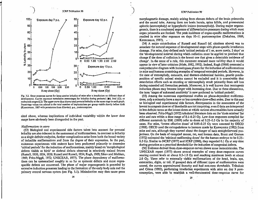
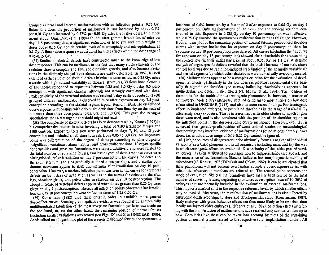
Fig. 3.2. Dose-response curves for intra-uterine lethality of mice after x irradiation on different days ofdevelopment. Curves represent cumulative percentages for lethality during perinatal (0), fetal (0), orembryonic stages (0). The upper curve thus shows total prenatal lethality at the same stage in each graph.Percentage values are related to the total number of implantations per group visible shortly before birth(Konermann. 1987 with permission from Elsevier). p.c., postconception.
cited above, whereas implications of individual variability within the lower doserange have obviously been disregarded in the past.
Aalformations in utero(53) Biological and experimental side factors taken into account for prenatal
lethality are also inherent in the assessment of malformations. In contrast to lethalityas a single definite endpoint, further complications arise from both the broad varietyof inducible malformations and from the degree of their expression. In the past,numerous experiments with rodents have been performed primarily to determine'critical periods' for the induction of malformations, mainly based on 'morphologicaldefects visible at birth' or skeletal defects observed in selectively stained fetuses(Russell, 1950, 1954, 1956; Russell and Russell, 1954; Rugh, 1969; Sikov and Mahlum,1969; Fritz-Niggli, 1972; UNSCEAR, 1977). The phase dependency of malforma-tions can be systematised roughly in so far as systemic defects and more organ-specific defects are concerned. The phase of major organogenesis is preceded byextensive induction processes leading to the formation of the early body axis and theprimary central nervous system (see Fig. 3.1). Misinduction may thus cause gross
28
morphogenetic damage, mainly arising from closure defects of the brain primordiaand the neural tube. Among them are brain hernia, spina bifida, and pronouncedaplastic (anencephaly) or hyperplastic lesions (exencephaly). During major organo-genesis, there is a condensed sequence of differentiation processes during which mostorgan primordia are formhed. The peak incidence of organ-specific malformations isreached in mice after exposure on days 10-11 postconception (Dekaban, 1969;Konermann, 1987). -
(54) A major contribution of Russell and Russell (cf. citations above) was toassociate the natural sequence of developmental steps with phase-specific irradiationdamage. For mice, they defined such 'critical periods of I or, more rarely, 2 days' asthe 'developmental interval during which radiation must be applied to produce thatchange if the dose of radiation is the lowest one that gives a detectable incidence ofchange'. In the sense of a rule, this statement retained more validity than it wouldappear in view of later criticism (Mole, 1982, 1992). Indeed, Rugh (1966) presented acomprehensive diagram with homologous phases for the induction of malformationsin mice and humans containing examples of temporal extended periods. However, inthe case of exencephaly, cataracts, and thoraco-abdominal hernias, genetic predis-position of specific animal strains cannot be excluded and it is conceivable thatretardation effects such as stunting or microcephaly result primarily from cell lossduring extended cell formation periods. Moreover, it is well known that teratogenicinduction phases may become longer with increasing dose. Due to these discussions,the term 'stages of enhanced sensitivity' is now preferred to 'critical periods'.
(55) Among the numerous experimental studies on phase-dependent malforma-tions, only a minority have a more or less complete dose-effect series. Due to this andto biological and experimental side factors, discrepancies in the assessment of thelowest teratogenic dose or of thresholds are not surprising, even if data are interpretedin less definite terms as the lowest doses at which various abnormalities have actuallybeen observed. Fritz-Niggli (1972) tabulated literature data on the 'dosis minima' formice and rats within a dose range of 0.1-0.25 Gy. Low dose responses compiled fordifferent mammals by SSK (1989) refer to doses of 0.25-0.5 Gy in the majority ofcases. For mice, 'lowest effective doses' of 0.05-0.25 Gy were assumed by OECD(1988). OECD used the extrapolations to humans made by Kameyama (1982) frommice and rats, although they warned about the danger of such straightforward pro-jections. On the basis of compiled mouse, rat, and human data, Brent and Gorson(1972) estimated the 'minimal malforming doses' for the human embryo to be 0.25-0.5 Gy. Similar to NCRP (1977) and ICRP (2000), they regarded 0.1 Gy at any timeduring gestation as a practical threshold for the induction of congenital defects.
(56) Evidence derived from dose-response curves shows some inconsistencies. TheUNSCEAR report (1977) shows several examples of steep dose-response curvesnearing control levels at about 0.5-1.0 Gy and reaching maximum levels at about2.0 Gy. These refer to externally visible malformations of the head, brain, eye,extremities, digits, or tail; If grouped data of different types of malformation werescored, the curves approximated linearity and had non-zero thresholds. Tribukaitand Cekan (1982), performing irradiation experiments with mice on day 9 post-conception, were able to establish a well-documented dose-response curve for
29
ICRP Publication 90 ICRP Publication 90
grouped external and internal malformations with an inflection point at 0.75 Gy.
Below this dose, the proportion of malformed fetuses increased by about 0.1%per 0.01 Gy and increased by.0.57% per 0.01 Gy after the higher doses. In a more
recent study, Uma Devi et al. (1994) found, after gamma irradiation of mice onday I 1.5 postconception, a significant reduction of-head size and brain weight at
doses above 0.15 Gy, and detectable levels of microcephaly and microphthalmia at0.1 Gy. A linear dose response was assumed for these effects within the dose range of
0.05-0.15 Gy. :-:(57) Studies on skeletal defects have contributed much to the knowledge of low
dose responses. This can be attributed to the fact that many single elements of theskeleton show a complex and thus radiosensitive genesis, and also because devia-tions in the distinctly shaped bone elements are easily detectable. In 1957, Russell
extended earlier studies on skeletal defects in mice to doses as low as 0.25 Gy, usinga strain with high natural variability in thoracal structures. Various bone elementsof the thorax responded to exposures between 0.25 and 1.0 Gy on day 8.5 post-conception with significant changes, although not strongly correlated with dose.
Peak sensitivity of the vertebral column was'reached I day earlier. Jacobson (1968)grouped different malformations observed'in mice after exposure on day 7.5 post-conception according to the skeletal regions (spine,' sternum, ribs). He establisheddose-response relationships with only slight deviations from linearity on the basis ofnot more than three dose points (0.05, 0.2, and 1.0 Gy). This gave rise to vaguespeculations that a teratogenic threshold might not exist.
(58) The complexity of skeletal defects has been demonstrated by Knauss (1978) in
his comprehensive studies in mice involving a total of 7100 irradiated fetuses and
1100 controls. Exposures to x rays were performed on days 7, 10, and 13 post-conception and included small .dose intervals from 0.05 to 3.0 Gy. An importantpoint was differentiation of the degree of damage according to physiologically
insignificant variations, abnormalities, and gross malformations. If organ-specificabnormalities and gross malformations were scored additively and were related tothe total number of surviving fetuses, two basic types of dose-response curve weredistinguished.. After irradiation on day 7 postconception, the curves for defects tothe skull, sternum, and ribs gradually attained a steeper slope, and a similar con-tinuous curvature applies to skull and sternum after irradiation on day 10 post-
conception. However, a marked inflection point was seen in the curves for vertebraldefects on both days of irradiation as well as in the curves for defects to the ribs,
legs, shoulder girdle, and pelvis after irradiation on day 10 postconception. The
abrupt increase of vertebral defects appeared when doses greater than 0.25 Gy were
given on day 7 postconception, whereas all inflection points observed after irradia-tion on day 10 postconception were shifted to doses of 1.25-1.50 Gy.
(59) Konermann (1982) used these data' in order to establish more general
dose-effect curves. Seemingly contradictive evidence was found if an anatomicallyundifferentiated tabulation of the most severe malformation per fetus was made onthe one hand, or, on the other hand, the remaining portion of normal fetuses(including smaller variations) was scored (see. Figs. IX and X in UNSCEAR, 1986).As visualised on a logarithmic plot of the severely malformed fetuses, the spontaneous
30
incidence of 0.6% increased by a factor of 3 after exposure to 0.05 Gy on day 7postconception. Only malformations of the skull and the cervical vertebra con-tributed to this. Exposure to 0.125 Gy on' day 10 postconception was ineffective,while 0.25 Gy doubled the spontaneous malformation rates at this'stage. However,from a linear plot of the remaining portion of normal fetuses, pronounced shouldercurves 'with steeper inclination for exposure on day 7 postconception than for
exposure on day 10 postconception were derived. All curves (including the flat curve'for exposure on day 13 postconception) showed clear thresholds for transcendingthe control level 'in their initial parts, i.e. at about 0.35, 0.9, or 1.1 Gy. A detailedanalysis of organ-specific defects revealed that the initial increase of normals abovecontrol level was due to radiation-induced stabilisation of the naturally variable riband sternal segments by which other deviations were numerically overcompensated.
(60) Malformations appear to be a complex criterion for the evaluation of devel-opmental effects, particularly in the low dose range>'Most experimental data basi-cally fit sigmoid or shoulder-type curves,'indicating thresholds as expected formulticellular, i.e. deterministic, effects (cf. Mizller ct al.,' '1994). The position ofthresholds for the multitudinous teratogenic phenomena is, however, a matter ofcontroversy.' Mole (1992) attributed detailed criticism to most entries on low doseeffects cited in UNSCEAR (1977), and also to more recent findings. For teratogeniceffects in implanted embryos, he postulated thresholds in the range of 0.40-2.0 Gyafter acute x-ray exposure. This is in agreement with older studies in which higherdoses were used, and is also consistent with the position of the shoulder region or
the inflection points in the dose-response curves mentioned. However, even if it isconceded that genetic predisposition of some animal strains or 'methodologicalshortcomings may interfere, evidence of malformations found at considerably lowerdoses, i.e.' within a dose range of 0.05-0.25 Gy,' cannot be ignored'.
(61) Major sources of disagreement arise obviously from: (i) neglect of individualvariability as a basic phenomenon in all organisms including man; and (ii) the wayin which teratogenic effects are evaluated. Discontinuity of the initial part of survi-val curves has been attributed to predisposition to radioresistance (see above), andthe occurrence of malformations likewise indicates less morphogenetic stability ofsubcohorts (cf. Knauss, 1978; Tribukait and Cekan, 1982). It can be anticipated thatthese phenomena will not become overt unless complete dose-response series withsubstantial observation numbers are 'referred to. The second point concerns themode of evaluation. Skeletal malformations have mainly been related to the totalnumber of surviving fetuses, neglecting spontaneous resorption rates of 10-20% ofembryos that are normally included in the evaluation of external malformations.This implies a marked shift in the respective reference levels by which smaller effectsmay be masked. Moreover, the manifestation of malformations is also affected byembryonic death according to dose and developmental stage (Konermann, 1987).Early embryos with gross inductive effects are thus more likely to be resorbed thanlocally malformed older embryos (Friedberg et al., 1987). Selection effects interfer-ing with the manifestation of malformations have received only scant attention up tonow. 'Corollaries like these can be taken into account by plots of the remainingportion of normal fetuses related to the respective total implantation number. All
*31
I
:) )ICRP Publication 90 ICRP Publication 90
U,
cm
CoEn5)
ID
with incomplete cell substitution, topographically normal but.overall retardedorgans or fetuses are 'reconstituted' (Rugh, 1962, 1963; Balla et al., 1983).
(63) Among the earliest quantitative studies in growth retardation are those ofRussell (1950), who found (in neonatal mice) that the strongest effects after exposureoccur during advanced organogenesis, i.e. on days 10.5-11.5 postconception. Dosesbetween 2.0 and 4.0 Gy evoked weight loss of 0.22 g/Gy (normal weight of newbornsis about 1.4 g). After lower doses (0.5-2.0 Gy), the highest relative effectiveness toinduce fetal weight loss was observed during this phase (Kriegel et al., 1962; Kriegel,1965). Further studies in mice and rats have shown that doses of 1.0-1.5 Gy maycause growth. retardation at any stage after implantation, but the phase of highestsensitivity is not unanimous. Sikov et al. (1969) irradiated rats on day 10 post-conception with doses of 0.2 or 1.0 Gy and with 0.5 or- 1.85 Gy on day 15 post-conception. Birth weight was reduced in all groups with the exception of 0.2 Gy, butpostnatal growth was only reduced in rats irradiated on day 15 postconception.Maximum reduction in birth weight together with the highest possible survival rateswas achieved in other experiments with rats after exposure to 2.2 Gy on day 18postconception (Murphree and Pace, 1960; Martin and Murphree, 1969). In con-trast, Brent (1977) assumed that rats reach highest sensitivity between implantationand the period of major organogenesis when a dose of 1.5 Gy was applied. Doses oflow-LET radiation below 0.3 Gy were not found to affect growth. In view ofinduction phases, Rugh et al. (1964) performed the most comprehensive study ingrowth retardation when exposing mice to 1.0 Gy every day between conception andbirth. Within an observation period of 4 months after birth, decreased body weightwas most pronounced after treatment on day 12 postconception in females and onday 13 postconception in males. The predominant pattern of phase dependency withthe highest sensitivity during late organogenesis does not exist after considerablyhigher doses. Nash and Gowen (1962) observed maximal growth retardation in 40-day-old mice when exposures up to 3.0 Gy were given on days 6.5, 17.5, 14.5, or 10.5postconception. The sequence of the exposure days corresponded with the degree ofretardation observed.
(64) There are only a few studies with a more or less complete dose-response ser-ies. Konermann (1982) exposed mice to doses of 0.05-3.0 Gy at various stagesbetween days 2 and 13 postconception and determined growth responses in fetusesnear term. Approximately linear relationships between weight loss and dosesexceeding 0.5 Gy were established for the different exposure days. Of particularinterest was the difference observed in the slopes of these curves, since the gradationwas opposite to the discussed susceptibility to lethality and gross induction defects.Exposure on days 5, 7, or 8 postconception thus led to a lower weight loss thanexposure on day 13 postconception. The greatest weight loss observed after expo-sure on days lO or 11 postconception, consistent with the majority of phase-depen-dent retardation effects cited above. Remarkably, a slight increase in fetal weightoccurred in animals irradiated with doses below 0.5 Gy on days 2-8 postconception,and this weight increase was even more pronounced after exposure to 3.0 Gy on day2 postconception. In this connection, two different phenomena of growth stimula-tion have been discussed (Konermann, 1987). Nutrition-dependent overgrowth is a
33
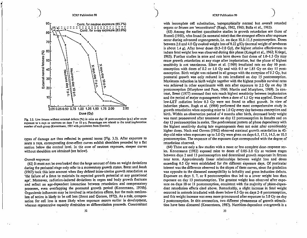
Fig. 3.3. Live fetuses without external defects (%) in mice on day 18 postconception (p.c.) after acuteexposure to x rays or neutrons on days 7 or 10 p.c. Percentages are related to the total implantationnumber of each group (Konermann, 1987 with permission from Elsevier).
types of damage are thus reflected in general terms (Fig. 3.3). After exposure toacute x rays, corresponding dose-effect curves exhibit shoulders preceded by a flatsection below the control level. In the case of neutron exposure, steeper curveswithout shoulders are obtained (see below).
Growth responses(62) It must not be overlooked that the large amount of data on weight deviations
during the perinatal stage only refer to a momentary growth status. Brent and Jensh(1967) took this into account when they defined intra-uterine growth retardation as'the failure of a fetus to maintain its expected growth potential at any gestationalage'. Moreover, radiation-induced deviations in organ and body growth fluctuateand reflect an age-dependent interaction between retardation and compensatoryprocesses, even overlapping the postnatal growth period (Konermann, 1976b).Organismic influences may be involved in retardation effects, but the main mechan-ism of action is likely to be cell loss (Brent and Gorson, 1972). As a rule, compen-sation for cell loss is more likely when exposure occurs earlier in development,whereas regenerative capacity diminishes as differentiation proceeds. Concomitant
32
ICRP Publication 90ICRP Publication 90
children from unexposed mothers. Parental socio-economic status was comparablein the three groups. The abdominal/pelvic x-ray examinations were to the stomachand upper gastrointestinal tract (usually including fluoroscopic examinations) for 20mothers and intravenous pyelography, lumbosicral spine, or genital tract for theremainder. Doses to the organs of interest were estimated to range from 0.5 to 10cGy, but it was difficult to evaluate fetal doses. The Bayley developmental scales'were administered to children'aged 1-;2.5 years and the McCarthy devlopmentalscales to those aged 3-5 years. No group differences were found on neurologicalexamination or on motor or cognitive scores fromr the Bayley or McCarthy devel-opmcntal scales. They noted that exposures occurred before week 8 postconceptionin most cases. -
5.7. Discussion
(245) The human data on the neurological effects of prenatal radiation exposurehave several constraints that limit the inferences that can be drawn and the precisionof the estimates of mental deficits. For one thing, there is almost total reliance onone study. Although the Japanese'atomic bomb study is an excellent one, scientificevidence'is m~ore persu'asive if it-is replicable in'differe'nt populations and circum-'stances. Second, the atomic bomb data are limited both by the small numbers ofstudy subjects who received high doses and the small numbers in the dose range 1-49 cGy, the critical range for defining the shape of the dose-response curve'or thepresence/magnitude of a dose threshold. Third, some' of the main endpoints havebeen fairly crude - severe mental retardation as diagnosed by clinical examination,or seizure disorder history obtained by parental report with medical confirmation(but with diagnostic workups that may not have given all the information desired).
(246) In spite of these limitations, two features of the results stand out. There is aclear constellation of effects of prenatal irradiation on ihe developing central ner-vous system - mental retardation, decreased intelligence scores and school perfor-mance, and seizure disorders. The first three factors showed strong associations withprenatal radiation exposure, while the association for seizure disorders was weaker,perhaps'owing to the sparseness and unreliability of the seizure data. The secondfeature of note is that, for all the endpoints, the period 'of weeks 8-15 postconcep-tion was the most radiosensitive, with the 16'-25-weck period quite consistentlyshowing sensitivity as well. No indication of mental deficits associated with radia-tion exposure was seen after week 25 postconception. The atomic bomb data gaveno indication of an effect for radiation exposure that-occurred during' weeks 0-7postconception, although a compilation of case reports of medical irradiation sug-gested that there might be such an effect, perhaps limited to very high doses (Deka-ban, 1968). However, experimental data do not support'an effect on the embryo. Astudy found no effects on the developing mouse nervous system at doses up to 3 Gyin the first 8 days after fertilisation, which corresponds to approximately the first 8weeks in humans (Hicks et al., 1952).
(247) The uncertainties in-the atomic bomb study estimates of radiation-relatedmental deficits include a variety of factors (cf. Schull et al., 1990): the sparse data,
Il
especially for mental retardation and convulsions; errors in the ascertainment anddiagnosis of mental disorders; errors in estimating doses and postconception age atexposure; the appropriateness of the comparison group; the impact of maternalinjury, disease,' and nutrition; possible selection factors in whether families -emi-grated from Hiroshima/Nagasaki; and lack of information about parental intelli-.gence and education. Although these factors may affect estimates of the magnitudeof risk in unknown ways,' their collective impact is not believed to be sufficient toinvalidate the study. -.
(248) Consideration should be given to the possibility that the mental effects might"be due to bias or some indirect mechanism - e.g. nutritional deprivation duringgestation or infancy, acute radiation sickness leading to a compromised immunesystem or depression of fetal haematopoiesis, genetic variation, or physical sensorydefects.
(249) Due to the nature of these endpoints (e.g. diagnosis of mental retardation)and of the sources from which they came, plus the fact they were seen only afterexposure in a delimited developmental period, it is most unlikely that a diagnostic orreporting bias could account for the results., '
(250) Nutritional deprivation is not likely to be the principal cause of the risk seen.for several reasons. (i) Although there are data showing that maternal malnutritionmay affect brain development,-it is primarily limited to marasmic infants who weresubstantially underweight at birth. There are no reports of underweight or marasmicinfants among the atomic bomb survivors (Schull and Otake, 1986). In fact, sup-plemental rations were provided for pregnant and nursing women in Japan duringand after the war, especially among those of >20 weeks gestation. (ii) One mightexpect nutritional effects to be at least as strong in the third trimester as the second,,which was not the pattern of mental deficits seen. (iii) One would expect maternalundernutrition to be relatively independent of radiation dose, in which case it couldnot account for a dose-dependent effect.
(251) Acute or subclinical radiation sickness has some plausibility as an explana-tion, particularly the possibility of reduced oxygen transport to the fetal braincaused by depressed haematopoiesis (Mole, 1990). However, there are several rea-sons why this is not likely to be -the main mechanism of damage to the brain.Maternal red blood cell counts may fall to 50-60% of the normal value and hae-moglobin levels, to 6-8 g,' but data on women with sickle cell anaemia who com-monly have haemoglobin values in this range do'not have an elevated frequency ofmentally retarded children (Schull and Otake, 1986). Between week 9 postconcep-tion and birth, 80-90% of the haemoglobin found in fetal red cells is fetal hac-moglobin which has biochemical characteristics that facilitate maternal oxygenunloading (UNSCEAR, 1993). This maximises oxygen transport to the fetus andprovides a protective mechanism against fetal hypoxia.:
. (252) A fraction of mental retardation is known to be caused by recessive geneticmutations. Consanguineous marriages, which, would increase the frequency ofexpression of these inherited disorders, were common in Hiroshima and Nagasaki atthe time of the bombs. Furthermore, the frequency of consanguineous marriages inNagasaki was inversely related to distance from the bomb hypocentre, which would
119
I ).

:..j) ICRP Publication 90
create a correlation with dose (Schull, 1958). Schull and Neel (1965) found thatchildren born to first cousins had an IQ that averaged five points less than childrenfrom unrelated parents in Hiroshima and Nagasaki. Hence, there is the potential foran indirect genetic effect. Even if one were to make the extreme assumption that anadditional 40% of those with higher doses had consanguineous marriages comparedwith the prevalence in the low-dose group, this would cause an average decrease inIQ of only about two points in the high-dose group, whereas a decrease of the orderof 20-30 IQ points was seen in the highest dose group. Thus consanguinity wouldbe, at most, a small contributor.
(253) Another hypothesis might be that possible effects of home environment -acting upon the child's motivation and socialisation - or of key physical impair-ments, such as visual or auditory defects, are dose related and are causing the effectson intelligence and school performance rather than radiation per se. The hypothesisabout home environment seems implausible in that the dose-related intelligence andschool deficits were seen only for those irradiated at weeks 8-25 postconception, notacross the board. The physical defects might be a possibility, although there is noevidence that this was the case.
(254) Although some of the indirect mechanisms, such as undernutrition, maternalill health, depressed fetal haematopoiesis, genetic susceptibility, or physical handi-caps, could have played some role in producing mental deficits, the severity of thedeficits and the critical induction period for them strongly suggests that the radia-tion insult itself plays the major role in the deficits.
(255) There are a number of gaps in our knowledge concerning the effects ofradiation on mental and neurological functioning. The types of potential deficitsthat have been evaluated are limited. There may be neurological processes that havedifferent critical periods than the 8-15- or 16-25-week periods. If so, endpoints thatdepend on those processes would likely show a different temporal pattern of inductionof radiation effects. For instance, Yoshimaru et al. (1995) noted that spatial memorydepends in part on the proper development of the hippocampus formation (dentategyrus) which arises relatively late in the development of the human brain. Theradiation effects for cognitive tests that measure spatial memory may, therefore, showa later critical period than the global assessments of mental functioning have shown.
(256) There are no exposed groups other than the Japanese atomic bomb survi-vors who have received sufficiently large in-utero brain doses to merit special studies.Among atomic bomb survivors, additional studies could be done. The number ofsubjects who received brain magnetic resonance imaging (MRI) scans is.very small(Schull, 1991); a larger group of people with mental retardation or perhaps low-normal IQ could be studied for structural brain defects. Ideally, MRI could beadministered to all of the in-utero-exposed survivors with estimated doses of 0.01Gy or more who were exposed between weeks 8 and 25 postconception. This wouldinvolve about 290 individuals. Examination of this group could provide importantinsights into the existence of a threshold, as well as the nature of radiation-impairedmechanisms culminating in cortical dysfunction. For example, the number of casesof mental retardation seen in the 16-25-week interval is small; only four with dosesof 0.01 Gy or more. Given this limited number, some of these cases, possibly even
120
ICRP Publication 90 7all, could have been exposed in the 8-15-week period, with the age at exposureoverestimated. This could occur among infants born prematurely. In this regard, itis worth noting that three of the four retarded cases in the 16-25-week period hadmaternal uterine doses of I Gy or more, and the mother undoubtedly experiencedsome of the symptoms of acute radiation illness. This could have precipitated apremature delivery. If this possibility could be excluded, the logical inference is thatthe cause of mental- retardation among the 8-15 week olds, presumed to be mis-managed neuronil migration, cannot account for the mental retardation seen at thelater ages since cortical migration has essentially ceased.
(257) Functional MRI with its capacity to identify activity centres and tracts ofneuronal transmission might identify alternative pathways or processes culminatingin mental retardation. The everincreasing sensitivity to and localisation of defectswith new equipment and techniques could provide valuable new insights. Similarly,a carefully designed series of modern neurocognitive tests administered to a samplefrom this population might help identify specific cognitive abilities that are com-promised by radiation, and this might vary by postconception exposure week.
(258) Nothing is known about individual differences in sensitivity to the effects ofradiation exposure on the developing brain. Insofar as the crude statistical indica-tors of differential sensitivity (namely, changes in variances or skewness) can deter-mine, there do not appear to be strong individual differences in predisposition tomental effects, but these crude indicators probably cannot rule out more subtle dif-ferential sensitivity effects.
(259) Little human information is available on the effects of dose protraction orfractionation, or on the effects of radionuclide exposures on the fetal brain. Twostudies have evaluated mental retardation among the offspring of mothers whoworked at Mayak while pregnant (Patrusheva et al., 1976; Buldakov et al., 1981),and neither found excess mental retardation. Similarly, a study of children exposedin utero to radiation contamination in the Techa River showed no elevation in theprevalence of mental retardation and no association between exposure and scholas-tic aptitude or achievement (Akleyev and !Cisselyov, 2000). Another study of thispopulation (Akleyev et al., 1997) found no association of exposure with neurologicalsigns or either of two neuropsychological tests. Several other studies -of mentalretardation, IQ (Nyagu et al., 1998; Kolominsky et al., 1999), seizure disorders(Tereschenko et al., 1991, 1992), psychomotor development (Lyaginskaya et al.,1992), and neurological signs (Patrusheva et al., 1973) showed putative associationswith prenatal exposure to Chernobyl or Mayak radiation, but the study designs andanalyses give too little detail, or methodological weaknesses were noted that limitthe confidence in these conclusions. In summary, no clear evidence was found indi-cating that protracted exposures cause mental sequelae within the limitations of thetotal radiation doses sustained and the sample sizes studied.
(260) No human information is available on the RBE of prenatal exposure toneutrons or other high-LET radiations in inducing mental deficits.
(261) Two studies have evaluated mental effects of in-utero diagnostic radiationexposure. A Chinese study (Hu and Yao, 1992) found no diminution in IQ followingdiagnostic radiation exposure to the fetus compared with a matched control group.
121
ICRP Publication 90 ICRP Publication 90
Another study of in-utero diagnostic radiation exposure (Ornoy et al., 1996) showedno deficits based on a neurological examination and two psychometric tests ofmotor and cognitive development. The null results are not surprising, given thesmall doses involved.
(262) Perhaps the most critical question at this point is whether there is a dosethreshold for the neurological and mental effects. The evidence in the atomic bombstudy is reasonably persuasive that there is a dose threshold for severe mentalretardation, although there is a good deal of uncertainty as to the dose at which thethreshold occurs. A formal test for a threshold has not been reported for intelligencescores. Inspection of the IQ data suggests that there might be a threshold in thevicinity of about 0.1 Gy, although this is by no means certain, and it is likely that theconfidence interval on any estimated threshold would encompass zero dose. Inspec-tion of the school performance data also suggests the possibility of a threshold at alow dose, but the statistical test for a threshold has not been reported. The atomicbomb data on unprovoked seizure disorders following in-utero irradiation are toosparse to permit a meaningful assessment of dose thresholds.'
(263) In-Publication 60, the summary indicated that the 'downward shift in IQ of30 points Sv1 . is consistent with the observation of an incidence (of seriousmental retardation) of 0.4 for a dose of I Sv' (ICRP, 1991, p. 5). The best estimate ofthe decrement in IQ at I Sv is 21 points for those irradiated at weeks 8-15 post-conception and 13 points at weeks 16-25 postconception (Schull, 1988). If oneassumes that the population IQ has a mean of 100 and a standard deviation of 15(which are typical values for IQ tests), then the expected proportion with an IQ of 65or under is slightly less than 1%. However, if the mean were shifted 21 points to 79,still with a standard deviation of 15, then about 18% would be expected to have IQsof 65 or less. If the mean were instead shifted only 13 points, then about 7% wouldbe expected to have an IQ of 65 or less. Thus fewer than 40% would be predicted toshow mental retardation, although the 40% value might be statistically compatiblewith these expectations given the sparseness of the data.
(264) Publicalion 60 also concluded that radiation-induced mental decrement 'isdeterministic with a threshold related to the minimum shift in IQ that can be mea-sured' (ICRP, 1991, p. 6). The dose-related shifts in IQ clearly point towards adeterministic effect. The existing analyses provide no'clear evidence for a dosethreshold with respect to IQ, and it seems likely that a definitive answer is beyondthe resolving power of the epidemiologic data that are available.
5.8. References
Akleyev. A.V., Yakovleva. V.P., Savostin. V.A. (1997) Late clinical effects of prenatal exposure. Problems
Radiat. Safety 1. 47-S0 (in Russian). :Akleyev, A.V., Kisselyov. I.F. (2000) AMedical-Biological and Ecological Impacts of Radioactive Con-
lamination of the Techa River. Moscow, Russia (in Russian).Buldakov, L.A., Ovcharenko, Y.P., Baklanov, P.S., et al. (1981) On the status of offspring born to women
exposed to a combined gamma-radiation and Pu-239. Bull. Radiat. Med. 1, 32-36.Dekaban, A.S. (1968) Abnormalities in children exposed to x-radiation during various stages of gestation:
tentative timetable of radiation injury to the human fetus, Part 1. J. NucI. Med. 9,471-477.
122
Dunn, K., Yoshimaru, H., Otake. M., ct al. (1990) Prenatal exposure to ionizing radiation and subsequent
development of seizures. Am. J. Epidemiol. 131, 114-123.Goerke, W., Goetze, W. (1971) EEG changes, psychomotor epilepsy and intracranial calcification occur-
ring as late results of radiation therapy. Clin. Electroencephal. 2, 146-153..Goldstein, L., Murphy, D.P. (1929) Microcephalic idiocy following radium therapy for uterine cancer
during pregnancy. Am. J. Obstet. Gynecol. 18, 189-195.Hicks, S.P., Schaufus, C.A., Williams, A.A.. ct al. (1952) Some effects of ionizing radiation and metabolic
inhibition of the developing mammalian nervous system. J. Pediatr. 40, 489-513.Hu. Y.M., Yao, J.X. (1992) Effect of prenatal exposure to diagnostic radiation on childhood physical and
intellectual development. Chin. J. Radiol. Med. Prot. 12, 2-6 (in Chinese).ICRP (1991) Recommendations of the International Commission on Radiological Protection. ICRP
Publication 60, Ann. ICRP 21.Igumnov, S., Drozdovitch, V. (2000) The intellectual development, mental and behavioural disorders in
children from Bclarus exposed in utero following the Chernobyl accident. J. Assoc. Eur. Psychiatrists
15,244-253.Kolominsky, Y., Igumnov, S., Drozdovitch, V. (1999) The psychological development of children from
. Belarus exposed in the prenatal period to radiation from the Chernobyl atomic power plant. J. Child
Psychol. Psychiatr. 40, 299-305.Lichter,' L., Bromet, EJ., Carlson, G., et al. (2000) School and neuropsychological performance of cvac-
uated children in Kiev 11 years after the Chemobyl disaster. J. Child Psych. Psychiat. 41. 291-299.Lyaginskaya, A.M., Buldakov, LA.. Osipov, V.A., ct al. (1992) Effects of Peoinatal Radiation Exposure as
a Result of the Chernobyl Accident. Institute of Biophysics, State Scientific Center of the Russian Fcd-
* ration, Moscow, Russia.Miller, R.W. (1956) Delayed cffects occurring within the first decade after exposure of young individuals
to the Hiroshima atomic bomb. Pediatrics 18, 1-18.Miller, R.W. (1990) Effects of prenatal exposure to ionizing radiation. Health Phys.'59, 57-61.
Mole, R.H. (1990) The effect of prenatal radiation exposure on the developing human brain. Int. J.
Radiat. Biol. 57, 647-663.Mole, R.H. (1992) ICRP and impairment of mental function following prenatal irradiation. J. Rad. Pro-
tect. 12, 93-105. .
NASINRC (1990) Health Effects of Exposure to Low Levels of Ionizing Radiation (BEIR V). NationalAcademy of Sciences/National Research Council, Washington, DC. USA.
Nyagu, A.l., Loganovsky, K.N., Loganovskaja, T.K. (1998) Psychophysiologic aftercffects.of prenatal
irradiation. Int. J. Psychophysiol. 30, 303-311.,Ornoy, A., Patlas, N;, Schwartz, L. (1996) The cffects of in utero diagnostic x-irradiation on the devel-
opment of preschool-agc children. Isr. J. Med. Sci. 32, 112-115.Otake, M., Schull, W. (1984) In utero exposure to A-bomb radiation and mental retardation: a reassess-
ment. Br. J. Radiol. 57. 409-414.Otake, M., Yoshimaru, H., Schull, WJ. (1987) Severe Mental Retardation among the Prenatally Exposed
Survivors of the Atomic Bombing of Hiroshima and NagasakiB a Comparison of the Old and New Dosi-merry Systems. Radiation Effects Research Foundation,'Hiroshima, Japan.
Otakc, M.. Schull, WJ., Fujikoshi, Y., et al. (1988) Effect on School Performance of Prenatal Exposure toIonizing Radiation in Hiroshima: a Comparison of the T65DR and DS86 Dosimetry Systems. RadiationEffects Research Foundation, Hiroshima, Japan.
Otake, bl., Schull, WJ., Lee, S. (1996) Threshold for radiation-related severe mental retardation in pre-
natally exposed A-bomb survivors: a re-analysis. Int. J. Radiat. Biol. 70, 755-763.Otake, M., Schull. WJ. (1998) Review: radiation-rclated brain damage and growth retardation among the
prenatally exposed atomic bomb survivors. Int. J. Radiat. Biol. 75, 159-171.Patrusheva, N.V., Doshchenko, V.N., Tokarskaya, Z.B., et al. (1973) On late cffects of prenatal exposure
in humans. Bull. Radiat. Med. 1, 78-83 (in Russian).Patrusheva, N.V., Voronin, Z.I., Voronina, N.E., et al. (1976) Morbidity among children exposed to
gamma-radiation in utero. Bull. Radiat. Med. 2,46-51 (in Russian).Plummer, G. (1952) Anomalies occurring in children exposed in utero to the atomic bomb in Hiroshima.
Pediatrics 10, 687-692.
123
) .2

?) ICRP Publication 90 )infancy by a period of hyperleukocytosis. Acute megakaryoblastic leukaemia israrely diagnosed in children who do not have trisomy 21 .but is the most commonform of leukaemia in this condition (Zipursky et al., 1997).'.Interestingly, there is apaucity of other childhood tumours and adult epithelial neoplasms in Down'ssyndrome (Hasle et al., 2000).
(308) Recessively inherited disorders leading to a defect in DNA repair, such asataxia telangiectasia, Bloom's syndrome, and xeroderma pigmentosum, are rare, butalmost inevitably lead to tumour in the homozygote during the first three decades oflife (ICRP, 1998). Of the recessive genes related to an increased cancer risk, ataxiatelangiectasia in the heterozygote may also increase cancer risk (Swift et al., 1987).Radiation has been shown to have a profound effect on the ability of ataxia tel.angiectasia cells to repair DNA damage, since they fail to halt their progressionthrough the cell cycle when exposed. Although this discussion will focus on theeffects of incidental or low-dose radiation as a carcinogen, one should consider thepossibility that some other gene or syndrome, in concert with radiation, couldpredispose individuals to neoplasia.
(309) Recent evidence has revealed another class of genes involved in DNA repair;the genes that predispose to syndromes associated with hereditary non-polyposiscolon (McLendon and Tien, 1998). It does not seem likely that these genes operatein childhood. The major mechanism of action of these genes involves an increase intarget tissues and an increase in mutation rate; a mechanism that would be impor-tant when multiple events are necessary to cause cancer. However, since embryonaltumours probably require only two events, and since these tissues are programmedfor a normal increase in target cell number, this mechanism would not affect theprobability of developing a clone of transformed cells (Knudson, 1986).
7.5. Second cancers
7.5.1. Radiation effects
(310) Children treated with radiation for a first cancer have an increased risk ofdeveloping additional neoplasms in the irradiated sites (Hawkins et al., 1987; Tuckeret al., 1987, 1991; Neglia et al., 1991; Breslow et al., 1993). Although therapeuticdoses have been shown to affect subsequent cancer risk, with higher doses increasingthe risk and young age at exposure being a significant variable for central nervoussystem and thyroid neoplasms, leukaemia was not amonig the radiation-associatedsecond cancers seen in many cohort studies. Doses in the therapeutic range are likelyto be lethal to haematopoietic stem cells.
(311) Variables determining the increased risk of neoplasia include age at irradia-tion, normal tissue in the field, and dose. Therapeutic doses range from 12 to 60 Gydepending on the sensitivity of the malignant cells and the underlying normal tissue.Adjacent tissues are often exposed to lower doses' because of internal scatter, andsecond neoplasnis often arise in these relatively low-dose sites. Lower doses (lessthan 30 Gy) are associated with thyroid and central nervous system tumours, whilebone and soft tissue sarcomas occur following doses greater than 30 Gy (Tucker
148
ICRP Publication 90 /
et al., 1987). Dose-response relationships have been observed with excesses rangingfrom three to 40 times expected. Tissues such as brain, thyroid, bone, and breastappear to be more susceptible if exposed during normal periods of rapid growth(i.e. early childhood or puberty) (Neglia et al., 1991; Tucker et al., 1991; Bhatia etal., 1996). However, embryonal neoplasms are not seen following radiation ther-apy, even when very young 'children are treated with doses in the therapeuticrange. , -
(312) Second cancers associated with radiation are considerably more frequent inchildren who are genetically predisposed. This was first seen in children with thegenetic form of retinoblastoma (Meadows et al., 1985; Draper et al., 1986). A reportof the largest series of long-term retinoblastoma survivors confirms the high risk ofnew cancers in those with the genetic form.(bilateral and familial cases) - 25% at 50years - and the elevated risk following radiation - 50% at 50 years (Wong et al.,1997). Multiple primary cancers have also been reported excessively in the Li-Frau.meni syndrome (Hisada et al., 1998). In a cohort. study of 200 patients with thissyndrome, 30 developed more than one cancer for a cumulative probability of asecond cancer of 57% (+ 10%) at 30 years, and a third cancer of 38% (± 12%) at 10years. Eight neoplasms occurred in the field of radiation. Evidence for an addedeffect of radiation in the aetiology of multiple tumours occurring at earlier ages thanexpected and with a shorter latent period comes from the study of patients withGorlin's syndrome or nevoid basal cell carcinoma syndrome (Meadows et al., 1985).This syndrome is characterised by tumours of the posterior fossa and basal cellcarcinomas, the latter often appearing within months following radiation.
7.5.2. Chemotherapy
(313) Chemotherapeutic agents such as alkylating agents and epipodophyllotoxinshave been associated with secondary leukaemias (Smith et al., 1994). Alkylatingagents and anthracyclines have also been implicated in affecting the risk for bonetumours associated with radiation therapy (Tucker et al., 1987; Newton et al., 1991).Different and characteristic chromosomal alterations accompany the leukaemiasthat occur in association with alkylators or epipodophyllotoxins. with deletions ofchromosomes 5 and 7 in the former, and translocations involving I Iq23, the locus ofthe myeloid leukaemia gene, in the latter (Bhatia et al., 1999). Alkylating-agent-associated secondary leukaemias occur within 7-8 years of exposure and are depen-dent upon dose and specific agent. For instance, nitrogen mustard, chlorambucil,and the nitrosoureas are more potent leukaemogens than is cyclophosphamide.Dose and schedule are critical in the development of the secondary leukaemiasassociated with the topoisorferase 11 inhibitors, such as epipodophyllotoxins, andthe usual latent period is between 6 months and 3 years (Pui, 1989). Leukaemias ininfants are most often associated with an abnormality involving the myeloid leu-kaemia gene at I lq23. A case-control study of infants with acute myelogenous leu-kaemia suggested a dose-response association with maternal consumption of dietarytopoisomerase 11 inhibitors (beans, soy, fruits, vegetables, wine, and black and greentea) (Ross et al., 1996).
149


