Embed Size (px)
Citation preview
Clinical Practice Guidelines
ROBROY L. MARTIN, PT, PhD • TODD E. DAVENPORT, DPT • STEPHEN PAULSETH, DPT, MSDANE K. WUKICH, MD • JOSEPH J. GODGES, DPT, MA
Ankle Stability and Movement Coordination Impairments:
Ankle Ligament SprainsClinical Practice Guidelines Linked to the
International Classification of Functioning, Disability and Health From the Orthopaedic Section
of the American Physical Therapy AssociationJ Orthop Sports Phys Ther. 2013;43(9):A1-A40. doi:10.2519/jospt.2013.0305
REVIEWERS: Roy D. Altman, MD • Anthony Delitto, PT, PhD • John DeWitt, DPTAmanda Ferland, DPT • Helene Fearon, PT • Joy MacDermid, PT, PhD • James W. Matheson, DPT
Thomas G. McPoil, PT, PhD • Stephen Reischl, DPT • Leslie Torburn, DPT • James Zachazewski, DPT
For author, coordinator, contributor, and reviewer a!liations, see end of text. Copyright ©2013 Orthopaedic Section, American Physical Therapy Association (APTA), Inc, and the Journal of Orthopaedic & Sports Physical Therapy®. The Orthopaedic Section, APTA, Inc, and the Journal of Orthopaedic & Sports Physical Therapy consent to the reproduction and distribution of this guideline for educational purposes. Address correspondence to: Joseph Godges, DPT, ICF Practice Guidelines Coordinator, Orthopaedic Section, APTA, Inc, 2920 East Avenue South, Suite 200, La Crosse, WI 54601. E-mail: [email protected]
RECOMMENDATIONS . . . . . . . . . . . . . . . . . . . . . . . . . . . . . . . . . . . . . . . . . . . . . . . . . . . A2
INTRODUCTION. . . . . . . . . . . . . . . . . . . . . . . . . . . . . . . . . . . . . . . . . . . . . . . . . . . . . . . . . . . . A3
METHODS . . . . . . . . . . . . . . . . . . . . . . . . . . . . . . . . . . . . . . . . . . . . . . . . . . . . . . . . . . . . . . . . . . . A4
CLINICAL GUIDELINES: Impairment/Function-Based Diagnosis . . . . . . . . . . . . . . . . . . A7
CLINICAL GUIDELINES:Examination . . . . . . . . . . . . . . . . . . . . . . . . . . . . . . . . . . . . . . . . . . . . . . . . . . . . . . . . . . . A16
CLINICAL GUIDELINES: Interventions . . . . . . . . . . . . . . . . . . . . . . . . . . . . . . . . . . . . . . . . . . . . . . . . . . . . . . . . . . A24
SUMMARY OF RECOMMENDATIONS . . . . . . . . . . . . . . . . . . . . . . . . . . . . . A29
AUTHOR/REVIEWER AFFILIATIONS AND CONTACTS . . . . . . A31
REFERENCES . . . . . . . . . . . . . . . . . . . . . . . . . . . . . . . . . . . . . . . . . . . . . . . . . . . . . . . . . . . . . A32
������*XLGHOLQHV�LQGG���� ��������������������30
Ankle Ligament Sprain: Clinical Practice Guidelines
a2 | september 2013 | volume 43 | number 9 | journal of orthopaedic & sports physical therapy
RISK FACTORS – ACUTE LATERAL ANKLE SPRAIN: Clinicians should recognize the increased risk of acute lateral ankle sprain in individuals who (1) have a history of a previous ankle sprain, (2) do not use an external support, (3) do not properly warm up with static stretching and dynamic move-ment before activity, (4) do not have normal ankle dorsiflex-ion range of motion, and (5) do not participate in a balance/proprioceptive prevention program when there is a history of a previous injury. (Recommendation based on moderate evidence.)
RISK FACTORS – ANKLE INSTABILITY: Clinicians should recog-nize the increased risk for developing ankle instability in pa-tients who (1) have an increased talar curvature, (2) are not using an external support, or (3) did not perform balance or proprioception exercises following an acute lateral ankle sprain. (Recommendation based on weak evidence.)
DIAGNOSIS/CLASSIFICATION – ACUTE LATERAL ANKLE SPRAIN: Clinicians should use the clinical findings of level of func-tion, ligamentous laxity, hemorrhaging, point tenderness, total ankle motion, swelling, and pain to classify a patient with acute ankle ligament sprain into the International Statistical Classification of Diseases and Related Health Problems (ICD) category of sprain and strain of ankle (S93.4), and the associated International Classification of Functioning, Disability and Health (ICF) impairment-based category of ankle stability (b7150 stability of a single joint) and movement coordination impairments (b7601 control of complex voluntary movements). (Recommendation based on moderate evidence.)
DIAGNOSIS/CLASSIFICATION – ANKLE INSTABILITY: Clinicians may incorporate a discriminative instrument, such as the Cumberland Ankle Instability Tool, to assist in identifying the presence and severity of ankle instability associated with the ICD category of disorder of ligament, instability second-ary to old ligament injury, ankle and foot (M24.27), and the associated ICF impairment-based category of ankle stability (b7150 stability of a single joint) and movement coordination impairments (b7601 control of complex voluntary move-ments). (Recommendation based on moderate evidence.)
DIFFERENTIAL DIAGNOSIS – ACUTE LATERAL ANKLE SPRAIN: Clinicians should use diagnostic classifications other than an acute lateral ankle sprain when the patient’s reported activity limitations or impairments of body function and structure are not consistent with those presented in the
Diagnosis/Classification section of this guideline. Particu-larly, the Ottawa and Bernese ankle rules should be used to determine whether a radiograph is required to rule out a fracture of the ankle and/or foot. (Recommendation based on strong evidence.)
DIFFERENTIAL DIAGNOSIS – ANKLE INSTABILITY: Clinicians should use diagnostic classifications other than ankle instability when the patient’s reported activity limitations or impairments of body function and structure are not con-sistent with those presented in the Diagnosis/Classification section of this guideline. (Recommendation based on expert opinion.)
EXAMINATION – OUTCOME MEASURES: Clinicians should incor-porate validated functional outcome measures, such as the Foot and Ankle Ability Measure and the Lower Extremity Functional Scale, as part of a standard clinical examina-tion. These should be utilized before and after interventions intended to alleviate the impairments of body function and structure, activity limitations, and participation restrictions associated with ankle sprain and instability. (Recommenda-tion based on strong evidence.)
EXAMINATION – ACTIVITY LIMITATION AND PARTICIPATION RESTRICTION MEASURES: When evaluating a patient in the postacute period following a recent or recurring lateral ankle sprain, assessment of activity limitation, participa-tion restriction, and symptom reproduction should include objective and reproducible measures, such as single-limb hop tests that assess performance with lateral movements, diagonal movements, and directional changes. (Recommen-dation based on moderate evidence.)
EXAMINATION – PHYSICAL IMPAIRMENT MEASURES: When evaluating a patient with an acute or subacute lateral ankle sprain over an episode of care, assessment of impairment of body function should include objective and reproducible measures of ankle swelling, ankle range of motion, talar translation and inversion, and single-leg balance. (Recom-mendation based on strong evidence.)
INTERVENTION – ACUTE/PROTECTED MOTION PHASE – EARLY WEIGHT BEARING WITH SUPPORT: Clinicians should advise patients with acute lateral ankle sprains to use external supports and to progressively bear weight on the a!ected limb. The type of external support and gait assistive device recommended should be based on the severity of the injury, phase of tissue healing, level of protection indicated, extent
Recommendations*
������*XLGHOLQHV�LQGG���� ��������������������30
Ankle Ligament Sprain: Clinical Practice Guidelines
journal of orthopaedic & sports physical therapy | volume 43 | number 9 | september 2013 | a3
of pain, and patient preference. In more severe injuries, immobilization ranging from semi-rigid bracing to below-knee casting may be indicated. (Recommendation based on strong evidence.)
INTERVENTION – ACUTE/PROTECTED MOTION PHASE – MANUAL THERAPY: Clinicians should use manual therapy procedures, such as lymphatic drainage, active and passive soft tis-sue and joint mobilization, and anterior-to-posterior talar mobilization procedures, within pain-free movement to reduce swelling, improve pain-free ankle and foot mobility, and normalize gait parameters in individuals with an acute lateral ankle sprain. (Recommendation based on moderate evidence.)
INTERVENTION – ACUTE/PROTECTED MOTION PHASE – PHYSICAL AGENTS: Cryotherapy: clinicians should use repeat-ed intermittent applications of ice to reduce pain, decrease the need for pain medication, and improve weight bearing following an acute ankle sprain. (Recommendation based on strong evidence.) Diathermy: clinicians can utilize pulsating shortwave diathermy for reducing edema and gait devia-tions associated with acute ankle sprains. (Recommendation based on weak evidence.) Electrotherapy: there is moderate evidence both for and against the use of electrotherapy for the management of acute ankle sprains. (Recommendation based on conflicting evidence.) Low-level laser therapy: there is moderate evidence both for and against the use of low-level laser therapy for the management of acute ankle sprains. (Recommendation based on conflicting evidence.) Ultrasound: clinicians should not use ultrasound for the management of acute ankle sprains. (Recommendation based on strong evidence.)
INTERVENTION – ACUTE/PROTECTED MOTION PHASE – THERAPEUTIC EXERCISES: Clinicians should implement re-habilitation programs that include therapeutic exercises for patients with severe lateral ankle sprains. (Recommendation based on strong evidence.)
INTERVENTION – PROGRESSIVE LOADING/SENSORIMOTOR TRAINING PHASE – MANUAL THERAPY: Clinicians should include manual therapy procedures, such as graded joint mobilizations, manipulations, and non–weight-bearing and weight-bearing mobilization with movement, to improve ankle dorsiflexion, proprioception, and weight-bearing tolerance in patients recovering from a lateral ankle sprain. (Recommendation based on strong evidence.)
INTERVENTION – PROGRESSIVE LOADING/SENSORIMOTOR TRAINING PHASE – THERAPEUTIC EXERCISE AND ACTIVITIES: Clinicians may include therapeutic exercises and activities, such as weight-bearing functional exercises and single-limb balance activities using unstable surfaces, to improve mobility, strength, coordination, and postural control in the postacute period of rehabilitation for ankle sprains. (Recommendation based on weak evidence.)
INTERVENTION – PROGRESSIVE LOADING/SENSORIMOTOR TRAINING PHASE – SPORT-RELATED ACTIVITY TRAINING: Clinicians can implement balance and sport-related activity training to reduce the risk for recurring ankle sprains in athletes. (Recommendation based on weak evidence.)
*These recommendations and clinical practice guidelines are based on the scientific literature accepted for publication prior to April 2012.
Recommendations* (continued)
AIM OF THE GUIDELINES
The Orthopaedic Section of the American Physical Therapy Association (APTA) has an ongoing e!ort to create evidence-based practice guidelines for orthopaedic physical therapy management of patients with musculoskeletal impairments described in the World Health Organization’s International Classification of Functioning, Disability and Health (ICF).286
The purpose of these clinical guidelines is to:
• Describe evidence-based physical therapy practice, includ-ing diagnosis, prognosis, intervention, and assessment of outcome, for musculoskeletal disorders commonly man-aged by orthopaedic physical therapists
• Classify and define common musculoskeletal conditions us-ing the World Health Organization’s terminology related to impairments of body function and body structure, activity limitations, and participation restrictions
• Identify interventions supported by current best evidence to address impairments of body function and structure, ac-
Introduction
������*XLGHOLQHV�LQGG���� ��������������������30
Ankle Ligament Sprain: Clinical Practice Guidelines
a4 | september 2013 | volume 43 | number 9 | journal of orthopaedic & sports physical therapy
Content experts were appointed by the Orthopaedic Section of the APTA as developers and authors of clinical practice guidelines for musculoskeletal conditions of the ankle and foot that are commonly treated by physical therapists. These content experts were given the task to identify impairments of body function and structure, activity limitations, and participation restrictions, described using ICF terminology, that could (1) categorize patients into mutually exclusive impairment patterns on which to base intervention strate-gies, and (2) serve as measures of changes in function over the course of an episode of care. The second task given to the content experts was to describe the supporting evidence for the identified impairment-pattern classification as well as interventions for patients with activity limitations and impairments of body function and structure consistent with the identified impairment-pattern classification. It was also acknowledged by the Orthopaedic Section, APTA content experts that only performing a systematic search and re-view of the evidence related to diagnostic categories based on International Statistical Classification of Diseases and Related Health Problems (ICD)286 terminology would not be su!cient for these ICF-based clinical practice guidelines, as most of the evidence associated with changes in levels of impairment or function in homogeneous populations is not readily searchable using the ICD terminology. Thus, the
authors of this guideline independently performed a sys-tematic search of MEDLINE, CINAHL, and the Cochrane Database of Systematic Reviews (1967 through April 2012) for any relevant articles related to classification, examina-tion, and intervention strategies for ankle sprains. Addition-ally, when relevant articles were identified, their reference lists were hand searched in an attempt to identify other relevant articles. Articles from the searches were compiled and reviewed for accuracy by the authors. This guideline was issued in 2013 based on publications in the scientific literature prior to April 2012. This guideline will be consid-ered for review in 2017, or sooner if new evidence becomes available. Any updates to the guideline in the interim period will be noted on the Orthopaedic Section, APTA website: www.orthopt.org.
LEVELS OF EVIDENCEIndividual clinical research articles were graded accord-ing to criteria described by the Centre for Evidence-Based Medicine, Oxford, UK (http://www.cebm.net) for diagnos-tic, prospective, and therapeutic studies.198 If the 2 content experts did not agree on a grade of evidence for a particular article, a third content expert was used to resolve the issue. An abbreviated version of the grading system is provided on the next page.
Methods
tivity limitations, and participation restrictions associated with common musculoskeletal conditions
• Identify appropriate outcome measures to assess changes resulting from physical therapy interventions in body func-tion and structure as well as in activity and participation of the individual
• Provide a description to policy makers, using internation-ally accepted terminology, of the practice of orthopaedic physical therapists
• Provide information for payers and claims reviewers re-garding the practice of orthopaedic physical therapy for common musculoskeletal conditions
• Create a reference publication for orthopaedic physical therapy clinicians, academic instructors, clinical instruc-tors, students, interns, residents, and fellows regarding the best current practice of orthopaedic physical therapy
STATEMENT OF INTENTThese guidelines are not intended to be construed or to
serve as a standard of medical care. Standards of care are determined on the basis of all clinical data available for an individual patient and are subject to change as scientific knowledge and technology advance and patterns of care evolve. These parameters of practice should be considered guidelines only. Adherence to them will not ensure a suc-cessful outcome in every patient, nor should they be con-strued as including all proper methods of care or excluding other acceptable methods of care aimed at the same results. The ultimate judgment regarding a particular clinical proce-dure or treatment plan must be made in light of the clinical data presented by the patient; the diagnostic and treatment options available; and the patient’s values, expectations, and preferences. However, we suggest that significant departures from accepted guidelines should be documented in the pa-tient’s medical records at the time the relevant clinical deci-sion is made.
Introduction (continued)
������*XLGHOLQHV�LQGG���� ��������������������30
Ankle Ligament Sprain: Clinical Practice Guidelines
journal of orthopaedic & sports physical therapy | volume 43 | number 9 | september 2013 | a5
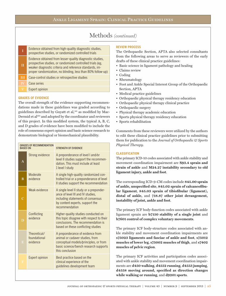
Methods (continued)
I Evidence obtained from high-quality diagnostic studies, prospective studies, or randomized controlled trials
II
Evidence obtained from lesser-quality diagnostic studies, prospective studies, or randomized controlled trials (eg, weaker diagnostic criteria and reference standards, im-proper randomization, no blinding, less than 80% follow-up)
III Case-control studies or retrospective studies
IV Case series
V Expert opinion
GRADES OF EVIDENCEThe overall strength of the evidence supporting recommen-dations made in these guidelines was graded according to guidelines described by Guyatt et al,101 as modified by Mac-Dermid et al160 and adopted by the coordinator and reviewers of this project. In this modified system, the typical A, B, C, and D grades of evidence have been modified to include the role of consensus expert opinion and basic science research to demonstrate biological or biomechanical plausibility.
GRADES OF RECOMMENDATION BASED ON STRENGTH OF EVIDENCE
A
Strong evidence A preponderance of level I and/or level II studies support the recommen-dation. This must include at least 1 level I study
BModerate evidence
A single high-quality randomized con-trolled trial or a preponderance of level II studies support the recommendation
C
Weak evidence A single level II study or a preponder-ance of level III and IV studies, including statements of consensus by content experts, support the recommendation
D
Conflicting evidence
Higher-quality studies conducted on this topic disagree with respect to their conclusions. The recommendation is based on these conflicting studies
E
Theoretical/ foundational evidence
A preponderance of evidence from animal or cadaver studies, from conceptual models/principles, or from basic science/bench research supports this conclusion
FExpert opinion Best practice based on the
clinical experience of the guidelines development team
REVIEW PROCESSThe Orthopaedic Section, APTA also selected consultants from the following areas to serve as reviewers of the early drafts of these clinical practice guidelines:• Basic science in ligament pathology and healing• Claims review• Coding• Rheumatology• Foot and Ankle Special Interest Group of the Orthopaedic
Section, APTA• Medical practice guidelines• Orthopaedic physical therapy residency education• Orthopaedic physical therapy clinical practice• Orthopaedic surgery• Physical therapy academic education• Sports physical therapy residency education• Sports rehabilitation
Comments from these reviewers were utilized by the authors to edit these clinical practice guidelines prior to submitting them for publication to the Journal of Orthopaedic & Sports Physical Therapy.
CLASSIFICATIONThe primary ICD-10 codes associated with ankle stability and movement coordination impairment are S93.4 sprain and strain of ankle and M24.27 instability secondary to old ligament injury, ankle and foot.
The corresponding ICD-9-CM codes include 845.00 sprain of ankle, unspecified site, 845.02 sprain of calcaneofibu-lar ligament, 845.03 sprain of tibiofibular (ligament), distal of ankle, and 718.87 other joint derangement, instability of joint, ankle and foot.
The primary ICF body-function codes associated with ankle ligament sprain are b7150 stability of a single joint and b7601 control of complex voluntary movements.
The primary ICF body-structure codes associated with an-kle stability and movement coordination impairments are s75023 ligaments and fasciae of ankle and foot, s75012 muscles of lower leg, s75002 muscles of thigh, and s7402 muscles of pelvic region.
The primary ICF activities and participation codes associ-ated with ankle stability and movement coordination impair-ments are d450 walking, d4552 running, d4553 jumping, d4558 moving around, specified as direction changes while walking or running, and d9201 sports.
������*XLGHOLQHV�LQGG���� ��������������������30
Ankle Ligament Sprain: Clinical Practice Guidelines
a6 | september 2013 | volume 43 | number 9 | journal of orthopaedic & sports physical therapy
Methods (continued)
This guideline has chosen to classify individuals with lat-eral ankle sprain into 2 categories: (1) acute lateral sprains and (2) ankle instability. The evidence related to the clas-sification of acute lateral sprain generally includes studies that enrolled subjects within 72 hours following injury, or subjects who demonstrated significant swelling, pain, lim-ited weight bearing, and overt gait deviations (ie, limited stance time, abbreviated/omitted terminal stance phase). Ankle instability relates to the postacute period and in-
cludes studies that enrolled subjects with primary concerns of instability, weakness, limited balance responses, and intermittent swelling. Chronic ankle instability is a term commonly applied to individuals with these complaints. However, definitive and uniformly applied criteria to di-agnose chronic ankle instability have not been developed. Therefore, the classification of “instability” is the best la-bel for these individuals and will be used throughout this guideline.
������*XLGHOLQHV�LQGG���� ��������������������30
Ankle Ligament Sprain: Clinical Practice Guidelines
journal of orthopaedic & sports physical therapy | volume 43 | number 9 | september 2013 | a7
INCIDENCEA review of emergency department records in the United States between 2002 and 2006 estimated the incidence rate of an ankle sprain to be 2.15 per 1000 person-years in the general population.266 The incidence of ankle sprain was highest in those between 15 and 19 years of age (7.2 per 1000 person-years). The overall incidence rate ratio for ankle sprain did not di!er between males and females. However, males between 14 and 24 years of age and females older than 30 years of age had a higher incidence rate compared to their respective counterparts.266 The black and white races were associated with higher rates of ankle sprain compared with the Hispanic race.266 Nearly half of all ankle sprains (49.3%) occurred during athletic activity, with basketball (41.1%), football (9.3%), and soccer (7.9%) being associated with the highest percentage of ankle sprains.266 Physically active indi-viduals, particularly those who participate in court and team sports,86 such as basketball,177 are at a higher risk than the general population.265 The ankle joint was found to account for 10% to 34% of all sport-related injuries, with lateral ankle sprain comprising 77% to 83% of these injuries.86 In prospec-tive studies that included physically active subjects, 20% of females281 and 18% of males280 sustained an inversion ankle sprain. In the United States Armed Services and Academies, the incidence rate is also higher than the general population and has been reported to vary between 35 and 58 per 1000 person-years.35,265
The rate of lateral ankle sprain reinjury is noteworthy.243 A systematic review noted that reinjury occurred in 3% to 34% of patients. This review found the time between initial injury and a second injury to vary greatly, with a time frame ranging from within 2 weeks to 96 months.259 A recent prospective study of track-and-field athletes noted a reinjury rate of 17% within 2 years.162 However, the reinjury rate may be greater in high-risk sports such as basketball, in which a reinjury rate of 73% was reported.177 The overall incidence of lateral ankle sprain may be underestimated because approximately 50% of those sustaining an ankle sprain do not seek medical attention after injury.12,177,224
PATHOANATOMICAL FEATURESThe hindfoot is composed of the distal tibiofibular syn-
desmosis, talocrural joint, and subtalar joint. The 3 major contributors to stability of the ankle joint are (1) osseous congruity and fit of the articular surfaces when the joints are loaded, (2) static ligamentous and capsular restraints, and (3) surrounding musculotendinous units.109 The lateral ligaments of the ankle complex are potentially injured with an inversion or supination mechanism. The most common mechanism of injury occurs with forefoot adduction, hind-foot internal rotation, ankle inversion in plantar flexion, and external rotation of the leg beyond anatomical constraints. This injury mechanism may result when landing from a jump, stepping into a hole, and/or landing on a competitor’s foot during sports. A lateral ankle sprain consists of partial or complete disruption of the lateral ankle ligaments. These ligaments consist of the anterior talofibular ligament, cal-caneofibular ligament, and posterior talofibular ligament. Up to 73% of lateral ankle sprains involve isolated anterior talofibular ligament injuries.86 Injury to the posterior talo-fibular ligament rarely occurs in isolation with an inversion mechanism of injury.
Combined subtalar, medial, and/or syndesmotic sprains can occur concurrently with lateral ankle sprain but are reported less often in the literature. Following an excessive ankle in-version injury, structures other than the lateral ligaments can be injured and may contribute to chronic concerns regarding pain, instability, and limitation in activities and participation. These structures include the lateral subtalar ligaments, fibu-lar (peroneal) tendon, nerve injury, extensor and peroneal retinaculum, inferior tibiofibular ligament, osteochondral lesions of the talus or tibial plafond, and neuromuscular ele-ments of the lower extremity.
Anterior Talofibular LigamentThe anterior talofibular ligament is an extra-articular liga-ment of the talocrural joint.219 Its fibers course laterally from the talus in the transverse plane and superiorly between the sagittal and frontal planes to attach on the anterior distal tip of the lateral malleolus.250 The anterior talofibular ligament can have single (38%), bifurcated (50%), or trifurcated (12%) fiber bundles.183 The anterior talofibular ligament provides the primary restraint to inversion movement when the ankle is in a plantar-flexed position.250 Maximal displacement of
CLINICAL GUIDELINES
Impairment/Function-Based Diagnosis
������*XLGHOLQHV�LQGG���� ��������������������30
Ankle Ligament Sprain: Clinical Practice Guidelines
a8 | september 2013 | volume 43 | number 9 | journal of orthopaedic & sports physical therapy
the talus from an applied anteriorly directed force was found to occur with the ankle in 10° of plantar flexion when com-pared to 0° or 20° of plantar flexion.240 Approximately half of the sprains involving the anterior talofibular ligament are avulsions from the fibula, with the other half being midsub-stance tears.250 Damage to the ligaments is dependent on the ankle and foot position at the time of injury and the velocity of the mechanism of injury. The anterior talofibular ligament demonstrates lower maximal load tolerance before failure as compared with the posterior talofibular ligament, calcaneo-fibular ligament, anterior inferior tibiofibular ligament, and deltoid ligament. The anterior talofibular ligament has the lowest modulus of elasticity, and injury to adjacent muscles (fibularis brevis, longus, and tertius) leaves the lateral ankle somewhat unprotected dynamically.8,83,208
Calcaneofibular LigamentThe calcaneofibular ligament is an extra-articular ligament of the talocrural joint that courses from the anterior distal tip of the fibula obliquely downward and backward to the lateral calcaneus. The location of the calcaneal insertion of the calcaneofibular ligament is highly variable.31 The fibers of the calcaneofibular ligament cross both the ankle and subtalar joints. The ligament is stronger and thicker than the anterior talofibular ligament and may be fan shaped in less than 2% of the population. Because the calcaneofibu-lar ligament crosses the subtalar joint and parallels its axis, subtalar joint motion can a!ect calcaneofibular ligament tension.219 Although tension within the ligament increases with dorsiflexion, it resists ankle inversion throughout the full range of ankle motion.119,122 Because the calcaneofibular ligament crosses both the ankle and subtalar joints, injury to this ligament may have a more profound functional e!ect on the ankle complex than isolated injuries to the anterior talofibular ligament.269
Posterior Talofibular LigamentThe posterior talofibular ligament runs from the posterior-medial portion of the fibula to the lateral tubercle on the posterior aspect of the talus. The posterior talofibular liga-ment is intracapsular but extrasynovial.250 It is the strongest of the lateral ligaments222 and primarily functions to provide transverse plane rotatory stability.232 Along with the calca-neofibular ligament, anterior talofibular ligament, and me-dial collateral ligaments, the posterior talofibular ligament assists to couple movements between the lower extremity and foot.119 Although the posterior talofibular ligament is rarely involved in a typical lateral ankle sprain, movements that in-volve extreme ankle dorsiflexion, foot external rotation and pronation, along with limb internal rotation may cause injury to the posterior talofibular ligament.32,219
Lateral Subtalar LigamentsThe fibers of the lateral talocalcaneal ligament are paral-lel to and blend in with the posterior fibers of the calca-neofibular ligament. The lateral talocalcaneal ligament crosses the posterior subtalar joint and is considered weaker and smaller than the calcaneofibular ligament.31 The lateral subtalar joint is further stabilized by the deep interosseous ligament located in the sinus tarsi and cer-vical ligaments, which are located laterally and insert on the inferolateral talar neck.227 The fibers of these ligaments run obliquely between the talus and calcaneus, subdivid-ing the subtalar joint into posterior and anterior chambers. These ligaments have a large modulus of elasticity and are considered stabilizers of the subtalar joint throughout the entire range of motion.136,210 The ligament of Rouvière, or fibulotalocalcaneal ligament, lies distinctly posterior to the calcaneofibular ligament and assists in resisting excessive supination.122 A combined injury of the anterior talofibular ligament and the interosseous talocalcaneal ligament can induce anterolateral rotatory instability of the ankle joint. After dissection of the interosseous talocalcaneal ligament, dorsiflexion of the talocalcaneal joint increased by 43%, with little e!ect on ankle supination motion.28,269 Subta-lar ligament sprains are reported after inversion injuries,14 with subtalar instability noted in 10% to 25% of those with lateral ankle instability.112,269 Unlike the anterior talofibu-lar ligament, calcaneofibular ligament, and posterior talo-fibular ligament, the lateral talocalcaneal ligament does not cross the ankle joint. However, with recurrent ankle sprains, greater loads may be placed on the lateral subtalar ligaments and contribute to chronic ankle symptoms, in-cluding instability.112
Extensor and Fibular RetinaculaThe extensor and fibular (peroneal) retinacula contribute to ankle and hindfoot stability primarily due to their anatomi-cal orientation. The inferior extensor retinaculum courses from the tip of the lateral malleolus to insert on the lateral calcaneus and sinus tarsi. The inferior extensor retinacu-lum also blends with the inferior fibular retinaculum and may improve evertor muscle function.94 Surgical augmen-tation of the inferior extensor retinaculum has been shown to provide protection to an anterior talofibular ligament repair in cadaveric9 and in clinical studies of the modified Broström procedure.82,155 The superior fibular retinaculum runs from the lateral malleolus to the calcaneus, parallel with the posterior fibers of the calcaneofibular ligament. The actual prevalence of injury to the retinacula is not well defined. However, the fibular and extensor retinacula may be injured in conjunction with lateral ankle sprains and po-tentially contribute to chronic pain, instability, and peroneal tendon subluxation.62
������*XLGHOLQHV�LQGG���� ��������������������30
Ankle Ligament Sprain: Clinical Practice Guidelines
journal of orthopaedic & sports physical therapy | volume 43 | number 9 | september 2013 | a9
Lower-Limb Neuromuscular StructuresA lateral ankle sprain may result in injuries to the lateral musculotendinous structures, resulting in tendon tearing, intramuscular strain, or tendon subluxation.68 Dynamic sta-bilization of the ankle complex is dependent on the adjacent musculature and laterally includes the fibularis (peroneus) longus and brevis. The tibialis anterior and extensor digito-rum longus and brevis are thought to eccentrically control ankle plantar flexion. Because lateral ankle sprains common-ly occur in plantar flexion, these muscles are also thought to protect against injury. However, both peripheral and central reactions of a muscle response are likely too slow to protect against a sudden inversion force.149 Therefore, anticipatory muscle contraction may be more important to protect against inversion ankle injuries than a reflexive response. Anticipa-tory muscle action may increase active muscle sti!ness, and hence joint sti!ness, while simultaneously increasing the sensitivity of the muscle spindle to stretch.
A lateral ankle sprain not only a!ects local musculature but may also lead to proximal muscle weakness of the bilateral gluteus maximus, biceps femoris, and lumbar erector spi-nae.30 Abnormal hip muscle activation has been found after ankle inversion movements in those with ankle hypermobil-ity after injury.18 Local sensory changes may also occur after a lateral ankle sprain.237 Sensory changes can occur in the joint receptors and cutaneous nerves, such as the sural nerve and distal superficial peroneal nerve. Nerve damage can alter a!erent cutaneous feedback receptors.119,149 This not only cre-ates local neurological changes but may also involve central neuromuscular pathways.30,237 Muscle spindles located within the adjacent lateral ankle muscles are involved in proprio-ception at the ankle and therefore may be involved in those with instability.121 Abnormal signals from the central nervous system could be present in individuals with chronic ankle symptoms and a!ect postural control. The role of the neuro-muscular elements in chronic pain and subjective instability is controversial and needs further study.
CLINICAL COURSEThe clinical course of acute lateral ankle sprains was inves-tigated in a systematic review that included 31 prospective studies.259 These studies generally noted a rapid decrease in pain and improvement in function the first 2 weeks after the injury. However, 5% to 33% of patients continued to have pain at 1-year or longer follow-up, with 5% to 25% still ex-periencing pain after 3 years.259 Residual problems included pain (30%), instability (20%), crepitus (18%), weakness (17%), sti!ness (15%), and swelling (14%).85 The percentage of individuals with a subjective report of full recovery ranged between 50% and 85% at approximately 3 years after the injury and seemed to be independent of sprain severity.259
When symptoms of instability continue after a lateral ankle injury, patients are commonly diagnosed as having ankle instability.
Acute lateral ankle sprains can vary greatly in their presen-tation with respect to the amount of edema, pain, range-of-motion limitation, and loss of function. In addition, those with acute lateral ankle sprains can present with sensorimo-tor deficits. Freeman and colleagues90 were among the first to describe a clinical presentation consistent with sensorimotor deficits associated with ligamentous disruption in individu-als with lateral ankle sprains. These sensorimotor functions have been outlined by Hertel110 to include proprioception, postural control, reflex reactions to inversion perturbation, alpha motor neuron pool excitability, and muscle strength. Proprioception allows for the detection of body movement or position and is purely an a!erent phenomenon.110 Postural control or balance requires the integration of somatosensory, visual, and vestibular a!erent information with an e!erent response to maintain an upright posture.110 Proprioceptive148 and postural control deficits178 have been identified in those with acute ankle sprains. A systematic review noted bal-ance is not only impaired on the injured extremity but may also be impaired on the uninjured extremity after an acute lateral ankle sprain.275 Decreased ankle eversion strength, noted shortly after injury, seems to resolve over time.123,148 Weakness after an acute sprain has also been identified in the gluteal muscles.30,91 No impairments have been identi-fied in fibularis (peroneal) muscle reaction time,148 and no studies have examined motor pool excitability after an acute ankle sprain.110
Once the acute symptoms have resolved, patients are catego-rized as being in the subacute phases of tissue healing, which include fibroplasia and remodeling. During these phases, pa-tients often experience weakness, impaired balance response, sti!ness, swelling, decreased function, and instability. These symptoms and signs can continue past the subacute phase, often for several years, and contribute to suboptimal out-comes. Individuals with these postacute clinical findings commonly receive the diagnosis of ankle instability. Symp-toms reported to be associated with ankle instability vary greatly in the literature. As noted above, recurrent sprains have been reported to occur in as high as 73% of athletes.177,288 However, in high-quality studies, continued reports of insta-bility were noted in 0% to 33% of patients in follow-up peri-ods of 3 years or less.259
Individuals with long-term symptoms and signs after acute lateral ankle injuries are commonly characterized as either having mechanical or functional ankle instability.109 Me-chanical ankle instability has been used to describe those who have excessive joint motion, whereas functional ankle
������*XLGHOLQHV�LQGG���� ��������������������30
Ankle Ligament Sprain: Clinical Practice Guidelines
a10 | september 2013 | volume 43 | number 9 | journal of orthopaedic & sports physical therapy
instability describes those who report instability but seem to have normal joint motion. Those with mechanical ankle instability may not only have laxity in the talocrural joint but also the subtalar joint, with both contributing to symptoms of instability.112 In contrast, it has been hypothesized that func-tional ankle instability results from sensorimotor and/or neu-romuscular deficits.88,110 However, defining what constitutes ankle instability and, furthermore, categorizing individuals into mechanical ankle instability or functional ankle instabil-ity have not been consistently performed in the literature.50 It has been hypothesized that ankle instability may be an inter-action between mechanical ankle instability and functional ankle instability, leading to multiple subgroups of individuals with ankle instability.114
The sensorimotor functions (ie, proprioception, postural con-trol, reflex reactions to inversion perturbation, alpha motor neuron pool excitability, and muscle strength) in those with ankle instability have been investigated. A recent systematic review noted impaired postural control when standing with eyes closed on unstable surfaces, prolonged time to stabilize after a jump, and decreased concentric inversion strength in those with chronic ankle instability.115 No di!erences were noted in ankle evertor muscle strength. This review115 also noted conflicting results for passive joint position sense, with no impairments in passive movement detection, reflex reac-tions to inversion perturbation, and fibularis reaction time. Impaired postural control has also been identified in other systematic reviews7,188,274 and is consistent with recently com-pleted research.206,272,276 The literature has noted altered al-pha motor neuron pool excitability in not only the muscles that cross the ankle but also in the proximal limb muscles.110 Decreased hip abduction and trunk strength91 and altered proximal lower extremity muscle activation patterns were also found in those with chronic ankle instability.254
The residual symptoms of pain after a lateral ankle sprain may be associated with concurrent pathology. Studies iden-tified that 64% to 77% of individuals with chronic ankle instability had current extra-articular conditions, most commonly associated with fibularis (peroneal) tendon dis-orders.62,233 Residual symptoms have also been associated with chondral damage.39,120,234,236,255 Because of this chon-dral damage, it has been hypothesized that repetitive ankle sprains may lead to the early onset of posttraumatic ankle arthritis.100,234,246
The factors that determine prognosis following an ankle sprain have largely been undefined. Only 1 study was iden-tified in a systematic review that investigated prognostic fac-tors in determining the clinical course after an acute lateral ankle sprain.259 This study found that having a high level of activity, defined as training 3 times or more per week, in-
creased the likelihood for residual symptoms. Similar find-ings were noted in a more recent study that noted that the number of individuals with ankle instability and reinjury was significantly greater in the high-activity group when compared to low-activity groups.106 Prognosis may also be related to not receiving appropriate treatment after injury, including bracing and rehabilitation.11,63,128,175,184,261,268
When nonsurgical intervention is ine!ective to address symptoms and disablement following lateral ankle sprain, surgical intervention may be indicated. Patients with me-chanical instability may undergo repair or reconstruction of the lateral ligament complex. Although those who sustain an acute lateral ankle sprain are commonly recommended to undergo conservative intervention, research supporting this practice is lacking. A review comparing conservative versus surgical interventions was able to pool outcomes from 12 tri-als.140 A statistically significantly higher incidence of insta-bility in conservatively treated patients was identified. There was some limited evidence for longer recovery times, and higher incidences of ankle sti!ness, impaired ankle mobil-ity, and complications in the surgical treatment group. The overall conclusion was that there was insu"cient evidence available from randomized controlled trials to recommend surgical or conservative treatment for those with acute lat-eral ligament sprains.140 A recent study200 compared surgery to functional treatment for acute grade III (severe) lateral ligament injuries. Physically active males (mean age, 20.4 years) with acute grade III injuries were randomly allocated to surgical (n = 25) or functional (n = 26) treatment. Long-term follow-up (mean, 14 years) found that both groups had recovered to preinjury activity level. The prevalence of reinjury was 1 of 15 in the surgical group and 7 of 18 in the functional treatment group. Stress radiographs revealed no di!erence between groups with anterior drawer or talar tilt tests. Grade II osteoarthritis was observed on magnetic resonance images in 4 of the 15 surgically treated patients.200 This study concluded that the long-term results of surgical treatment of acute lateral ligament rupture of the ankle are comparable with functional treatment. Surgery appeared to decrease the prevalence of reinjury, potentially at the expense of increasing the risk of developing posttraumatic osteoarthritis.200
RISK FACTORSRisk factors for acute lateral ankle sprain are categorized as being intrinsic or extrinsic. Intrinsic factors describe the char-acteristic of an individual that increases their risk for a lateral ankle sprain and include the history of previous sprains, age, gender, physical characteristics (ie, height, weight, and body mass index), and musculoskeletal characteristics (ie, bal-ance, proprioception, range of motion, strength, anatomic
������*XLGHOLQHV�LQGG����� ��������������������30
Ankle Ligament Sprain: Clinical Practice Guidelines
journal of orthopaedic & sports physical therapy | volume 43 | number 9 | september 2013 | a11
alignment, and ligament laxity). Extrinsic factors describe features outside/external to the individual that may put an individual at risk for lateral ankle sprain, and generally in-clude the use of external support, sport, level of competition, and participation in neuromuscular training. The risk fac-tors for an acute lateral ankle sprain may be di!erent from the risk factors for developing ankle instability, and therefore will be described separately. Following a prospective cohort of subjects from preinjury, or even after an acute lateral ankle sprain, to the development of ankle instability has not been well demonstrated in the literature.
Acute Lateral Ankle Sprain: Intrinsic Risk Factors
Previous InjuryPrevious ankle sprains have been identified as a risk factor for a future sprain in the majority of
prospective cohort studies.6,11,56,77,79,118,145,146,177,182,229,235,244,245,262 This includes a subanalysis in a level I intervention study that included 765 high school–aged male and female soccer and basketball athletes.175 In this prospective cohort, the risk of sustaining an ankle sprain was twice as high (risk ratio = 2.14) in those with a previous sprain.175 Some of the studies that have not found previous injury as a risk factor81,102,176,241 were noted to have deficiencies, such as small number of in-jured subjects102 and subjects having their ankle taped during the study.241
Physical CharacteristicsGenerally, age6,11,56,79,145,175-177,229,280,281 and gen-der11,77,175-177,262 were not found to be risk factors for
an ankle sprain. However, in the United States Military Acad-emy35 and Armed Services,265 females were noted to be at high-er risk for an ankle sprain. Conversely, a study by Lindenfeld et al158 found male soccer players to be at higher risk for injury than females. It may be that age, gender, and grade of injury are interrelated. It was noted that males between the ages of 15 and 24 and females older than 30 years of age were found to have a higher incidence of ankle sprain than their respec-tive counterparts.266 Additionally, females had a higher risk for grade I (less severe) injury, whereas no gender di!erence was noted for grade II or grade III (more severe) injuries.124
Studies have mostly noted height 6,17,20,56,79,102,145,175,177,229,280 and weight6,17,20,56,79,102,175,177,280 not to be risk factors for an ankle sprain. However,
2 studies did find male cadets who were taller and heavier to be at greater risk.182,265 The evidence for body mass index is less definitive, as there is evidence both for96,176,245,265 and against79,145,175,280,281 body mass index as a risk factor for an acute lateral ankle sprain.
Musculoskeletal CharacteristicsTwo systematic reviews have investigated postural control as a predictor for an ankle sprain.58,178 Al-though McKeon and Hertel178 noted disagreement
in the literature, their consensus was that poor postural control (as assessed through instrumented force plate test-ing) was generally associated with an increased risk of ankle sprain. A review by de Noronha et al58 found methodologi-cal di!erences and flaws in studies and therefore noted that conclusions regarding postural sway as a predictor for future ankle sprains should be interpreted with caution. Studies completed since this review have had inconsistent findings. Some have noted deficiencies in postural sway and ability to balance as predictors,56,176,241 whereas others have found these not to be predictors,79,118,265 for future ankle sprains. de Noronha et al58 noted that conflicting evidence and meth-odological flaws were substantiated in studies assessing proprioception as a predictor for future ankle sprains. In ad-dition, an association between reaction time and future ankle sprain has not been identified.20,280,281
Potential roles for range of motion and strength as risk factors for ankle sprain also have been inves-tigated in the systematic review by de Noronha et
al.58 This review noted that limited dorsiflexion was a pre-dictor for a lateral ankle sprain.58 The study with the best quality score in the systematic review by de Noronha et al58 noted that individuals with an inflexible ankle (average dor-siflexion of 34° measured in weight bearing) were 5 times more likely to su!er an ankle sprain compared to those with an average dorsiflexion range of motion of 45°.203 However, this finding was not supported in a recent study where the average dorsiflexion measured in weight bearing was 44.9° and 43.7° for those who sustained and those who did not sustain an ankle sprain, respectively.56 Findings related to subtalar17,20,118,280,281 and first metatarsophalangeal extension range of motion118,280,281 as predictors for an ankle sprain have been inconsistent. The systematic review by de Noronha et al58 noted strength not to be a predictor for an ankle sprain. It should be noted that a study not included in the review by de Noronha et al58 also found hip strength not to be a predictor of future ankle sprains.176
Characteristics related to anatomic alignment, in-cluding tibial varum,17 foot type,20,79 arch type,175 forefoot position,17,20 rearfoot position,17,20,79,280,281
and toe deformity,79 have not been identified as risk factors in prospective cohort studies. The exceptions to these find-ings noted that tibial varum in females20 and a mobile foot type (as measured with computerized assessment)279 were associated with ankle sprains. Additionally, a wider foot has been associated with lateral ankle sprains in male military recruits.182
I
II
II
II
II
II
������*XLGHOLQHV�LQGG����� ��������������������30
Ankle Ligament Sprain: Clinical Practice Guidelines
Foot type (visually classified as pronated, supinated, or neutral),47 Q angle,195 and tibiofemoral angle194 were not associated with an ankle sprain.
General ligament laxity,17,20,118,176 ankle ligament laxity,17,79,118 and functional instability56,118 were not found to be predictors of a future ankle sprain.
However, Beynnon et al20 identified increased talar tilt as a risk factor in males but not females.
Better cardiovascular condition as assessed through functional performance was found to be a predictor of ankle sprains in males265,280 but not females.265
In contrast, Arnason et al6 found that maximal oxygen con-sumption was not a predictor of ankle sprains.
Acute Lateral Ankle Sprain: Extrinsic Risk FactorsAthletes who did not use a lace-up ankle brace when participating in high school football174 or bas-ketball173 had a higher incidence of ankle injuries,
irrespective of previous injury.
Systematic reviews by Aaltonen et al1 and Dizon and Reyes63 have also noted that the use of exter-nal supports, including both taping and bracing,
reduces the incidence of ankle sprains. Most of the evidence presented in these reviews indicated that external support is most e!ective in those with previous injuries.1,63
High school–aged basketball and soccer players with a history of previous ankle sprain who did not partic-ipate in a balance training program were at greater
risk for ankle sprain.175 Similarly, athletes who had a previous injury were found to have a higher rate of ankle sprains when they did not participate in a proprioception program when compared to those who completed the program.128,184 Balance and proprioceptive prevention programs have generally con-sisted of ankle disc or wobble board activities.
An increased risk of injury has been noted in ath-letes with a previous injury who did not perform a balance program261 and in those who did not
participate in a general stretching program as part of their pregame warm-up.177 There is also an increased rate of in-jury in athletes who did not participate in a proprioceptive exercise program11,268 and in those who did not participate in neuromuscular warm-up activities.150 Two studies77,78 did not support the use of a neuromuscular prevention program to reduce the incidence of ankle sprains. In these studies, a clinically significant reduction in ankle sprains may have been observed, but statistical significance was not achieved secondary to underpowered research designs.77,78
Wearing an air-cell shoe was identified as a risk factor for ankle sprain,177 whereas no di!erence in injury rate was noted when comparing high- versus
low-top shoes.15
An increased risk of ankle injury when playing on third- and fourth-generation artificial turf was noted in the systematic review by Williams et al.283
Clinicians should recognize the increased risk of acute lateral ankle sprain in individuals who (1) have a history of a previous ankle sprain, (2) do not
use an external support, (3) do not properly warm up with static stretching and dynamic movement before activity, (4) do not have normal ankle dorsiflexion range of motion, and (5) do not participate in a balance/proprioceptive prevention program when there is a history of a previous injury.
Ankle Instability: Risk FactorsA systematic review by Hiller et al115 identified a larger talar curvature, inverted heel at heel strike of gait, decreased foot clearance when walking,
prolonged time to stabilize after jumping, increased postural sway, and decreased concentric ankle inversion strength as characteristics associated with ankle instability. Additionally, laxity with anterior and inversion ankle testing has been asso-ciated with ankle instability in a separate systematic review.45 Studies not included in these reviews have supported osseous characteristics, including increased talar curvature,161,248 an-terior positioning of the talus,161,273 and decreased dorsiflexion range of motion during jogging,69 as potential risk factors for developing ankle instability.
Based on the information presented for acute lat-eral ankle sprain, a risk for developing ankle insta-bility could include not wearing an external support
or not performing balance and proprioception activities as part of an appropriate rehabilitation program following an acute lateral ankle sprain.
Most studies have compared those with ankle instability to normal individuals. Therefore, it is unclear if some of the characteristics associated with ankle instability are true risk factors for developing recurrent ankle sprains or con-sequences of a previous ankle sprain, regardless of whether it is a recurrent injury. The authors of this clinical guideline suggest that neuromusculoskeletal characteristics, such as prolonged time to stabilize after jumping, increased postur-al sway, and decreased concentric ankle inversion strength, are associated with ankle instability and are likely a conse-quence of a previous ankle sprain. In contrast, the authors of this guideline suggest that the osseous characteristics
III
II
II
I
II
I
II
II
II
B
III
II
a12 | september 2013 | volume 43 | number 9 | journal of orthopaedic & sports physical therapy
������*XLGHOLQHV�LQGG����� ��������������������30
Ankle Ligament Sprain: Clinical Practice Guidelines
journal of orthopaedic & sports physical therapy | volume 43 | number 9 | september 2013 | a13
identified through imaging, such as increased talar curva-ture and anterior positioning of the talus, may represent true risk factors.
Clinicians should recognize the increased risk for developing ankle instability in patients who (1) have an increased talar curvature, (2) are not using
an external support, or (3) did not perform balance or pro-prioception exercises following an acute lateral ankle sprain.
DIAGNOSIS/CLASSIFICATION
Acute Lateral Ankle SprainAcute lateral ankle sprains are often characterized based on the severity of the injury. Traditionally, lateral ankle sprains are graded I, II, and III to represent the extent and severity of ligament damage, with grade I being the least and grade III being the most severe type of injury. Grading scales can incorporate multiple static and dynamic measures to describe the severity of injury.85 Static measures include an assessment for the presence of ligament laxity, hemorrhaging, swelling, and tenderness. Dynamic measures have included range of motion, strength, and ability to perform functional tests. These grading scales do not have evidence to support their use. Tests to assess ligament stability (ie, anterior drawer and talar tilt) have not shown desirable diagnostic accuracy when done in isolation.112,205,256 Diagnostic testing, including stress radiographs, magnetic resonance imaging, arthrography, computed tomography, ultrasonography, and bone scan, has also been used to define the severity of injury.85 While inter-vention strategies and recovery time are often linked to sever-ity of injury,49,163 clear data on recovery rates in high-quality studies are lacking.259
A method to grade acute lateral ankle sprains has been de-fined as follows163:
• Grade I: no loss of function, no ligamentous laxity (ie, nega-tive anterior drawer and talar tilt tests), little or no hemor-rhaging, no point tenderness, decreased total ankle motion of 5° or less, and swelling of 0.5 cm or less.
• Grade II: some loss of function, positive anterior drawer test (anterior talofibular ligament involvement), negative talar tilt test (no calcaneofibular ligament involvement), hemorrhaging, point tenderness, decreased total ankle mo-tion greater than 5° but less than 10°, and swelling greater than 0.5 cm but less than 2.0 cm.
• Grade III: near total loss of function, positive anterior drawer and talar tilt tests, hemorrhaging, extreme point tenderness, decreased total ankle motion greater than 10°, swelling greater than 2.0 cm. Grade III injuries have been further divided according to stress radiograph results, with
anterior drawer movement of 3 mm or less being IIIA and greater than 3 mm of movement being IIIB.
This grading method was used in a prospective study with 272 track-and-field athletes grouped ac-cording to the grade of their lateral ankle sprain.163
Those with grades I, II, IIIA, and IIIB were significantly dif-ferent with respect to total ankle range-of-motion loss and volume of edema. A significant di!erence was noted between stress radiographs with IIIA and IIIB. The groups could also be di!erentiated in the length of time it took to return to full athletic activity. Those with grades I, II, IIIA, and IIIB required a mean ! SD of 7.2 ! 1.6, 15.0 ! 2.1, 30.7 ! 3.1, and 55.4 ! 4.9 days, respectively, to fully recover.163
A function score was developed to describe the se-verity of an acute ankle sprain and predict the out-come of subjects.49 This score developed from the
Lysholm knee scale239 consists of 5 items: pain, instability, ability to bear weight, swelling, and gait pattern, each with multiple responses. Scores range from 0 (worst) to 100 (best). An initial score greater than 35 was able to predict a “cured” outcome, whereas 35 or less predicted an “injured” outcome at 2 weeks postinjury, with a sensitivity and specificity of 0.97 and 1.0, respectively.49
Systems to grade those who sustained a lateral an-kle sprain have been reviewed.85 Many of these sys-tems incorporate static and/or dynamic measures
with criteria to assist in score interpretation. Clinical find-ings of tenderness are felt to be important. Grade III injuries frequently have tenderness along the medial malleolus, as a complete tear of the anterior talofibular ligament is accom-panied by a capsule tear, sprain of the posterior deltoid liga-ment, and/or impaction of the talus and medial malleolus.
Clinicians should use the clinical findings of level of function, ligamentous laxity, hemorrhaging, point tenderness, total ankle motion, swelling, and pain
to classify a patient with acute ankle ligament sprain into the ICD category of sprain and strain of ankle (S93.4), and the associated ICF impairment-based category of ankle sta-bility (b7150 stability of a single joint) and movement coor-dination impairments (b7601 control of complex voluntary movements).
Ankle InstabilityWhen symptoms of instability continue after a lateral ankle injury, patients are commonly diagnosed as having mechani-cal or functional ankle instability. However, there has been discrepancy on how to objectively categorize individuals into these 2 groups.50 Discriminative instruments have been rec-
CII
III
V
B
������*XLGHOLQHV�LQGG����� ��������������������30
Ankle Ligament Sprain: Clinical Practice Guidelines
a14 | september 2013 | volume 43 | number 9 | journal of orthopaedic & sports physical therapy
ommended to help identify individuals with mechanical and/or functional forms of ankle instability.50
The Cumberland Ankle Instability Tool is a 9-item questionnaire with multiple responses designed to evaluate the severity of functional ankle instability.116
Eight of the 9 items ask individuals to describe their instability or “rolling over” of their ankle during sport and daily activities. The other item queries when individuals have pain. Scores range from 0 (worst) to 30 (best). Evidence for reliability and validity has been provided.116 The test-retest intraclass corre-lation coe!cient (ICC) was 0.96. Evidence for discriminative validity identified a score of 28 or higher as having a sensitivity and specificity of 85.5 and 82.6, respectively, in di"erentiating between those who had experienced an ankle sprain or not. A score of 28 or higher also had a sensitivity and specificity of 82.9 and 74.7, respectively, in di"erentiating between indi-viduals with and without functional ankle instability.116
The Ankle Instability Instrument consists of 12 items, 9 of which are scored in a yes/no fashion.65 The instrument was developed to identify and de-
termine the severity of functional ankle instability using ex-ploratory factor analysis of 4 items relating to the severity of injury, 5 items relating to the history of ankle instability, and 3 items relating to instability during activities of daily living. Test-retest reliability ranged from 0.70 to 0.98 for the individual items and 0.95 for the instrument overall.65 No information was given on score interpretation.
The Functional Ankle Instability Questionnaire contains 10 items, answered in a yes/no fashion. Eight of these items are used to identify individu-
als with functional ankle instability.125 To be categorized as having functional ankle instability, subjects need to answer “yes” for the 3 items related to a feeling of instability and the 1 item related to the need for immobilization or crutches after injury. They also need to answer “no” to the 4 items that indicate a more severe injury.125
Clinicians may incorporate a discriminative instru-ment, such as the Cumberland Ankle Instability Tool, to assist in identifying the presence and se-
verity of ankle instability associated with the ICD category of instability secondary to old ligament injury, ankle and foot (M24.27), and the associated ICF impairment-based category of ankle stability (b7150 stability of a single joint) and movement coordination impairments (b7601 control of complex voluntary movements).
DIFFERENTIAL DIAGNOSISIn addition to the lateral ligaments, there are many structures
around the ankle complex that may be traumatized with an inversion force, including osseous, chondral, neural, muscular, and vascular structures. Each of these structures may be in-jured depending on the magnitude of the force, the direction of the force, and the position of the lower extremity during the injury. Because of the nature of chronic ankle instability, his-tory, concerns, and di"erential diagnosis are generally di"er-ent from those associated with an acute lateral ankle sprain.
Acute Lateral Ankle SprainThe Ottawa ankle rules have become well estab-lished for determining the appropriate level of concern for excluding fracture without the use of
radiographs. The Ottawa ankle rules state that radiographs are indicated if there was pain in the malleolar zone and any of the following criteria are met: (1) tenderness along the tip of the posterior edge of the distal 6 cm of the lateral malleo-lus, (2) tenderness along the medial malleolus, and/or (3) an inability to bear weight for 4 steps.231 Also, the Ottawa ankle rules state that radiographs are indicated if there was pain in the midfoot area and any of the following criteria are met: (1) tenderness at the base of the fifth metatarsal, (2) tenderness over the navicular bone, and/or (3) an inability to bear weight for 4 steps.231 A meta-analysis of 27 studies demonstrated a negative likelihood ratio of less than 1.4%, indicating that very few fractures are missed with the application of these rules.10 Specificity, however, was found to be low to modest.
The Bernese ankle rules were developed to improve on the specificity of the Ottawa ankle rules in iden-tifying a fracture after low-energy malleolar and/or
midfoot trauma. This examination consists of 3 consecutive steps: indirect fibular stress applied 10 cm proximal to the fibular tip, direct medial malleolar stress, and simultaneous compression of the midfoot and hindfoot. In a prospective cohort of 364 patients who had sustained a low-energy su-pination-type injury, sensitivity and specificity were 1.0 and 0.91, respectively.74
Syndesmotic sprains4 and cuboid syndrome130 have been described in 2 separate case series. Diagnosis and grading of a syndesmosis sprain include an as-
sessment of the pain and extent of edema between the distal tibia/fibula and posteromedial ankle regions. The palpation, external rotation, squeeze, and dorsiflexion-compression tests can be used for diagnosis and help determine prog-nosis.4 Female dancers with hyperpronated feet have been reported to be at the greatest risk for developing cuboid syn-drome,130 which often results in pain and localized swelling on the dorsolateral foot region, adjacent to the cuboid. Those with cuboid syndrome may also have limited weight-bearing ability and a positive midtarsal adduction test.130
I
II
IV
B
I
I
IV
������*XLGHOLQHV�LQGG����� ��������������������30
Ankle Ligament Sprain: Clinical Practice Guidelines
journal of orthopaedic & sports physical therapy | volume 43 | number 9 | september 2013 | a15
In addition to fractures, syndesmotic sprains, and cuboid syndrome, the following should also be considered:
• Fibularis (peroneal) tendon tendinitis/tendinopathy• Fibularis (peroneal) sensory nerve injury• Medial collateral ligament ankle sprain• Lisfranc fracture/dislocation• Subtalar sprain• Spring or bifurcate ligament injury• Achilles tendon rupture• Lateral talar process injury• Anterior process of the calcaneus injury
Clinicians should use diagnostic classifications other than an acute lateral ankle sprain when the patient’s reported activity limitations or impair-
ments of body function and structure are not consistent with those presented in the Diagnosis/Classification section of this guideline. Particularly, the Ottawa and Bernese ankle rules should be used to determine whether a radiograph is required to rule out a fracture of the ankle and/or foot.
Ankle InstabilityThe di!erential diagnosis for those with ankle in-stability includes:
• Osteochondral lesions of the talus• Fibularis (peroneal) tendon pathology• Accessory ossicles• Tarsal coalition• Sinus tarsi syndrome
• Subtalar sprains with or without instability• Spring or bifurcate ligament injury• Ankle impingement
Clinicians should use diagnostic classifications oth-er than ankle instability when the patient’s reported activity limitations or impairments of body func-
tion and structure are not consistent with those presented in the Diagnosis/Classification section of this guideline.
IMAGING STUDIESHistory and clinical examination are usually su"cient to di-agnose an acute lateral ankle sprain. However, radiographs may be useful in acute cases when indicated by the Ottawa and Bernese ankle rules. Generally, patients with suspected ankle sprains are treated conservatively for 4 to 6 weeks. For those with persistent symptoms, including those consistent with ankle instability, radiographs, stress radiographs, mag-netic resonance imaging, arthrography, computerized to-mography, ultrasonography, and/or bone scan/scintigraphy can be used to assess the integrity of the soft tissue and/or osseous anatomy. Stress radiographs typically measure the distance from the posterior lip of the distal tibia and the talar dome when anterior stress is applied to the talocrural joint. Magnetic resonance imaging can be a useful tool to assess for integrity of the lateral ligamentous complex and morphologi-cal alterations in adjacent tissues. It has been recommended that magnetic resonance imaging be used for di!erential diagnosis purposes,36 specifically to rule out osteochondral lesions in patients who have no or limited improvement af-ter 4 to 6 weeks of nonsurgical treatment with concerns re-garding persistent pain, instability, crepitus, catching, and/or locking.36
V
A
V
F
������*XLGHOLQHV�LQGG����� ��������������������30
Ankle Ligament Sprain: Clinical Practice Guidelines
a16 | september 2013 | volume 43 | number 9 | journal of orthopaedic & sports physical therapy
CLINICAL GUIDELINES
ExaminationOUTCOME MEASURESReviews of evaluative outcome instruments to assess change in status over time for those with foot- and ankle-related pathologies have been completed.33,73,108,167,169 Three instruments have evidence supporting their use after lateral ankle ligament reconstruction: the Foot and Ankle Out-come Score,212 Karlsson Ankle Function Score,137 and Kai-kkonen score.133 Out of all instruments identified, 6 have evidence to support their use for individuals with impair-ments of body function and structure, activity limitations, and participation restrictions associated with lateral ankle sprains.
The Foot and Ankle Ability Measure (FAAM) is a region-specific instrument designed to assess ac-tivity limitations and participation restrictions for
individuals with general musculoskeletal foot and ankle dis-orders.167 This includes those who sustained an ankle sprain. It consists of the 21-item activities of daily living (ADL) and separately scored 8-item sports subscales. The FAAM has strong evidence for content validity, construct validity, test-retest reliability, and responsiveness with general musculo-skeletal foot and ankle disorders.168 There is also evidence for validity in those with chronic ankle instability.37 Test-retest ICCs and minimal detectable change values at 95% confidence (MDC95) were 0.89 and 5.7 for the ADL subscale and 0.87 and 12.5 for the sports subscale. The minimal clini-cally important di!erence was reported to be 8 and 9 points over a 4-week time frame for the ADL and sports subscales, respectively.168
The Foot and Ankle Disability Index (FADI) is a former version of the FAAM. The 2 instruments are identical, with the exception of an additional
5 items found on the FADI. Four of these items assess pain and the other item assesses an individual’s ability to sleep.166 These 5 items were subsequently removed after factor and item response theory analyses.168 The FADI, therefore, is composed of a 26-item ADL subscale and an 8-item sports subscale.166 Evidence for validity, reliability, and responsive-ness was reported using subjects with chronic ankle instabili-ty.103 Test-retest ICCs and standard errors of measure (SEMs) were 0.89 and 2.6 for the ADL subscale and 0.84 and 5.3 for the sports subscale. Scores significantly increased after 4 weeks of rehabilitation, demonstrating responsiveness of the scale, with e!ect sizes of 0.52 and 0.71 for the ADL and sports subscales, respectively.103
The Lower Extremity Functional Scale (LEFS) was created to be a broad region-specific measure ap-propriate for individuals with musculoskeletal dis-
orders of the hip, knee, ankle, or foot.21 The LEFS consists of 20 items that assess activity limitations and participation re-strictions. Test-retest reliability was r = 0.87, with an MDC90 of 9.4 over a 1-week interval with subjects who sustained an acute ankle sprain.3 A significant di!erence between changes in scores over a 1-week period was noted when comparing those 6 or more days to those less than 6 days post–ankle sprain.3 In a group of subjects with hip, knee, ankle, and foot pathologies, the minimal clinically important di!erence was reported to be 9 points over a 4-week interval.21 The LEFS also has evidence to support the interpretation of scores with computerized adaptive testing.264
The Chronic Ankle Instability Scale was developed to quantify the multidimensional profile of patients with chronic ankle instability.70 The Chronic Ankle
Instability Scale contains 4 subscales with a total of 14 items. The subscales are defined as impairment, disability, partici-pation, and emotion. Evidence for validity and reliability was reported using subjects with chronic ankle instability. The test-retest ICC was 0.84, with an MDC95 of 4.7 points over a 1-week interval.70
The Sports Ankle Rating System was developed as a region-specific measure consisting of both self-reported and clinician-completed outcome
measures.282 This system consists of the quality-of-life mea-sure, clinical rating score, and single-assessment numeric evaluation. The system was developed so that the 3 outcome measures could be either used together or independently. The quality-of-life measure is a self-reported questionnaire designed to assess an athlete’s quality of life after an ankle injury. This questionnaire contains 5 items in each of 5 subscales that pertain to symptoms, work/school activities, recreation/sports activities, ADL, and lifestyle. The clinical rating score has both clinician- and patient-completed items. The patient-completed items assess the severity of concerns related to pain, swelling, sti!ness, and giving way. The clini-cian-completed items assess gait, motion, strength, stability, single-limb balance, and lateral hopping distance. In a test-retest assessment using normal subjects, the coe"cient of variation was reported to be less than 1%. Scores from the ankle sprain group were reported to be significantly di!erent across the 4-week evaluation interval.282
I
I
II
II
II
������*XLGHOLQHV�LQGG����� ��������������������30

Ankle Ligament Sprain: Clinical Practice Guidelines
journal of orthopaedic & sports physical therapy | volume 43 | number 9 | september 2013 | a17
The Ankle Joint Functional Assessment Tool is a region-specific instrument that contains 6 items generally related to impairment and 6 generally
related to activity. Significant score improvement was noted in subjects receiving treatment after an ankle sprain.217 The test-retest ICC was 0.94, with a SEM of 1.5 points.215 It was also able to distinguish between those with functional ankle instability and normal individuals.215
In summary, evidence for validity, reliability, and responsive-ness to assess outcomes in patients with lateral ankle sprains and chronic ankle instability is available for the FADI, FAAM, Sports Ankle Rating System, and LEFS. Values for minimal clinically important di!erence, which allows one to objec-tively assess meaningful changes in score over time, have also been reported for the FAAM and LEFS.
Clinicians should incorporate validated function-al outcome measures, such as the FAAM and the LEFS, as part of a standard clinical examination.
These should be utilized before and after interventions in-tended to alleviate the impairments of body function and structure, activity limitations, and participation restrictions associated with ankle sprain and instability.
ACTIVITY LIMITATION AND PARTICIPATION RESTRICTION MEASURESMeasures of activity and participation that quantify lower extremity function for individuals who have sustained a lat-eral ankle sprain integrate neuromuscular control, strength, range of motion, and proprioception as part of the assess-ment process. Studies evaluating kinematics, kinetics, and muscle function in subjects with ankle instability have iden-tified abnormalities in ankle movement and neuromuscular control during sport-related activities.51-53,57,117 Often, those with ankle instability function at a high level and only have limitation during sports. Therefore, while measures of activ-ity and performance may be a component of the evaluation process for individuals with acute injuries, these measures may be more relevant in detecting limitations in individuals with ankle instability. For example, in individuals with ankle instability, a key characteristic in determining the usefulness of a test, such as the side hop or the 6-m crossover hop, may relate to whether or not the symptoms of instability are re-produced during the test.
Following an acute lateral ankle sprain, evidence of validity and responsiveness for lateral hopping for distance has been noted. Over a 6-month period
after an ankle sprain, the percentage of subjects who were able to perform lateral hopping within 20% of the distance for their uninvolved limb improved from 77% to 97%. Also,
75% of subjects who could not perform a lateral hop within 80% of their uninjured limb reported a decrease in ankle function or pain with activity.95 Lateral hopping for distance contributed to predicting scores on the Sports Ankle Rating System132 and was responsive to change over a 2-week pe-riod.282 The e!ect sizes of lateral hopping for distance from initial to 2-week evaluations and 2-week to 4-week evalua-tions were 5.14 and 0.96, respectively.282
In individuals with acute lateral ankle sprains, measures of activities and participation along with a self-report of athletic ability were accurate in
predicting time from injury to return to full participation in sports.285 The measures included in the activity-participation score for the regression model were the 40-m walk time, 40-m run time, figure-of-eight run, single-limb forward hop, crossover hop, and stair hop.285
Tests that were studied and did not demonstrate ev-idence of usefulness in those with ankle instability included the cocontraction,54 shuttle run,54,186 up/
down hop,64 triple crossover hop,186,277 single-limb hurdle,27 single-limb forward hop for distance,64,277,287 single-limb 6-m hop for time,287 and single-limb 30-m hop for time.287
There is evidence to support the side hop,34 figure-of-eight hop,34 6-m crossover hop,34 square hop,34 and hopping course,27 but only when instability was
noted during the test, in those with ankle instability. When comparing individuals with ankle instability to normal indi-viduals or to the uninjured limb, a significant di!erence was not noted for any of the 5 tests.27,34,131,277,287 There is additional evidence for the side hop and figure-of-eight hop, as perfor-mance on these single-limb hop tests correlated to scores on the Ankle Instability Index.64
In those with ankle instability, there is conflicting evidence to support the use of the agility multiple hop test. Those with ankle instability had more bal-
ance errors and needed more time to complete the test when compared to their una!ected limb and normal individuals.71 The number of balance errors correlated with the time need-ed to perform the test and the perceived di"culty.71,72 In con-trast, times to complete the hop test were not di!erent when comparing those with ankle instability to healthy controls.54
When evaluating a patient in the postacute period following a recent or recurring lateral ankle sprain, assessment of activity limitation, participation re-
striction, and symptom reproduction should include objec-tive and reproducible measures, such as single-limb hop tests that assess performance with lateral movements, diagonal movements, and directional changes.
II
A
II
II
II
II
II
B
������*XLGHOLQHV�LQGG����� ��������������������30
Ankle Ligament Sprain: Clinical Practice Guidelines
a18 | september 2013 | volume 43 | number 9 | journal of orthopaedic & sports physical therapy
Lateral Hop for Distance• ICF category: measurement of activity limitation, jumping• Description: the distance an individual travels laterally in
3 continuous hops on a single limb• Measurement method: the patient stands on the involved
limb and hops as far as possible in a lateral direction with 3 continuous hops on the same leg. The distance between the lateral heel at the starting point and the position of the lateral heel after the third hop is measured.282 For those with an acute injury, if a patient is unable to perform the test, they are given a score of zero.
• Nature of variable: continuous• Units of measurement: centimeters• Measurement properties: no studies have evaluated the
reliability of this measure. Seventy-five percent of 104 sub-jects who could not perform a lateral hop within 80% of their uninjured limb reported a decrease in ankle function or pain with activity.95
Side Hop• ICF category: measurement of activity limitation, jumping• Description: the amount of time needed to hop laterally
back and forth over a 30-cm distance for 10 repetitions• Measurement method: the patient stands on the involved
limb to the side of a starting line. A second line is locat-ed 30 cm laterally to the starting line. The patient is in-structed to hop as fast as possible on the involved limb laterally back and forth over these lines for 10 repetitions. One repetition is counted when the subject hops from the starting line over the second line, back over the start line. Three practice trials are completed before 3 maximal-e!ort test trials are timed. The 3 test times are averaged. A trial is discarded if the patient touches down with the contralateral limb or does not completely clear the 30-cm distance.34
• Nature of variable: continuous• Units of measurement: seconds• Measurement properties: test-retest reliability with an
electronic timer: combined group of healthy individuals (n = 30) and individuals with functional ankle instability (n = 30): ICC = 0.84; MDC95, 5.82 seconds.34 Combined group of nondisabled individuals (n = 24), individuals with chronic ankle instability (n = 24), and individuals with a history of an ankle sprain but who were able to maintain a high level of activity (n = 24): reliability calculated using symmetry ratio of uninvolved and involved sides. ICC = 0.28; SEM, 7.2%.277
Figure-of-Eight• ICF category: measurement of activity limitation, jumping• Description: the amount of time needed to hop on involved
limb twice around a standardized figure-of-eight patterned course
• Measurement method: the patient stands on the involved limb behind a starting line designated by 1 of 2 cones. The second cone is placed 5 m away. Subjects are instructed to hop as fast as possible in a figure-of-eight pattern, twice around the 2 cones. Three practice trials are completed be-fore 3 maximal-e!ort test trials. The 3 test times are aver-aged. A trial is discarded if an individual does not complete the pattern or touches down the contralateral limb.34
• Nature of variable: continuous• Units of measurement: seconds• Measurement properties: test-retest reliability with an elec-
tronic timer: combined group of nondisabled individuals (n = 30) and individuals with functional ankle instability (n = 30): ICC = 0.95; MDC95, 4.59 seconds.34 Combined group of healthy individuals (n = 24), individuals with chronic ankle instability (n = 24), and individuals with a history of an ankle sprain but who were able to maintain a high level of activity (n = 24): reliability calculated using symmetry ratio of uninvolved and involved sides. ICC = 0.21; SEM, 8.3%.277
6-m Crossover Hop• ICF category: measurement of activity limitation, jumping• Description: the amount of time needed to hop diagonally
a distance of 6 m• Measurement method: the subject stands on the involved
limb behind a starting point medial to a line 6 m long and 15 cm wide. The individual is instructed to hop as fast as possible on the involved limb diagonally from side to side over the line for the distance of 6 m. Three practice trials are completed before 3 maximal-e!ort test trials. The 3 test times are averaged. A trial is discarded if an individual touches down with the contralateral limb or does not com-pletely clear the 15-cm width of the line.34
• Nature of variable: continuous• Units of measurement: seconds• Measurement properties: test-retest reliability with an
electronic timer: combined group of healthy individuals (n = 30) and individuals with functional ankle instability (n = 30). ICC = 0.96; MDC95, 1.03 seconds.34
Square Hop• ICF category: measurement of activity limitation, jumping• Description: the amount of time needed to hop in and out
of a 40 " 40-cm square in a clockwise or counterclockwise direction, depending on if the right or left limb is being tested, respectively, 5 times around the square
• Measurement method: a square 40 " 40 cm is marked on the floor with tape. Starting outside the square, partici-pants are instructed to hop, as fast as possible, in the square and then hop out the side of the square that is clockwise if the right limb is being tested. If the left limb is being tested, the individual would hop out the square to the side that
������*XLGHOLQHV�LQGG����� ��������������������30
Ankle Ligament Sprain: Clinical Practice Guidelines
journal of orthopaedic & sports physical therapy | volume 43 | number 9 | september 2013 | a19
is counterclockwise to the start side. This is repeated for each of the 4 sides of the square so that the participant will return to the start position in 8 hops. The subject repeats this so that they jump around the square 5 times. Three practice trials are completed before 3 maximal-e!ort test trials. The 3 test times are averaged. The trial is discarded if an individual touches down with the contralateral limb or does not completely clear the tape outlining the square.34
• Nature of variable: continuous• Units of measurement: seconds• Measurement properties: test-retest reliability with an
electronic timer: combined group of healthy individuals (n = 24) and individuals with functional ankle instability (n = 24). ICC = 0.90; MDC95, 3.88 seconds.34
Hopping Course• ICF category: measurement of activity limitation, jumping• Description: the amount of time needed to hop through the
course of 8 squares• Measurement method: the course consists of 8 separate
33.02 " 33.02-cm (13-inch) squares positioned in 2 rows of 4. In each row, the first and last squares are level, whereas the middle 2 squares are sloped. The 2 middle squares in the first row are sloped 15° laterally, whereas the middle 2 squares in the second row are sloped at a 15° incline and decline, respectively.38 The subject is instructed to hop from square to square, moving through 4 squares in row 1 to the 4 squares in row 2 as fast as possible. Three practice tri-als are completed before 5 maximal-e!ort test trials. The 5 test times are averaged. A trial is discarded if an individual hops out of sequence, o! the course, or touches down the contralateral limb.27
• Nature of variable: continuous• Units of measurement: seconds• Measurement properties: test-retest reliability with an
electronic timer: individuals with functional ankle insta-bility (n = 20): ICC = 0.93; SEM, 0.18 seconds.27 Healthy individuals (n = 20): ICC = 0.97; SEM, 1.10 seconds.221
PHYSICAL IMPAIRMENT MEASURES
Swelling• ICF category: measurement of impairment of body struc-
ture, ankle, and foot• Description: a measure of the amount of fluid in the leg,
ankle, and foot• Measurement method: the zero end point of a tape measure
is positioned at the groove on the edge of the lateral mal-leolus, midway between the prominence of the lateral mal-leolus and tibialis anterior tendon. The tape is then drawn medially around the foot to then cross the plantar aspect to-ward and behind the base of the fifth metatarsal. The tape is then drawn toward and under the medial malleolus, across
the Achilles tendon toward and under the lateral malleolus of the fibula, and finally to meet the original zero end point. The ankle is positioned in either neutral, comfortable, or a 20° plantar flexion position. Foot position used for the initial testing should be replicated for future reassessments.
• Nature of variable: continuous• Units of measurement: millimeters• Measurement properties: test-retest reliability: 30 patients
with observable swelling after unilateral malleolar fracture, measured with the ankle positioned in 20° of plantar flex-ion. Intratester ICC>0.99; MDC95, 6.8 mm.211 Twenty-nine subjects with acute lateral ankle sprains, measured with the ankle positioned in neutral. Interrater reliability: ICC = 0.99. Intrarater reliability: ICC = 0.93 to 0.98.204 Fifteen patients with observable ankle edema from leg injury, mea-sured with the ankle positioned in neutral. Intratester ICC = 0.99.172 Twenty-nine patients with ankle swelling (83% with the diagnosis of ankle sprain), measured with the ankle in a “comfortable position.” Intrarater and interrater ICC = 0.98.197 Fifty healthy individuals, measured with the ankle in neutral plantar flexion/dorsiflexion. Intratester ICC = 0.99.238 Validity: patients with observable ankle edema from leg injury, measured with the ankle positioned in neutral. Correlation to volumetric measures: r = 0.90.172
• Instrument variations: water displacement volumetry has also been used to quantify the amount of fluid in the leg, ankle, and foot.
Ankle Range of Motion• ICF category: measurement of impairment of body func-
tion, mobility of a single joint• Description: passive non–weight-bearing goniometric
measure of ankle dorsiflexion with the knee extended to 0° and flexed to 45°. Measures with the knee extended are intended to be descriptive of gastrocnemius flexibility, whereas those with the knee flexed are thought to reveal soleus flexibility.
• Measurement method: the patient assumes either a supine or prone position on examination table with ankle and foot suspended over the end of the table. The stationary arm of the goniometer is aligned with the fibular head. The axis of the goniometer is placed just distal to the lateral malleolus and the movable arm of the goniometer is aligned parallel with the plantar aspect of the calcaneus and fifth metatar-sal. When measuring ankle dorsiflexion, the subtalar joint should be maintained in neutral.
• Nature of variable: continuous• Units of measurement: degrees• Measurement properties: Martin and McPoil170 published
a review of the literature for goniometric ankle measures of dorsiflexion and plantar flexion. Most of the identified works in this review reported intratester reliability mea-sures greater than 0.90, with intertester reliability gener-
������*XLGHOLQHV�LQGG����� ��������������������30
Ankle Ligament Sprain: Clinical Practice Guidelines
a20 | september 2013 | volume 43 | number 9 | journal of orthopaedic & sports physical therapy
ally being around 0.70. The reliability of plantar flexion measurements was consistently lower than that for dor-siflexion.170 Test-retest reliability ankle dorsiflexion: 22 patients with acute lateral ankle sprain.289 Intratester ICC = 0.91; MDC95, 6°. Improvement in dorsiflexion range of motion after a 6-week home exercise program was between 16° and 19°.
• Instrument variations: fluid-filled bubble inclinometers have also been used to reliably assess ankle motion, in-cluding non–weight-bearing and weight-bearing dor-siflexion55,189 and posterior talar glide.55 Weight-bearing dorsiflexion has also been assessed using a tape measure to assess the maximal distance the great toe could be moved from a wall while still maintaining contact of the anterior knee on the wall and heel on the floor during a lunging movement.263
Subtalar Joint Range of Motion• ICF category: measurement of impairment of body func-
tion, mobility of a single joint• Description: passive non–weight-bearing goniometric
measure of rearfoot inversion and eversion range of motion• Measurement method: the stationary arm of the goniom-
eter is held over a bisection of the distal one third of the posterior tibia and fibula. The axis is placed over the sub-talar joint while the movable arm is placed over a bisection of the posterior aspect of the calcaneus.
• Nature of variable: continuous• Units of measurement: degrees• Measurement properties: test-retest reliability: 37 patients
with orthopaedic conditions.76 Intratester ICC = 0.74 to 0.79. Intertester ICC = 0.17 to 0.32. Twenty patients with ankle pathology.225 Intertester ICC = 0.42 for inversion and 0.25 for eversion. Thirty healthy individuals (n = 60 ankles).181 Intratester ICC = 0.83 to 0.94; MDC95, 8° for inversion and 6° for eversion. Intertester ICC = 0.41 for in-version and 0.54 for eversion. Thirty healthy individuals.257 Intertester ICC = 0.28 for inversion and 0.49 for eversion.
Ankle and Foot Supination and Pronation• ICF category: measurement of impairment of body func-
tion, mobility of multiple joints• Description: active non–weight-bearing goniometric mea-
sure of supination and pronation. These movements are intended to describe the combined motion of inversion, adduction, and plantar flexion and eversion, abduction, and dorsiflexion, respectively. These movements occur to varying extent at the ankle, subtalar, midtarsal, and tarso-metatarsal joints.
• Measurement method: the patient assumes a sitting posi-tion with the knee flexed to 90°, the leg o! the table and unsupported. The stationary arm of the goniometer is aligned with the tibial tuberosity down the anterior mid-
line of the leg. The axis of the goniometer is placed over the anterior aspect of the ankle midway between the malleoli and the movable arm, aligned with the anterior midline of the second metatarsal.
• Nature of variable: continuous• Units of measurement: degrees• Measurement properties: 30 healthy individuals (n = 60
ankles).181 Intratester ICC = 0.82 to 0.96; MDC95, 9° for both inversion and eversion. Intertester ICC = 0.62 to 0.73.
Anterior Drawer• ICF category: measurement of impairment of body struc-
ture, stability of a single joint• Description: the amount of anterior talar translation in
respect to the ankle mortise. A variation of this has been described as the anterolateral drawer test.207 With intact deltoid ligaments, the anterolateral rotatory motion is spe-cifically assessed as the lateral talus moves in relation to the medial talus in positive testing conditions.
• Measurement method: the patient is positioned sitting in 90° of knee flexion, with the leg relaxed and unsupported. One hand of the examiner is placed on the distal tibia while palpating the articulation between the lateral surface of the talus and the anterior aspect of the distal fibula. The second hand grasps the posterior aspect of the calcaneus. The test is performed by pulling the calcaneus and subse-quently the talus in an anterior direction while the distal tibia is stabilized. A number of ankle positions can be used between 10° and 20° of plantar flexion. A variation to this test emphasizes the rotatory component by including an internal rotation force to the talus in addition to the an-teriorly directed force. The test has also been described as being performed in full knee extension with the patient positioned supine or prone.
• Nature of variable: ordinal• Units of measurement: normal: no side-to-side di!er-
ences. Abnormal: increased motion on the involved side compared to the uninvolved side.
• Measurement properties: 160 subjects with acute injury to the lateral ankle ligaments, of which 122 had surgically confirmed rupture of at least 1 ligament. The remaining 38 had a negative arthrogram and normal ankle at 6-month re-examination.256
• Diagnostic accuracy: sensitivity, 0.80 (95% confidence interval [CI]: 0.71, 0.86). Specificity: 0.74 (95% CI: 0.57, 0.85). Positive likelihood ratio = 3.01 (95% CI: 1.71, 5.31). Negative likelihood ratio = 0.28 (95% CI: 0.18, 0.42) (7 subjects with uncertain results were excluded from the calculation). The combination of pain with palpation of the anterior talofibular ligament, lateral hematoma, and a positive anterior drawer on examination 5 days after injury had a sensitivity of 100%, specificity of 75%, positive likeli-hood ratio of 4.13, and negative likelihood ratio of 0.01 to
������*XLGHOLQHV�LQGG����� ��������������������30
Ankle Ligament Sprain: Clinical Practice Guidelines
journal of orthopaedic & sports physical therapy | volume 43 | number 9 | september 2013 | a21
identify lateral ligament rupture.256 Intertester reliability of this examination ranged between 0.5 and 1.0 (moder-ate to perfect)153 among 5 investigators.256 Twelve subjects with a history of more than 1 unilateral ankle sprain and 8 healthy controls were examined.112 Sensitivity, 0.58 (95% CI: 0.32, 0.80). Specificity, 0.94 (95% CI: 0.63, 0.99). Posi-tive likelihood ratio = 10.39 (95% CI: 0.68, 159.81). Nega-tive likelihood ratio = 0.45 (95% CI: 0.23, 0.86) (using the Altman convention for diagnostic studies with a zero count in the 2-by-2 contingency table, adding 0.5 to all 4 cells). One hundred eighty-eight subjects with acute ankle sprains were included. Arthrography found 55 (29%) normal, 85 (45%) with a rupture of the anterior talofibular ligament, 46 (25%) with a rupture of both the anterior talofibular and calcaneofibular ligaments, and 2 (1%) with an isolated rupture of the calcaneofibular ligament.205
• Diagnostic accuracy: sensitivity, 0.67 (95% CI: 0.58, 0.76). Specificity, 0.82 (95% CI: 0.69, 0.91). Positive likelihood ratio = 3.79 (95% CI: 1.99, 7.20). Negative likelihood ratio = 0.40 (95% CI: 0.29, 0.54) (39 subjects with uncertain results were excluded from the calculation).
• Cadaver model: when tested at between 10° and 20° of plantar flexion, anterior talar translation with inversion and internal rotation forces produced the greatest amount of talar translation/ligament laxity.13,92,122 The anterior drawer performed in 20° of plantar flexion was not able to di!erentiate between intact and sectioned anterior talo-fibular ligament or between intact and sectioned anterior talofibular and calcaneofibular ligaments.93 The anterior drawer test that emphasized rotatory instability had a cor-relation of 0.93 to direct anatomical measurements.199
• Instrument variations: joint arthrometers, such as the LigMaster,66 Dynamic Anterior Ankle Tester,139 Quasi-static Anterior Ankle Tester,139 Telos,165 and ankle meter,226 have been developed to quantify the amount of anterior translation.
Talar Tilt• ICF category: measurement of impairment of body struc-
ture, stability of a single joint• Description: assess the amount of talar inversion occurring
within the ankle mortise• Measurement method: the test is performed with the pa-
tient sitting in 90° of knee flexion with the legs relaxed and unsupported. One hand of the examiner grasps the distal tibia and fibula while the second hand grasps the calcaneus, holding the ankle in a neutral position. The test is performed by inverting the calcaneus, and subsequently the talus, relative to the ankle mortise. Variations of this test have been described with the patient in supine and sidelying positions.
• Nature of variable: ordinal• Units of measurement: normal: no side-to-side di!er-
ences. Abnormal: increased motion on the involved side compared to the uninvolved side.
• Measurement properties: 12 subjects with a history of more than 1 unilateral ankle sprain and 8 healthy controls were examined.112
• Diagnostic accuracy: sensitivity, 0.50 (95% CI: 0.25, 0.75). Specificity, 0.88 (95% CI: 0.53, 0.98). Positive likelihood ratio = 4.00 (95% CI: 0.59, 27.25). Negative likelihood ra-tio = 0.57 (95% CI: 0.31, 1.07).
• Cadaver model: an inversion movement with the ankle in neutral dorsiflexion produced the greatest tension on the calcaneofibular ligament.13,42,122,144
• Instrument variations: joint arthrometers, such as the Lig-Master66 and Telos,165 have been developed to quantify the degree of talar tilt.
Isokinetic Muscle Strength of Inversion and Eversion• ICF category: measurement of impairment of body func-
tion, power of isolated muscles and muscle groups• Description: assessment of inversion and eversion force
production at a controlled speed• Measurement method: inversion and eversion torque (both
average and peak torque) assessed with an isokinetic dy-namometer with the ankle positioned between 0° and 20° of plantar flexion. Velocities of 30°/s, 60°/s, 120°/s, and 180°/s are tested using both concentric and eccentric contractions.
• Nature of variable: continuous• Units of measurement: Newton meters• Measurement properties: test-retest reliability: 24 patients
with functional ankle instability.221 Velocity of 120°/s with concentric and eccentric inversion/eversion movements. The ankle was positioned in 10° to 15° of plantar flex-ion. ICC = 0.91 (range, 0.82-0.98); SEM, 0.7 to 0.8 Nm. Eleven patients with chronic ankle instability: the ankle was positioned in 0° of plantar flexion. ICC = 0.92 and 0.89 at 120°/s and 0.90 and 0.71 at 30°/s for inversion and eversion, respectively.5 Eight to 35 nondisabled individu-als.135,157,249 Velocities of 30°/s, 60°/s, 120°/s, and 180°/s in 0°, 10°, 15°, or 20° of plantar flexion: ICC = 0.54 to 0.96. Invertor and evertor strength deficits in individuals with chronic ankle instability remain controversial. Some stud-ies have identified invertor strength deficits,107,187,202,218,278 whereas others have not.156,180 Evertor strength deficits have also been identified in some studies107,202,242 but not others.156,180,218,278
Single-Limb Balance• ICF category: measurement of impairment of body func-
tion, proprioception function• Description: maintain balance on 1 limb• Measurement method: Freeman et al90 initially described
a balance assessment, using a modification of the Rom-
������*XLGHOLQHV�LQGG����� ��������������������30
Ankle Ligament Sprain: Clinical Practice Guidelines
a22 | september 2013 | volume 43 | number 9 | journal of orthopaedic & sports physical therapy
berg test, for individuals with ankle instability. The simple balance test is performed by having an individual stand on 1 leg for 1 minute with eyes open and 1 minute with eyes closed. Each surface contact with the contralateral leg was counted a “touch” or failure point. Alternately, the time the individual is able to maintain balance up to 60 seconds has also been described as the single-limb balance test (SLBT).40
• Nature of variable: continuous• Units of measurement: simple balance test: number of
“touches.” SLBT: seconds.• Measurement properties: the SLBT was done in 230 ath-
letes (mean age, 18 years) in a preseason examination. Dur-ing the course of the study, 28 ankle sprains were noted. A significant association between positive SLBT and ankle sprain was noted (Ȥ2 = 5.83, P = .016). The relative risk of ankle sprain with positive SLBT was 2.43 (95% CI: 1.15, 5.14).241 Intertester reliability in this study was high (ț = 0.89).241
• Diagnostic accuracy: identifying those at risk for an ankle sprain. Sensitivity, 0.68 (95% CI: 0.49, 0.82). Specific-ity, 0.56 (95% CI: 0.50, 0.63). Positive likelihood ratio = 1.56 (95% CI: 1.16, 2.10). Negative likelihood ratio = 0.57 (95% CI: 0.33, 0.99). Validity: performances on the simple balance test and SLBT were di!erent between individu-als with and without unstable ankles, as well as between injured and uninjured ankles in individuals with ankle in-stability.40,87,131,156 Simple balance test scores did not corre-late with functional outcome measures in individuals with ankle instability.60 Normative data: average single-limb balance times for individuals between 20 and 49 years of age ranged between 29.7 and 30.0 seconds with eyes open and between 24.2 and 28.8 seconds with eyes closed. The average times decreased to between 14.2 and 29.4 seconds with eyes open and between 4.3 and 21.0 seconds with eyes closed for those between the ages of 50 and 79 years.25
• Instrument variations: postural sway in single-limb bal-ance testing can be measured with a force plate. These force plate measures have been used to detect sensorimo-tor deficits.7,178,188
Balance Error Scoring System Test• ICF category: measurement of impairment of body func-
tion, proprioception function• Description: maintain balance in 6 conditions: double
limb, single limb, and tandem stances on both firm and foam surfaces
• Measurement method: the Balance Error Scoring System (BESS) test consists of counting the number of deviations from a standardized stance position or “errors” in a 20-sec-ond time period for each of the 6 conditions. The stance po-sition requires the individuals to stand with their hands on iliac crests, head in neutral, and eyes closed. An “error” is
counted when the individual (1) opens their eyes; (2) steps, stumbles, or falls out of test position; (3) removes hands from their hips; (4) moves into more than 30° of hip flexion or abduction; (5) lifts the toes or heels; or (6) remains out of the test position for more than 5 seconds.
• Nature of variable: continuous• Units of measurement: number of “errors”• Measurement properties: test-retest reliability, healthy
individuals (n = 30-111) for the 6 conditions and total score.84,209,247 Intratester ICC = 0.50 to 0.98.84,209,247 Inter-tester ICC = 0.44 to 0.83.84 MDC95, 7.3 “errors” for intra-tester and 9.4 “errors” for intertester assessments.84 Using generalizability theory analysis with healthy individuals (n = 48): 3 trials were found to provide acceptable reliability.26 The reliability of 3 trials with 4 conditions (double-limb stance removed) was r = 0.88 with healthy individuals (n = 78).127 Validity: individuals with functional ankle instabil-ity (n = 30) scored more “errors” on the single-limb firm, tandem foam, single-limb foam, and total BESS score than healthy individuals.67 BESS scores were found to correlate with force plate measures in healthy individuals (n = 111).209 Normative data: average total BESS scores ranged from 11 to 13 “errors” for those between the ages of 20 and 54 years and 15 to 21 “errors” for those between the ages of 55 and 69 years (n = 589).129
Star Excursion Balance Test• ICF category: measurement of impairment of body func-
tion, control of voluntary movement functions• Description: maintain balance on 1 lower extremity while
reaching as far as possible in 8 di!erent directions with the other
• Measurement method: the Star Excursion Balance Test (SEBT) layout consists of 8 lines from a center point ar-ranged at 45° angles. The lines can be labeled according to their position in a counterclockwise direction as follows: anterior, anterolateral, lateral, posterolateral, posterior, posteromedial, medial, and anteromedial. The test consists of having the subject stand with the lower extremity being tested in the center while the maximum reach distance of the contralateral lower extremity achieved along each of the direction lines is measured. Subjects are not allowed to move the support foot and should keep their hand on their hips. The test consists of 6 practice and 3 test trials in each of the 8 directions. Reach distances can be normalized by dividing excursion distance by lower extremity length.98
• Nature of variable: continuous• Units of measurement: centimeters• Measurement properties: test-retest reliability in healthy
individuals (n = 16-20): intratester ICC = 0.67 to 0.96; SEM, 1.77 to 4.78 cm.113,143 Intertester ICC = 0.35 to 0.94; SEM, 2.27 to 4.96 cm.113 Validity: individuals with ankle instability had decreased reach distances compared to their
������*XLGHOLQHV�LQGG����� ��������������������30
Ankle Ligament Sprain: Clinical Practice Guidelines
journal of orthopaedic & sports physical therapy | volume 43 | number 9 | september 2013 | a23
uninvolved side and healthy individuals.2,99,111,192 Decreased anterior and posteromedial reach distances on the involved side were predictive of ankle instability.126 Anteromedial, medial, and posteromedial directions were found to rep-resent the best directions to discriminate those with ankle instability from healthy individuals.111 The posteromedial direction had the highest correlation to overall SEBT per-formance with factor analysis (Į = .96).111 Individuals with an anterior reach distance di!erence greater than 4 cm between lower extremities were 2.5 times more likely to sustain a lower extremity injury.201 Females with a compos-ite reach distance less than 94% of their lower extremity
length were 6.5% more likely to sustain a lower extrem-ity injury.201 Individuals with ankle instability who under-went rehabilitation had improved SEBT reach distances with concurrent improvement in function.104 Di!erences in anteromedial, medial, and posteromedial reach distances were not found between those with ankle instability and healthy individuals.220 A di!erence in SEBT total score was not found between those with recurrent ankle sprains and a control group.190
• Modifications: the Y-balance test is an adaptation of the SEBT that involves performing the reach only for the ante-rior, posterolateral, and posteromedial directions.201
������*XLGHOLQHV�LQGG����� ��������������������30
Ankle Ligament Sprain: Clinical Practice Guidelines
a24 | september 2013 | volume 43 | number 9 | journal of orthopaedic & sports physical therapy
This synthesis of evidence for interventions intended to al-leviate the impairments of body function and structure, activ-ity limitations, and participation restrictions associated with lateral ankle sprain is divided into 2 sections. The first section describes the evidence for interventions for patients in the protected motion phase of rehabilitation following a lateral ankle sprain. Studies that enrolled subjects within 72 hours following injury, or subjects who demonstrated significant edema, pain, limited weight bearing, and overt gait devia-tions (ie, limited stance time, abbreviated/omitted terminal stance phase), are included in this section. The protected mo-tion phase of rehabilitation generally is associated with the acute phase of tissue healing.
The second section discusses the evidence for interventions in the progressive loading and sensorimotor training phase of rehabilitation following a recent or recurring lateral ankle sprain. The progressive loading and sensorimotor training phase generally corresponds to the postacute period of re-habilitation. Studies that enrolled subjects with injuries that were in the postacute period, with primary concerns of insta-bility, weakness, limited balance responses, and intermittent edema, are included in this section. Also, this section includes studies that enrolled subjects with mechanical and/or func-tional ankle instability.
ACUTE/PROTECTED MOTION PHASE OF REHABILITATION
Early Weight Bearing With SupportKerkho!s et al141 conducted a systematic review involving 22 studies, which concluded there was a significant benefit to weight bearing as tolerat-
ed compared to non–weight-bearing cast immobilization. Crutches or other gait assistive devices are commonly used in the early stages of tissue healing to prevent reinjury and to minimize pain as weight-bearing capacity progresses. The review did not identify any outcome studies that fa-vored non–weight-bearing cast immobilization. Studies generally favored mobilization compared to immobilization in outcome measures that reflect return to sport (weighted mean di!erence, 4.6 days; 95% CI: 1.5, 7.6 days), return to work (weighted mean di!erence, 7.1 days; 95% CI: 5.6, 8.7 days), and instability (weighted mean di!erence, 2.5 days; 95% CI: 1.3, 3.6 days). There also were small e!ect sizes of treatment that favored the early mobilization group for
ankle range of motion and swelling. One study reported that 44% of subjects had persistent symptoms at 1-year follow-up; however, these symptoms did not reduce their ability to work.59
The systematic review by Kerkho!s et al141 includ-ed 9 studies that specifically compared di!erent types of external support used in conjunction with
functional weight bearing. The use of lace-up bracing led to significantly better results for short-term swelling than semi-rigid bracing. However, semi-rigid ankle support was associ-ated with significantly shorter time to return to work and sport, as well as decreased incidence of subjective instability, compared to an elastic wrap. External support from tape was associated with the most complications, such as skin irrita-tion, compared to an elastic wrap.141
Lamb et al151 noted reduced symptoms and dis-ability in short- and intermediate-term outcomes with below-knee casting and semi-rigid support
compared to elastic wrap in individuals with severe ankle sprains. Duration for use of the external support was deter-mined by the brace manufacturer’s recommendations and a nationwide practice survey in the UK.43,152 Subsequently, Cooke et al44 reanalyzed data from the sample in the study by Lamb et al151 and established no long-term di!erences in symptoms, disability, and costs (considering indirect costs) comparing those who received elastic wrap, semi-rigid brace, rigid brace, or below-knee cast. However, in the short term, semi-rigid bracing and below-knee casting appeared most cost-e!ective.44
Kemler et al138 recently conducted a systematic re-view to compare the use of ankle braces with other external supports on symptoms and functional
outcomes in patients with acute lateral ankle sprains. Eight studies met inclusion criteria. Data pooling was not possible to assess the study objectives, so qualitative best-practice syn-thesis was undertaken. The authors’ recommendations were that improved functional outcomes and decreased economic expenditures could be attributed to use of an ankle brace compared to other external supports.138
Freeman89 conducted a cohort study to determine the e!ectiveness of strapping and mobilization, cast immobilization, and suture of the lateral an-
kle ligaments in individuals with acute lateral ankle sprains.
CLINICAL GUIDELINES
Interventions
II
I
I
III
������*XLGHOLQHV�LQGG����� ��������������������30
Ankle Ligament Sprain: Clinical Practice Guidelines
journal of orthopaedic & sports physical therapy | volume 43 | number 9 | september 2013 | a25
Although recovery duration among patients who reported recovery was fastest in the early mobilization group (12 weeks) compared to the immobilization group (22 weeks) and suture group (26 weeks), immobilization and sutur-ing were associated with improved mechanical stability on stress radiography. Subsequently, Smith and Reischl223 completed a cadaver study to determine the optimal posi-tion for immobilization of severe ankle sprains. The authors sectioned the lateral ankle ligaments of 3 nonembalmed human cadaver specimens to simulate a grade III lateral ankle sprain, and then measured the critical angle of ankle dorsiflexion necessary to reduce the resulting anterior sub-luxation of the talocrural joint according to stress radiog-raphy. The authors established that a range of dorsiflexion angles between 5° and 15° reduced the anterior talocrural subluxation. However, the specific dorsiflexion angle varied based on specimen.
Clinicians should advise patients with acute lat-eral ankle sprains to use external supports and to progressively bear weight on the a!ected limb.
The type of external support and gait assistive device recom-mended should be based on the severity of the injury, phase of tissue healing, level of protection indicated, extent of pain, and patient preference. In more severe injuries, immobiliza-tion ranging from semi-rigid bracing to below-knee casting may be indicated.
Manual TherapyA single session of manual therapy in the emergen-cy department was associated with improvement in edema and pain in individuals presenting with
acute ankle sprains.75 Ankle sprain severity was not carefully described in this study. Manual treatment included soft tis-sue mobilization, joint mobilization, isometric mobilization, contract/relax, positional release, and lymphatic drainage procedures that were directed to individually identified im-pairments in body structures and functions. A separate study found significant di!erences favoring subjects with acute ankle sprains who received low-grade accessory joint mobi-lization procedures, specifically posterior glide of the talus relative to the tibia and fibula in the pain-free ranges. These subjects achieved full ankle dorsiflexion and step symmetry within the first 2 to 3 treatments.97
Clinicians should use manual therapy procedures, such as lymphatic drainage, active and passive soft tissue and joint mobilization, and anterior-to-pos-
terior talar mobilization procedures, within pain-free move-ment to reduce swelling, improve pain-free ankle and foot mobility, and normalize gait parameters in individuals with an acute lateral ankle sprain.
Physical Agents
CryotherapyBleakley et al22 conducted a systematic review that yielded marginal evidence for the use of ice in addi-tion to exercise for the management of acute-phase
healing after lower extremity sprain and minor surgery. The use of ice demonstrated a favorable e!ect compared to no ice on pain, weight bearing, and use of prescription and nonpre-scription analgesic medications. A longer duration of icing appeared to be most beneficial. However, the optimal mode and dosage for ice application could not be identified from the reviewed studies.
Bleakley et al23 subsequently identified signifi-cant improvement in activity at 1 week following intermittent immersion cryotherapy compared to
a single 20-minute bout of cryotherapy in individuals with acute ankle sprains.
Clinicians should use repeated intermittent appli-cations of ice to reduce pain, decrease the need for pain medication, and improve weight bearing fol-
lowing an acute ankle sprain.
DiathermyPasila et al193 found a significant reduction in edema and examiner’s subjective assessment of limping in individuals with acute ankle sprains who received
pulsating shortwave diathermy compared to sham control treatment. Neither range of motion nor strength appeared to benefit from shortwave diathermy.
Clinicians can utilize pulsating shortwave diather-my for reducing edema and gait deviations associ-ated with acute ankle sprains.
ElectrotherapyWilson284 reported greater improvement in clini-cal grading that considered swelling, pain, and gait deviation in individuals with acute ankle sprains
who received pulsed electrical stimulation.
Man et al164 found no significant di!erence in ankle and foot volume, girth, and self-perceived function-ing in individuals with acute ankle sprains receiving
electrical stimulation at either motor or submotor intensity.
There is moderate evidence both for and against the use of electrotherapy for the management of acute ankle sprains.
II
II
II
II
A
B
I
A
C
II
D
������*XLGHOLQHV�LQGG����� ��������������������30
Ankle Ligament Sprain: Clinical Practice Guidelines
a26 | september 2013 | volume 43 | number 9 | journal of orthopaedic & sports physical therapy
Low-Level Laser TherapyStergioulas230 reported a significant decrease in foot and ankle volume at 24, 48, and 72 hours following initiation of low-level laser treatment in individuals
during the acute phase of tissue healing after ankle sprains.
In a study by de Bie et al,48 no significant di!er-ences were observed in measures of pain and func-tion between individuals with acute ankle sprains
who received low-level laser therapy and placebo treatment.
There is moderate evidence both for and against the use of low-level laser therapy for the manage-ment of acute ankle sprains.
UltrasoundSystematic reviews by van der Windt et al253 and van den Bekerom et al251 identified no significant e!ect among studies comparing active ultrasound treat-
ment to sham ultrasound treatment following ankle sprains.
Clinicians should not use ultrasound for the man-agement of acute ankle sprains.
Therapeutic ExercisesBleakley et al24 found a significant increase in lower extremity function for subjects with acute grade I or II ankle sprains who received exercise in addition
to advice for early progressive weight-bearing mobility. Hale et al104 identified significant improvement in squat reach and self-reported lower extremity functional ability as a result of formal rehabilitation intervention. Center-of-pressure veloc-ity in single-limb standing with eyes open and closed was not significantly di!erent as a result of treatment. van Rijn et al260 reported no significant improvement in subjective re-covery, laxity, subjective instability, and recurrence of injury at 3 months and 1 year following injury in patients with acute ankle sprains receiving conventional medical treatment com-bined with supervised rehabilitation compared to patients receiving only conventional medical treatment.
van Rijn et al258 later reanalyzed the data from this original trial and established significant improve-ment in functioning in individuals with severe
ankle sprains (n = 102) who received physical therapy in-tervention and conventional medical treatment compared to control subjects who only received conventional medical treatment. In this analysis, subjects were further stratified based on severity of ankle injury according to the ankle func-tion score.49 Significant improvement was noted in the most functionally impaired subset of the intervention group, con-
sisting of subjects with ankle function score outcomes of 40 or less on self-reported pain and stability. Intervention con-sisted of a standardized protocol of exercises, such as active range-of-motion exercises for the ankle and foot and progres-sive resistive exercises for the ankle and foot incorporating progressive weight bearing, based on guidelines established by the Royal Dutch Society for Physical Therapy.252
Holme et al123 found a significant decrease in 1-year recurrence rate without corresponding significant changes in postural control and ankle muscle
strength attributable to treatment initiated in the acute period following ankle sprain. Bassett and Prapavessis16 documented significant improvement in self-reported ankle function with both home- and clinic-based physical therapy interventions. Self-reported ankle function was not signifi-cantly di!erent between groups. Rate of attendance at clinic appointments was significantly higher in the group receiving primarily home-based intervention.
Clinicians should implement rehabilitation pro-grams that include therapeutic exercises for pa-tients with severe lateral ankle sprains.
PROGRESSIVE LOADING/SENSORIMOTOR TRAINING PHASE OF REHABILITATION
Manual TherapyIn a systematic review, van der Wees et al252 identi-fied a clinically significant initial e!ect of manual therapy to improve ankle dorsiflexion range of mo-
tion in individuals with postacute ankle sprains. Köhne et al147 also reported significant improvement in ankle dorsi-flexion range of motion and error measured during active maintenance of a learned position in individuals receiving repeated bouts of long-axis distraction manipulation directed at the talocrural joint.
Pellow and Brantingham196 documented a sig-nificant e!ect of talocrural manipulation on pain, ankle range of motion, and self-reported function
secondary to manual therapy in individuals with low-severity postacute ankle sprains (ie, grade I or II), which carried over to 1 month after discharge from the study. López-Rodríguez et al159 identified significant changes in plantar foot weight bearing in response to talocrural long-axis distraction ma-nipulation and posterior glide manipulation in individuals with subacute ankle sprains.
Collins et al41 established significant improve-ment in ankle dorsiflexion without corresponding changes in pressure and thermal pain thresholds
I
I
II
II
D
A
I
II
A
I
II
III
������*XLGHOLQHV�LQGG����� ��������������������30
Ankle Ligament Sprain: Clinical Practice Guidelines
journal of orthopaedic & sports physical therapy | volume 43 | number 9 | september 2013 | a27
in a cohort with postacute ankle sprains receiving weight-bearing mobilization with movement directed at the talo-crural joint. These techniques were previously described by others.80,185 Vicenzino et al263 documented a significant improvement in posterior talar glide and weight-bearing ankle dorsiflexion resulting from both non–weight-bearing and weight-bearing mobilization with movement directed to the talocrural joint.
Whitman et al271 found successful outcome of manual therapy and a range-of-motion exercise program within 3 clinic visits for 64 of 85 (75%)
subjects with postacute lateral ankle sprains. A 4-factor clinical prediction rule was identified to predict likely rapid responders to manual therapy. Subjects meeting at least 3 of 4 criteria were up to 95% likely to respond favorably to inter-vention within 3 treatment sessions. Criteria included worse symptoms with standing, worse symptoms in the evening, navicular drop test of 5 mm or more, and hypomobility of the distal tibiofibular joint. Intervention included a pragmatic approach to thrust and nonthrust manipulation directed to the ankle, foot, and proximal and distal tibiofibular joints in addition to a general range-of-motion exercise program for a maximum of 3 visits.
Clinicians should include manual therapy pro-cedures, such as graded joint mobilizations, manipulations, and non–weight-bearing and
weight-bearing mobilization with movement, to improve ankle dorsiflexion, proprioception, and weight-bearing tol-erance in patients recovering from a lateral ankle sprain.
Therapeutic Exercise and ActivitiesIn a systematic review, van der Wees et al252 iden-tified no e!ects of exercise interventions on pos-tural sway in individuals with functional ankle
instability. In this review, 13 studies were included that used either balance retraining or strengthening interventions. Nonsignificant standardized mean di!erences were iden-tified for all postural sway outcome measures under study. However, ankle passive range of motion demonstrated sig-nificant improvement. A systematic review by de Vries et al61 noted inconclusive findings regarding the e!ectiveness of balance retraining activities; however, this form of neu-romuscular training was thought to demonstrate short-term e!ectiveness. Low methodological quality was recognized as a deficiency in the identified studies. Webster and Gribble267 conducted a systematic review of studies involving functional (weight bearing) rehabilitation strategies and outcomes mea-surements. These authors concluded that functional exercises and activities, especially utilizing unstable surfaces, promote improvement in dynamic postural control.
Included in the systematic review by Webster and Gribble,267 Rozzi et al217 identified significant im-provement in stability index measures of postural
sway in individuals with functional ankle instability who underwent a progressive balance retraining program. Also included in the systematic review by Webster and Gribble,267 McKeon et al179 demonstrated significant improvement in single-limb postural control with eyes closed and squat reach measures in subjects with functional ankle instability who received a balance retraining program. McKeon et al179 sub-sequently reported a significant decrease in rearfoot/low-er-leg coupling variability during walking but not rearfoot inversion/eversion and lower-leg rotation in individuals with functional ankle instability following a balance retraining intervention. Kidgell et al142 indicated significant improve-ment in center-of-pressure sway path in patients perform-ing a single-leg balance retraining program utilizing both ankle disc and mini-trampoline activities, but not in patients receiving the control intervention. Han et al105 recently de-scribed significant improvement in center-of-pressure ex-cursion during stabilometric measurement in response to a balance retraining program in individuals with functional ankle instability.
Tropp and colleagues243 documented a reduced pro-portion of recurrent sprains in soccer players with a previous history of lateral ankle sprains who re-
ceived ankle disc training (5%) or orthosis (3%) compared to orthosis use and no training (25%). Wester and colleagues270 also documented a reduction in self-reported symptoms and decreased incidence of recurrent sprains in response to a 12-week wobble board training program in a cohort of indi-viduals with postacute lateral ankle sprains. There were no significant di!erences between groups at any follow-up time point in volumetric edema measurements and time taken to become symptom free with walking or other activities.
Coughlan and Caulfield46 found no significant dif-ferences in ankle kinematics during treadmill walk-ing and running as a result of balance retraining
intervention within a cohort of individuals with postacute ankle sprains. Kaminski et al134 found no significant change in isokinetic strength measurements of the ankle invertor and evertor muscle groups following a 6-week strengthen-ing program.
Moderate-quality evidence suggests adjunctive interventions can increase the rate of improve-ment observed as a result of therapeutic exercise
and activity programs, such as taping171 and external atten-tional focus.154,216 Evidence supporting the use of stochastic resonance, a form of subsensory electrical stimulation, is conflicting.19,213,214,267
III
A
I
II
II
III
III
������*XLGHOLQHV�LQGG����� ��������������������30

Ankle Ligament Sprain: Clinical Practice Guidelines
a28 | september 2013 | volume 43 | number 9 | journal of orthopaedic & sports physical therapy
Postural strategies to correct balance perturbations during sudden lower extremity movements involve musculature that crosses the hip joint,191 and de-
ficient hip strength has been identified as a risk factor for sustaining an inversion-mechanism ankle sprain.176 In indi-viduals with functional ankle instability, hip muscle recruit-ment patterns during perturbation are altered compared to nondisabled individuals.18,29,30 Thus, therapeutic exercise and activities targeting potential hip and trunk muscle coordi-nation, strength, and endurance deficits appear to have an important role in comprehensive rehabilitation programs for patients following ankle sprains.
Clinicians may include therapeutic exercises and activities, such as weight-bearing functional ex-ercises and single-limb balance activities using
unstable surfaces, to improve mobility, strength, coordi-nation, and postural control in the postacute period of rehabilitation for ankle sprains.
Sport-Related Activity TrainingStasinopoulos228 documented significant reduc-tion in incidence of ankle sprains utilizing balance training and sport-related activity training, when
compared to utilization of an ankle stirrup brace, in volley-ball players (n = 52) who had a history of ankle sprain in the previous season that necessitated loss of playing time. There was no significant di!erence in ankle sprain incidence between groups receiving balance training11,243 and sport-related activity training11 and the number of ankle sprains in the subsequent season.
Clinicians can implement balance and sport-relat-ed activity training to reduce the risk for recurring ankle sprains in athletes.
V
C
II
C
������*XLGHOLQHV�LQGG����� ��������������������30
Ankle Ligament Sprain: Clinical Practice Guidelines
journal of orthopaedic & sports physical therapy | volume 43 | number 9 | september 2013 | a29
RISK FACTORS – ACUTE LATERAL ANKLE SPRAIN
Clinicians should recognize the increased risk of acute lateral ankle sprain in individuals who (1) have a history of a previous ankle sprain, (2) do not use an external support, (3) do not properly warm up with static stretching and dynamic movement before activity, (4) do not have normal ankle dorsiflexion range of motion, and (5) do not par-ticipate in a balance/proprioceptive prevention program when there is a history of a previous injury.
RISK FACTORS – ANKLE INSTABILITY
Clinicians should recognize the increased risk for developing ankle instability in patients who (1) have an increased talar curvature, (2) are not using an external support, or (3) did not perform balance or proprioception exercises following an acute lateral ankle sprain.
DIAGNOSIS/CLASSIFICATION – ACUTE LATERAL ANKLE SPRAIN
Clinicians should use the clinical findings of level of function, liga-mentous laxity, hemorrhaging, point tenderness, total ankle motion, swelling, and pain to classify a patient with acute ankle ligament sprain into the ICD category of sprain and strain of ankle (S93.4), and the associated ICF impairment-based category of ankle stability (b7150 stability of a single joint) and movement coordination impair-ments (b7601 control of complex voluntary movements).
DIAGNOSIS/CLASSIFICATION – ANKLE INSTABILITY
Clinicians may incorporate a discriminative instrument, such as the Cumberland Ankle Instability Tool, to assist in identifying the presence and severity of ankle instability associated with the ICD category of instability secondary to old ligament injury, ankle and foot (M24.27), and the associated ICF impairment-based category of an-kle stability (b7150 stability of a single joint) and movement coordina-tion impairments (b7601 control of complex voluntary movements).
DIFFERENTIAL DIAGNOSIS – ACUTE LATERAL ANKLE SPRAIN
Clinicians should use diagnostic classifications other than an acute lateral ankle sprain when the patient’s reported activity limitations or impairments of body function and structure are not consistent with those presented in the Diagnosis/Classification section of this guide-line. Particularly, the Ottawa and Bernese ankle rules should be used to determine whether a radiograph is required to rule out a fracture of the ankle and/or foot.
DIFFERENTIAL DIAGNOSIS – ANKLE INSTABILITY
Clinicians should use diagnostic classifications other than ankle
instability when the patient’s reported activity limitations or impair-ments of body function and structure are not consistent with those presented in the Diagnosis/Classification section of this guideline.
EXAMINATION – OUTCOME MEASURES
Clinicians should incorporate validated functional outcome mea-sures, such as the FAAM and the LEFS, as part of a standard clinical examination. These should be utilized before and after interventions intended to alleviate the impairments of body function and structure, activity limitations, and participation restrictions associated with ankle sprain and instability.
EXAMINATION – ACTIVITY LIMITATION AND PARTICIPATION RESTRICTION MEASURES
When evaluating a patient in the postacute period following a recent or recurring lateral ankle sprain, assessment of activity limitation, participation restriction, and symptom reproduction should include objective and reproducible measures, such as single-limb hop tests that assess performance with lateral movements, diagonal move-ments, and directional changes.
EXAMINATION – PHYSICAL IMPAIRMENT MEASURES
When evaluating a patient with an acute or subacute lateral ankle sprain over an episode of care, assessment of impairment of body function should include objective and reproducible measures of ankle swelling, ankle range of motion, talar translation and inversion, and single-leg balance.
INTERVENTION – ACUTE/PROTECTED MOTION PHASE – EARLY WEIGHT BEARING WITH SUPPORT
Clinicians should advise patients with acute lateral ankle sprains to use external supports and to progressively bear weight on the a!ected limb. The type of external support and gait assistive de-vice recommended should be based on the severity of the injury, phase of tissue healing, level of protection indicated, extent of pain, and patient preference. In more severe injuries, immobiliza-tion ranging from semi-rigid bracing to below-knee casting may be indicated.
INTERVENTION – ACUTE/PROTECTED MOTION PHASE – MANUAL THERAPY
Clinicians should use manual therapy procedures, such as lymphatic drainage, active and passive soft tissue and joint mobilization, and anterior-to-posterior talar mobilization procedures, within pain-free movement, to reduce swelling, improve pain-free ankle and foot mobility and normalize gait parameters in individuals with an acute lateral ankle sprain.
CLINICAL GUIDELINES
Summary of RecommendationsB
C
F
A
A
A
A
B
B
B
B
������*XLGHOLQHV�LQGG����� ��������������������30
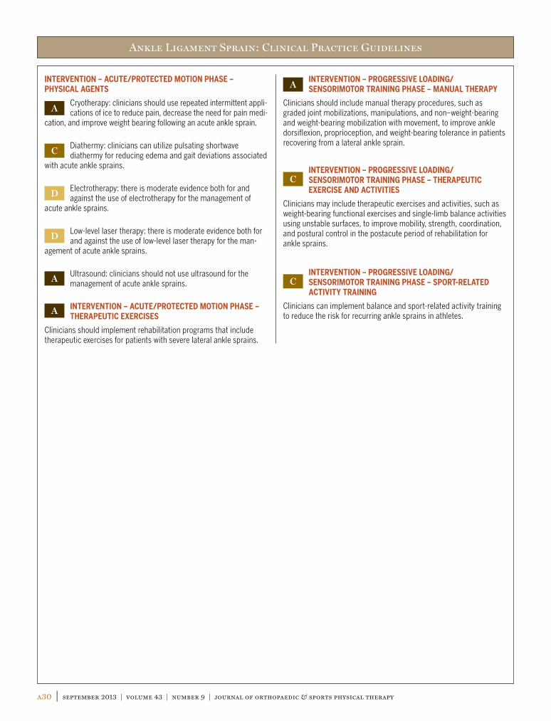
Ankle Ligament Sprain: Clinical Practice Guidelines
a30 | september 2013 | volume 43 | number 9 | journal of orthopaedic & sports physical therapy
INTERVENTION – ACUTE/PROTECTED MOTION PHASE – PHYSICAL AGENTS
Cryotherapy: clinicians should use repeated intermittent appli-cations of ice to reduce pain, decrease the need for pain medi-
cation, and improve weight bearing following an acute ankle sprain.
Diathermy: clinicians can utilize pulsating shortwave diathermy for reducing edema and gait deviations associated
with acute ankle sprains.
Electrotherapy: there is moderate evidence both for and against the use of electrotherapy for the management of
acute ankle sprains.
Low-level laser therapy: there is moderate evidence both for and against the use of low-level laser therapy for the man-
agement of acute ankle sprains.
Ultrasound: clinicians should not use ultrasound for the management of acute ankle sprains.
INTERVENTION – ACUTE/PROTECTED MOTION PHASE – THERAPEUTIC EXERCISES
Clinicians should implement rehabilitation programs that include therapeutic exercises for patients with severe lateral ankle sprains.
INTERVENTION – PROGRESSIVE LOADING/ SENSORIMOTOR TRAINING PHASE – MANUAL THERAPY
Clinicians should include manual therapy procedures, such as graded joint mobilizations, manipulations, and non–weight-bearing and weight-bearing mobilization with movement, to improve ankle dorsiflexion, proprioception, and weight-bearing tolerance in patients recovering from a lateral ankle sprain.
INTERVENTION – PROGRESSIVE LOADING/ SENSORIMOTOR TRAINING PHASE – THERAPEUTIC EXERCISE AND ACTIVITIES
Clinicians may include therapeutic exercises and activities, such as weight-bearing functional exercises and single-limb balance activities using unstable surfaces, to improve mobility, strength, coordination, and postural control in the postacute period of rehabilitation for ankle sprains.
INTERVENTION – PROGRESSIVE LOADING/ SENSORIMOTOR TRAINING PHASE – SPORT-RELATED ACTIVITY TRAINING
Clinicians can implement balance and sport-related activity training to reduce the risk for recurring ankle sprains in athletes.A
A
A
A
C
C
C
D
D
������*XLGHOLQHV�LQGG����� ��������������������30
Ankle Ligament Sprain: Clinical Practice Guidelines
journal of orthopaedic & sports physical therapy | volume 43 | number 9 | september 2013 | a31
AUTHORSRobRoy L. Martin, PT, PhDAssociate ProfessorDepartment of Physical
TherapyDuquesne UniversityPittsburgh, [email protected]! Physical TherapistCenters for Rehab Services/
Center for Sports MedicineUniversity of Pittsburgh
Medical CenterPittsburgh, Pennsylvania
Todd E. Davenport, DPTAssociate ProfessorDepartment of Physical TherapyUniversity of the PacificStockton, [email protected]
Stephen Paulseth, DPT, MSPaulseth and Associates
Physical TherapyLos Angeles, CaliforniaClinical FacultyOrthopedic Physical Therapy
Residency ProgramDivision of Biokinesiology
and Physical TherapyUniversity of Southern
CaliforniaLos Angeles, [email protected]
Dane K. Wukich, MDChief, Division of Foot and
Ankle SurgeryAssistant Professor of Orthopaedic
SurgeryUniversity of Pittsburgh
Comprehensive Foot and Ankle Center
Pittsburgh, [email protected]
Joseph J. Godges, DPT, MAICF Practice Guidelines CoordinatorOrthopaedic Section, APTA, IncLa Crosse, [email protected] ProfessorDivision of Biokinesiology
and Physical TherapyUniversity of Southern CaliforniaLos Angeles, [email protected]
REVIEWERSRoy D. Altman, MDProfessor of MedicineDivision of Rheumatology
and ImmunologyDavid Ge!en School of Medicine
at UCLALos Angeles, [email protected]
Anthony Delitto, PT, PhDProfessor and Chair
School of Health and Rehabilitation Sciences
University of PittsburghPittsburgh, [email protected]
John DeWitt, DPTDirector of Physical Therapy Sports
and Orthopaedic ResidenciesThe Ohio State UniversityColumbus, [email protected]
Amanda Ferland, DPTClinic Relationship ManagerOptimisPTMurrieta, [email protected]
Helene Fearon, PTPrincipal and ConsultantFearon/Levine ConsultingPhoenix, [email protected]
Joy MacDermid, PT, PhDAssociate ProfessorSchool of Rehabilitation ScienceMcMaster UniversityHamilton, Ontario, [email protected]
James W. Matheson, DPTPresident and Clinic DirectorCatalyst Sports Medicine
Hudson, Wisconsinjwmatheson@catalystsportsmedicine.
com
Thomas G. McPoil, PT, PhDProfessorSchool of Physical TherapyRegis UniversityDenver, [email protected]
Stephen Reischl, DPTAdjunct Associate Professor
of Clinical Physical TherapyDivision of Biokinesiology and
Physical TherapyUniversity of Southern CaliforniaLos Angeles, [email protected]
Leslie Torburn, DPTPrincipal and ConsultantSilhouette Consulting, IncRedwood City, [email protected]
James Zachazewski, DPTClinical DirectorDepartment of Physical and
Occupational TherapyMassachusetts General HospitalBoston, [email protected]
AFFILIATIONS AND CONTACTS
������*XLGHOLQHV�LQGG����� ��������������������30

Ankle Ligament Sprain: Clinical Practice Guidelines
a32 | september 2013 | volume 43 | number 9 | journal of orthopaedic & sports physical therapy
1. Aaltonen S, Karjalainen H, Heinonen A, Parkkari J, Kujala UM. Preven-tion of sports injuries: systematic review of randomized controlled trials. Arch Intern Med. 2007;167:1585-1592. http://dx.doi.org/10.1001/archinte.167.15.1585
2. Akbari M, Karimi H, Farahini H, Faghihzadeh S. Balance problems after unilateral lateral ankle sprains. J Rehabil Res Dev. 2006;43:819-824.
3. Alcock GK, Stratford PW. Validation of the Lower Extremity Func-tional Scale on athletic subjects with ankle sprains. Physiother Can. 2002;54:233-240.
4. Alonso A, Khoury L, Adams R. Clinical tests for ankle syndesmosis injury: reliability and prediction of return to function. J Orthop Sports Phys Ther. 1998;27:276-284.
5. Amaral De Noronha M, Borges NG, Jr. Lateral ankle sprain: isokinetic test reliability and comparison between invertors and evertors. Clin Biomech (Bristol, Avon). 2004;19:868-871. http://dx.doi.org/10.1016/j.clinbiomech.2004.05.011
6. Arnason A, Sigurdsson SB, Gudmundsson A, Holme I, Engebretsen L, Bahr R. Risk factors for injuries in football. Am J Sports Med. 2004;32:5S-16S. http://dx.doi.org/10.1177/0363546503258912
7. Arnold BL, De La Motte S, Linens S, Ross SE. Ankle instability is associ-ated with balance impairments: a meta-analysis. Med Sci Sports Exerc. 2009;41:1048-1062. http://dx.doi.org/10.1249/MSS.0b013e318192d044
8. Attarian DE, McCrackin HJ, DeVito DP, McElhaney JH, Garrett WE, Jr. Biomechanical characteristics of human ankle ligaments. Foot Ankle. 1985;6:54-58.
9. Aydogan U, Glisson RR, Nunley JA. Extensor retinaculum aug-mentation reinforces anterior talofibular ligament repair. Clin Orthop Relat Res. 2006;442:210-215. http://dx.doi.org/10.1097/01.blo.0000183737.43245.26
10. Bachmann LM, Kolb E, Koller MT, Steurer J, ter Riet G. Accuracy of Ottawa ankle rules to exclude fractures of the ankle and mid-foot: systematic review. BMJ. 2003;326:417. http://dx.doi.org/10.1136/bmj.326.7386.417
11. Bahr R, Bahr IA. Incidence of acute volleyball injuries: a prospective cohort study of injury mechanisms and risk factors. Scand J Med Sci Sports. 1997;7:166-171.
12. Bahr R, Karlsen R, Lian O, Ovrebø RV. Incidence and mechanisms of acute ankle inversion injuries in volleyball. A retrospective cohort study. Am J Sports Med. 1994;22:595-600.
13. Bahr R, Pena F, Shine J, et al. Mechanics of the anterior drawer and ta-lar tilt tests. A cadaveric study of lateral ligament injuries of the ankle. Acta Orthop Scand. 1997;68:435-441.
14. Barg A, Tochigi Y, Amendola A, Phisitkul P, Hintermann B, Saltzman CL. Subtalar instability: diagnosis and treatment. Foot Ankle Int. 2012;33:151-160. http://dx.doi.org/10.3113/FAI.2012.0151
15. Barrett JR, Tanji JL, Drake C, Fuller D, Kawasaki RI, Fenton RM. High- versus low-top shoes for the prevention of ankle sprains in bas-ketball players. A prospective randomized study. Am J Sports Med. 1993;21:582-585.
16. Bassett SF, Prapavessis H. Home-based physical therapy intervention with adherence-enhancing strategies versus clinic-based management for patients with ankle sprains. Phys Ther. 2007;87:1132-1143. http://dx.doi.org/10.2522/ptj.20060260
17. Baumhauer JF, Alosa DM, Renström AF, Trevino S, Beynnon B. A prospective study of ankle injury risk factors. Am J Sports Med. 1995;23:564-570.
18. Beckman SM, Buchanan TS. Ankle inversion injury and hypermobility: e!ect on hip and ankle muscle electromyography onset latency. Arch Phys Med Rehabil. 1995;76:1138-1143.
19. Bernier JN, Perrin DH. E!ect of coordination training on propriocep-tion of the functionally unstable ankle. J Orthop Sports Phys Ther. 1998;27:264-275.
20. Beynnon BD, Renström PA, Alosa DM, Baumhauer JF, Vacek PM. Ankle ligament injury risk factors: a prospective study of college athletes. J Orthop Res. 2001;19:213-220. http://dx.doi.org/10.1016/S0736-0266(00)90004-4
21. Binkley JM, Stratford PW, Lott SA, Riddle DL. The Lower Extremity Functional Scale (LEFS): scale development, measurement properties, and clinical application. North American Orthopaedic Rehabilitation Research Network. Phys Ther. 1999;79:371-383.
22. Bleakley C, McDonough S, MacAuley D. The use of ice in the treat-ment of acute soft-tissue injury: a systematic review of randomized controlled trials. Am J Sports Med. 2004;32:251-261. http://dx.doi.org/10.1177/0363546503260757
23. Bleakley CM, McDonough SM, MacAuley DC, Bjordal J. Cryotherapy for acute ankle sprains: a randomised controlled study of two di!erent icing protocols. Br J Sports Med. 2006;40:700-705; discussion 705. http://dx.doi.org/10.1136/bjsm.2006.025932
24. Bleakley CM, O’Connor SR, Tully MA, et al. E!ect of accelerated reha-bilitation on function after ankle sprain: randomised controlled trial. BMJ. 2010;340:c1964. http://dx.doi.org/10.1136/bmj.c1964
25. Bohannon RW, Larkin PA, Cook AC, Gear J, Singer J. Decrease in timed balance test scores with aging. Phys Ther. 1984;64:1067-1070.
26. Broglio SP, Zhu W, Sopiarz K, Park Y. Generalizability theory analysis of Balance Error Scoring System reliability in healthy young adults. J Athl Train. 2009;44:497-502. http://dx.doi.org/10.4085/1062-6050-44.5.497
27. Buchanan AS, Docherty CL, Schrader J. Functional performance testing in participants with functional ankle instability and in a healthy control group. J Athl Train. 2008;43:342-346. http://dx.doi.org/10.4085/1062-6050-43.4.342
28. Budny A. Subtalar joint instability: current clinical concepts. Clin Podiatr Med Surg. 2004;21:449-460. http://dx.doi.org/10.1016/j.cpm.2004.03.003
29. Bullock-Saxton JE. Local sensation changes and altered hip muscle function following severe ankle sprain. Phys Ther. 1994;74:17-28; dis-cussion 28-31.
30. Bullock-Saxton JE, Janda V, Bullock MI. The influence of ankle sprain injury on muscle activation during hip extension. Int J Sports Med. 1994;15:330-334. http://dx.doi.org/10.1055/s-2007-1021069
31. Burks RT, Morgan J. Anatomy of the lateral ankle ligaments. Am J Sports Med. 1994;22:72-77.
32. Butler AM, Walsh WR. Mechanical response of ankle ligaments at low loads. Foot Ankle Int. 2004;25:8-12.
33. Button G, Pinney S. A meta-analysis of outcome rating scales in foot and ankle surgery: is there a valid, reliable, and responsive system? Foot Ankle Int. 2004;25:521-525.
34. Ca!rey E, Docherty CL, Schrader J, Klossner J. The ability of 4 single-limb hopping tests to detect functional performance deficits in indi-viduals with functional ankle instability. J Orthop Sports Phys Ther. 2009;39:799-806. http://dx.doi.org/10.2519/jospt.2009.3042
35. Cameron KL, Owens BD, DeBerardino TM. Incidence of ankle sprains among active-duty members of the United States Armed Services from 1998 through 2006. J Athl Train. 2010;45:29-38. http://dx.doi.
REFERENCES
������*XLGHOLQHV�LQGG����� ��������������������30
Ankle Ligament Sprain: Clinical Practice Guidelines
journal of orthopaedic & sports physical therapy | volume 43 | number 9 | september 2013 | a33
org/10.4085/1062-6050-45.1.29
36. Campbell SE, Warner M. MR imaging of ankle inversion injuries. Magn Reson Imaging Clin N Am. 2008;16:1-18. http://dx.doi.org/10.1016/j.mric.2008.02.001
37. Carcia CR, Martin RL, Drouin JM. Validity of the Foot and Ankle Abil-ity Measure in athletes with chronic ankle instability. J Athl Train. 2008;43:179-183. http://dx.doi.org/10.4085/1062-6050-43.2.179
38. Chambers RB, Cook TM, Cowell HR. Surgical reconstruction for cal-caneonavicular coalition. Evaluation of function and gait. J Bone Joint Surg Am. 1982;64:829-836.
39. Choi WJ, Lee JW, Han SH, Kim BS, Lee SK. Chronic lateral ankle instability: the e!ect of intra-articular lesions on clinical outcome. Am J Sports Med. 2008;36:2167-2172. http://dx.doi.org/10.1177/0363546508319050
40. Chrintz H, Falster O, Roed J. Single-leg postural equilib-rium test. Scand J Med Sci Sports. 1991;1:244-246. http://dx.doi.org/10.1111/j.1600-0838.1991.tb00305.x
41. Collins N, Teys P, Vicenzino B. The initial e!ects of a Mulligan’s mobili-zation with movement technique on dorsiflexion and pain in subacute ankle sprains. Man Ther. 2004;9:77-82. http://dx.doi.org/10.1016/S1356-689X(03)00101-2
42. Colville MR, Marder RA, Boyle JJ, Zarins B. Strain measurement in lat-eral ankle ligaments. Am J Sports Med. 1990;18:196-200.
43. Cooke MW, Lamb SE, Marsh J, Dale J. A survey of current consultant practice of treatment of severe ankle sprains in emergency depart-ments in the United Kingdom. Emerg Med J. 2003;20:505-507.
44. Cooke MW, Marsh JL, Clark M, et al. Treatment of severe ankle sprain: a pragmatic randomised controlled trial comparing the clinical ef-fectiveness and cost-e!ectiveness of three types of mechanical ankle support with tubular bandage. The CAST trial. Health Technol Assess. 2009;13:1-121. http://dx.doi.org/10.3310/hta13130
45. Cordova ML, Sefton JM, Hubbard TJ. Mechanical joint laxity associ-ated with chronic ankle instability: a systematic review. Sports Health. 2010;2:452-459. http://dx.doi.org/10.1177/1941738110382392
46. Coughlan G, Caulfield B. A 4-week neuromuscular training program and gait patterns at the ankle joint. J Athl Train. 2007;42:51-59.
47. Dahle LK, Mueller MJ, Delitto A, Diamond JE. Visual assessment of foot type and relationship of foot type to lower extremity injury. J Orthop Sports Phys Ther. 1991;14:70-74.
48. de Bie RA, de Vet HC, Lenssen TF, van den Wildenberg FA, Kootstra G, Knipschild PG. Low-level laser therapy in ankle sprains: a randomized clinical trial. Arch Phys Med Rehabil. 1998;79:1415-1420.
49. de Bie RA, de Vet HC, van den Wildenberg FA, Lenssen T, Knipschild PG. The prognosis of ankle sprains. Int J Sports Med. 1997;18:285-289. http://dx.doi.org/10.1055/s-2007-972635
50. Delahunt E, Coughlan GF, Caulfield B, Nightingale EJ, Lin CW, Hiller CE. Inclusion criteria when investigating insu"ciencies in chronic ankle instability. Med Sci Sports Exerc. 2010;42:2106-2121. http://dx.doi.org/10.1249/MSS.0b013e3181de7a8a
51. Delahunt E, Monaghan K, Caulfield B. Altered neuromuscular control and ankle joint kinematics during walking in subjects with functional instability of the ankle joint. Am J Sports Med. 2006;34:1970-1976. http://dx.doi.org/10.1177/0363546506290989
52. Delahunt E, Monaghan K, Caulfield B. Ankle function dur-ing hopping in subjects with functional instability of the ankle joint. Scand J Med Sci Sports. 2007;17:641-648. http://dx.doi.org/10.1111/j.1600-0838.2006.00612.x
53. Delahunt E, Monaghan K, Caulfield B. Changes in lower limb kinemat-
ics, kinetics, and muscle activity in subjects with functional instabil-ity of the ankle joint during a single leg drop jump. J Orthop Res. 2006;24:1991-2000. http://dx.doi.org/10.1002/jor.20235
54. Demeritt KM, Shultz SJ, Docherty CL, Gansneder BM, Perrin DH. Chronic ankle instability does not a!ect lower extremity functional per-formance. J Athl Train. 2002;37:507-511.
55. Denegar CR, Hertel J, Fonseca J. The e!ect of lateral ankle sprain on dorsiflexion range of motion, posterior talar glide, and joint laxity. J Orthop Sports Phys Ther. 2002;32:166-173.
56. de Noronha M, França LC, Haupenthal A, Nunes GS. Intrinsic predictive factors for ankle sprain in active university students: a prospective study. Scand J Med Sci Sports. In press. http://dx.doi.org/10.1111/j.1600-0838.2011.01434.x
57. de Noronha M, Refshauge KM, Crosbie J, Kilbreath SL. Relationship between functional ankle instability and postural control. J Orthop Sports Phys Ther. 2008;38:782-789. http://dx.doi.org/10.2519/jospt.2008.2766
58. de Noronha M, Refshauge KM, Herbert RD, Kilbreath SL, Hertel J. Do voluntary strength, proprioception, range of motion, or pos-tural sway predict occurrence of lateral ankle sprain? Br J Sports Med. 2006;40:824-828; discussion 828. http://dx.doi.org/10.1136/bjsm.2006.029645
59. Dettori JR, Basmania CJ. Early ankle mobilization, part II: a one-year follow-up of acute, lateral ankle sprains (a randomized clinical trial). Mil Med. 1994;159:20-24.
60. de Vries JS, Kingma I, Blankevoort L, van Dijk CN. Di!erence in balance measures between patients with chronic ankle instability and patients after an acute ankle inversion trauma. Knee Surg Sports Traumatol Ar-throsc. 2010;18:601-606. http://dx.doi.org/10.1007/s00167-010-1097-1
61. de Vries JS, Krips R, Sierevelt IN, Blankevoort L. Interventions for treating chronic ankle instability. Cochrane Database Syst Rev. 2006:CD004124. http://dx.doi.org/10.1002/14651858.CD004124.pub2
62. DiGiovanni BF, Fraga CJ, Cohen BE, Shere! MJ. Associated injuries found in chronic lateral ankle instability. Foot Ankle Int. 2000;21:809-815. http://dx.doi.org/10.1177/107110070002101003
63. Dizon JM, Reyes JJ. A systematic review on the e!ectiveness of external ankle supports in the prevention of inversion ankle sprains among elite and recreational players. J Sci Med Sport. 2010;13:309-317. http://dx.doi.org/10.1016/j.jsams.2009.05.002
64. Docherty CL, Arnold BL, Gansneder BM, Hurwitz S, Gieck J. Functional-performance deficits in volunteers with functional ankle instability. J Athl Train. 2005;40:30-34.
65. Docherty CL, Gansneder BM, Arnold BL, Hurwitz SR. Develop-ment and reliability of the ankle instability instrument. J Athl Train. 2006;41:154-158.
66. Docherty CL, Rybak-Webb K. Reliability of the anterior drawer and talar tilt tests using the LigMaster joint arthrometer. J Sport Rehabil. 2009;18:389-397.
67. Docherty CL, Valovich McLeod TC, Shultz SJ. Postural control deficits in participants with functional ankle instability as measured by the Bal-ance Error Scoring System. Clin J Sport Med. 2006;16:203-208.
68. Dombek MF, Lamm BM, Saltrick K, Mendicino RW, Catanzariti AR. Peroneal tendon tears: a retrospective review. J Foot Ankle Surg. 2003;42:250-258.
69. Drewes LK, McKeon PO, Kerrigan DC, Hertel J. Dorsiflexion defi-cit during jogging with chronic ankle instability. J Sci Med Sport. 2009;12:685-687. http://dx.doi.org/10.1016/j.jsams.2008.07.003
70. Eechaute C, Vaes P, Duquet W. The chronic ankle instability scale:
������*XLGHOLQHV�LQGG����� ��������������������30
Ankle Ligament Sprain: Clinical Practice Guidelines
a34 | september 2013 | volume 43 | number 9 | journal of orthopaedic & sports physical therapy
clinimetric properties of a multidimensional, patient-assessed instru-ment. Phys Ther Sport. 2008;9:57-66. http://dx.doi.org/10.1016/j.ptsp.2008.02.001
71. Eechaute C, Vaes P, Duquet W. The dynamic postural control is im-paired in patients with chronic ankle instability: reliability and validity of the multiple hop test. Clin J Sport Med. 2009;19:107-114. http://dx.doi.org/10.1097/JSM.0b013e3181948ae8
72. Eechaute C, Vaes P, Duquet W. Functional performance deficits in patients with CAI: validity of the multiple hop test. Clin J Sport Med. 2008;18:124-129. http://dx.doi.org/10.1097/JSM.0b013e31816148d2
73. Eechaute C, Vaes P, Van Aerschot L, Asman S, Duquet W. The clinimet-ric qualities of patient-assessed instruments for measuring chronic ankle instability: a systematic review. BMC Musculoskelet Disord. 2007;8:6. http://dx.doi.org/10.1186/1471-2474-8-6
74. Eggli S, Sclabas GM, Eggli S, Zimmermann H, Exadaktylos AK. The Ber-nese ankle rules: a fast, reliable test after low-energy, supination-type malleolar and midfoot trauma. J Trauma. 2005;59:1268-1271.
75. Eisenhart AW, Gaeta TJ, Yens DP. Osteopathic manipulative treatment in the emergency department for patients with acute ankle injuries. J Am Osteopath Assoc. 2003;103:417-421.
76. Elveru RA, Rothstein JM, Lamb RL. Goniometric reliability in a clinical setting. Subtalar and ankle joint measurements. Phys Ther. 1988;68:672-677.
77. Emery CA, Meeuwisse WH. The e!ectiveness of a neuromuscular prevention strategy to reduce injuries in youth soccer: a cluster-randomised controlled trial. Br J Sports Med. 2010;44:555-562. http://dx.doi.org/10.1136/bjsm.2010.074377
78. Emery CA, Rose MS, McAllister JR, Meeuwisse WH. A prevention strat-egy to reduce the incidence of injury in high school basketball: a cluster randomized controlled trial. Clin J Sport Med. 2007;17:17-24. http://dx.doi.org/10.1097/JSM.0b013e31802e9c05
79. Engebretsen AH, Myklebust G, Holme I, Engebretsen L, Bahr R. Intrinsic risk factors for acute ankle injuries among male soccer players: a prospective cohort study. Scand J Med Sci Sports. 2010;20:403-410. http://dx.doi.org/10.1111/j.1600-0838.2009.00971.x
80. Exelby L. Peripheral mobilisations with movement. Man Ther. 1996;1:118-126. http://dx.doi.org/10.1054/math.1996.0259
81. Faude O, Junge A, Kindermann W, Dvorak J. Risk factors for injuries in elite female soccer players. Br J Sports Med. 2006;40:785-790. http://dx.doi.org/10.1136/bjsm.2006.027540
82. Ferkel RD, Chams RN. Chronic lateral instability: arthroscopic findings and long-term results. Foot Ankle Int. 2007;28:24-31. http://dx.doi.org/10.3113/FAI.2007.0005
83. Ferran NA, Oliva F, Ma!ulli N. Ankle instability. Sports Med Arthrosc. 2009;17:139-145. http://dx.doi.org/10.1097/JSA.0b013e3181a3d790
84. Finno! JT, Peterson VJ, Hollman JH, Smith J. Intrarater and inter-rater reliability of the Balance Error Scoring System (BESS). PM R. 2009;1:50-54. http://dx.doi.org/10.1016/j.pmrj.2008.06.002
85. Fong DT, Chan YY, Mok KM, Yung P, Chan KM. Understanding acute ankle ligamentous sprain injury in sports. Sports Med Arthrosc Rehabil Ther Technol. 2009;1:14. http://dx.doi.org/10.1186/1758-2555-1-14
86. Fong DT, Hong Y, Chan LK, Yung PS, Chan KM. A systematic review on ankle injury and ankle sprain in sports. Sports Med. 2007;37:73-94.
87. Forkin DM, Koczur C, Battle R, Newton RA. Evaluation of kinesthetic def-icits indicative of balance control in gymnasts with unilateral chronic ankle sprains. J Orthop Sports Phys Ther. 1996;23:245-250.
88. Freeman MA. Instability of the foot after injuries to the lateral ligament of the ankle. J Bone Joint Surg Br. 1965;47:669-677.
89. Freeman MA. Treatment of ruptures of the lateral ligament of the ankle. J Bone Joint Surg Br. 1965;47:661-668.
90. Freeman MA, Dean MR, Hanham IW. The etiology and prevention of functional instability of the foot. J Bone Joint Surg Br. 1965;47:678-685.
91. Friel K, McLean N, Myers C, Caceres M. Ipsilateral hip abductor weak-ness after inversion ankle sprain. J Athl Train. 2006;41:74-78.
92. Fujii T, Kitaoka HB, Luo ZP, Kura H, An KN. Analysis of ankle-hindfoot stability in multiple planes: an in vitro study. Foot Ankle Int. 2005;26:633-637.
93. Fujii T, Luo ZP, Kitaoka HB, An KN. The manual stress test may not be su"cient to di!erentiate ankle ligament injuries. Clin Biomech (Bristol, Avon). 2000;15:619-623.
94. Geppert MJ, Sobel M, Bohne WH. Lateral ankle instability as a cause of superior peroneal retinacular laxity: an anatomic and biomechanical study of cadaveric feet. Foot Ankle. 1993;14:330-334.
95. Gerber JP, Williams GN, Scoville CR, Arciero RA, Taylor DC. Persistent disability associated with ankle sprains: a prospective examination of an athletic population. Foot Ankle Int. 1998;19:653-660.
96. Gomez JE, Ross SK, Calmbach WL, Kimmel RB, Schmidt DR, Dhanda R. Body fatness and increased injury rates in high school football linemen. Clin J Sport Med. 1998;8:115-120.
97. Green T, Refshauge K, Crosbie J, Adams R. A randomized controlled trial of a passive accessory joint mobilization on acute ankle inversion sprains. Phys Ther. 2001;81:984-994.
98. Gribble PA, Hertel J. Considerations for normalization of measures of the Star Excursion Balance Test. Meas Phys Educ Sci. 2003;7:89-100.
99. Gribble PA, Hertel J, Denegar CR, Buckley WE. The e!ects of fatigue and chronic ankle instability on dynamic postural control. J Athl Train. 2004;39:321-329.
100. Gross P, Marti B. Risk of degenerative ankle joint disease in volleyball players: study of former elite athletes. Int J Sports Med. 1999;20:58-63. http://dx.doi.org/10.1055/s-2007-971094
101. Guyatt GH, Sackett DL, Sinclair JC, Hayward R, Cook DJ, Cook RJ. Us-ers’ guides to the medical literature. IX. A method for grading health care recommendations. Evidence-Based Medicine Working Group. JAMA. 1995;274:1800-1804.
102. Hägglund M, Waldén M, Ekstrand J. Previous injury as a risk factor for injury in elite football: a prospective study over two consecutive seasons. Br J Sports Med. 2006;40:767-772. http://dx.doi.org/10.1136/bjsm.2006.026609
103. Hale SA, Hertel J. Reliability and sensitivity of the Foot and Ankle Disability Index in subjects with chronic ankle instability. J Athl Train. 2005;40:35-40.
104. Hale SA, Hertel J, Olmsted-Kramer LC. The e!ect of a 4-week compre-hensive rehabilitation program on postural control and lower extremity function in individuals with chronic ankle instability. J Orthop Sports Phys Ther. 2007;37:303-311. http://dx.doi.org/10.2519/jospt.2007.2322
105. Han K, Ricard MD, Fellingham GW. E!ects of a 4-week exercise program on balance using elastic tubing as a perturbation force for individuals with a history of ankle sprains. J Orthop Sports Phys Ther. 2009;39:246-255. http://dx.doi.org/10.2519/jospt.2009.2958
106. Haraguchi N, Tokumo A, Okamura R, et al. Influence of activity level on the outcome of treatment of lateral ankle ligament rupture. J Orthop Sci. 2009;14:391-396. http://dx.doi.org/10.1007/s00776-009-1346-7
107. Hartsell HD, Spaulding SJ. Eccentric/concentric ratios at selected ve-locities for the invertor and evertor muscles of the chronically unstable ankle. Br J Sports Med. 1999;33:255-258.
������*XLGHOLQHV�LQGG����� ��������������������30
Ankle Ligament Sprain: Clinical Practice Guidelines
journal of orthopaedic & sports physical therapy | volume 43 | number 9 | september 2013 | a35
108. Haywood KL, Hargreaves J, Lamb SE. Multi-item outcome measures for lateral ligament injury of the ankle: a struc-tured review. J Eval Clin Pract. 2004;10:339-352. http://dx.doi.org/10.1111/j.1365-2753.2003.00435.x
109. Hertel J. Functional anatomy, pathomechanics, and pathophysiology of lateral ankle instability. J Athl Train. 2002;37:364-375.
110. Hertel J. Sensorimotor deficits with ankle sprains and chronic ankle instability. Clin Sports Med. 2008;27:353-370. http://dx.doi.org/10.1016/j.csm.2008.03.006
111. Hertel J, Braham RA, Hale SA, Olmsted-Kramer LC. Simplifying the star excursion balance test: analyses of subjects with and without chronic ankle instability. J Orthop Sports Phys Ther. 2006;36:131-137. http://dx.doi.org/10.2519/jospt.2006.2103
112. Hertel J, Denegar CR, Monroe MM, Stokes WL. Talocrural and subta-lar joint instability after lateral ankle sprain. Med Sci Sports Exerc. 1999;31:1501-1508.
113. Hertel J, Miller SJ, Denegar CR. Intratester and intertester reli-ability during the Star Excursion Balance Tests. J Sport Rehabil. 2000;9:104-116.
114. Hiller CE, Kilbreath SL, Refshauge KM. Chronic ankle instability: evolution of the model. J Athl Train. 2011;46:133-141. http://dx.doi.org/10.4085/1062-6050-46.2.133
115. Hiller CE, Nightingale EJ, Lin CW, Coughlan GF, Caulfield B, Delahunt E. Characteristics of people with recurrent ankle sprains: a systematic review with meta-analysis. Br J Sports Med. 2011;45:660-672. http://dx.doi.org/10.1136/bjsm.2010.077404
116. Hiller CE, Refshauge KM, Bundy AC, Herbert RD, Kilbreath SL. The Cumberland Ankle Instability Tool: a report of validity and reliability testing. Arch Phys Med Rehabil. 2006;87:1235-1241. http://dx.doi.org/10.1016/j.apmr.2006.05.022
117. Hiller CE, Refshauge KM, Herbert RD, Kilbreath SL. Balance and recov-ery from a perturbation are impaired in people with functional ankle instability. Clin J Sport Med. 2007;17:269-275. http://dx.doi.org/10.1097/JSM.0b013e3180f60b12
118. Hiller CE, Refshauge KM, Herbert RD, Kilbreath SL. Intrinsic predictors of lateral ankle sprain in adolescent dancers: a prospective cohort study. Clin J Sport Med. 2008;18:44-48. http://dx.doi.org/10.1097/JSM.0b013e31815f2b35
119. Hintermann B. Biomechanics of the unstable ankle joint and clinical implications. Med Sci Sports Exerc. 1999;31:S459-S469.
120. Hintermann B, Boss A, Schafer D. Arthroscopic findings in patients with chronic ankle instability. Am J Sports Med. 2002;30:402-409.
121. Hla KM, Ishii T, Sakane M, Hayashi K. E!ect of anesthesia of the sinus tarsi on peroneal reaction time in patients with functional instability of the ankle. Foot Ankle Int. 1999;20:554-559.
122. Hollis JM, Blasier RD, Flahi! CM. Simulated lateral ankle ligamentous injury. Change in ankle stability. Am J Sports Med. 1995;23:672-677.
123. Holme E, Magnusson SP, Becher K, Bieler T, Aagaard P, Kjaer M. The e!ect of supervised rehabilitation on strength, postural sway, position sense and re-injury risk after acute ankle ligament sprain. Scand J Med Sci Sports. 1999;9:104-109. http://dx.doi.org/10.1111/j.1600-0838.1999.tb00217.x
124. Hosea TM, Carey CC, Harrer MF. The gender issue: epidemiology of ankle injuries in athletes who participate in basketball. Clin Orthop Relat Res. 2000:45-49.
125. Hubbard TJ, Kaminski TW. Kinesthesia is not a!ected by functional ankle instability status. J Athl Train. 2002;37:481-486.
126. Hubbard TJ, Kramer LC, Denegar CR, Hertel J. Contributing factors
to chronic ankle instability. Foot Ankle Int. 2007;28:343-354. http://dx.doi.org/10.3113/FAI.2007.0343
127. Hunt TN, Ferrara MS, Bornstein RA, Baumgartner TA. The reliability of the modified Balance Error Scoring System. Clin J Sport Med. 2009;19:471-475. http://dx.doi.org/10.1097/JSM.0b013e3181c12c7b
128. Hupperets MD, Verhagen EA, van Mechelen W. E!ect of unsupervised home based proprioceptive training on recurrences of ankle sprain: randomised controlled trial. BMJ. 2009;339:b2684.
129. Iverson GL, Kaarto ML, Koehle MS. Normative data for the Balance Er-ror Scoring System: implications for brain injury evaluations. Brain Inj. 2008;22:147-152. http://dx.doi.org/10.1080/02699050701867407
130. Jennings J, Davies GJ. Treatment of cuboid syndrome secondary to lateral ankle sprains: a case series. J Orthop Sports Phys Ther. 2005;35:409-415. http://dx.doi.org/10.2519/jospt.2005.1596
131. Jerosch J, Bischof M. Proprioceptive capabilities of the ankle in stable and unstable joints. Sports Exerc Inj. 1996;2:167-171.
132. Johnson MR, Stoneman PD. Comparison of a lateral hop test versus a forward hop test for functional evaluation of lateral ankle sprains. J Foot Ankle Surg. 2007;46:162-174. http://dx.doi.org/10.1053/j.jfas.2006.12.007
133. Kaikkonen A, Kannus P, Järvinen M. A performance test protocol and scoring scale for the evaluation of ankle injuries. Am J Sports Med. 1994;22:462-469. http://dx.doi.org/10.1177/036354659402200405
134. Kaminski TW, Buckley BD, Powers ME, Hubbard TJ, Ortiz C. E!ect of strength and proprioception training on eversion to inversion strength ratios in subjects with unilateral functional ankle instability. Br J Sports Med. 2003;37:410-415; discussion 415.
135. Kaminski TW, Dover GC. Reliability of inversion and eversion peak- and average-torque measurements from the Biodex System 3 dynamom-eter. J Sport Rehabil. 2001;10:205-220.
136. Karlsson J, Eriksson BI, Renström P. Subtalar instability of the foot. A review and results after surgical treatment. Scand J Med Sci Sports. 1998;8:191-197.
137. Karlsson J, Peterson L. Evaluation of ankle joint function: the use of a scoring scale. Foot. 1991;1:15-19. http://dx.doi.org/10.1016/0958-2592(91)90006-W
138. Kemler E, van de Port I, Backx F, van Dijk CN. A systematic review on the treatment of acute ankle sprain: brace versus other func-tional treatment types. Sports Med. 2011;41:185-197. http://dx.doi.org/10.2165/11584370-000000000-00000
139. Kerkho!s GM, Blankevoort L, Sierevelt IN, Corvelein R, Janssen GH, van Dijk CN. Two ankle joint laxity testers: reliability and validity. Knee Surg Sports Traumatol Arthrosc. 2005;13:699-705. http://dx.doi.org/10.1007/s00167-005-0644-7
140. Kerkho!s GM, Handoll HH, de Bie R, Rowe BH, Struijs PA. Surgical versus conservative treatment for acute injuries of the lateral liga-ment complex of the ankle in adults. Cochrane Database Syst Rev. 2007:CD000380. http://dx.doi.org/10.1002/14651858.CD000380.pub2
141. Kerkho!s GM, Rowe BH, Assendelft WJ, Kelly KD, Struijs PA, van Dijk CN. Immobilisation for acute ankle sprain. A systematic review. Arch Orthop Trauma Surg. 2001;121:462-471.
142. Kidgell DJ, Horvath DM, Jackson BM, Seymour PJ. E!ect of six weeks of dura disc and mini-trampoline balance training on postural sway in athletes with functional ankle instability. J Strength Cond Res. 2007;21:466-469. http://dx.doi.org/10.1519/R-18945.1
143. Kinzey SJ, Armstrong CW. The reliability of the star-excursion test in assessing dynamic balance. J Orthop Sports Phys Ther.
������*XLGHOLQHV�LQGG����� ��������������������30
Ankle Ligament Sprain: Clinical Practice Guidelines
a36 | september 2013 | volume 43 | number 9 | journal of orthopaedic & sports physical therapy
1998;27:356-360.
144. Kjaersgaard-Andersen P, Frich LH, Madsen F, Helmig P, Søgård P, Søjb-jerg JO. Instability of the hindfoot after lesion of the lateral ankle liga-ments: investigations of the anterior drawer and adduction maneuvers in autopsy specimens. Clin Orthop Relat Res. 1991:170-179.
145. Kofotolis N, Kellis E. Ankle sprain injuries: a 2-year prospective cohort study in female Greek professional basketball players. J Athl Train. 2007;42:388-394.
146. Kofotolis ND, Kellis E, Vlachopoulos SP. Ankle sprain injuries and risk factors in amateur soccer players during a 2-year period. Am J Sports Med. 2007;35:458-466. http://dx.doi.org/10.1177/0363546506294857
147. Köhne E, Jones A, Korporaal C, Price JL, Brantingham JW, Globe G. A prospective, single-blinded, randomized, controlled clinical trial of the e!ects of manipulation on proprioception and ankle dorsiflexion in chronic recurrent ankle sprain. J Am Chiropr Assoc. 2007;44:7-17.
148. Konradsen L, Olesen S, Hansen HM. Ankle sensorimotor control and eversion strength after acute ankle inversion injuries. Am J Sports Med. 1998;26:72-77.
149. Konradsen L, Voigt M, Hojsgaard C. Ankle inversion injuries. The role of the dynamic defense mechanism. Am J Sports Med. 1997;25:54-58.
150. LaBella CR, Huxford MR, Grissom J, Kim KY, Peng J, Christo!el KK. E!ect of neuromuscular warm-up on injuries in female soccer and basketball athletes in urban public high schools: cluster randomized controlled trial. Arch Pediatr Adolesc Med. 2011;165:1033-1040. http://dx.doi.org/10.1001/archpediatrics.2011.168
151. Lamb SE, Marsh JL, Hutton JL, Nakash R, Cooke MW. Mechanical supports for acute, severe ankle sprain: a pragmatic, multicentre, randomised controlled trial. Lancet. 2009;373:575-581. http://dx.doi.org/10.1016/S0140-6736(09)60206-3
152. Lamb SE, Nakash RA, Withers EJ, et al. Clinical and cost e!ective-ness of mechanical support for severe ankle sprains: design of a randomised controlled trial in the emergency department [ISRCTN 37807450]. BMC Musculoskelet Disord. 2005;6:1. http://dx.doi.org/10.1186/1471-2474-6-1
153. Landis JR, Koch GG. The measurement of observer agreement for cat-egorical data. Biometrics. 1977;33:159-174.
154. Laufer Y, Rotem-Lehrer N, Ronen Z, Khayutin G, Rozenberg I. E!ect of attention focus on acquisition and retention of postural control fol-lowing ankle sprain. Arch Phys Med Rehabil. 2007;88:105-108. http://dx.doi.org/10.1016/j.apmr.2006.10.028
155. Lee KT, Park YU, Kim JS, Kim JB, Kim KC, Kang SK. Long-term results after modified Brostrom procedure without calcaneofibular liga-ment reconstruction. Foot Ankle Int. 2011;32:153-157. http://dx.doi.org/10.3113/FAI.2011.0153
156. Lentell G, Katzman LL, Walters MR. The relationship between muscle function and ankle stability. J Orthop Sports Phys Ther. 1990;11:605-611.
157. Leslie M, Zachazewski JE, Browne P. Reliability of isokinetic torque values for ankle-invertors and evertors. J Orthop Sports Phys Ther. 1990;11:612-616.
158. Lindenfeld TN, Schmitt DJ, Hendy MP, Mangine RE, Noyes FR. Inci-dence of injury in indoor soccer. Am J Sports Med. 1994;22:364-371.
159. López-Rodríguez S, Fernández de-las-Peñas C, Alburquerque-Sendín F, Rodríguez-Blanco C, Palomeque-del-Cerro L. Immediate e!ects of ma-nipulation of the talocrural joint on stabilometry and baropodometry in patients with ankle sprain. J Manipulative Physiol Ther. 2007;30:186-192. http://dx.doi.org/10.1016/j.jmpt.2007.01.011
160. MacDermid JC, Walton DM, Law M. Critical appraisal of research evi-
dence for its validity and usefulness. Hand Clin. 2009;25:29-42. http://dx.doi.org/10.1016/j.hcl.2008.11.003
161. Magerkurth O, Frigg A, Hintermann B, Dick W, Valderrabano V. Frontal and lateral characteristics of the osseous configuration in chronic ankle instability. Br J Sports Med. 2010;44:568-572. http://dx.doi.org/10.1136/bjsm.2008.048462
162. Malliaropoulos N, Ntessalen M, Papacostas E, Longo UG, Maf-fulli N. Reinjury after acute lateral ankle sprains in elite track and field athletes. Am J Sports Med. 2009;37:1755-1761. http://dx.doi.org/10.1177/0363546509338107
163. Malliaropoulos N, Papacostas E, Papalada A, Ma!ulli N. Acute lateral ankle sprains in track and field athletes: an expanded classifica-tion. Foot Ankle Clin. 2006;11:497-507. http://dx.doi.org/10.1016/j.fcl.2006.05.004
164. Man IO, Morrissey MC, Cywinski JK. E!ect of neuromuscular electrical stimulation on ankle swelling in the early period after ankle sprain. Phys Ther. 2007;87:53-65. http://dx.doi.org/10.2522/ptj.20050244
165. Martin DE, Kaplan PA, Kahler DM, Dussault R, Randolph BJ. Retrospec-tive evaluation of graded stress examination of the ankle. Clin Orthop Relat Res. 1996:165-170.
166. Martin R, Burdett R, Irrgang J. Development of the Foot and Ankle Disability Index (FADI) [abstract]. J Orthop Sports Phys Ther. 1999;29:A32-A33.
167. Martin RL, Irrgang JJ. A survey of self-reported outcome instruments for the foot and ankle. J Orthop Sports Phys Ther. 2007;37:72-84. http://dx.doi.org/10.2519/jospt.2007.2403
168. Martin RL, Irrgang JJ, Burdett RG, Conti SF, Van Swearingen JM. Evi-dence of validity for the Foot and Ankle Ability Measure (FAAM). Foot Ankle Int. 2005;26:968-983.
169. Martin RL, Irrgang JJ, Lalonde KA, Conti S. Current concepts review: foot and ankle outcome instruments. Foot Ankle Int. 2006;27:383-390.
170. Martin RL, McPoil TG. Reliability of ankle goniometric measurements: a literature review. J Am Podiatr Med Assoc. 2005;95:564-572.
171. Matsusaka N, Yokoyama S, Tsurusaki T, Inokuchi S, Okita M. E!ect of ankle disk training combined with tactile stimulation to the leg and foot on functional instability of the ankle. Am J Sports Med. 2001;29:25-30.
172. Mawdsley RH, Hoy DK, Erwin PM. Criterion-related validity of the figure-of-eight method of measuring ankle edema. J Orthop Sports Phys Ther. 2000;30:149-153.
173. McGuine TA, Brooks A, Hetzel S. The e!ect of lace-up ankle braces on injury rates in high school basketball players. Am J Sports Med. 2011;39:1840-1848. http://dx.doi.org/10.1177/0363546511406242
174. McGuine TA, Hetzel S, Wilson J, Brooks A. The e!ect of lace-up ankle braces on injury rates in high school football players. Am J Sports Med. 2012;40:49-57. http://dx.doi.org/10.1177/0363546511422332
175. McGuine TA, Keene JS. The e!ect of a balance training program on the risk of ankle sprains in high school athletes. Am J Sports Med. 2006;34:1103-1111. http://dx.doi.org/10.1177/0363546505284191
176. McHugh MP, Tyler TF, Tetro DT, Mullaney MJ, Nicholas SJ. Risk factors for noncontact ankle sprains in high school athletes: the role of hip strength and balance ability. Am J Sports Med. 2006;34:464-470. http://dx.doi.org/10.1177/0363546505280427
177. McKay GD, Goldie PA, Payne WR, Oakes BW. Ankle injuries in basket-ball: injury rate and risk factors. Br J Sports Med. 2001;35:103-108.
178. McKeon PO, Hertel J. Systematic review of postural control and lateral ankle instability, part I: can deficits be detected with in-strumented testing. J Athl Train. 2008;43:293-304. http://dx.doi.org/10.4085/1062-6050-43.3.293
������*XLGHOLQHV�LQGG����� ��������������������30
Ankle Ligament Sprain: Clinical Practice Guidelines
journal of orthopaedic & sports physical therapy | volume 43 | number 9 | september 2013 | a37
179. McKeon PO, Ingersoll CD, Kerrigan DC, Saliba E, Bennett BC, Hertel J. Balance training improves function and postural control in those with chronic ankle instability. Med Sci Sports Exerc. 2008;40:1810-1819. http://dx.doi.org/10.1249/MSS.0b013e31817e0f92
180. McKnight CM, Armstrong CW. The role of ankle strength in functional ankle instability. J Sport Rehabil. 1997;6:21-29.
181. Menadue C, Raymond J, Kilbreath SL, Refshauge KM, Adams R. Reli-ability of two goniometric methods of measuring active inversion and eversion range of motion at the ankle. BMC Musculoskelet Disord. 2006;7:60. http://dx.doi.org/10.1186/1471-2474-7-60
182. Milgrom C, Shlamkovitch N, Finestone A, et al. Risk factors for lateral ankle sprain: a prospective study among military recruits. Foot Ankle. 1991;12:26-30.
183. Milner CE, Soames RW. Anatomy of the collateral ligaments of the hu-man ankle joint. Foot Ankle Int. 1998;19:757-760.
184. Mohammadi F. Comparison of 3 preventive methods to re-duce the recurrence of ankle inversion sprains in male soc-cer players. Am J Sports Med. 2007;35:922-926. http://dx.doi.org/10.1177/0363546507299259
185. Mulligan BR. Mobilisations with movement (MWM’s). J Man Manip Ther. 1993;1:154-156.
186. Munn J, Beard D, Refshauge K, Lee RJ. Do functional-performance tests detect impairment in subjects with ankle instability? J Sport Re-habil. 2002;11:40-50.
187. Munn J, Beard DJ, Refshauge KM, Lee RY. Eccentric muscle strength in functional ankle instability. Med Sci Sports Exerc. 2003;35:245-250. http://dx.doi.org/10.1249/01.MSS.0000048724.74659.9F
188. Munn J, Sullivan SJ, Schneiders AG. Evidence of sensorimotor deficits in functional ankle instability: a systematic review with meta-analysis. J Sci Med Sport. 2010;13:2-12. http://dx.doi.org/10.1016/j.jsams.2009.03.004
189. Munteanu SE, Strawhorn AB, Landorf KB, Bird AR, Murley GS. A weightbearing technique for the measurement of ankle joint dorsiflex-ion with the knee extended is reliable. J Sci Med Sport. 2009;12:54-59. http://dx.doi.org/10.1016/j.jsams.2007.06.009
190. Nakagawa L, Ho!man M. Performance in static, dynamic, and clinical tests of postural control in individuals with recurrent ankle sprains. J Sport Rehabil. 2004;13:255-268.
191. Neptune RR, Wright IC, van den Bogert AJ. Muscle coordination and function during cutting movements. Med Sci Sports Exerc. 1999;31:294-302.
192. Olmsted LC, Carcia CR, Hertel J, Shultz SJ. E"cacy of the Star Excur-sion Balance Tests in detecting reach deficits in subjects with chronic ankle instability. J Athl Train. 2002;37:501-506.
193. Pasila M, Visuri T, Sundholm A. Pulsating shortwave diathermy: value in treatment of recent ankle and foot sprains. Arch Phys Med Rehabil. 1978;59:383-386.
194. Pefanis N, Karagounis P, Tsiganos G, Armenis E, Baltopoulos P. Tibio-femoral angle and its relation to ankle sprain occurrence. Foot Ankle Spec. 2009;2:271-276. http://dx.doi.org/10.1177/1938640009349502
195. Pefanis N, Papaharalampous X, Tsiganos G, Papadakou E, Baltopoulos P. The e!ect of Q angle on ankle sprain occurrence. Foot Ankle Spec. 2009;2:22-26. http://dx.doi.org/10.1177/1938640008330769
196. Pellow JE, Brantingham JW. The e"cacy of adjusting the ankle in the treatment of subacute and chronic grade I and grade II ankle inversion sprains. J Manipulative Physiol Ther. 2001;24:17-24. http://dx.doi.org/10.1067/mmt.2001.112015
197. Petersen EJ, Irish SM, Lyons CL, et al. Reliability of water volumetry
and the figure of eight method on subjects with ankle joint swelling. J Orthop Sports Phys Ther. 1999;29:609-615.
198. Phillips B, Ball C, Sackett D, et al. Oxford Centre for Evidence-Based Medicine - Levels of Evidence (March 2009). Available at: http://www.cebm.net/index.aspx?o=4590. Accessed August 4, 2009.
199. Phisitkul P, Chaichankul C, Sripongsai R, Prasitdamrong I, Teng-trakulcharoen P, Suarchawaratana S. Accuracy of anterolateral drawer test in lateral ankle instability: a cadaveric study. Foot Ankle Int. 2009;30:690-695. http://dx.doi.org/10.3113/FAI.2009.0690
200. Pihlajamäki H, Hietaniemi K, Paavola M, Visuri T, Mattila VM. Surgical versus functional treatment for acute ruptures of the lateral ligament complex of the ankle in young men: a randomized controlled trial. J Bone Joint Surg Am. 2010;92:2367-2374. http://dx.doi.org/10.2106/JBJS.I.01176
201. Plisky PJ, Rauh MJ, Kaminski TW, Underwood FB. Star Excursion Bal-ance Test as a predictor of lower extremity injury in high school bas-ketball players. J Orthop Sports Phys Ther. 2006;36:911-919. http://dx.doi.org/10.2519/jospt.2006.2244
202. Pontaga I. Ankle joint evertor-invertor muscle torque ratio de-crease due to recurrent lateral ligament sprains. Clin Biomech (Bristol, Avon). 2004;19:760-762. http://dx.doi.org/10.1016/j.clinbiomech.2004.05.003
203. Pope R, Herbert R, Kirwan J. E!ects of ankle dorsiflexion range and pre-exercise calf muscle stretching on injury risk in Army recruits. Aust J Physiother. 1998;44:165-172.
204. Pugia ML, Middel CJ, Seward SW, et al. Comparison of acute swelling and function in subjects with lateral ankle injury. J Orthop Sports Phys Ther. 2001;31:384-388.
205. Raatikainen T, Putkonen M, Puranen J. Arthrography, clinical exami-nation, and stress radiograph in the diagnosis of acute injury to the lateral ligaments of the ankle. Am J Sports Med. 1992;20:2-6. http://dx.doi.org/10.1177/036354659202000102
206. Rahnama L, Salavati M, Akhbari B, Mazaheri M. Attentional demands and postural control in athletes with and without functional ankle instability. J Orthop Sports Phys Ther. 2010;40:180-187. http://dx.doi.org/10.2519/jospt.2010.3188
207. Rasmussen O, Tovborg-Jensen I. Anterolateral rotational instability in the ankle joint. An experimental study of anterolateral rotational in-stability, talar tilt, and anterior drawer sign in relation to injuries to the lateral ligaments. Acta Orthop Scand. 1981;52:99-102.
208. Renström P, Wertz M, Incavo S, et al. Strain in the lateral ligaments of the ankle. Foot Ankle. 1988;9:59-63. http://dx.doi.org/10.1177/107110078800900201
209. Riemann BL, Guskiewicz KM, Shields EW. Relationship between clini-cal and forceplate measures of postural stability. J Sport Rehabil. 1999;8:71-82.
210. Ringleb SI, Dhakal A, Anderson CD, Bawab S, Paranjape R. E!ects of lateral ligament sectioning on the stability of the ankle and subtalar joint. J Orthop Res. 2011;29:1459-1464. http://dx.doi.org/10.1002/jor.21407
211. Rohner-Spengler M, Mannion AF, Babst R. Reliability and minimal detectable change for the figure-of-eight-20 method of measurement of ankle edema. J Orthop Sports Phys Ther. 2007;37:199-205. http://dx.doi.org/10.2519/jospt.2007.2371
212. Roos EM, Brandsson S, Karlsson J. Validation of the Foot and Ankle Outcome Score for ankle ligament reconstruction. Foot Ankle Int. 2001;22:788-794. http://dx.doi.org/10.1177/107110070102201004
213. Ross SE, Arnold BL, Blackburn JT, Brown CN, Guskiewicz KM. En-
������*XLGHOLQHV�LQGG����� ��������������������30
Ankle Ligament Sprain: Clinical Practice Guidelines
a38 | september 2013 | volume 43 | number 9 | journal of orthopaedic & sports physical therapy
hanced balance associated with coordination training with stochastic resonance stimulation in subjects with functional ankle instability: an experimental trial. J Neuroeng Rehabil. 2007;4:47. http://dx.doi.org/10.1186/1743-0003-4-47
214. Ross SE, Guskiewicz KM. E!ect of coordination training with and without stochastic resonance stimulation on dynamic postural stability of subjects with functional ankle instability and subjects with stable ankles. Clin J Sport Med. 2006;16:323-328.
215. Ross SE, Guskiewicz KM, Gross MT, Yu B. Assessment tools for identifying functional limitations associated with func-tional ankle instability. J Athl Train. 2008;43:44-50. http://dx.doi.org/10.4085/1062-6050-43.1.44
216. Rotem-Lehrer N, Laufer Y. E!ect of focus of attention on transfer of a postural control task following an ankle sprain. J Orthop Sports Phys Ther. 2007;37:564-569. http://dx.doi.org/10.2519/jospt.2007.2519
217. Rozzi SL, Lephart SM, Sterner R, Kuligowski L. Balance training for persons with functionally unstable ankles. J Orthop Sports Phys Ther. 1999;29:478-486.
218. Ryan L. Mechanical stability, muscle strength and proprioception in the functionally unstable ankle. Aust J Physiother. 1994;40:41-47.
219. Safran MR, Benedetti RS, Bartolozzi AR, 3rd, Mandelbaum BR. Lat-eral ankle sprains: a comprehensive review: part 1: etiology, patho-anatomy, histopathogenesis, and diagnosis. Med Sci Sports Exerc. 1999;31:S429-S437.
220. Sefton JM, Hicks-Little CA, Hubbard TJ, et al. Sensorimotor func-tion as a predictor of chronic ankle instability. Clin Biomech (Bristol, Avon). 2009;24:451-458. http://dx.doi.org/10.1016/j.clinbiomech.2009.03.003
221. Sekir U, Yildiz Y, Hazneci B, Ors F, Saka T, Aydin T. Reliability of a functional test battery evaluating functionality, proprioception, and strength in recreational athletes with functional ankle instability. Eur J Phys Rehabil Med. 2008;44:407-415.
222. Siegler S, Block J, Schneck CD. The mechanical characteristics of the collateral ligaments of the human ankle joint. Foot Ankle. 1988;8:234-242.
223. Smith RW, Reischl S. The influence of dorsiflexion in the treatment of severe ankle sprains: an anatomical study. Foot Ankle. 1988;9:28-33.
224. Smith RW, Reischl SF. Treatment of ankle sprains in young athletes. Am J Sports Med. 1986;14:465-471.
225. Smith-Oricchio K, Harris BA. Interrater reliability of subtalar neu-tral, calcaneal inversion and eversion. J Orthop Sports Phys Ther. 1990;12:10-15.
226. Spahn G. The ankle meter: an instrument for evaluation of anterior talar drawer in ankle sprain. Knee Surg Sports Traumatol Arthrosc. 2004;12:338-342. http://dx.doi.org/10.1007/s00167-003-0477-1
227. Standring S, Gray H. Gray’s Anatomy: The Anatomical Basis of Clinical Practice. 40th ed. St Louis, MO: Churchill Livingstone; 2008.
228. Stasinopoulos D. Comparison of three preventive methods in order to reduce the incidence of ankle inversion sprains among female volley-ball players. Br J Sports Med. 2004;38:182-185.
229. Ste!en K, Myklebust G, Andersen TE, Holme I, Bahr R. Self-reported injury history and lower limb function as risk factors for injuries in fe-male youth soccer. Am J Sports Med. 2008;36:700-708. http://dx.doi.org/10.1177/0363546507311598
230. Stergioulas A. Low-level laser treatment can reduce edema in second degree ankle sprains. J Clin Laser Med Surg. 2004;22:125-128. http://dx.doi.org/10.1089/104454704774076181
231. Stiell IG, Greenberg GH, McKnight RD, Wells GA. Ottawa ankle rules for
radiography of acute injuries. N Z Med J. 1995;108:111.
232. Stormont DM, Morrey BF, An KN, Cass JR. Stability of the loaded ankle. Relation between articular restraint and primary and secondary static restraints. Am J Sports Med. 1985;13:295-300.
233. Strauss JE, Forsberg JA, Lippert FG, 3rd. Chronic lateral ankle instabil-ity and associated conditions: a rationale for treatment. Foot Ankle Int. 2007;28:1041-1044. http://dx.doi.org/10.3113/FAI.2007.1041
234. Sugimoto K, Takakura Y, Okahashi K, Samoto N, Kawate K, Iwai M. Chondral injuries of the ankle with recurrent lateral instability: an arthroscopic study. J Bone Joint Surg Am. 2009;91:99-106. http://dx.doi.org/10.2106/JBJS.G.00087
235. Surve I, Schwellnus MP, Noakes T, Lombard C. A fivefold reduction in the incidence of recurrent ankle sprains in soccer players using the Sport-Stirrup orthosis. Am J Sports Med. 1994;22:601-606.
236. Takao M, Uchio Y, Naito K, Fukazawa I, Ochi M. Arthroscopic as-sessment for intra-articular disorders in residual ankle disability after sprain. Am J Sports Med. 2005;33:686-692. http://dx.doi.org/10.1177/0363546504270566
237. Takebayashi T, Yamashita T, Minaki Y, Ishii S. Mechanosensitive a!er-ent units in the lateral ligament of the ankle. J Bone Joint Surg Br. 1997;79:490-493.
238. Tatro-Adams D, McGann SF, Carbone W. Reliability of the figure-of-eight method of ankle measurement. J Orthop Sports Phys Ther. 1995;22:161-163.
239. Tegner Y, Lysholm J. Rating systems in the evaluation of knee ligament injuries. Clin Orthop Relat Res. 1985:43-49.
240. Tohyama H, Yasuda K, Ohkoshi Y, Beynnon BD, Renström PA. Anterior drawer test for acute anterior talofibular ligament injuries of the ankle. How much load should be applied during the test? Am J Sports Med. 2003;31:226-232.
241. Trojian TH, McKeag DB. Single leg balance test to identify risk of ankle sprains. Br J Sports Med. 2006;40:610-613; discussion 613. http://dx.doi.org/10.1136/bjsm.2005.024356
242. Tropp H. Pronator muscle weakness in functional instability of the ankle joint. Int J Sports Med. 1986;7:291-294. http://dx.doi.org/10.1055/s-2008-1025777
243. Tropp H, Askling C, Gillquist J. Prevention of ankle sprains. Am J Sports Med. 1985;13:259-262.
244. Tropp H, Ekstrand J, Gillquist J. Stabilometry in functional instability of the ankle and its value in predicting injury. Med Sci Sports Exerc. 1984;16:64-66.
245. Tyler TF, McHugh MP, Mirabella MR, Mullaney MJ, Nicholas SJ. Risk factors for noncontact ankle sprains in high school football players: the role of previous ankle sprains and body mass index. Am J Sports Med. 2006;34:471-475. http://dx.doi.org/10.1177/0363546505280429
246. Valderrabano V, Hintermann B, Horisberger M, Fung TS. Ligamentous posttraumatic ankle osteoarthritis. Am J Sports Med. 2006;34:612-620. http://dx.doi.org/10.1177/0363546505281813
247. Valovich McLeod TC, Perrin DH, Guskiewicz KM, Shultz SJ, Diamond R, Gansneder BM. Serial administration of clinical concussion assess-ments and learning e!ects in healthy young athletes. Clin J Sport Med. 2004;14:287-295.
248. Van Bergeyk AB, Younger A, Carson B. CT analysis of hindfoot align-ment in chronic lateral ankle instability. Foot Ankle Int. 2002;23:37-42.
249. van Cingel R, van Melick N, van Doren L, Aufdemkampe G. Intra-examiner reproducibility of ankle inversion-eversion isokinetic strength in healthy subjects. Isokinet Exerc Sci. 2009;17:181-188. http://dx.doi.org/10.3233/IES-2009-0351
������*XLGHOLQHV�LQGG����� ��������������������30
Ankle Ligament Sprain: Clinical Practice Guidelines
journal of orthopaedic & sports physical therapy | volume 43 | number 9 | september 2013 | a39
250. van den Bekerom MP, Oostra RJ, Alvarez PG, van Dijk CN. The anato-my in relation to injury of the lateral collateral ligaments of the ankle: a current concepts review. Clin Anat. 2008;21:619-626. http://dx.doi.org/10.1002/ca.20703
251. van den Bekerom MP, van der Windt DA, ter Riet G, van der Heijden GJ, Bouter LM. Therapeutic ultrasound for acute ankle sprains. Cochrane Database Syst Rev. 2011:CD001250. http://dx.doi.org/10.1002/14651858.CD001250.pub2
252. van der Wees PJ, Lenssen AF, Hendriks EJ, Stomp DJ, Dekker J, de Bie RA. E!ectiveness of exercise therapy and manual mobilisation in ankle sprain and functional instability: a systematic review. Aust J Physio-ther. 2006;52:27-37.
253. van der Windt DA, van der Heijden GJ, van den Berg SG, ter Riet G, de Winter AF, Bouter LM. Ultrasound therapy for musculoskeletal disor-ders: a systematic review. Pain. 1999;81:257-271.
254. Van Deun S, Staes FF, Stappaerts KH, Janssens L, Levin O, Peers KK. Relationship of chronic ankle instability to muscle activation patterns during the transition from double-leg to single-leg stance. Am J Sports Med. 2007;35:274-281. http://dx.doi.org/10.1177/0363546506294470
255. van Dijk CN, Bossuyt PM, Marti RK. Medial ankle pain after lateral liga-ment rupture. J Bone Joint Surg Br. 1996;78:562-567.
256. van Dijk CN, Lim LS, Bossuyt PM, Marti RK. Physical examination is su"cient for the diagnosis of sprained ankles. J Bone Joint Surg Br. 1996;78:958-962.
257. Van Gheluwe B, Kirby KA, Roosen P, Phillips RD. Reliability and ac-curacy of biomechanical measurements of the lower extremities. J Am Podiatr Med Assoc. 2002;92:317-326.
258. van Rijn RM, van Heest JA, van der Wees P, Koes BW, Bierma-Zeinstra SM. Some benefit from physiotherapy intervention in the subgroup of patients with severe ankle sprain as determined by the ankle function score: a randomised trial. Aust J Physiother. 2009;55:107-113.
259. van Rijn RM, van Os AG, Bernsen RM, Luijsterburg PA, Koes BW, Bierma-Zeinstra SM. What is the clinical course of acute ankle sprains? A systematic literature review. Am J Med. 2008;121:324-331.e7. http://dx.doi.org/10.1016/j.amjmed.2007.11.018
260. van Rijn RM, van Os AG, Kleinrensink GJ, et al. Supervised exercises for adults with acute lateral ankle sprain: a randomised controlled trial. Br J Gen Pract. 2007;57:793-800.
261. Verhagen E, van der Beek A, Twisk J, Bouter L, Bahr R, van Mech-elen W. The e!ect of a proprioceptive balance board training program for the prevention of ankle sprains: a prospective con-trolled trial. Am J Sports Med. 2004;32:1385-1393. http://dx.doi.org/10.1177/0363546503262177
262. Verhagen EA, Van der Beek AJ, Bouter LM, Bahr RM, Van Mechelen W. A one season prospective cohort study of volleyball injuries. Br J Sports Med. 2004;38:477-481. http://dx.doi.org/10.1136/bjsm.2003.005785
263. Vicenzino B, Branjerdporn M, Teys P, Jordan K. Initial changes in posterior talar glide and dorsiflexion of the ankle after mobilization with movement in individuals with recurrent ankle sprain. J Orthop Sports Phys Ther. 2006;36:464-471. http://dx.doi.org/10.2519/jospt.2006.2265
264. Wang YC, Hart DL, Stratford PW, Mioduski JE. Clinical interpretation of computerized adaptive test outcome measures in patients with foot/ankle impairments. J Orthop Sports Phys Ther. 2009;39:753-764. http://dx.doi.org/10.2519/jospt.2009.3122
265. Waterman BR, Belmont PJ, Jr., Cameron KL, Deberardino TM, Owens BD. Epidemiology of ankle sprain at the United States Mili-tary Academy. Am J Sports Med. 2010;38:797-803. http://dx.doi.
org/10.1177/0363546509350757
266. Waterman BR, Owens BD, Davey S, Zacchilli MA, Belmont PJ, Jr. The epidemiology of ankle sprains in the United States. J Bone Joint Surg Am. 2010;92:2279-2284. http://dx.doi.org/10.2106/JBJS.I.01537
267. Webster KA, Gribble PA. Functional rehabilitation interventions for chronic ankle instability: a systematic review. J Sport Rehabil. 2010;19:98-114.
268. Wedderkopp N, Kaltoft M, Lundgaard B, Rosendahl M, Froberg K. Prevention of injuries in young female players in European team handball. A prospective intervention study. Scand J Med Sci Sports. 1999;9:41-47.
269. Weindel S, Schmidt R, Rammelt S, Claes L, Campe A, Rein S. Subtalar instability: a biomechanical cadaver study. Arch Orthop Trauma Surg. 2010;130:313-319. http://dx.doi.org/10.1007/s00402-008-0743-2
270. Wester JU, Jespersen SM, Nielsen KD, Neumann L. Wobble board training after partial sprains of the lateral ligaments of the ankle: a prospective randomized study. J Orthop Sports Phys Ther. 1996;23:332-336.
271. Whitman JM, Cleland JA, Mintken PE, et al. Predicting short-term response to thrust and nonthrust manipulation and exercise in patients post inversion ankle sprain. J Orthop Sports Phys Ther. 2009;39:188-200. http://dx.doi.org/10.2519/jospt.2009.2940
272. Wikstrom EA, Fournier KA, McKeon PO. Postural control di!ers be-tween those with and without chronic ankle instability. Gait Posture. 2010;32:82-86. http://dx.doi.org/10.1016/j.gaitpost.2010.03.015
273. Wikstrom EA, Hubbard TJ. Talar positional fault in persons with chronic ankle instability. Arch Phys Med Rehabil. 2010;91:1267-1271. http://dx.doi.org/10.1016/j.apmr.2010.04.022
274. Wikstrom EA, Naik S, Lodha N, Cauraugh JH. Balance capabilities after lateral ankle trauma and intervention: a meta-analysis. Med Sci Sports Exerc. 2009;41:1287-1295. http://dx.doi.org/10.1249/MSS.0b013e318196cbc6
275. Wikstrom EA, Naik S, Lodha N, Cauraugh JH. Bilateral balance im-pairments after lateral ankle trauma: a systematic review and meta-analysis. Gait Posture. 2010;31:407-414. http://dx.doi.org/10.1016/j.gaitpost.2010.02.004
276. Wikstrom EA, Tillman MD, Chmielewski TL, Cauraugh JH, Naugle KE, Borsa PA. Dynamic postural control but not mechanical stabil-ity di!ers among those with and without chronic ankle instabil-ity. Scand J Med Sci Sports. 2010;20:e137-e144. http://dx.doi.org/10.1111/j.1600-0838.2009.00929.x
277. Wikstrom EA, Tillman MD, Chmielewski TL, Cauraugh JH, Naugle KE, Borsa PA. Self-assessed disability and functional performance in individuals with and without ankle instability: a case control study. J Orthop Sports Phys Ther. 2009;39:458-467. http://dx.doi.org/10.2519/jospt.2009.2989
278. Wilkerson GB, Pinerola JJ, Caturano RW. Invertor vs. evertor peak torque and power deficiencies associated with lateral ankle ligament injury. J Orthop Sports Phys Ther. 1997;26:78-86.
279. Willems T, Witvrouw E, Delbaere K, De Cock A, De Clercq D. Relation-ship between gait biomechanics and inversion sprains: a prospective study of risk factors. Gait Posture. 2005;21:379-387. http://dx.doi.org/10.1016/j.gaitpost.2004.04.002
280. Willems TM, Witvrouw E, Delbaere K, Mahieu N, De Bourdeaudhuij I, De Clercq D. Intrinsic risk factors for inversion ankle sprains in male subjects: a prospective study. Am J Sports Med. 2005;33:415-423.
281. Willems TM, Witvrouw E, Delbaere K, Philippaerts R, De Bourdeaud-huij I, De Clercq D. Intrinsic risk factors for inversion ankle sprains in
������*XLGHOLQHV�LQGG����� ��������������������30
Ankle Ligament Sprain: Clinical Practice Guidelines
a40 | september 2013 | volume 43 | number 9 | journal of orthopaedic & sports physical therapy
females – a prospective study. Scand J Med Sci Sports. 2005;15:336-345. http://dx.doi.org/10.1111/j.1600-0838.2004.00428.x
282. Williams GN, Molloy JM, DeBerardino TM, Arciero RA, Taylor DC. Evalu-ation of the Sports Ankle Rating System in young, athletic individuals with acute lateral ankle sprains. Foot Ankle Int. 2003;24:274-282.
283. Williams S, Hume PA, Kara S. A review of football injuries on third and fourth generation artificial turfs compared with natural turf. Sports Med. 2011;41:903-923. http://dx.doi.org/10.2165/11593190-000000000-00000
284. Wilson DH. Treatment of soft-tissue injuries by pulsed electrical en-ergy. Br Med J. 1972;2:269-270.
285. Wilson RW, Gansneder BM. Measures of functional limitation as pre-dictors of disablement in athletes with acute ankle sprains. J Orthop Sports Phys Ther. 2000;30:528-535.
286. World Health Organization. International Classification of Functioning, Disability and Health: ICF. Geneva, Switzerland: World Health Organi-zation; 2001.
287. Worrell TW, Booher LD, Hench KM. Closed kinetic chain assessment following inversion ankle sprain. J Sport Rehabil. 1994;3:197-203.
288. Yeung MS, Chan KM, So CH, Yuan WY. An epidemiological survey on ankle sprain. Br J Sports Med. 1994;28:112-116.
289. Youdas JW, McLean TJ, Krause DA, Hollman JH. Changes in active ankle dorsiflexion range of motion after acute inversion ankle sprain. J Sport Rehabil. 2009;18:358-374.
@ MORE INFORMATIONWWW.JOSPT.ORG
NOTIFY JOSPT of Changes in Address
Please remember to let JOSPT know about changes in your mailing address. The US Postal Service typically will not forward second-class periodical mail. Journals are destroyed, and the USPS charges JOSPT for sending them to the wrong address. You may change your address online at www.jospt.org. Visit “INFORMATION FOR READERS”, click “Change of Address”, and select and complete the online form. We appreciate your assistance in keeping JOSPT’s mailing list up to date.
������*XLGHOLQHV�LQGG����� ��������������������30