Embed Size (px)
Citation preview

Ankle Joint


Ankle Joint
• Tibia is the larger bone and the true weight bearing bone of the leg.
• Medial and lateral malleoli are at the distal end of the tibia and tibia and fibula.
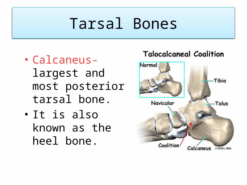
Tarsal Bones
• Calcaneus- largest and most posterior tarsal bone.
• It is also known as the heel bone.

• Calcaneal tuberosity- projection of posterior side of the calcaneus, where the achilles tendon attaches.
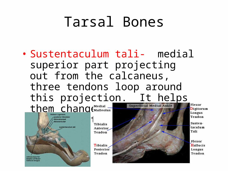
Tarsal Bones
• Sustentaculum tali- medial superior part projecting out from the calcaneus, three tendons loop around this projection. It helps them change direction from posterior to plantar foot.

Tarsal Bones
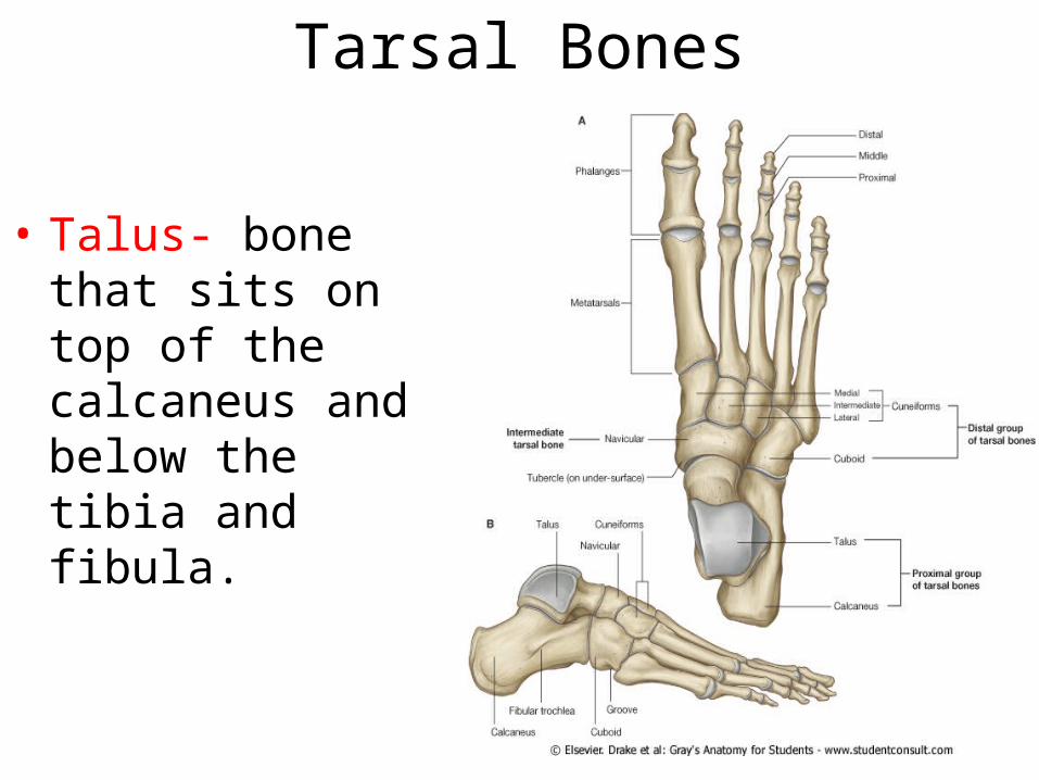
• Talus- bone that sits on top of the calcaneus and below the tibia and fibula.

Tarsal Bones
• Navicular- medial side of the talus and proximal to the cuniform bones.

Tarsal Bones
• Cuboid and Cuniforms- the most distal row of tarsal bones in the foot.
Foot bones
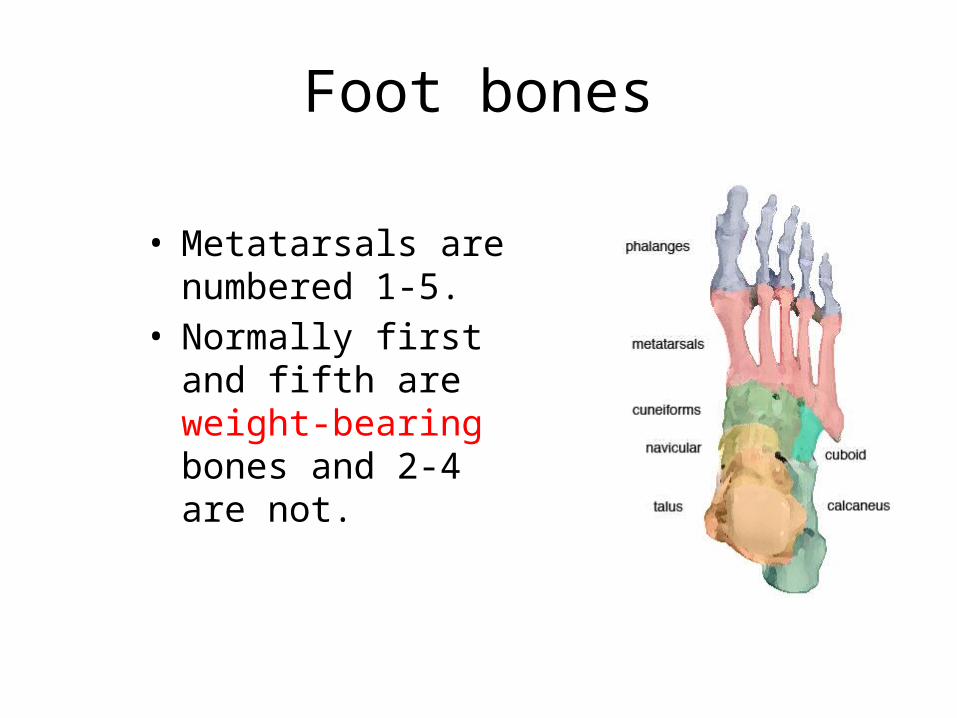
• Metatarsals are numbered 1-5.
• Normally first and fifth are weight-bearing bones and 2-4 are not.
Foot Bones
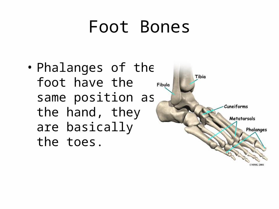
• Phalanges of the foot have the same position as the hand, they are basically the toes.

Joints of the Ankle/Foot
• Talocrural Joint-joint in the ankle found between the tibia, fibula, and talus. Dorsi/plantar flexion
• Subtalar Joint -joint in the ankle found between the talus and calcaneus.
• The subtalar joint allows gliding and rotation, which are involved in inversion and eversion of the foot.
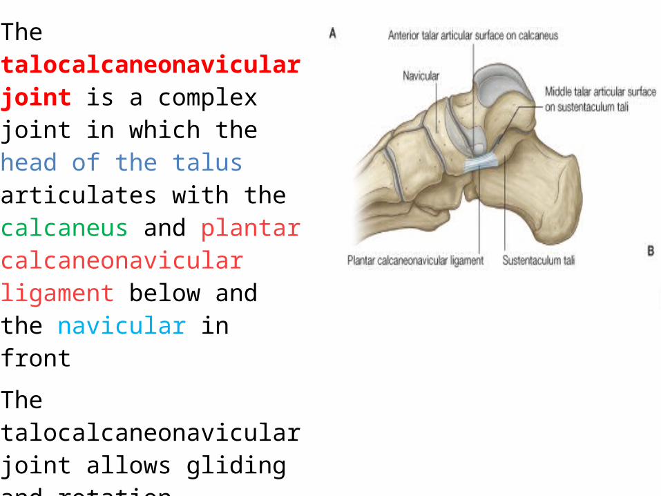
The talocalcaneonavicular joint is a complex joint in which the head of the talus articulates with the calcaneus and plantar calcaneonavicular ligament below and the navicular in front
The talocalcaneonavicular joint allows gliding and rotation movements, which together with similar movements of the subtalar joint are involved with inversion and eversion of the foot. It also participates in pronation and supination.

Joints of the foot and ankle
• Inferior tibiofibular joint is a syndesmosis joint.
• This is not a synovial joint, but one covered by a fibrous tissue that holds the joint together.
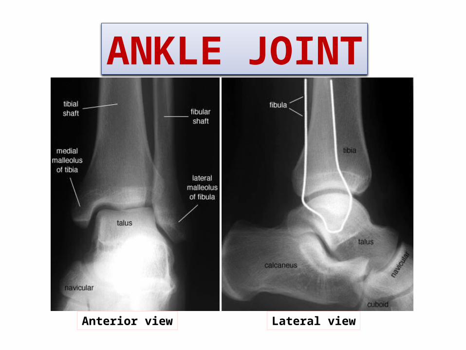
ANKLE JOINT
Anterior view Lateral view

Type & Articular Surfaces
ARTICULAR SURFACES:UPPER:A socket formed by: Lateral malleolus. the lower end of tibia & medial malleolus.
LOWER:Body of talus.
TYPE: synovial, hinge joint.
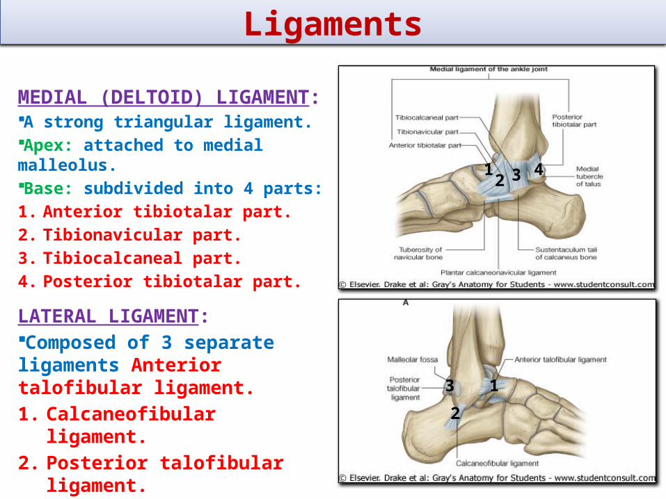
Ligaments
MEDIAL (DELTOID) LIGAMENT:A strong triangular ligament.Apex: attached to medial malleolus.Base: subdivided into 4 parts:1. Anterior tibiotalar part.2. Tibionavicular part.3. Tibiocalcaneal part.4. Posterior tibiotalar part.
LATERAL LIGAMENT:Composed of 3 separate ligaments Anterior talofibular ligament.1. Calcaneofibular ligament.2. Posterior talofibular
ligament.
1
2
3
12 3 4
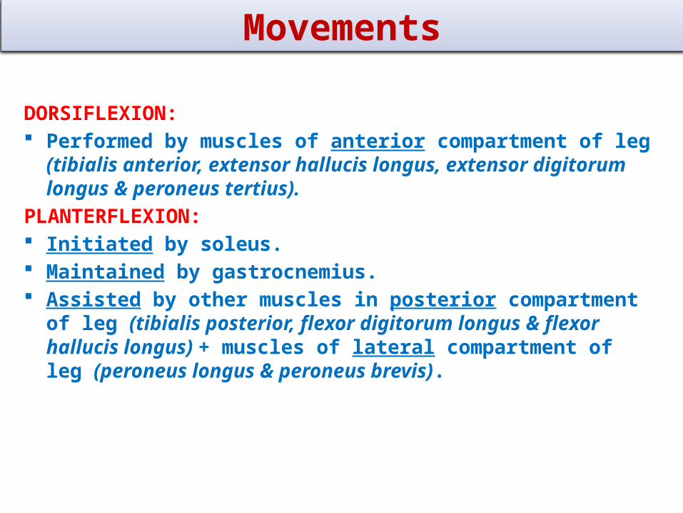
Movements
DORSIFLEXION: Performed by muscles of anterior compartment of leg (tibialis
anterior, extensor hallucis longus, extensor digitorum longus & peroneus tertius).
PLANTERFLEXION: Initiated by soleus. Maintained by gastrocnemius. Assisted by other muscles in posterior compartment of leg (tibialis
posterior, flexor digitorum longus & flexor hallucis longus) + muscles of lateral compartment of leg (peroneus longus & peroneus brevis).


![Subperiosteal bone proliferation at the tibia in ... · who have neurofibromatosis also have a congenital pseudarthrosis of the tibia [1]. is a subperiosteal haematoma which may lead](https://img.dokumen.tips/doc/110x75/604103a2383053274b34db04/subperiosteal-bone-proliferation-at-the-tibia-in-who-have-neurofibromatosis.jpg)


![Analysis of bone remodeling in the tibia after total knee ...€¦ · of tibia, adjacent to the implant, after TKA [9]. The prosthesis related bone loss is a concern about the success](https://img.dokumen.tips/doc/110x75/5f79a67bb377441fff433cf6/analysis-of-bone-remodeling-in-the-tibia-after-total-knee-of-tibia-adjacent.jpg)