Embed Size (px)
Citation preview
DDTEC-314; No of Pages 9
TECHNOLOGIES
DRUG DISCOVERY
TODAY
Animal models of Alzheimer’s diseaseand drug developmentBart Laurijssens1,*, Fabienne Aujard2, Anisur Rahman2
1BEL Pharm Consulting, Moulin d’Ozil, 07140 Chambonas, France2CNRS UMR 7179, MNHN, 1 Av du Petit Chateau, 91800 Brunoy, France
Drug Discovery Today: Technologies Vol. xxx, No. xx 2012
Editors-in-Chief
Kelvin Lam – Harvard University, USA
Henk Timmerman – Vrije Universiteit, The Netherlands
Translational pharmacology
Animal disease models are considered important in the
development of drugs for Alzheimer’s disease. This
brief review will discuss possible reasons why their
success in identifying efficacious treatments has been
limited, and will provide some thoughts on the role of
animal experimentation in drug development. Specifi-
cally, none of the current models of Alzheimer’s dis-
ease have either construct or predictive validity, and no
model probably ever will. Clearly, specific animal
experiments contribute to our understanding of the
disease and generate hypotheses. Ultimately, however,
the hypothesis can only be tested in human patients
and only with the proper tools. These tools are a
pharmacologically active intervention (in humans)
and a clinical trial suited to evaluate the mechanism
of action. Integration of knowledge in quantitative
(sub) models is considered important if not essential
in this process.
Introduction
Alzheimer’s disease (AD) is a neurodegenerative disease
affecting an estimated 5.4 million people globally, mainly
the elderly. There is currently no cure for AD, but some
symptomatic treatments are available. The disease is charac-
terized by its hallmark histopathological findings of extra-
cellular b-amyloid (Ab) plaques and intracellular
neurofibrillary tangles of tau and by neuronal and synaptic
Please cite this article in press as: Laurijssens, B. et al. Animal models of Alzheimer’s dis
10.1016/j.ddtec.2012.04.001
*Corresponding author.: B. Laurijssens ([email protected])
1740-6749/$ � 2012 Elsevier Ltd. All rights reserved. http://dx.doi.org/10.1016/j.ddtec.2012
Section editor:Oscar Della Pasqua – Leiden/Amsterdam Center for DrugResearch, Leiden, The Netherlands.
loss in brain regions involved in learning and memory pro-
cesses (http://www.alz.org/) [1].
The interest in finding a cure or prevention for AD is
understandably great. Proper animal models of human AD
are considered desirable if not essential in this process and
much research effort has been put into that effect. As no
perfect model exists, the question becomes whether ‘the best
models available’ are good enough. What exactly can be
inferred from the results and what not? Or, differently put,
how do they contribute to our understanding and decision-
making.
The objective, therefore, of this brief review is to discuss the
potential role of the current animal disease models for AD in
drug development. Specifically, the aim is to discuss why the
animal models of AD should have such a limited success in
predicting successful treatments in the clinic (or rather,
clinical trials), and to provide some thoughts on how to
use animal experiments in drug development.
Animal models of Alzheimer’s disease
The aetiology of AD is unknown, but there is still a general
consensus in favour of the ‘amyloid hypothesis’ [1,2], even if
it has been questioned [3]. A wide range of animal models
have been developed to mimic the human context of the
disease for the purpose of developing therapeutics or disease
modifying agents. In fact, in most of the animal models the
first goal is to simulate the neuropathological findings of AD
ease and drug development, Drug Discov Today: Technol (2012), http://dx.doi.org/
.04.001 e1
Drug Discovery Today: Technologies | Translational pharmacology Vol. xxx, No. xx 2012
DDTEC-314; No of Pages 9
followed by the correlation of cognitive function without
knowing whether the neuropathological agents have similar
biological consequences in humans and in animal models. It
is beyond the scope of this review to discuss all the different
models. Here we will briefly summarize some potential ani-
mal models and their translation towards the clinical settings
(Table 1). Animal models used in AD can be broadly divided
into three categories: Natural models, Genetic models and
Interventional models.
Natural models
Several animals including polar bears, dogs, cats, goats and
sheep and some non-human primates spontaneously develop
some AD-related neuropathological features [4]. The few
Please cite this article in press as: Laurijssens, B. et al. Animal models of Alzheimer’s dis
10.1016/j.ddtec.2012.04.001
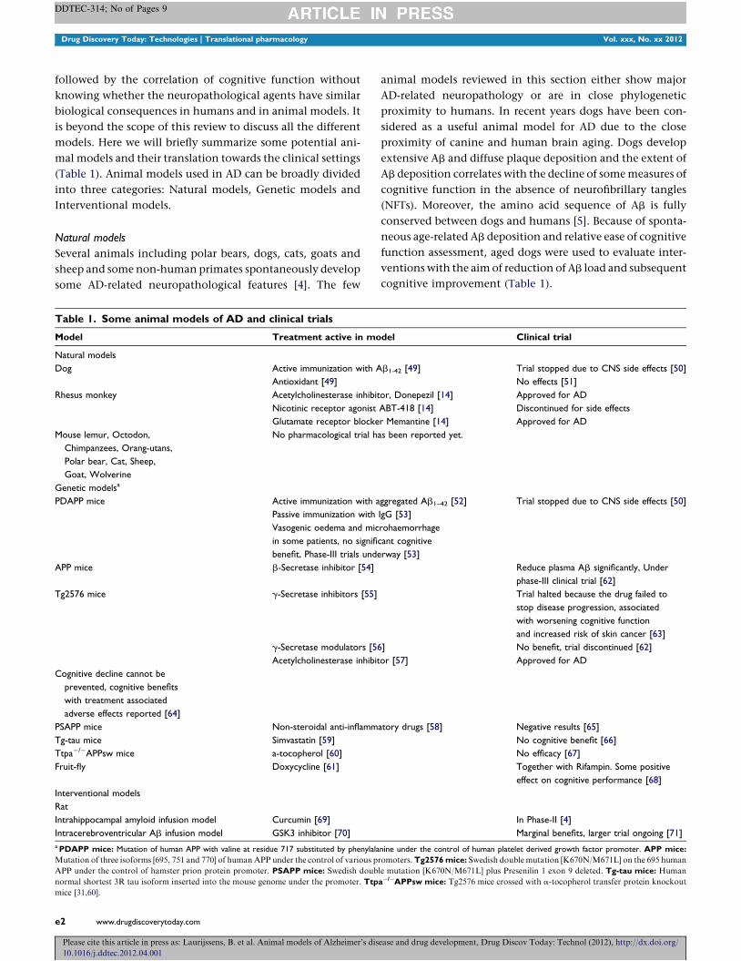
Table 1. Some animal models of AD and clinical trials
Model Treatment active in mo
Natural models
Dog Active immunization with A
Antioxidant [49]
Rhesus monkey Acetylcholinesterase inhibit
Nicotinic receptor agonist
Glutamate receptor blocke
Mouse lemur, Octodon,
Chimpanzees, Orang-utans,
Polar bear, Cat, Sheep,
Goat, Wolverine
No pharmacological trial ha
Genetic modelsa
PDAPP mice Active immunization with a
Passive immunization with
Vasogenic oedema and mic
in some patients, no signific
benefit, Phase-III trials unde
APP mice b-Secretase inhibitor [54]
Tg2576 mice g-Secretase inhibitors [55]
g-Secretase modulators [56
Acetylcholinesterase inhibit
Cognitive decline cannot be
prevented, cognitive benefits
with treatment associated
adverse effects reported [64]
PSAPP mice Non-steroidal anti-inflamm
Tg-tau mice Simvastatin [59]
Ttpa�/�APPsw mice a-tocopherol [60]
Fruit-fly Doxycycline [61]
Interventional models
Rat
Intrahippocampal amyloid infusion model Curcumin [69]
Intracerebroventricular Ab infusion model GSK3 inhibitor [70]
a PDAPP mice: Mutation of human APP with valine at residue 717 substituted by phenylala
Mutation of three isoforms [695, 751 and 770] of human APP under the control of various p
APP under the control of hamster prion protein promoter. PSAPP mice: Swedish doub
normal shortest 3R tau isoform inserted into the mouse genome under the promoter. Ttp
mice [31,60].
e2 www.drugdiscoverytoday.com
animal models reviewed in this section either show major
AD-related neuropathology or are in close phylogenetic
proximity to humans. In recent years dogs have been con-
sidered as a useful animal model for AD due to the close
proximity of canine and human brain aging. Dogs develop
extensive Ab and diffuse plaque deposition and the extent of
Ab deposition correlates with the decline of some measures of
cognitive function in the absence of neurofibrillary tangles
(NFTs). Moreover, the amino acid sequence of Ab is fully
conserved between dogs and humans [5]. Because of sponta-
neous age-related Ab deposition and relative ease of cognitive
function assessment, aged dogs were used to evaluate inter-
ventions with the aim of reduction of Ab load and subsequent
cognitive improvement (Table 1).
ease and drug development, Drug Discov Today: Technol (2012), http://dx.doi.org/
del Clinical trial
b1-42 [49] Trial stopped due to CNS side effects [50]
No effects [51]
or, Donepezil [14] Approved for AD
ABT-418 [14] Discontinued for side effects
r Memantine [14] Approved for AD
s been reported yet.
ggregated Ab1–42 [52] Trial stopped due to CNS side effects [50]
IgG [53]
rohaemorrhage
ant cognitive
rway [53]
Reduce plasma Ab significantly, Under
phase-III clinical trial [62]
Trial halted because the drug failed to
stop disease progression, associated
with worsening cognitive function
and increased risk of skin cancer [63]
] No benefit, trial discontinued [62]
or [57] Approved for AD
atory drugs [58] Negative results [65]
No cognitive benefit [66]
No efficacy [67]
Together with Rifampin. Some positive
effect on cognitive performance [68]
In Phase-II [4]
Marginal benefits, larger trial ongoing [71]
nine under the control of human platelet derived growth factor promoter. APP mice:
romoters. Tg2576 mice: Swedish double mutation [K670N/M671L] on the 695 human
le mutation [K670N/M671L] plus Presenilin 1 exon 9 deleted. Tg-tau mice: Human
a�/�APPsw mice: Tg2576 mice crossed with a-tocopherol transfer protein knockout
Vol. xxx, No. xx 2012 Drug Discovery Today: Technologies | Translational pharmacology
DDTEC-314; No of Pages 9
Among the non-human primate models, mouse lemurs
seem to be a potential animal model that exhibit amyloid
plaque, NFTs and some other AD related neuropathology [6].
This small prosimian primate lives 8–10 years in captivity and
shows age-related changes similar to those of aging humans
[6]. About 20% of mouse lemurs aged five years or older show
significant brain atrophy [7], extensive accumulation of amy-
loid plaques, neurodegeneration [8] and/or loss of cholinergic
neurons [9]. The genes responsible for the formation of senile
plaques are highly similar between mouse lemurs and
humans [8]. The presence of amyloid deposition has been
recently linked to cerebral atrophy in aged individuals, giving
new insight into the understanding of pathological aging in
this non-human primate species [10]. Moreover, a compar-
able decline in declarative memory and executive function
has been reported in both aged human and aged mouse lemur
whereas procedural memory appears to be conserved in both
species [11]. In a recent study it has been shown that cogni-
tively impaired aged mouse lemurs have cerebral atrophy
especially those brain regions that are responsible for cogni-
tive functions [12]. It should be noted that there is a differ-
ence in distribution of Ab deposits and plaques between
human and mouse lemur. In humans the Ab depositions
usually start in the hippocampus but in mouse lemur they
appear first in cortical regions [13]. As with human AD,
currently no diagnostic tools are available that can predict
which adult mouse lemur will develop AD-like symptoms.
However, this model provides an opportunity to search for
these predictors.
Rhesus monkeys and humans share many diseases of aging
and probably are the most successful models to identify
diagnostic markers and the development of safe and effective
treatments for human brain disorders. Monkeys of over 19
years old showed significant amyloid-plaque-like lesions in
areas of brain responsible for cognitive function. However,
most of the aged monkeys do not show an Alzheimer’s-like
syndrome and age-related rapid cognitive decline commonly
found in AD patients is not usually found in them either [14].
Moreover, their long developmental period, low reproductive
output, long captive life and risk of serious zoonotic disease
transmission are major disadvantages for using these animals
in a wider range of AD research [15].
Octodon degu, a rodent of South American origin has
recently been found to have spontaneous development of
AD-related neuropathology at older age. Localisation of both
intracellular and extracellular amyloid deposits and NFTs-like
intracellular deposition has been detected in different layers
of cortex and hippocampus of old animals. Adult octodon
cortex shows the presence of cholinergic neurons as in
humans but with a different distribution. They also demon-
strate age-associated cognitive impairment but whether these
cognitive deficits are also associated with cholinergic neuro-
degeneration similar to AD patients is not known yet.
Please cite this article in press as: Laurijssens, B. et al. Animal models of Alzheimer’s dis
10.1016/j.ddtec.2012.04.001
Another interesting finding is the presence of extensive astro-
gliosis in the aged octodon brain, which is a characteristic
feature of human AD brain. It has been hypothesized that
high homology (97.5%) of octodon Ab and human Ab might
be an important factor in the appearance of AD markers in
this rodent. Breeding difficulty and comparatively longer life-
span are limitations of this rodent model [16,17].
Genetic models
Transgenic technology provides unique opportunity to repro-
duce the cause of familial AD by transfecting a mutant human
amyloid precursor protein (APP). Mice have extensively been
used as transgenic models and facilitated our understanding of
the molecular mechanisms associated with Ab-production,
deposition and clearance and the effects of Ab on neuronal
network and synapses that play important role in cognitive
function. The APP mouse model successfully produced a wide
range of parenchymal and vascular amyloid deposits similar to
those of human AD [18]. Although morphological similarity
does exist, there is a difference in biochemical composition of
deposited Ab between mouse models and AD brain [19]. More-
over, these transgenic mouse models failed to develop neuro-
fibrillary tangles (NFTs), an important histopathological
hallmark of AD result from intraneuronal aggregation of
hyperphosphorylated tau protein [20]. Oddo and colleagues
[21] presented for the first time a triple transgenic mouse model
where both plaques and NFTs were found in AD-relevant brain
regions. These mice also developed extracellular amyloid beta
deposits before the formation of NFTs and exhibited impaired
synaptic plasticity including long-term potentiation, the key
basis for cognitive function. Furthermore, transgenic mouse
models also provided valuable information regarding the role
of inflammation, oxidative stress and mitochondrial dysfunc-
tion in the pathogenesis of AD [22,23]. However, progressive
neuronal loss in hippocampus and specific neocortical regions
of the human AD brain [24] is not evident in most of the
transgenic mouse models, and this is a major limitation of
these murine models. In addition, these transgenic mice repre-
sent only those who are suffering from familial AD, which is
<1% of all AD patients. There is no mouse model that can fully
reproduce the features of disease progression of vast majority of
AD cases that is sporadic/late-onset AD.
The fruit-fly Drosophila melanogaster is a widely used and
well-appreciated animal model of neurodegeneration includ-
ing AD [25]. The fruit-fly is small in size, has a simple well-
studied anatomy and possesses a well-organized brain.
Although the fruit-fly brain has only a fraction of the cells
of the human brain and a different neuroanatomical organi-
zation, it is similar in the fundamental aspects of cell biology,
in terms of regulation of gene expression, membrane traffick-
ing, neuronal connectivity, cell signalling, synaptogenesis
and cell death [26,27]. Moreover, its short life cycle and
completely sequenced genomes are added experimental
ease and drug development, Drug Discov Today: Technol (2012), http://dx.doi.org/
www.drugdiscoverytoday.com e3

Drug Discovery Today: Technologies | Translational pharmacology Vol. xxx, No. xx 2012
DDTEC-314; No of Pages 9
Box 1. Validation criteria for animal models [36]
Face validity: The animal model resembles the human disease condition
on a superficial level, for example, biochemistry or symptomatology.
Predictive validity: The animal model can successfully discriminate
between successful and unsuccessful treatments for the human disease
condition.
Construct validity: The animal model is based on a sound theoretical
rationale, requiring good understanding of the human disease condition.
Notably, construct validity does not require superficial similarity.
advantages of this model. Importantly, the transparent cuti-
cle of the larvae of fruit-fly allows the study of the disease
progression in living intact animals which is rather difficult
in vertebrates [28]. The task for associative learning and
memory such as Pavlovian olfactory conditioning can be
measured in this model, which is homologous to the classical
conditioning of the eyeblink response found to be impaired
in patients diagnosed with AD [26]. The fact that hippocam-
pal-dependant cognitive functions, which are impaired early
in human AD, cannot be tested in invertebrates due to the
lack of these brain structures is a major limitation of this
animal model. In spite of the lack in homology between fruit-
fly and human brain, this simple invertebrate animal pro-
vided insight in the disease mechanism ranging from genetics
to some cognitive functions that could be followed in living
intact animals.
Interventional models
Introduction of pharmacological or chemical substances into
the brain or the induction of lesions in specific brain regions
may replicate some of the characteristic features of AD. Many
models involve the introduction of Ab peptide into the brain
of, for example, the rat [29] or rhesus monkey [30]. Although
these models induce some of the clinical signs, they do not
directly resemble AD pathology [31]. Other chemical inter-
ventional models include scopolamine-induced amnesia,
introduction of inflammation with endotoxins or interfer-
ence with brain metabolism [4].
The lesion models involve the chemical or physical
destruction of specific brain areas, which are generally either
cholinergic (i.e. the nucleus basalis magnocellularis in
rodents, e.g. [32]) or involved in cognition (i.e. hippocampus,
striatal and cortical brain regions). Major disadvantages of the
lesion models include non-specificity of the lesion, and their
failure to capture the disease progression and the more global
aspects of the disease (too specific) [4].
As a model of disease, interventional models would gen-
erally be better at identifying symptomatic or corrective
treatments, rather than disease modifying therapies that halt
or slow down progression, unless the ‘intervention’ repre-
sents a damage early in the disease progression.
Furthermore, a wide range of variability including species,
animal husbandry, site of injection or induction, model
protocol and concentration and volume of substances may
influence the experimental outcome. However, these models
can provide important insights such as scopolamine-induced
amnesia model contributed to the role of cholinergic system
in cognition [33], specific brain lesions induced memory
deficit models and the neuronal mechanism underlying
memory dysfunction [34] and the Ab/pharmaco-chemical
substance induced model and the understanding of inflam-
mation, neurotoxicity, neurodegeneration and synaptic
function [35].
Please cite this article in press as: Laurijssens, B. et al. Animal models of Alzheimer’s dis
10.1016/j.ddtec.2012.04.001
e4 www.drugdiscoverytoday.com
Animal models of disease
It is obvious that many of the animal models of AD have
contributed, and continue to contribute, to our understand-
ing of the processes that may or may not underlie human AD.
But can they really be called animal models of AD, and can
they therefore be used as such?
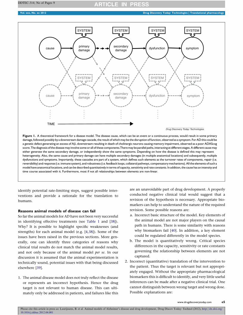
The perfect animal model of a disease would be a scaled
down replica of the human disease, representing all impor-
tant components from cause via structural damage to symp-
toms (Fig. 1). It would be structurally and quantitatively
identical to the human. For this, both the animal model
and the human disease need to be very well understood.
Such a model would have face-, predictive- and construct
validity [36] (see Box 1). None of the models above satisfy this
criterion and no animal model probably ever will, especially
for a disease such as AD, given its complexity [36,37]. It could
be argued, therefore, that such a model should not even be an
objective.
Even predictive validity, although very useful, is not trivial
to establish. It requires effective drugs in the clinic, ideally
with different mechanisms, and a good understanding of the
false positives and false negatives. The latter is particularly
hard to evaluate because compounds that do not work in the
animal model are often not tested in the clinic (or reported).
In a way, one could say that the availability of an animal
model with established good predictive validity is unfortu-
nately inversely correlated with the unmet medical need.
All the disease models for AD listed have at best face
validity, where the animal model merely shares phenomen-
ological similarities with the disorder under study. Hence the
problem of ‘translation’ and recent interest in ‘translational
sciences’.
The schematic in Fig. 1 illustrates the complexity and the
many possible pitfalls in trying to create an animal analogue
for human disease. A single relevant difference, whether
structural or quantitative, can ‘invalidate’ the model. This
is, when it is not understood and considered in the transla-
tion to the human condition.
By contrast, many of the single components of the sche-
matic in Fig. 1 will be preserved across species (normal
physiology). Thus, whereas it is probably impossible to repli-
cate the full sequence from cause to symptoms in animals, the
study of components of the hypothesized sequence could
ease and drug development, Drug Discov Today: Technol (2012), http://dx.doi.org/
Vol. xxx, No. xx 2012 Drug Discovery Today: Technologies | Translational pharmacology
DDTEC-314; No of Pages 9
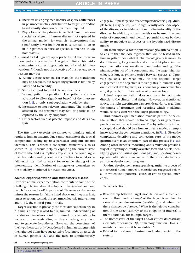
primarydamage
symptomsecondarydamage
cause dysfunction
causeprimarydamage
secondarydamage
dysfunction symptom
SYSTEM SYSTEMSYSTEM SYSTEM
SYSTEM SYSTEM SYSTEM SYSTEM
TIME
Drug Discovery Today: Technologies
Figure 1. A theoretical framework for a disease model. The disease cause, which can be an event or a continuous process, would result in some primary
damage, followed possibly by a downstream damage cascade, the result of which may be the disruption of function, observed as a symptom. For AD this could be
a genetic defect generating an excess of Ab, downstream resulting in death of cholinergic neurons causing memory impairment, observed as a poor ADAScog
score. The diagnosis of the disease may involve some or all of these components.There may be parallel paths, interacting at different stages. A different cause may
either generate the same secondary damage, or independently show the same symptoms. Depending on how the disease is defined this may represent
heterogeneity. Also, the same cause and primary damage can have multiple secondary damages (in multiple anatomical locations) and subsequently, multiple
dysfunctions and symptoms. Importantly, these cascades are part of a system, which defines such elements as the turnover rates of components, repair (i.e.
reversibility) and response (i.e. immune system), and robustness (i.e. feedback loops, collateral pathways, compensatory mechanisms). All the elements of such a
model have anatomical locations, and can be described quantitatively in terms of capacity, sensitivity and rate constants. In addition, the cause has an intensity and
time course associated with it. Furthermore, most if not all relationships between elements are non-linear.
identify potential rate-limiting steps, suggest possible inter-
ventions and provide a rationale for the translation to
humans.
Reasons animal models of disease can fail
So far the animal models for AD have not been very successful
in identifying effective treatments (see Table 1 and [38]).
Why? It is possible to highlight specific weaknesses (and
strengths) for each animal model (e.g. [4,38]). Some of the
issues have been raised in the previous sections. More gen-
erally, one can identify three categories of reasons why
clinical trial results do not match the animal model results,
and not only because of the animal model per se. In our
discussion it is assumed that the animal experimentation is
technically sound, potential issues with that being discussed
elsewhere [39].
1. The animal disease model does not truly reflect the disease
or represents an incorrect hypothesis. Hence the drug
target is not relevant to human disease. This can ulti-
mately only be addressed in patients, and failures like this
Please cite this article in press as: Laurijssens, B. et al. Animal models of Alzheimer’s dis
10.1016/j.ddtec.2012.04.001
are an unavoidable part of drug development. A properly
conducted negative clinical trial would suggest that a
revision of the hypothesis is necessary. Appropriate bio-
markers can help to understand the nature of the required
revision. Some possible reasons are:
a. Incorrect basic structure of the model. Key elements of
the animal model are not major players on the causal
path in humans. There is some similarity with reasons
why biomarkers fail [40]. In addition, a key element
could be regulated differently in the model species.
b. The model is quantitatively wrong. Critical species
differences in the capacity, sensitivity or rate constants
governing the relationship between elements are not
captured.
2. Incorrect (quantitative) translation of the intervention to
the patient. Thus the target is relevant but not appropri-
ately engaged. Without the appropriate pharmacological
biomarkers this is difficult to identify, and very little useful
inferences can be made after a negative clinical trial. One
cannot distinguish between wrong target and wrong dose.
Possible explanations are:
ease and drug development, Drug Discov Today: Technol (2012), http://dx.doi.org/
www.drugdiscoverytoday.com e5
Drug Discovery Today: Technologies | Translational pharmacology Vol. xxx, No. xx 2012
DDTEC-314; No of Pages 9
a. Incorrect dosing regimen because of species differences
in pharmacokinetics, distribution to target site and/or
target affinity, duration of treatment required.
b. Physiology of the primary target is different between
species, or altered in human disease (not captured in
the animal model), for example, interventions that
significantly lower brain Ab in mice can fail to do so
in AD patients because of species differences in Ab
homeostasis.
3. Clinical trial design not appropriate to test the interven-
tion under investigation. A negative clinical trial risks
abandoning a correct hypothesis and a beneficial inter-
vention. Although not the objective of this review, some
reasons may be:
a. Wrong dosing regimen. For example, the translation
may be adequate, but target engagement is limited by
safety and tolerability.
b. Study too short to be able to notice effects
c. Wrong patient population. The patients are too
advanced to allow significant benefit of the interven-
tion [41], or only a subpopulation would benefit.
d. Insensitive or not relevant endpoints. The modality
affected by the treatment may not, or poorly so, be
captured by the study endpoints.
e. Other factors such as placebo response and data ana-
lysis.
The first two categories are failures to translate animal
results to human patients. One cannot translate if the crucial
components leading up to a particular endpoint are not
identified. This is where a conceptual framework such as
shown in Fig. 1 would help by capturing the current state
of knowledge and assumptions explicitly. One could argue
that this understanding could also contribute to avoid some
failures of the third category, for example, timing of the
intervention, identification of surrogate or biomarkers or
the modality monitored for treatment effect.
Animal experimentation and Alzheimer’s disease
How can animal experimentation contribute to some of the
challenges facing drug development in general and our
search for a cure for AD in particular? Three major challenges
mirror the reasons for failure listed above and they are: first,
target selection, second, the (pharmacological) intervention
and third, the clinical patient trials.
Target selection is probably the most difficult challenge in
AD and is directly related to our, limited, understanding of
the disease. An obvious role of animal experiments is to
increase this understanding, as they already greatly have,
and to generate hypotheses. However, the correctness of
the hypothesis can only be addressed in human patients with
the right tool. Some have suggested to focus more on research
in human patients [37] and the need to simultaneously
Please cite this article in press as: Laurijssens, B. et al. Animal models of Alzheimer’s dis
10.1016/j.ddtec.2012.04.001
e6 www.drugdiscoverytoday.com
engage multiple targets to treat complex disorders [38]. Multi-
ple targets may be required to significantly affect one aspect
of the disease, or to address the multidimensionality of the
disorder. In addition, animal models can be used to screen
series of compounds, and identify potential targets by their
ability to modulate an aspect of the hypothesized disease
model.
The main objective for the pharmacological intervention is
to ensure that the dose regimen that will be tested in the
human patient does what it pharmacologically is meant to
do: sufficiently, long enough and at the right place. Animal
experimentation can contribute to our understanding of the
pharmacokinetics of the compound and the target pharma-
cology, as long as properly scaled between species, and pro-
vide guidance on what may be the required target
engagement. One objective is to verify this in humans early
on in clinical development, as is done for pharmacokinetics
and, if possible, with biomarkers of pharmacology.
Animal experimentation does not seem to contribute
directly to the clinical trial design. However, as mentioned
above, the right experiments can provide guidance regarding
the timing of treatment and regarding which modalities
would be consistent with the target pharmacology.
Thus, animal experimentation remains part of the scien-
tific method that iterates between hypothesis generation,
predictions and experimentation. The hypothesis is in fact
conceptual and should be a human disease model, attempt-
ing to address the components mentioned in Fig. 1. Given the
complexity, describing and connecting parts of the model
quantitatively is an important part of this process [41–45].
Among other benefits, modelling and simulation provide a
way of integrating currently available facts and beliefs, iden-
tifying gaps and raising questions [45] and, for drug devel-
opment, ultimately some sense of the uncertainties of a
particular development program.
For drug development some specific quantitative aspects of
a theoretical human model to consider are suggested below,
all of which are a potential source of critical species differ-
ences.
Target selection:
� Relationship between target modulation and subsequent
events. How much ‘change’ of the target is required to
cause changes downstream (sensitivity) and when can
these changes be observed? What is the relative contribu-
tion of the target pathway to the endpoint of interest? Is
there a rationale for multiple targets?
� The homeostasis of the target and/or critical downstream
elements, for example, Ab, or memory function. How is it
maintained and can it be modulated?
� Related to the above, robustness and redundancies in the
system.
ease and drug development, Drug Discov Today: Technol (2012), http://dx.doi.org/
Vol. xxx, No. xx 2012 Drug Discovery Today: Technologies | Translational pharmacology
DDTEC-314; No of Pages 9
Evaluation of the intervention:
� Pharmacokinetics
� Can the intervention modulate the component(s) of inter-
est (target engagement), and if so, what is the required
timing of the intervention? And, again, how long does it
take before these changes can be observed, which also
depends on the endpoint? In other words, pharmacoki-
netic–pharmacodynamic relationships for the target phar-
macology.
� Are there bio- or surrogate markers that can inform on the
target pharmacology, and, if so, what is the relationship
between target function and biomarker level? Again, con-
sidering relative contributions and time courses. A recent
Please cite this article in press as: Laurijssens, B. et al. Animal models of Alzheimer’s dis
10.1016/j.ddtec.2012.04.001
0
0.0
0.2
0.4
0.6
0.8
1.0
2 4 6 8 10
Time (weeks)Blood Brain
Plasmaantibody
D Ke
Koff · Cx
PCo
Kon· D · P
K01 + Kon · P
Effect of QPB Variability
Fra
ctio
n of
Pla
sma
or B
rain
Bas
al A
β42
BrainPlasma
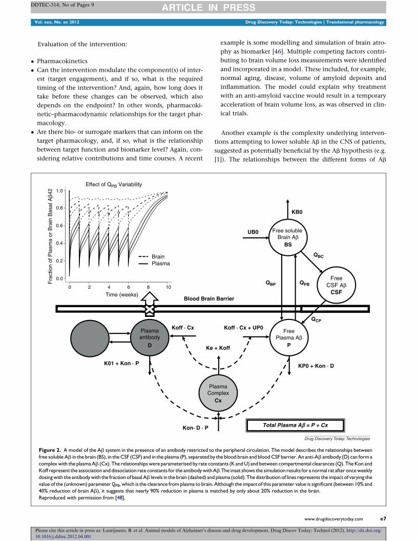
Figure 2. A model of the Ab system in the presence of an antibody restricted to
free soluble Ab in the brain (BS), in the CSF (CSF) and in the plasma (P), separated b
complex with the plasma Ab (Cx). The relationships were parameterised by rate co
Koff represent the association and dissociation rate constants for the antibody with
dosing with the antibody with the fraction of basal Ab levels in the brain (dashed) and
value of the (unknown) parameter QPB, which is the clearance from plasma to brain.
40% reduction of brain Ab), it suggests that nearly 90% reduction in plasma is m
Reproduced with permission from [48].
example is some modelling and simulation of brain atro-
phy as biomarker [46]. Multiple competing factors contri-
buting to brain volume loss measurements were identified
and incorporated in a model. These included, for example,
normal aging, disease, volume of amyloid deposits and
inflammation. The model could explain why treatment
with an anti-amyloid vaccine would result in a temporary
acceleration of brain volume loss, as was observed in clin-
ical trials.
Another example is the complexity underlying interven-
tions attempting to lower soluble Ab in the CNS of patients,
suggested as potentially beneficial by the Ab hypothesis (e.g.
[1]). The relationships between the different forms of Ab
ease and drug development, Drug Discov Today: Technol (2012), http://dx.doi.org/
FreeCSF Aβ
CSF
QCP
QBP QPB
QBC
Free soluble Brain A β
BS
KB0
UB0
FreePlasma Aβ
P
Barrier
+ Koff
lasmamplexCx
Koff · Cx + UP0
Total Plasma Aβ = P + Cx
KP0 + Kon · D
Drug Discovery Today: Technologies
the peripheral circulation. The model describes the relationships between
y the blood brain and blood CSF barrier. An anti-Ab antibody (D) can form a
nstants (K and U) and between compartmental clearances (Q). The Kon and
Ab.The inset shows the simulation results for a normal rat after once weekly
plasma (solid). The distribution of lines represents the impact of varying the
Although the impact of this parameter value is significant (between 10% and
atched by only about 20% reduction in the brain.
www.drugdiscoverytoday.com e7
Drug Discovery Today: Technologies | Translational pharmacology Vol. xxx, No. xx 2012
DDTEC-314; No of Pages 9
across the different compartments of interest, for example,
brain, CSF and plasma, are not immediately obvious. This
issue was highlighted for the rational selection of biomarkers
by Thompson and Lockhart [47], for example Ab concentra-
tions in the CSF as marker for CNS concentrations, but is
equally important for the rationalization of interventions.
Preliminary work attempting to address this was presented by
Simeoni et al. [48], and an example of the model, with an
intervention included, is shown in Fig. 2. Interestingly, the
preliminary simulations with the model suggest that an anti-
body against Ab, restricted to the systemic circulation, would
only modestly lower CNS levels of Ab, even if plasma levels
were greatly reduced (Fig. 2). This is clearly a red flag when in
the process of developing such an antibody, and merits
further investigation of the system and assumptions. The
model made specific assumptions about the structure of
the model and some of the parameters and ignored some
non-linearities that must be present in the system. Never-
theless, this work could guide specific future experiments and
the impact of parameter assumptions can be explored by
sensitivity analysis (as was done for this presentation).
Thus, even if animal models of AD cannot be considered
models of disease with construct (or even predictive) validity,
it is not to so that animal experiments have not been or will
not be useful in understanding of the disease or that the
hypotheses they aim to reflect are incorrect. The challenge is
to identify which part of the overall human disease model a
particular animal experiment addresses.
Lastly, although not specifically addressed here, anticipat-
ing safety issues of the intervention under study is obviously
important. In particular, adverse pharmacology associated
with the primary and/or secondary targets of the compound
can, should, be addressed applying similar principles as for
efficacy.
Conclusions
The value of many of the so-called animal models for AD lies
not in the fact that they attempt to replicate the full spectrum
of the human disease, but in that they could help us, together
with clinical data, to generate hypotheses and understand the
complex physiology relevant to the human disease. In that
sense, they are not really animal models of disease but ‘just’
animal experiments. These experiments, then, need to
address specific questions that allow the construction of
the hypotheses for the whole or parts of a quantitative human
disease model. The conceptual human model is where inter-
ventions should be evaluated: for example, target selection,
dosing regimen and timing, biomarker and clinical endpoint
selection and safety. Integration of information and ideas
into a quantitative conceptual (sub) model of human disease
and pharmacology is therefore considered essential.
For drug development, whereas there is likely to remain
great uncertainty around target selection and the associated
Please cite this article in press as: Laurijssens, B. et al. Animal models of Alzheimer’s dis
10.1016/j.ddtec.2012.04.001
e8 www.drugdiscoverytoday.com
hypotheses, it is crucial to identify a pharmacologically active
dosing regimen to test in patients. Then a negative clinical
patient study, although disappointing, will at least increase
our knowledge regarding the disease.
Acknowledgements
FA and AR are funded as part of the Pharma-Cog consortium
by the European Community’s Seventh Framework Pro-
gramme for the Innovative Medicine Initiative under Grant
Agreement no. 115009. For further information please refer
to http://www.pharmacog.org/.
References1 Mattson, M.P. (2004) Pathways towards and away from Alzheimer’s
disease. Nature 430, 631–639
2 Hardy, J.A. and Higgins, G.A. (1992) Alzheimer’s disease: the amyloid
cascade hypothesis. Science 256, 184–185
3 Schnabel, J. (2011) Little proteins, big clues. Nature 475, S12–S14
4 Van Dam, D. and De Deyn, P.P. (2011) Animal models in the drug
discovery pipeline for Alzheimer’s disease. Br. J. Pharmacol. 164,
1285–1300
5 Johnstone, E. et al. (1991) Conservation of the sequence of the Alzheimer’s
disease amyloid peptide in dog, polar bear and five other mammals by
cross-species polymerase chain reaction analysis. Mol. Brain Res. 10,
299–305
6 Languille, S. et al. (2012) The grey mouse lemur: a non human primate
model for ageing studies. Ageing Res. Rev. 11, 150–162
7 Dhenain, M. et al. (2003) Regional atrophy in the brain of lissencephalic
mouse lemur primates: measurement by automatic histogram-based
segmentation of MR images. Magn. Reson. Med. 50, 984–992
8 Bons, N. et al. (2006) Microcebus murinus: a useful primate model for
human cerebral aging and Alzheimer’s disease? Genes Brain Behav. 5,
120–130
9 Mestre, N. and Bons, N. (1993) Age-related cytological changes and
neuronal loss in basal forebrain cholinergic neurons in Microcebus murinus
(Lemurian, Primate). Neurodegeneration 2, 25–32
10 Kraska, A. et al. (2011) Age-associated cerebral atrophy in mouse lemur
primates. Neurobiol. Aging 32, 894–906
11 Picq, J.L. (2007) Aging affects executive functions and memory in mouse
lemur primates. Exp. Gerontol. 42, 223–232
12 Picq, J-L. et al. (2012) Age-related cerebral atrophy in nonhuman primates
predicts cognitive impairments. Neurobiol. Aging 33, 1096–1109
13 Giannakopoulos, P. et al. (1997) Quantitative analysis of tau protein-
immunoreactive accumulations and b amyloid protein deposits in the
cerebral cortex of the mouse lemur, Microcebus murinus. Acta Neuropathol.
94, 131–139
14 Buccafusco, J.J. (2008) Estimation of working memory in macaques for
studying drugs for the treatment of cognitive disorders. J. Alzheimer’s Dis.
15, 709–720
15 De Magalhaes, J. and Costa, J. (2009) A database of vertebrate longevity
records and their relation to other life-history traits. J. Evol. Biol. 22,
1770–1774
16 Braidy, N. et al. (2012) Recent rodent models for Alzheimer’s disease:
clinical implications and basic research. J. Neural Transm. 119, 173–195
17 Inestrosa, N.C. et al. (2005) Human-like rodent amyloid-b-peptide
determines Alzheimer pathology in aged wild-type Octodon degu.
Neurobiol. Aging 26, 1023–1028
18 Games, D. et al. (1995) Alzheimer-type neuropathology in transgenic mice
overexpressing V717F b-amyloid precursor protein. Nature 373, 523–527
19 Kalback, W. et al. (2002) APP transgenic mice Tg2576 accumulate Ab
peptides that are distinct from the chemically modified and insoluble
peptides deposited in Alzheimer’s disease senile plaques. Biochemistry 41,
922–928
ease and drug development, Drug Discov Today: Technol (2012), http://dx.doi.org/
Vol. xxx, No. xx 2012 Drug Discovery Today: Technologies | Translational pharmacology
DDTEC-314; No of Pages 9
20 Goedert, M. et al. (2006) Tau protein, the paired helical filament and
Alzheimer’s disease. J. Alzheimer’s Dis. 9, 195–207
21 Oddo, S. et al. (2003) Triple-transgenic model of Alzheimer’s disease with
plaques and tangles: intracellular A [beta] and synaptic dysfunction.
Neuron 39, 409–421
22 Du, H. et al. (2008) Cyclophilin D deficiency attenuates mitochondrial and
neuronal perturbation and ameliorates learning and memory in
Alzheimer’s disease. Nat. Med. 14, 1097–1105
23 Lee, Y.J. et al. (2010) Inflammation and Alzheimers disease. Arch. Pharm.
Res. 33, 1539–1556
24 Gomez-Isla, T. et al. (1996) Profound loss of layer II entorhinal cortex
neurons distinguishes very mild Alzheimers disease from nondemented
aging. J. Neurosci. 16, 4450–4491
25 Driscoll, M. and Gerstbrein, B. (2003) Dying for a cause: invertebrate
genetics takes on human neurodegeneration. Nat. Rev. Genet. 4,
181–194
26 Iijima, K. and Iijima-Ando, K. (2008) Drosophila models of Alzheimer’s
amyloidosis: the challenge of dissecting the complex mechanisms of
toxicity of amyloid-b 42. J. Alzheimers Dis. 15, 523–540
27 Sang, T.K. and Jackson, G.R. (2005) Drosophila models of
neurodegenerative disease. NeuroRx 2, 438–446
28 Sinadinos, C. et al. (2009) Live axonal transport disruption by mutant
huntingtin fragments in Drosophila motor neuron axons. Neurobiol. Dis.
34, 389–395
29 Nakamura, S. et al. (2001) Progressive brain dysfunction following
intracerebroventricular infusion of beta1-42-amyloid peptide. Brain Res.
912, 128–136
30 Li, W. et al. (2010) A nonhuman primate model of Alzheimer’s disease
generated by intracranial injection of amyloid-beta42 and thiorphan.
Metab. Brain Dis. 25, 277–284
31 Duyckaerts, C. et al. (2008) Alzheimer disease models and human
neuropathology: similarities and differences. Acta Neuropathol. (Berl.) 115,
5–38
32 Fine, A. et al. (1985) Cholinergic ventral forebrain grafts into the neocortex
improve passive avoidance memory in a rat model of Alzheimer disease.
Proc. Natl. Acad. Sci. U.S.A. 82, 5227–5230
33 Ebert, U. and Kirch, W. (1998) Scopolamine model of dementia:
electroencephalogram findings and cognitive performance. Eur. J. Clin.
Invest. 28, 944–949
34 Castane, A. et al. (2010) Selective lesions of the dorsomedial striatum
impair serial spatial reversal learning in rats. Behav. Brain Res. 210,
74–83
35 Frautschy, S. et al. (2001) Phenolic anti-inflammatory antioxidant reversal
of A [beta]-induced cognitive deficits and neuropathology. Neurobiol.
Aging 22, 993–1005
36 Willner, P. (1986) Validation criteria for animal models of human mental
disorders: learned helplessness as a paradigm case. Prog.
Neuropsychopharmacol. Biol. Psychiatry 10, 677–690
37 Horrobin, D.F. (2003) Modern biomedical research: an internally self-
consistent universe with little contact with medical reality? Nat. Rev. Drug
Discov. 2, 151–154
38 Geerts, H. (2009) Of mice and men: bridging the translational disconnect
in CNS drug discovery. CNS Drugs 23, 915–926
39 van der Worp, H.B. et al. (2010) Can animal models of disease reliably
inform human studies? PLoS Med. 7, e1000245
40 Frank, R. and Hargreaves, R. (2003) Clinical biomarkers in drug discovery
and development. Nat. Rev. Drug Discov. 2, 566–580
41 Dollery, C.T. (2010) The challenge of complexity. Clin. Pharmacol. Ther.
88, 13–15
42 Aderem, A. (2005) Systems biology: its practice and challenges. Cell 121,
511–513
43 Butcher, E.C. et al. (2004) Systems biology in drug discovery. Nat.
Biotechnol. 22, 1253–1259
44 Boxenbaum, H. (1992) Pharmacokinetics: philosophy of modeling. Drug
Metab. Rev. 24, 89–120
45 Massoud, T.F. et al. (1998) Principles and philosophy of modeling in
biomedical research. FASEB J. 12, 275–285
Please cite this article in press as: Laurijssens, B. et al. Animal models of Alzheimer’s dis
10.1016/j.ddtec.2012.04.001
46 Cedarbaum, J.M. and Wagg, J.K. (2011) Predicting initial changes in brain
volume with effective anti-amyloid agents: just ask alice. Alzheimer’s
Demen. S46
47 Thompson, P.W. and Lockhart, A. (2009) Monitoring the amyloid beta-
peptide in vivo-caveat emptor. Drug Discov. Today 14, 241–251
48 Simeoni M, et al. (2008) Modelling beta amyloid system: sensitivity
analysis at steady state and in dynamic conditions. Population Analysis
Group Europe (PAGE) Meeting, Marseille, France (Abstract 1396)
49 Cotman, C.W. and Head, E. (2008) The canine (dog) model of human
aging and disease: dietary, environmental and immunotherapy
approaches. J. Alzheimers Dis. 15, 685–707
50 Check, E. (2002) Nerve inflammation halts trial for Alzheimer’s drug.
Nature 415, 462
51 Fillenbaum, G.G. et al. (2005) Dementia and Alzheimer’s disease in
community-dwelling elders taking vitamin C and/or vitamin E. Ann.
Pharmacother. 39, 2009–2014
52 Schenk, D. (2002) Amyloid-b immunotherapy for Alzheimer’s disease: the
end of the beginning. Nat. Rev. Neurosci. 3, 824–828
53 Morgan, D. (2011) Immunotherapy for Alzheimers disease. J. Intern. Med.
269, 54–63
54 Hussain, I. et al. (2007) Oral administration of a potent and selective non-
peptidic BACE-1 inhibitor decreases b-cleavage of amyloid precursor
protein and amyloid-b production in vivo. J. Neurochem. 100, 802–809
55 Townsend, M. et al. (2010) Oral treatment with a g-secretase inhibitor
improves long-term potentiation in a mouse model of Alzheimer’s disease.
J. Pharmacol. Exp. Ther. 333, 110–119
56 Kounnas, M.Z. et al. (2010) Modulation of [gamma]-secretase reduces
[beta]-amyloid deposition in a transgenic mouse model of Alzheimer’s
disease. Neuron 67, 769–780
57 Dong, H. et al. (2009) Effects of donepezil on amyloid-[beta] and synapse
density in the Tg2576 mouse model of Alzheimer’s disease. Brain Res.
1303, 169–178
58 Choi, J.K. et al. (2010) Anti-inflammatory treatment in AD mice protects
against neuronal pathology. Exp. Neurol. 223, 377–384
59 Boimel, M. et al. (2009) Statins reduce the neurofibrillary tangle burden in
a mouse model of tauopathy. J. Neuropathol. Exp. Neurol. 68, 314–325
60 Nishida, Y. et al. (2009) Depletion of vitamin E increases Amyloid b
accumulation by decreasing its clearances from brain and blood in a
mouse model of Alzheimer disease. J. Biol. Chem. 284, 33400–33408
61 Costa, R. et al. (2011) Testing the therapeutic potential of doxycycline in a
Drosophila melanogaster model of Alzheimer’s disease. J. Biol. Chem. 286,
41647–41655
62 Golde, T.E. et al. (2010) Targeting A [beta] and tau in Alzheimer’s disease,
an early interim report. Exp. Neurol. 223, 252–266
63 Extance, A. (2010) Alzheimer’s failure raises questions about disease-
modifying strategies. Nat. Rev. Drug Discov. 9, 749–751
64 Sabbagh, M. and Cummings, J. (2011) Progressive cholinergic decline in
Alzheimer’s disease: consideration for treatment with donepezil 23 mg in
patients with moderate to severe symptomatology. BMC Neurol. 11, 21
65 Imbimbo, B.P. (2009) An update on the efficacy of non-steroidal anti-
inflammatory drugs in Alzheimer’s disease. Expert Opin. Invest. Drugs 18,
1147–1168
66 Feldman, H. et al. (2010) Randomized controlled trial of atorvastatin in
mild to moderate Alzheimer disease. Neurology 74, 956–964
67 Isaac, M. et al. (2008) Vitamin E for Alzheimer’s disease and mild cognitive
impairment. Cochrane Database Syst Rev (Online) CD002854
68 Loeb, M.B. et al. (2004) A randomized, controlled trial of doxycycline and
rifampin for patients with Alzheimer’s disease. J. Am. Geriatr. Soc. 52,
381–387
69 Ahmed, T. et al. (2010) Curcuminoids enhance memory in an amyloid-
infused rat model of Alzheimer’s disease. Neuroscience 169, 1296–1306
70 Hu, S. et al. (2009) GSK3 inhibitors show benefits in an Alzheimer’s disease
(AD) model of neurodegeneration but adverse effects in control animals.
Neurobiol. Dis. 33, 193–206
71 Martinez, A. et al. (2011) Glycogen synthase kinase 3 inhibitors in the next
horizon for Alzheimer’s disease treatment. Int. J. Alzheimers Dis. 2011,
280502
ease and drug development, Drug Discov Today: Technol (2012), http://dx.doi.org/
www.drugdiscoverytoday.com e9


![Drug Therapy: Alzheimer's Disease AD 2004 NEJM.pdfDrug Therapy: Alzheimer's Disease [Review Article] Cummings, Jeffrey L. ... Without advances in therapy, the number of ... laboratory](https://img.dokumen.tips/doc/110x75/5f7513d1a253f67f283141e1/drug-therapy-alzheimers-disease-ad-2004-nejmpdf-drug-therapy-alzheimers-disease.jpg)