Embed Size (px)
Citation preview
262International Journal of Scientific Study | October 2015 | Vol 3 | Issue 7
Angiomatous Meningioma Arising in Tonsil: A Rare Tumor at Rare SiteRajeev K Mishra1, Shilpa U Vahikar2, Mastraj Singh3, G N Lal4, Kanchan Shrivastava5
1Professor and Head, Department of Pathology, Baba Raghav Das Medical College, Gorakhpur, Uttar Pradesh, India, 2Assistant Professor, Department of Pathology, Baba Raghav Das Medical College, Gorakhpur, Uttar Pradesh, India, 3Associate Professor, Department of Path ology, Purvanchal Institute of Dental Sciences, GIDA, Gorakhpur, Uttar Pradesh, India, 4Managing Director, Nandan ENT Clinic, Gorakhpur, Uttar Pradesh, India, 5Lecturer, Department of Pathology, Baba Raghav Das Medical College, Gorakhpur, Uttar Pradesh, India
They have histologic features comparable to those observed in the CNS, including meningothelial, transitional, psammomatous, fibrous, angiomatous, and chordoid variants.5,6
They have been reported to occur in a variety of locations including calvarium, orbit, middle ear, paranasal sinuses, nasopharynx, neck, skin, lungs, mediastinum, and retroperitoneum.7
CASE REPORT
We present the case of 45-year-old male patient who came to the ear-nose-throat outpatient department with the complaints of difficulty in swallowing, persistent cough, and cold with a slight change in voice. His complaints began 1-year ago with slowly growing soft reddish brown mass over rectal tonsil. The case underwent tonsillectomy on the lesional side.
Pre-operatively the tumor was highly vascular present within the parenchyma of the tonsil. Complete surgical excision of the rectal tonsil done and the specimen was sent for histopathological examination in 10% buffered
INTRODUCTION
Neoplasms of the meninges are derived from mesenchymal elements normally present at this site and the most common tumor is meningioma.1 This distinctive group of tumor accounts for approximately 35% of all primary central nervous system (CNS) neoplasms, 15% of intracranial tumors, and about 25% of intraspinal tumors, with an estimated incidence rate of 6.29/100,000 person - years.2
Angiomatous meningioma (AM) is a rare subgroup of meningiomas, constitute 2.1% of all meningiomas.3 It is World Health Organization (WHO) Grade-I histological subtype of meningioma which has a predominance of blood vessels over that of the tumor cells.4 Vast majority of these tumors are histologically and clinically benign.3 Primary extradural meningioma is a far more rare entity.
Case Report
AbstractAngiomatous meningioma (AM) is a rare histological variant of meningioma, accounts for 2.1% of all meningioma, accounts for 2.1% of all meningiomas. It is World Health Organization Grade-I histological subtype of meningioma having predominance of blood vessels over that of tumor cells. We report a case of 45-year-old male presented with extradural AM in very unusual site, i.e. ,rectal tonsil, the great diagnostic difficulty was noticed due to its unusual site. Histopathology of this tumor showed thin walled vascular channels and cells with bland morphology in the background. The diagnosis was confirmed by immunohistochemistry. Tumor cells showed positivity for epithelial membrane antigen, vimentin, CD31, and progesterone. Primary extradural meningioma is a rare entity, extradural AM being rarer still. We present this rare case to highlight the unusual ectopic site of AM and its histopathological features to distinguished it from closely mimicking vascular tumors such as hemangioblastoma and hemangiopericytoma.
Key words: Angiomatous meningioma, Extradural, Tonsil
Access this article online
www.ijss-sn.com
Month of Submission : 08-2015 Month of Peer Review : 09-2015 Month of Acceptance : 10-2015 Month of Publishing : 10-2015
Corresponding Author: Dr. Shilpa U Vahikar, Type IV/144, BRD Medical College, Gorakhpur, Uttar Pradesh, India. Phone: +91-9794180080. E-mail: [email protected]
DOI: 10.17354/ijss/2015/491
Mishra, et al.: A Rare Case of Angiomatous Mengioma in Tonsil
263 International Journal of Scientific Study | October 2015 | Vol 3 | Issue 7
formalin to histopathology laboratory for routine paraffin sectioning. Hematoxylin and eosin (H and E) stained sections on examination revealed numerous small and medium sized thin walled vascular channels exceeding 50% of the area of the tumor. Most of them are small with hyalinized walls with intervening areas showing oval cells with abundant cytoplasm and vesicular nuclei (Figure 1). Some cells showed vacuolated and clear cytoplasm. The mitotic activity was inconspicuous. Mild degenerative nuclear atypia was present in few cells.
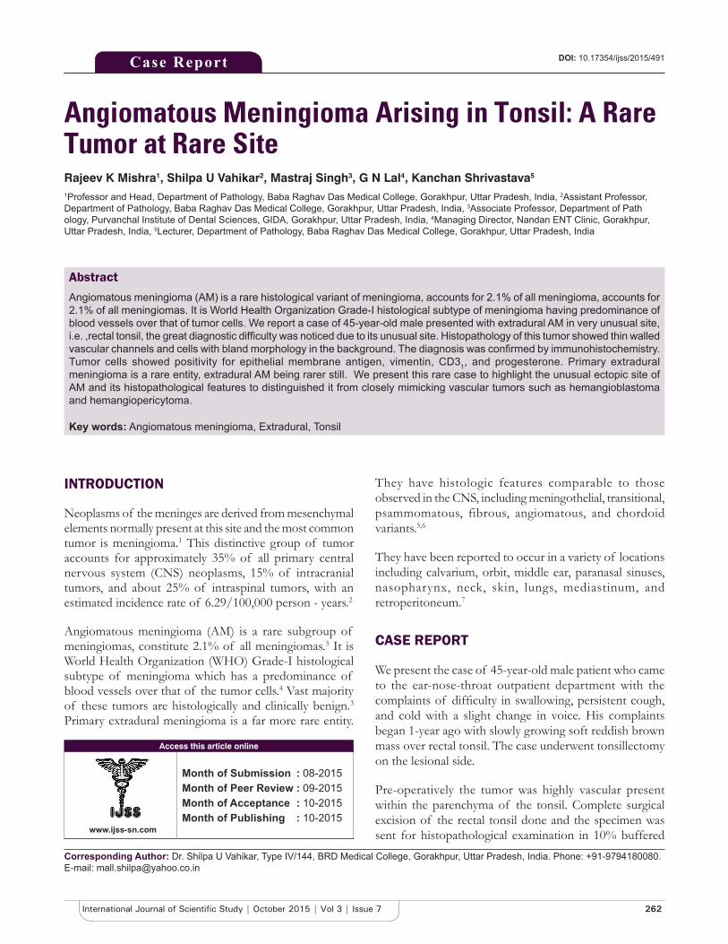
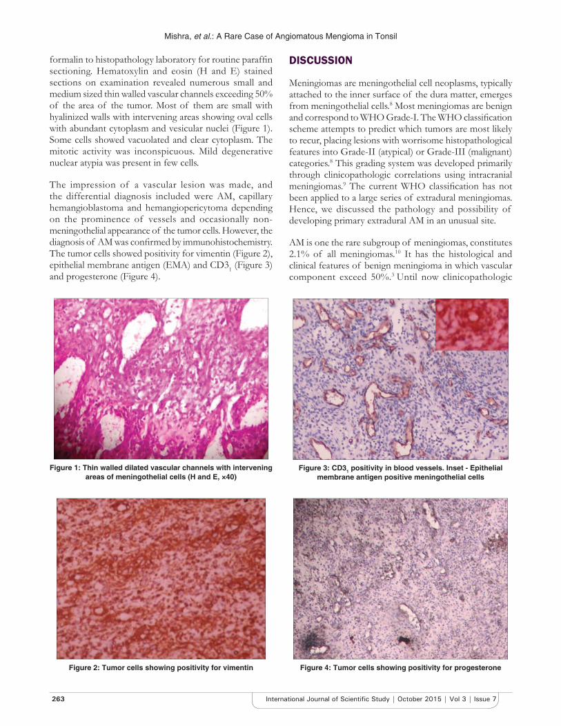
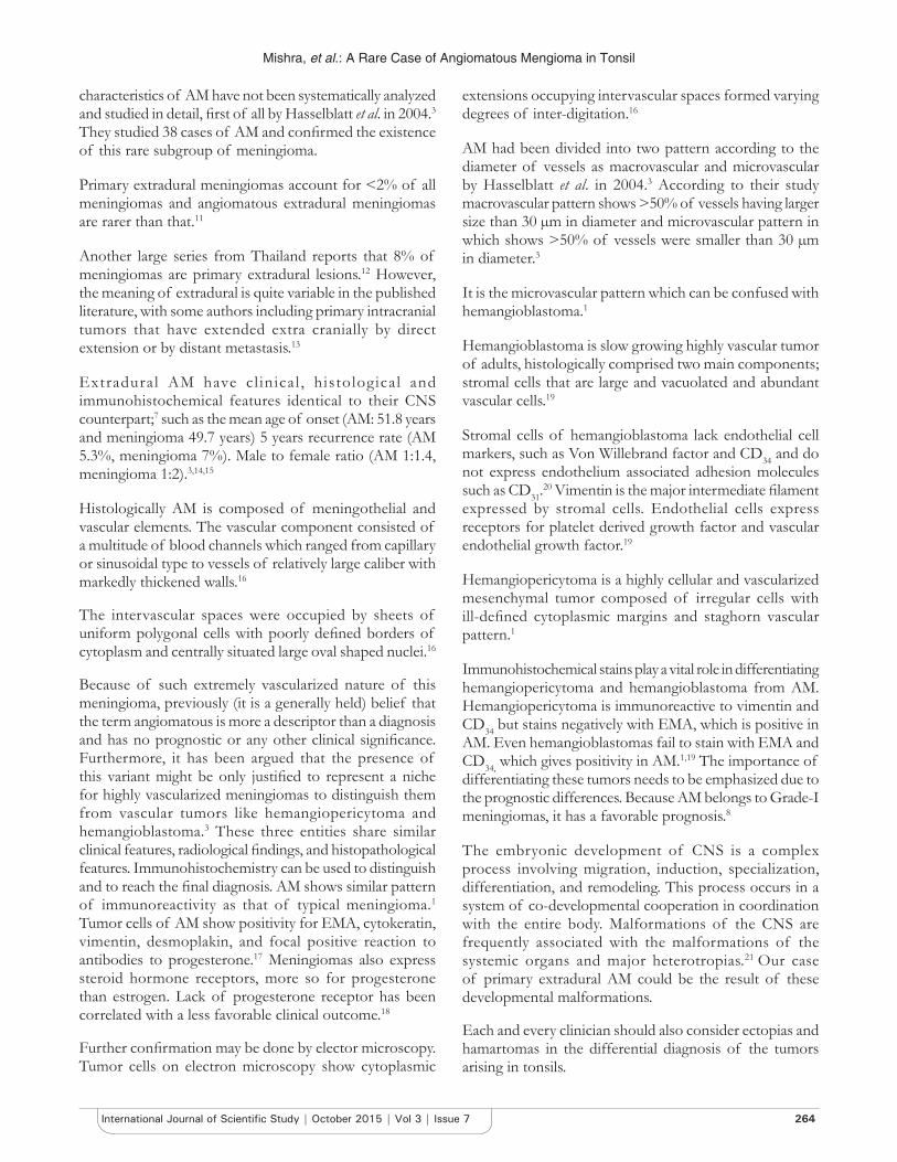
The impression of a vascular lesion was made, and the differential diagnosis included were AM, capillary hemangioblastoma and hemangiopericytoma depending on the prominence of vessels and occasionally non-meningothelial appearance of the tumor cells. However, the diagnosis of AM was confirmed by immunohistochemistry. The tumor cells showed positivity for vimentin (Figure 2), epithelial membrane antigen (EMA) and CD31 (Figure 3) and progesterone (Figure 4).
DISCUSSION
Meningiomas are meningothelial cell neoplasms, typically attached to the inner surface of the dura matter, emerges from meningothelial cells.8 Most meningiomas are benign and correspond to WHO Grade-I. The WHO classification scheme attempts to predict which tumors are most likely to recur, placing lesions with worrisome histopathological features into Grade-II (atypical) or Grade-III (malignant) categories.8 This grading system was developed primarily through clinicopathologic correlations using intracranial meningiomas.9 The current WHO classification has not been applied to a large series of extradural meningiomas. Hence, we discussed the pathology and possibility of developing primary extradural AM in an unusual site.
AM is one the rare subgroup of meningiomas, constitutes 2.1% of all meningiomas.10 It has the histological and clinical features of benign meningioma in which vascular component exceed 50%.3 Until now clinicopathologic
Figure 1: Thin walled dilated vascular channels with intervening areas of meningothelial cells (H and E, ×40)
Figure 2: Tumor cells showing positivity for vimentin
Figure 3: CD31 positivity in blood vessels. Inset - Epithelial membrane antigen positive meningothelial cells
Figure 4: Tumor cells showing positivity for progesterone
Mishra, et al.: A Rare Case of Angiomatous Mengioma in Tonsil
264International Journal of Scientific Study | October 2015 | Vol 3 | Issue 7
characteristics of AM have not been systematically analyzed and studied in detail, first of all by Hasselblatt et al. in 2004.3 They studied 38 cases of AM and confirmed the existence of this rare subgroup of meningioma.
Primary extradural meningiomas account for <2% of all meningiomas and angiomatous extradural meningiomas are rarer than that.11
Another large series from Thailand reports that 8% of meningiomas are primary extradural lesions.12 However, the meaning of extradural is quite variable in the published literature, with some authors including primary intracranial tumors that have extended extra cranially by direct extension or by distant metastasis.13
Extradural AM have cl inical , histological and immunohistochemical features identical to their CNS counterpart;7 such as the mean age of onset (AM: 51.8 years and meningioma 49.7 years) 5 years recurrence rate (AM 5.3%, meningioma 7%). Male to female ratio (AM 1:1.4, meningioma 1:2).3,14,15
Histologically AM is composed of meningothelial and vascular elements. The vascular component consisted of a multitude of blood channels which ranged from capillary or sinusoidal type to vessels of relatively large caliber with markedly thickened walls.16
The intervascular spaces were occupied by sheets of uniform polygonal cells with poorly defined borders of cytoplasm and centrally situated large oval shaped nuclei.16
Because of such extremely vascularized nature of this meningioma, previously (it is a generally held) belief that the term angiomatous is more a descriptor than a diagnosis and has no prognostic or any other clinical significance. Furthermore, it has been argued that the presence of this variant might be only justified to represent a niche for highly vascularized meningiomas to distinguish them from vascular tumors like hemangiopericytoma and hemangioblastoma.3 These three entities share similar clinical features, radiological findings, and histopathological features. Immunohistochemistry can be used to distinguish and to reach the final diagnosis. AM shows similar pattern of immunoreactivity as that of typical meningioma.1 Tumor cells of AM show positivity for EMA, cytokeratin, vimentin, desmoplakin, and focal positive reaction to antibodies to progesterone.17 Meningiomas also express steroid hormone receptors, more so for progesterone than estrogen. Lack of progesterone receptor has been correlated with a less favorable clinical outcome.18
Further confirmation may be done by elector microscopy. Tumor cells on electron microscopy show cytoplasmic
extensions occupying intervascular spaces formed varying degrees of inter-digitation.16
AM had been divided into two pattern according to the diameter of vessels as macrovascular and microvascular by Hasselblatt et al. in 2004.3 According to their study macrovascular pattern shows >50% of vessels having larger size than 30 µm in diameter and microvascular pattern in which shows >50% of vessels were smaller than 30 µm in diameter.3
It is the microvascular pattern which can be confused with hemangioblastoma.1
Hemangioblastoma is slow growing highly vascular tumor of adults, histologically comprised two main components; stromal cells that are large and vacuolated and abundant vascular cells.19
Stromal cells of hemangioblastoma lack endothelial cell markers, such as Von Willebrand factor and CD34 and do not express endothelium associated adhesion molecules such as CD31.
20 Vimentin is the major intermediate filament expressed by stromal cells. Endothelial cells express receptors for platelet derived growth factor and vascular endothelial growth factor.19
Hemangiopericytoma is a highly cellular and vascularized mesenchymal tumor composed of irregular cells with ill-defined cytoplasmic margins and staghorn vascular pattern.1
Immunohistochemical stains play a vital role in differentiating hemangiopericytoma and hemangioblastoma from AM. Hemangiopericytoma is immunoreactive to vimentin and CD34 but stains negatively with EMA, which is positive in AM. Even hemangioblastomas fail to stain with EMA and CD34, which gives positivity in AM.1,19 The importance of differentiating these tumors needs to be emphasized due to the prognostic differences. Because AM belongs to Grade-I meningiomas, it has a favorable prognosis.8
The embryonic development of CNS is a complex process involving migration, induction, specialization, differentiation, and remodeling. This process occurs in a system of co-developmental cooperation in coordination with the entire body. Malformations of the CNS are frequently associated with the malformations of the systemic organs and major heterotropias.21 Our case of primary extradural AM could be the result of these developmental malformations.
Each and every clinician should also consider ectopias and hamartomas in the differential diagnosis of the tumors arising in tonsils.
Mishra, et al.: A Rare Case of Angiomatous Mengioma in Tonsil
265 International Journal of Scientific Study | October 2015 | Vol 3 | Issue 7
CONCLUSION
Although AM is a rare variant of meningioma, it shares similar clinical features and prognosis with benign meningiomas and has some unique clinical, radiological, histopathological, and immunohistochemical features. Primary extradural AM rarely presents with tonsilar involvement hence clinician should consider this entity in the differential diagnosis of tonsilar tumors. It may mimic other vascular neoplasms like hemangioblastoma or hemangiopericytoma creating a diagnostic dilemma which requires immunohistochemistry for final diagnosis.
REFERENCES
1. Rao S, Rajkumar A, Kuruvilla S. Angiomatous meningioma: A diagnostic dilemma. Indian J Pathol Microbiol 2008;51:53-5.
2. Central Brain Tumour Registry of the United States 2012 Statistical Report: Primary Brain Tumours in United States, 2004-2008. Available from: http://www.cbtrus.org. [Last accessed on 2012 Mar 23].
3. Hasselblatt M, Nolte KW, Paulus W. Angiomatous meningioma: A clinicopathologic study of 38 cases. Am J Surg Pathol 2004;28:390-3.
4. Liu Z, Wang C, Wang H, Wang Y, Li JY, Liu Y. Clinical characteristics and treatment of angiomatous meningioma: A report of 27 cases. Int J Clin Exp Pathol 2013;6:695-702.
5. Tokgoz N, Oner YA, Kaymaz M, Ucar M, Yilmaz G, Tali TE. Primary intra-osseous meningioma: CT and MRI appearance. AJNR Am J Neuroradiol 2005;26:2053-6.
6. Sadar ES, Conomy JP, Benjamin SP, Levine HL. Meningiomas of the paranasal sinuses, benign and malignant. Neurosurgery 1979;4:227-32.
7. Gibson SE, Prayson RA. Primary ectopic meningiomas. Meningiomas 2009;7:573-83.
8. Perry A, Louis DN, Scheithauer BW, Budka H, Von Deimling A. Meningiomas. In: Lous DN, Oghaki H, Wiestler OD, Cavenee WK, editors.
WHO Classification of Tumors of the Central Nervous System. 4th ed. Lyon, France: IARC Press; 2007. p. 164-72.
9. Jain D, Ebrahimi KB, Miller NR, Eberhart CG. Intra-orbital meningiomas: A pathologic review using current World Health Organization criteria. Arch Pathol Lab Med 2010;134:766-70.
10. Chaushev N, Topalov N, Milanov I. A case report of angiomatous meningioma. J Clin Med 2010;3:60-3.
11. Whicker JH, Devine KD, MacCarty CS. Diagnostic and therapeutic problems in extra-cranial meningiomas. Am J Surg 1973;126:452-7.
12. Shuangshoti S, Panyathanya R. Neural neoplasms in Thailand: A study of 2,897 cases. Neurology 1974;24:1127-34.
13. Chen KT, Dehner LP. Primary tumors of the external and middle ear. II. A clinic-pathologic study of 14 paragangliomas and three meningiomas. Arch Otolaryngol 1978;104:253-9.
14. Ayerbe J, Lobato RD, de la Cruz J, Alday R, Rivas JJ, Gómez PA, et al. Risk factors predicting recurrence in patients operated on for intra-cranial meningioma. A multivariate analysis. Acta Neurochir (Wien) 1999;141:921-32.
15. Roser F, Nakamura M, Ritz R, Bellinzona M, Dietz K, Samii M, et al. Proliferation and progesterone receptor status in benign meningiomas are not age dependent. Cancer 2005;104:598-601.
16. Popoff NA, Malinin TI, Rosomoff HL. Fine structure of intracranial hemangiopericytoma and angiomatous meningioma. Cancer 1974;34:1187-97.
17. Mills SE. Sternberg’s Diagnostic Surgical Pathology. 4th ed. Philadelphia: Lippincott William and Wilkins; 2004. p. 460-81.
18. Hsu DW, Efird JT, Hedley-Whyte ET. Progesterone and estrogen receptors in meningiomas: Prognostic considerations. J Neurosurg 1977;86:113-20.
19. Aldape KD, Plate KH, Vortmeyer AO, Zagzay D, Neumann HP. Hemangioblastoma. In: Louis DN, Ohgaki H, Wiestler OD, Cavenee WK, editors. WHO Classification of Tumors of the Central Nervous System. Lyon, France: IARC Press; 2007. p. 184-6.
20. Böhling T, Mäenpää A, Timonen T, Vantunen L, Paetau A, Haltia M. Different expression of adhesion molecules on stromal cells and endothelial cells of capillary hemangioblastoma. Acta Neuropathol 1996;92:461-6.
21. Mclendon RE, Cunnings TJ. Ectopias and hamartomas. In: McLendon RE, Rosenblum MK, Binger DD, editors. Russell and Rubinsteins Pathology of Tumors of the Nervous System. 7th ed. Boca Raton, FL: Taylor and Francis Group; 2006. p. 616.
How to cite this article: Mishra RK, Vahikar SU, Singh M, Lal GN, Shrivastava K. Angiomatous Meningioma Arising in Tonsil: A Rare Tumor at Rare Site. Int J Sci Stud 2015;3(7):262-265.
Source of Support: Nil, Conflict of Interest: None declared.